


Transcriptomic analyses of human and murine muscle identify insulin receptor mRNA reduction in insulin resistance associated with hyperinsulinemia - Evidence review
The study shows that prolonged exposure to insulin reduces the insulin receptor gene and protein in murine myotubes, attenuating the AKT response and glucose uptake. Independent studies confirm muscle desensitization, including in humans; however, receptor reduction remains dependent on the model, dose, and duration of chronic exposure.
- Published 16/11/2026
- Updated 17/11/2026
- Article history
ABSTRACT
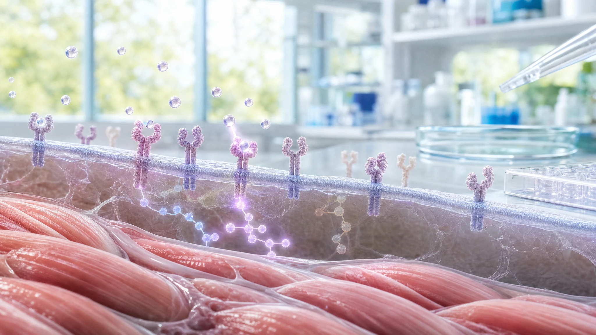
The analyzed study evaluated whether prolonged exposure to high insulin concentrations can directly reduce the response of muscle cells to the hormone. By integrating transcriptomic data from 488 human skeletal muscle samples with experiments on murine C2C12 myotubes and observations in mice, the authors found an inverse association between fasting insulin and INSR gene expression. In cells exposed to insulin for 16 hours, INSR, the total amount of the receptor, and the portion exposed on the cell surface decreased; after re-stimulation with insulin, AKT and ERK signaling were attenuated, and the glucose uptake response was reduced.
The research contributes to the passage from "The Choice to Live in Health or Illness" by Dr. A. Colonnese and R. Panzironi, which suggests that persistent excess insulin can reprogram components of its own signaling pathway and promote muscle desensitization. Independent studies in humans and muscle cells confirm that prolonged hyperinsulinemia can reduce insulin response and glucose transport. However, not all models show the same reduction in AKT or the receptor; the picture is therefore highly consistent on a mechanistic level, but it does not alone prove that this process causes diabetes in every person.
Analyzed study
Human and mouse muscle transcriptomic analyses identify insulin receptor mRNA downregulation in hyperinsulinemia-associated insulin resistance
PMID: 34921686
PMCID: PMC9255858
The research question was whether hyperinsulinemia, i.e., persistent exposure to high amounts of insulin, could directly contribute to skeletal muscle insulin resistance rather than just being a consequence of it. The hypothesis was that sustained high insulin levels alter the gene expression program of the muscle cell, including INSR, the gene that provides the instructions for producing the insulin receptor.
The insulin receptor is the first element of communication: it receives the hormone's signal on the cell surface and initiates a sequence that promotes glucose utilization. In simple terms, if the available receptors decrease, the cell may have less capacity to perceive a new demand from insulin. This is the precise point for which the publication was used in paragraphs 3.5 and 14.9 of “The Choice to Live in Health or Illness” by Dr. A. Colonnese and R. Panzironi: it documents that chronic exposure to insulin can reduce initial components of insulin signaling in the muscle and attenuate the response to subsequent stimulation.
The study combines an association observed in human muscle with experiments that allow for testing a direct effect of insulin in cells. The cellular component can support a causal relationship in the experimental model because the cells differed intentionally by insulin treatment. The correlations in human samples, however, do not prove that circulating insulin is the sole cause of the lower INSR expression: obesity, glycemia, muscle composition, medications, and other metabolic conditions may contribute to the observed relationship.
Materials and methods of the original study
The authors conducted a transcriptomic meta-analysis of three independent human skeletal muscle datasets, totaling 488 samples. The datasets included sedentary individuals with a high body mass index and/or impaired glucose tolerance, subjects with normal glucose tolerance, prediabetes, or type 2 diabetes, and a small case-control group comprising people with diabetes and severe insulin resistance treated with insulin. Muscle gene expression was correlated with fasting insulin; insulin values were mathematically transformed prior to analysis, and the analyses corrected for the risk of false-positive results due to the large number of genes examined.
To isolate the effect of insulin, C2C12 myotubes—differentiated mouse muscle cells in culture—were used. The cells were treated for 16 hours with 0, 2, or 200 nM of human insulin; 2 nM was a high concentration but closer to experimental physiological conditions, while 200 nM was supraphysiological. After treatment, some cells were maintained for six hours without serum and without insulin, a procedure used to observe how long alterations might persist after the stimulus was removed.
The insulin response was measured by evaluating the phosphorylation of AKT and ERK1/2. Phosphorylation is the addition of a phosphate group to a protein, a modification that can activate or alter its function. AKT is an important signaling node that promotes glucose uptake and utilization; ERK1/2 participates in other branches of the cellular response. Glucose uptake was also measured using labeled 2-deoxy-glucose, along with glycogen content, Insr messenger RNA, total and surface receptor quantity, and the overall gene profile via RNA sequencing.
The RNA sequencing experiments included four groups, with five biological replicates per group: cells with or without 200 nM of insulin, analyzed either before or after the serum-free phase. In mice, the authors examined gastrocnemius muscle lysates and correlated the amount of insulin receptor with fasting insulin, glycemia, and body weight. After technical exclusions and missing data, subgroup analyses included 7 to 11 animals. The available material does not report a single sample size for all cellular measurements, but generally indicates three to ten biological replicates depending on the experiment.
Results
The most relevant human data for the book's narrative was the inverse trend between fasting insulin and INSR expression in skeletal muscle. In all three datasets, as fasting insulin increased, INSR messenger RNA decreased. This relationship was also observed in the subgroup with normal glucose tolerance from the FUSION dataset. This result indicates consistency across different human samples, but it remains an association and does not establish the direction of causality in humans.
In the cellular model, 16 hours of exposure to high insulin induced a less effective response to subsequent acute stimulation. After the serum-free period, acute phosphorylation of AKT at the Thr308 and Ser473 sites and that of ERK1/2 were reduced in a dose-dependent manner based on the chronic treatment. The practical significance is that myotubes previously exposed to high levels of insulin transmitted a new insulin signal less effectively. With 200 nM of chronic insulin, basal glucose uptake increased, but the proportional increase caused by a new exposure to insulin decreased. Glycogen synthesis showed wide variability and did not differ in a statistically significant way between groups.
The amount of Insr RNA decreased markedly for both isoform A and isoform B of the receptor; their relative proportion did not change. After six hours without serum, expression showed only partial recovery. Consistently, the total amount of INSR protein decreased with chronic exposure to 2 and 200 nM of insulin, and the absolute amount of receptor available on the cell surface also decreased. The serum-free phase only slightly improved the reduction of the receptor in the group treated with 200 nM.
The work did not show a net loss in the ability of the individual residual receptor to autophosphorylate. After acute stimulation, the ratio between phosphorylated receptor and total receptor was not significantly different; however, because the total number of receptors was lower, the overall number of phosphorylation events per cell decreased. Receptor internalization, i.e., its movement from the surface to the inside of the cell, also did not appear to be drastically altered. The authors therefore interpret the lower abundance of the receptor, together with modifications of components located downstream of the receptor, as the most important explanation for the reduced insulin response in their model.
In the muscle of male mice, the amount of INSR was inversely correlated with fasting insulin both on a low-fat diet and a high-fat diet. In multiple models, insulin, glycemia, and weight explained approximately 95% of the variability of INSR in the male group on a low-fat diet and approximately 66% in the group on a high-fat diet. In females, the relationship was not detected, partly because insulin values were close to the lower limit of measurement. This data limits the possibility of generalizing the association to both sexes and all nutritional contexts without reservations.
Interpretation
The central biological significance is that persistent insulin stimulation can make muscle cells less responsive to new signals. In the studied model, this occurs at least in part because the cell produces less Insr RNA and, consequently, possesses fewer total insulin receptors and fewer receptors on its surface. Downstream of the receptor, the subsequent activation of AKT is attenuated, and glucose transport responds less to insulin.
The most directly documented chain of events is: prolonged insulin exposure → reduced Insr transcription → reduction of INSR protein and surface receptors → attenuation of acute AKT-mediated signaling and glucose transport response. RNA sequencing also indicated changes in genes belonging to the insulin receptor, FOXO, and glucose metabolism pathways. FOXO1 is a transcription factor, meaning it is a protein that helps regulate the switching on or off of genes. Insulin increased FOXO1 phosphorylation, a condition associated with lower activity of this protein in the nucleus; the result is compatible with reduced Insr transcription, but it does not prove that FOXO1 is solely responsible.
The authors also identified SIN3A as a possible negative regulator of Insr: experimentally reducing Sin3a RNA increased Insr RNA. This is an interesting mechanistic element, but it was obtained in cells and has not yet been proven to be a determining factor in humans. Similarly, the reduction of Insr does not exclude other mechanisms, such as receptor degradation, alterations in IRS-1, PI3K, or AKT, and contributions from lipids, inflammation, glycemia, and body composition.
Regarding the book's sections, the publication does not directly demonstrate that chronic mTOR activation is the sole cause of the reduction in INSR. Its specific contribution is to document the functional desensitization associated with chronic insulin exposure and the remodeling of insulin receptor expression. The link to the broader PI3K/AKT/mTOR sequence remains biologically plausible, but it requires studies targeting individual molecular nodes and cannot be attributed entirely to this work.
Independent confirmatory evidence
Confirmation in humans of the general idea that sustained hyperinsulinemia can precede and promote a reduced insulin response comes from a study by Del Prato and colleagues. Fifteen healthy young individuals were subjected to physiological hyperinsulinemia protocols while maintaining normal glycemia via a euglycemic clamp, a technique that allows for the control of blood glucose levels while measuring how much glucose the body uses in response to insulin. After 72–96 hours, systemic glucose utilization decreased by 20–40%. The defect primarily concerned the non-oxidative portion, which is largely glycogen synthesis, while the suppression of hepatic glucose production remained unchanged. This study does not measure INSR in the muscle, but it confirms the functional downstream step of the narrative directly in humans: persistently elevated insulin, even without hyperglycemia, can reduce insulin sensitivity [1].
Turner and colleagues studied primary muscle cells derived from healthy human donors. After three days with 100 nM of insulin, subsequent acute stimulation produced lower glucose uptake and reduced phosphorylation of IRS-1 at Tyr612 and AKT at Ser473. IRS-1 is an adapter protein that transmits the signal initiated by the receptor to the rest of the cell. This independent model therefore confirms the functional sequence described in the book, from prolonged insulin exposure to a reduced response of IRS-1, AKT, and glucose transport. The limitation is that these are cultured cells, with a high insulin concentration and without interaction with the liver, adipose tissue, immune system, or physical activity [2].
A previous experiment on human myoblasts cultured for three weeks with high insulin provided further confirmation of desensitization. After a brief new insulin stimulation, phosphorylation of the receptor, IRS-1, PI3K activity, and AKT phosphorylation increased only in cells cultured with low insulin; in cells chronically exposed to high insulin, these increases did not occur. Glucose transport also increased after acute stimulation only in the low-insulin group. This study supports the segment of the narrative concerning the attenuation of the receptor → IRS-1 → PI3K → AKT cascade and the resulting lower glucose response. Unlike the study analyzed, total levels of receptor and IRS-1 did not differ between conditions: this shows that desensitization can develop even without a measurable decrease in the total amount of receptor [3].
Taken together, these independent studies reinforce a specific point: prolonged exposure to insulin can reduce the response to subsequent stimulation in muscle or muscle cells. Confirmation of the specific reduction in INSR expression is stronger in experimental models compared to human intervention studies, whereas in humans, the functional consequence on insulin sensitivity is documented more directly.
Conflicting evidence
A relevant finding that calls for caution comes from an experiment on C2C12 cells exposed to insulin for 72 hours. In cells treated during the differentiation process, acute AKT phosphorylation at Ser473 and insulin-induced glucose uptake decreased. In already differentiated myotubes, however, chronic insulin exposure did not alter AKT phosphorylation at Ser473, although it did reduce GLUT4 and hexokinase II RNA, two components involved in glucose utilization. This result does not rule out the possibility of desensitization, but it indicates that the effect on AKT depends on the cellular stage, the protocol, and likely the dose and duration of the treatment [4].
This difference is relevant because the analyzed study treated already differentiated murine myotubes for 16 hours and observed a reduction in the AKT response after the serum-free phase. The heterogeneity between models therefore does not allow us to state that any chronic insulin exposure always reduces AKT in the same way. Furthermore, the study on human myoblasts exposed to high insulin for three weeks documented a desensitization of glucose transport and the PI3K/AKT response without a reduction in total insulin receptor levels [3].
The conflicting evidence does not concern the general fact that high insulin exposure can alter cellular response, but rather the dominant mechanism and its quantitative reproducibility. No directly conflicting evidence sufficiently relevant has been identified that demonstrates, under comparable conditions, an improvement in muscle insulin sensitivity caused by persistent hyperinsulinemia; this does not prove the absence of contrary results.
Summary table of evidence
The table distinguishes the publication under review from the independent research used to verify the book's narrative.
| Reference | Study type | Population or model | Main result | Direction of evidence | Strength of evidence |
|---|---|---|---|---|---|
| Original study | Human, cellular, and animal transcriptomics | 488 human samples; C2C12 myotubes; mouse muscle | Fasting insulin inversely associated with human INSR; chronic insulin reduces Insr, INSR, and acute AKT response in cells | direct confirmation | moderate |
| [1] | Human metabolic intervention | 15 healthy young individuals subjected to euglycemic hyperinsulinemia for 72–96 hours | 20–40% reduction in glucose utilization, primarily the non-oxidative component | direct confirmation | moderate |
| [2] | Human cellular | Primary muscle cells from healthy donors, 100 nM insulin for three days | Reduction in glucose uptake and acute phosphorylation of IRS-1 and AKT | direct confirmation | moderate |
| [3] | Human cellular | Human myoblasts cultured for three weeks with low or high insulin | High insulin attenuates the acute PI3K/AKT response and glucose transport without reducing total receptor proteins | indirect confirmation | moderate |
| [4] | Murine cellular | C2C12 myoblasts and myotubes exposed to insulin for 72 hours | Reduction of AKT and glucose uptake in cells treated during differentiation, but not of AKT phosphorylation in already differentiated myotubes | conflicting evidence | low |
Confirmation of the evidence
Claim in the book
In paragraphs 3.5 and 14.9, “The Choice to Live in Health or Illness” by Dr. A. Colonnese and R. Panzironi states that chronic exposure to insulin can reprogram components of its own signaling pathway. Specifically, it can progressively reduce the muscle's ability to perceive the hormone, decreasing insulin receptor expression and attenuating the AKT response to subsequent stimulation.
Persistent hyperinsulinemia → reduction of INSR and other signaling components → reduced IRS-1/PI3K/AKT response → reduced glucose uptake and utilization → muscle insulin resistance
The publication subject to the Evidence Review was used in the book to support the following passage: chronic excess of insulin can reduce the expression of the INSR gene, the total and surface amount of the insulin receptor in myotubes, attenuating the acute AKT response and the glucose uptake response.
The correspondence between the study and the book's text is precise. The analyzed study documents an inverse association in human muscle between fasting insulin and INSR RNA, and demonstrates in murine muscle cells that prolonged experimental exposure to insulin precedes the reduction of Insr, the receptor protein, and the acute insulin response. The work therefore primarily supports the initial segment of the chain: persistent hyperinsulinemia → reduced receptor availability → less effective cellular signaling.
Independent research completes the narrative at different levels. In humans, maintaining elevated insulin with controlled glycemia reduced glucose utilization and, in particular, its non-oxidative destination, consistent with lower glycogen synthesis [1]. In primary human muscle cells, prolonged exposure to insulin reduced subsequent phosphorylation of IRS-1 and AKT and glucose uptake [2]. In human myoblasts, long exposure to high insulin attenuated the response of the receptor → IRS-1 → PI3K → AKT pathway and glucose transport [3].
These findings converge in supporting desensitization induced by prolonged insulin exposure, but they do not define a single universal mechanism. The fact that some models show an unchanged AKT response in already differentiated myotubes [4], or desensitization without a reduction in the total amount of receptor [3], indicates that INSR represents an important and biologically consistent mechanism, but not necessarily the only one. The link with mTOR discussed in chapter 14 of “The Choice to Live in Health or Illness” by Dr. A. Colonnese and R. Panzironi must therefore be read as a chain composed of multiple pieces of evidence: this study documents the receptor and AKT with greater immediacy, while the specific role of mTOR requires dedicated evidence.
Overall level of confirmation
STRONG CONFIRMATION
The original publication directly documents the decrease of Insr, INSR, and the acute AKT response in the cellular model; independent studies in humans and muscle cells confirm the reduction in insulin sensitivity and glucose transport following prolonged exposure to insulin. The specific reduction of INSR in humans remains primarily associative, and the mechanism varies between models.
Conclusions
The analyzed publication directly supports, in a murine myotube model, that prolonged exposure to insulin can reduce insulin receptor expression and attenuate the cell's response to a subsequent stimulus. The inverse association observed in human muscle makes the result biologically relevant, but it does not, by itself, demonstrate causality in humans.
Independent evidence confirms that sustained hyperinsulinemia can decrease insulin sensitivity and glucose utilization even in healthy individuals and human muscle cells. The framework is therefore well-supported for functional desensitization and moderately supported for receptor reduction as a predominant mechanism. Longitudinal human validation is still needed to measure insulin, the receptor, muscle signaling, and metabolic consequences together over time.
Overall assessment
The narrative is reasonably well-supported: prolonged hyperinsulinemia can induce insulin desensitization in muscle and reduce the AKT response and glucose transport. The decrease in INSR is confirmed at a mechanistic level and is consistent with associative human data, but it depends on the experimental model, duration of exposure, and cellular context.
References
[1] Effect of sustained physiologic hyperinsulinaemia and hyperglycaemia on insulin secretion and insulin sensitivity in man
[2] Characterising hyperinsulinemia-induced insulin resistance in human skeletal muscle cells
[3] Continually high insulin levels impair Akt phosphorylation and glucose transport in human myoblasts
Editorial note
Artificial Intelligence was used solely as a tool to support editorial and documentary work. All content, interpretations, bibliographic citations and conclusions have been verified, reviewed and approved by the author, who assumes full scientific responsibility for them.
The manuscript was also subject to independent peer review prior to publication.
To ensure maximum transparency, the article's complete history — including the dates of submission, review, acceptance and publication, as well as information on the peer review process — is available on the dedicated Article History page.