


Chronic hyperinsulinemia induces a reduction in post-receptor insulin signal transduction in Hep G2 cells - Review of the evidence
The study shows that sixteen hours of insulin exposure reduces, in Hep G2 liver cells, activation of the receptor, IRS-1, and PI3K recruitment. Independent studies support desensitization induced by persistent exposure, including in humans, but different cellular models and tissues limit definitive clinical conclusions and require further targeted prospective confirmation.
- Published 16/11/2026
- Updated 17/11/2026
- Article history
ABSTRACT
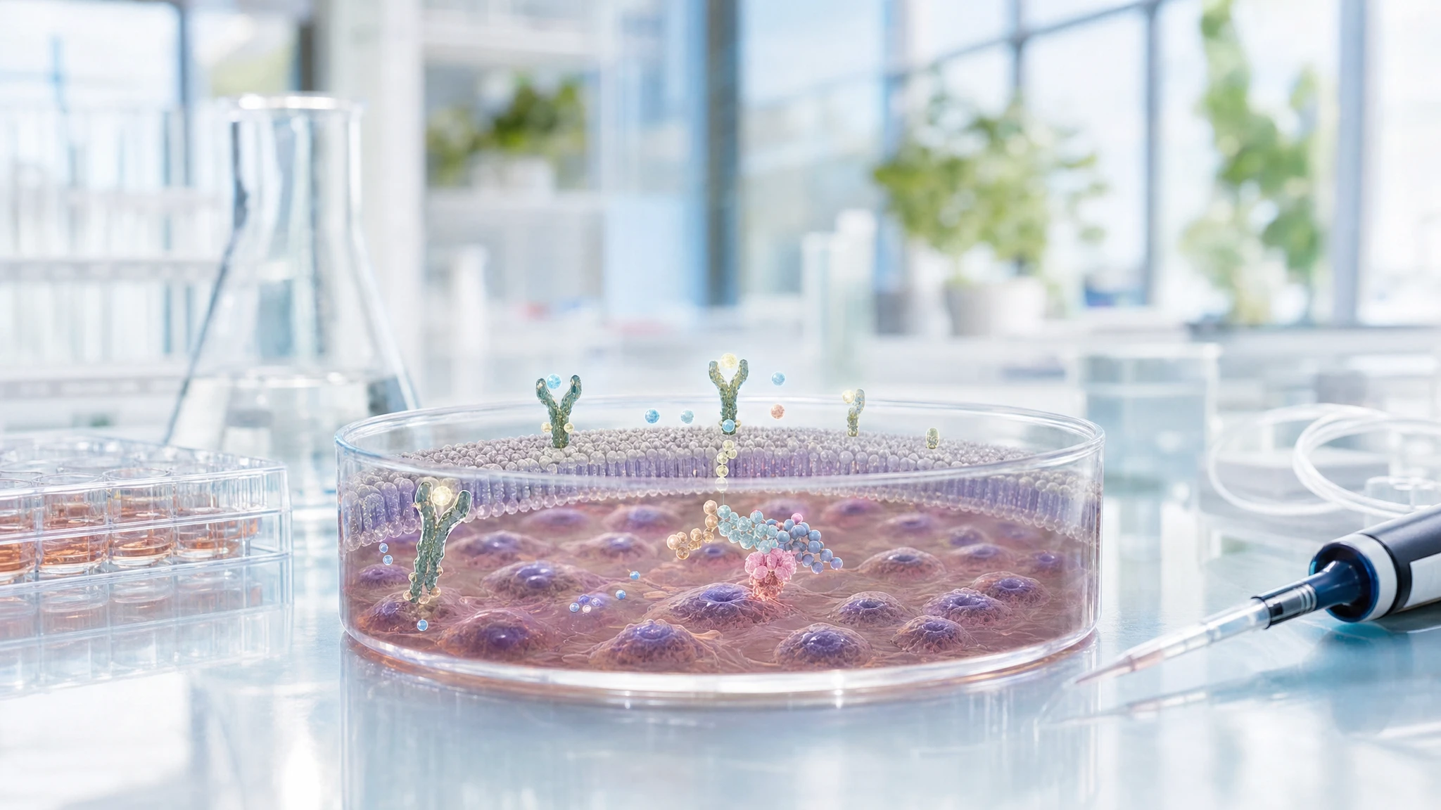
This article analyzes a cellular study that evaluated whether prolonged exposure to insulin can reduce the response to subsequent stimulation with the same hormone. In Hep G2 liver cells, derived from a human liver tumor, sixteen hours of insulin treatment reduced the quantity of the insulin receptor and, above all, its activation. The activation of IRS-1 and the association between IRS-1 and PI3K were also reduced, which are initial steps necessary for the insulin signal to proceed within the cell.
The work contributes to the passage from "The Choice to Live in Health or Illness" by Dr. A. Colonnese and R. Panzironi, according to which hyperinsulinemia can simultaneously compromise the receptor, IRS-1, and the recruitment of PI3K. Independent studies confirm that persistent exposure to insulin can attenuate receptor autophosphorylation, reduce downstream signals, and decrease glucose uptake in human muscle cells. Experiments in humans also indicate a reduction in insulin sensitivity after hyperinsulinemia maintained for days. However, the Hep G2 model is not a healthy liver and does not demonstrate on its own that the same mechanism occurs identically in all tissues or in people. The overall confirmation is moderate.
Analyzed study
Chronic hyperinsulinism induced down-regulation of insulin post-receptor signaling transduction in Hep G2 cells
DOI: 10.1007/bf02896773
PMID: 12674767
The research question was whether acute and chronic exposure to insulin produced different effects on the early steps of insulin signaling in Hep G2 cells. Under normal conditions, insulin binds to its receptor on the cell surface. The receptor is activated through autophosphorylation—meaning it adds phosphate groups to itself—and then transfers the signal to IRS-1 (insulin receptor substrate 1), a protein that helps recruit PI3K (phosphoinositide 3-kinase), an enzyme involved in the transmission of the metabolic signal.
The hypothesis was that prolonged exposure to high concentrations of insulin would make this cascade less reactive to a new stimulus. This is the reason for its relevance to section 3.5: the publication does not directly evaluate clinical insulin resistance or glycemia in people, but it documents a possible cellular segment of desensitization. Since Hep G2 is a liver tumor cell line cultured in the laboratory, the study can show molecular modifications in that model, but it cannot establish on its own how present these are in the healthy liver, muscle, or the entire human organism.
Materials and methods of the original study
The available material consists only of the abstract. The authors used Hep G2 cells cultured in serum-free medium. The cells were incubated for 16 hours in the absence of insulin or with different insulin concentrations ranging from 1 to 100 nmol/L. At the end of the chronic treatment, the cells were subjected to a new acute stimulation with 100 nmol/L of insulin for one minute.
The total amount of the insulin receptor beta subunit, IRS-1, and the p85 subunit of PI3K was measured using Western immunoblot, a technique that detects specific proteins in a cell sample. The phosphorylation of the receptor and IRS-1, along with the interaction between IRS-1 and PI3K, was evaluated by immunoprecipitation, a procedure that allows for the isolation of a protein and the molecules associated with it. The number of experimental replicates, exclusion criteria, detailed statistical procedures, and complete analysis values are not reported in the available material.
Results
Acute one-minute stimulation rapidly activated the insulin receptor and IRS-1: both showed an increase in tyrosine phosphorylation, a chemical modification necessary to transmit the signal. This activation was accompanied by the association between IRS-1 and PI3K, consistent with the initiation of the insulin cascade.
After 16 hours of chronic insulin treatment, the amount of the receptor's beta subunit decreased in a dose-dependent manner: higher insulin concentrations were associated with a lower amount of receptor. The total amount of IRS-1 was only slightly reduced. However, the functional effect was more pronounced: after treatment with 100 nmol/L of insulin, receptor phosphorylation dropped to 22% of the control level, and IRS-1 phosphorylation dropped to 15% of the control; both differences were statistically significant, with P < 0,01.
In simple terms, the cells did not just have fewer receptors: the remaining receptor and IRS-1 responded much less to new insulin exposure. The association between IRS-1 and PI3K also decreased by 66% compared to the control, with P < 0,01. The total amount of PI3K, however, did not change significantly. The result therefore indicates a reduction in the recruitment of PI3K into the signaling complex, not a decrease in the overall availability of the enzyme.
Interpretation
The main biological significance is that continuous insulin signaling can make the cellular response to a subsequent dose of insulin less effective. In this model, the compromised chain is initial: insulin receptor → IRS-1 → PI3K recruitment. If these steps function less effectively, subsequent metabolic signals may be attenuated.
The segment specifically documented by the publication is the reduction in receptor activation, IRS-1 phosphorylation, and IRS-1-PI3K association following chronic insulin exposure. The authors' interpretation, according to which these modifications contribute to insulin resistance, is biologically plausible but remains an inference: the study did not directly measure hepatic glucose production, glycogen synthesis, or insulin sensitivity in humans.
The result therefore supports the book's narrative at a mechanistic level. It does not prove that hyperinsulinemia is always the initial cause of insulin resistance, because in real life, elevated insulin, increased glycemia, excess lipids, inflammation, and individual predisposition can coexist and influence each other.
Independent supporting evidence
Direct confirmation of the first part of the chain comes from Catalano and colleagues. In a cell model subjected to chronic insulin exposure, subsequent stimulation produced reduced autophosphorylation of the insulin receptor. After removing the hormone, the alteration appeared reversible, but it reappeared rapidly upon re-exposure. The study therefore confirms that desensitization can begin at the receptor itself; the limitation is the use of cultured cells, which do not reproduce the metabolic complexity of the organism [1].
Turner and colleagues examined primary muscle cells obtained from healthy human donors and differentiated in culture. After three days with 100 nmol/L of insulin, subsequent acute stimulation resulted in lower glucose uptake and reduced phosphorylation of IRS-1 and AKT (a protein that transmits many of insulin's metabolic effects downstream). This confirms, in a different tissue closer to human muscle, that prolonged exposure can attenuate the insulin signaling response and the final function of glucose absorption. However, it remains an in vitro study [2].
Cen and colleagues added a further level of evidence. In a transcriptomic meta-analysis of 488 human skeletal muscle samples, higher fasting insulin concentrations were associated with lower messenger RNA expression of INSR, the insulin receptor gene. In experiments on murine myotubes, prolonged insulin exposure reduced Insr expression, the amount of total and surface receptor, and attenuated AKT signaling and glucose uptake. The human part is observational and does not demonstrate causality; the experimental part, however, demonstrates causality in the murine model, not in humans [3].
The physiological relevance of this narrative is also supported by a controlled experiment in 15 healthy young adults. Del Prato and colleagues maintained physiological hyperinsulinemia for 72–96 hours, with normal or slightly elevated glycemia. Euglycemic hyperinsulinemia reduced insulin sensitivity, and the defect primarily involved non-oxidative glucose metabolism, i.e., the pathway that mainly includes glycogen synthesis. This study did not measure the receptor, IRS-1, or PI3K in the liver; however, it confirms, directly in humans, that persistent exposure to insulin can reduce the metabolic efficacy of the hormone [4].
Conflicting evidence
No directly conflicting evidence of sufficient relevance has been identified; this does not prove the absence of contrary results.
The main caveats stem from the heterogeneity of the models. The response to insulin depends on the tissue, the duration of exposure, the hormone concentration, glycemia, and the presence of lipids or inflammation. Furthermore, the liver responds to insulin differently than skeletal muscle, and Hep G2 cells are tumor cells, not normal human hepatocytes. For these reasons, the convergence of studies supports the existence of a desensitization mechanism, but it does not authorize the automatic transfer of every molecular variation observed in culture to the clinical physiology of all individuals.
Summary table of evidence
The table distinguishes the molecular data obtained in the original study from independent confirmations obtained in cellular models, human muscle tissue, and healthy individuals.
| Reference | Study type | Population or model | Main result | Direction of evidence | Strength of evidence |
|---|---|---|---|---|---|
| Original study | Cellular | Human Hep G2 cells | After 16 hours of insulin: receptor reduction, receptor phosphorylation at 22% and IRS-1 at 15% of control; IRS-1-PI3K association reduced by 66%. | direct confirmation | preliminary |
| [1] | Cellular | Cell model with insulin receptor | Chronic insulin exposure reduced receptor autophosphorylation after re-stimulation. | direct confirmation | moderate |
| [2] | Cellular | Primary muscle cells from healthy human donors | Three days of insulin reduced glucose uptake and phosphorylation of IRS-1 and AKT. | indirect confirmation | moderate |
| [3] | Observational and cellular | 488 human muscle samples; murine myotubes | Higher fasting insulin associated with lower INSR; chronic insulin reduced receptor, AKT signaling, and glucose uptake in myotubes. | biologically consistent evidence | moderate |
| [4] | Physiological trial in humans | 15 healthy young adults | Physiological hyperinsulinemia for 72–96 hours reduced insulin sensitivity, especially non-oxidative glucose metabolism. | indirect confirmation | moderate |
Confirmation of the evidence
Claim in the book
In section 3.5 of “The Choice to Live in Health or Illness” by Dr. A. Colonnese and R. Panzironi, the relevant statement is that hyperinsulinemia can simultaneously compromise the insulin receptor, IRS-1, and the pathway to PI3K, interrupting the cascade in its initial stages.
Persistent insulin exposure → reduced receptor activation or quantity → reduced IRS-1 phosphorylation → reduced PI3K recruitment → attenuated cellular insulin response
The publication subject to the Evidence Review was used in the book to support the following passage: in Hep G2 cells, chronic insulin exposure reduces the quantity and, above all, the activation of the insulin receptor, attenuates IRS-1 phosphorylation, and reduces the association between IRS-1 and PI3K, without significantly decreasing the total amount of PI3K.
The correspondence between the original study and the paragraph in “The Choice to Live in Health or Illness” by Dr. A. Colonnese and R. Panzironi is direct at the molecular level: the work precisely documents the attenuation of the receptor → IRS-1 → PI3K segment following prolonged exposure to insulin. Independent literature extends this narrative without requiring every study to reproduce the entire chain in the same tissue.
The first step, the reduced reactivity of the receptor, is experimentally confirmed by an independent cell model [1]. The subsequent step, namely the decrease in the response downstream of IRS-1, is consistent with human muscle cells exposed to insulin for three days, in which the phosphorylation of IRS-1, AKT, and glucose uptake are reduced [2]. A further line of evidence links hyperinsulinemia to lower receptor expression in human muscle and experimentally demonstrates a reduction in surface receptor, signaling, and glucose uptake in myotubes [3]. Finally, the physiological study on healthy volunteers shows that sustained hyperinsulinemia can reduce metabolic sensitivity to insulin in the entire organism [4].
The chain is therefore convergent but not complete in humans at the level of every single protein. The data on the receptor, IRS-1, and PI3K are derived from cell cultures, while the direct human data concerns systemic insulin sensitivity and not hepatic signaling measured protein by protein.
Overall level of confirmation
MODERATE CONFIRMATION
The original publication directly documents the molecular pathway for which it is cited. Independent studies confirm receptor desensitization, the reduction of downstream signals, and lower metabolic efficacy of insulin in humans as well. The confirmation remains moderate because the complete receptor → IRS-1 → PI3K detail has not been demonstrated in the same way in the healthy human liver.
Conclusions
The analyzed study clearly supports, in the Hep G2 model, that chronic exposure to insulin can attenuate three early steps of insulin signaling: the receptor, IRS-1, and PI3K recruitment. Independent evidence is consistent with desensitization induced by persistent exposure to the hormone and includes cellular, muscular, and physiological data in humans.
The narrative that hyperinsulinemia and insulin resistance can reinforce each other is therefore moderately supported. It remains necessary to distinguish the cellular mechanism from its individual clinical relevance: not all tissues respond in the same way, and hyperinsulinemia can be both a consequence and a possible contributing factor to the progression of insulin resistance.
Overall assessment
The mechanism of insulin signaling desensitization following persistent exposure to the hormone is quite well supported at the cellular level. The consistency with the book's paragraph is high. Data in humans confirm a reduction in insulin sensitivity, but they do not fully reconstruct the same molecular hepatic cascade.
References
[1] Insulin Resistance Induced by Hyperinsulinemia Coincides with a Persistent Alteration at the Insulin Receptor Tyrosine Kinase Domain
[2] Characterising hyperinsulinemia-induced insulin resistance in human skeletal muscle cells
[3] Human and mouse muscle transcriptomic analyses identify insulin receptor mRNA downregulation in hyperinsulinemia‐associated insulin resistance
Editorial note
Artificial Intelligence was used solely as a tool to support editorial and documentary work. All content, interpretations, bibliographic citations and conclusions have been verified, reviewed and approved by the author, who assumes full scientific responsibility for them.
The manuscript was also subject to independent peer review prior to publication.
To ensure maximum transparency, the article's complete history — including the dates of submission, review, acceptance and publication, as well as information on the peer review process — is available on the dedicated Article History page.