


Insulin-activated signaling pathway and GLUT4 translocation to the membrane in hiPSC-derived cardiomyocytes - Evidence review
In cardiomyocytes derived from human stem cells, insulin increased the activation of AKT and AS160 and moved GLUT4 to the membrane, without increasing its total quantity. The result supports a cellular trafficking mechanism, but it is derived from a single cell line. Independent studies confirm this, with important contextual experimental limitations.
- Published 17/11/2026
- Updated 17/11/2026
- Article history
ABSTRACT
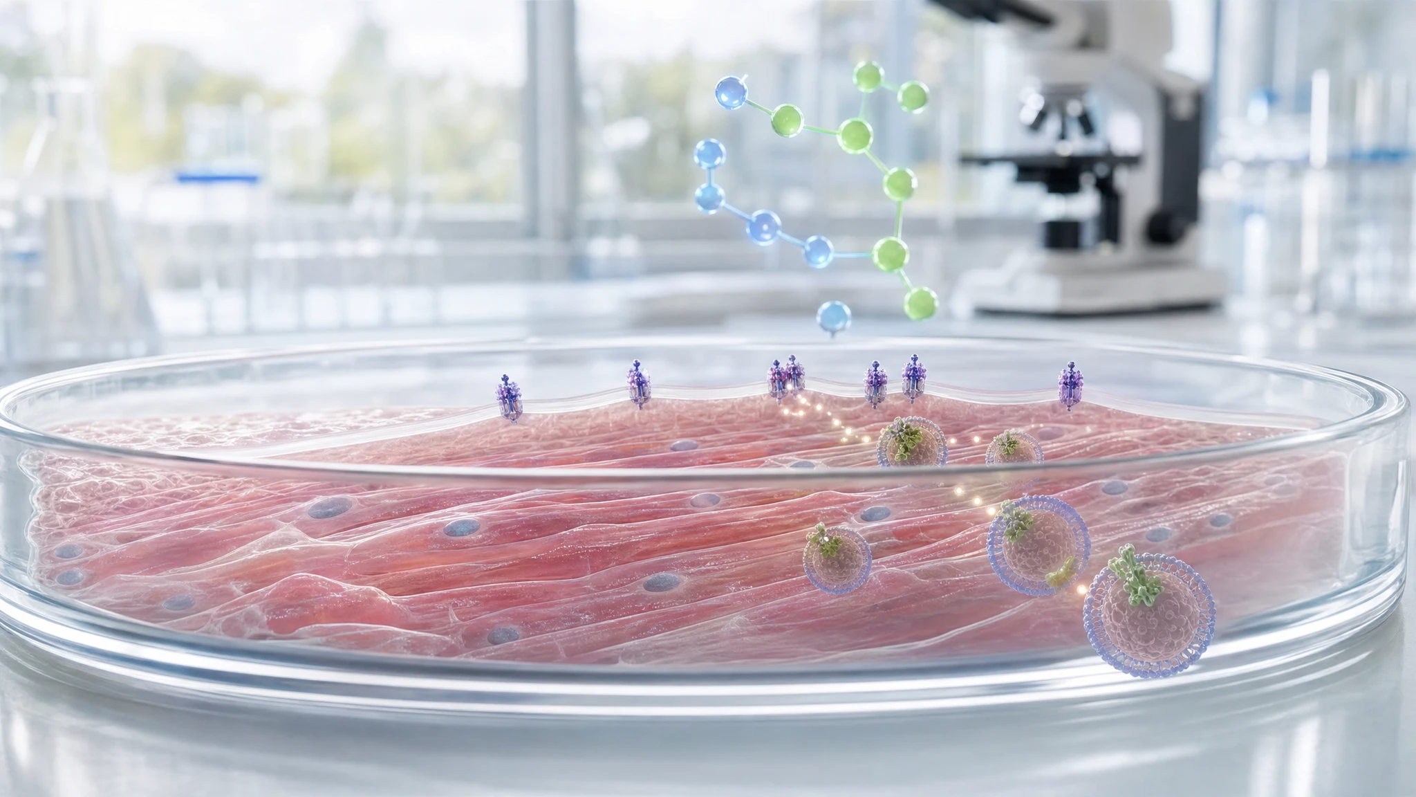
The analyzed study evaluated whether cardiomyocytes derived from human induced pluripotent stem cells respond to insulin in a manner similar to mature heart cells. After 30 minutes of stimulation, insulin increased the phosphorylation of AKT and AS160 and increased the presence of GLUT4 in the membrane fraction, without significantly changing the total amount of GLUT4. The result therefore indicates an intracellular translocation of the transporter, rather than its new production.
In the narrative of “The Choice to Live in Health or Illness” by Dr. A. Colonnese and R. Panzironi, the study contributes to the explanation that the amount of GLUT4 exposed on the membrane, rather than the total cellular amount, is the relevant factor for glucose entry. Independent studies in adipocytes, muscle, and adult cardiomyocytes support the role of the AKT-AS160-GLUT4 axis. However, a previous study on other stem cell-derived cardiomyocytes did not observe a functional insulin response, indicating that the phenomenon also depends on the maturation and the cell line used.
Analyzed study
Insulin-Activated Signaling Pathway and GLUT4 Membrane Translocation in hiPSC-Derived Cardiomyocytes
DOI: 10.3390/ijms25158197
PMID: 39125765
PMCID: PMC11312081
The research question was whether cardiomyocytes derived from human induced pluripotent stem cells, known as hiPSC-CMs, possess an insulin response capable of mobilizing GLUT4. GLUT4 is a transporter: a protein that allows glucose to cross the cell membrane. In insulin-sensitive cells, the transporter is largely stored in internal compartments and is brought to the surface when the hormonal signal arrives.
The hypothesis was that insulin activates the AKT-AS160 sequence and produces an increase of GLUT4 in the plasma membrane. AKT is a protein that transmits signals within the cell. AS160, also known as TBC1D4, is a protein that regulates the trafficking of vesicles containing GLUT4. This work is relevant to section 4.4 of “The Choice to Live in Health or Illness” by Dr. A. Colonnese and R. Panzironi because it directly documents that the total amount of GLUT4 can remain unchanged while the portion localized to the membrane increases.
The study demonstrates a molecular and localization response of GLUT4 in a human cell model cultured in the laboratory. It does not directly measure glucose uptake, it does not prove that all hiPSC-CM lines respond in the same way, and it cannot establish how the phenomenon occurs in the intact human heart.
Materials and methods of the original study
Researchers used a single human induced pluripotent stem cell line, UKBi015-B, obtained from healthy donors and differentiated into cardiomyocytes. The cells began to contract spontaneously around the eighth day of differentiation. Between the eleventh and fourteenth day, they were maintained in a medium containing lactate as the sole energy source, a procedure used to enrich the preparation in cardiomyocytes.
On the fourteenth day, a portion of the cells was exposed to insulin at a concentration of 100 nM for 30 minutes; untreated cells served as the control. The authors performed Western blot, a technique that measures specific proteins, on both total cell lysates and membrane fractions separated by centrifugation. They measured phosphorylated AKT at Ser473, phosphorylated AS160 at Thr642, GLUT4, and GLUT1. Phosphorylation is a chemical modification often associated with the activation or regulation of a protein.
The localization of GLUT4 was also evaluated using immunofluorescence, a technique that makes proteins labeled with fluorescent signals visible. The Western blot data were derived from three independent experiments. The immunofluorescence analysis included 34 alpha-actinin-positive cells for each condition. Data were expressed as mean ± standard error of the mean and analyzed using an unpaired t-test or analysis of variance. Information on biological replicates from different cell lines or donors is not reported beyond the use of the UKBi015-B line alone.
Results
Insulin triggered cellular signals consistent with the initiation of GLUT4 trafficking. After treatment, the ratio of phosphorylated AKT to total AKT rose from 100.00 in the control to 136.86 ± 9.80 in the treated cells. The ratio of phosphorylated AS160 to total AS160 also increased, from 100.00 to 137.93 ± 8.12. The authors report statistical significance with p < 0.05 and p < 0.01, respectively. These data indicate a relative increase in the phosphorylated form of the two proteins, not an increase in their total quantity.
The result most relevant to the book concerns GLUT4. In total cell lysates, insulin did not lead to a detectable increase in GLUT4: the normalized value was 100.00 in the control and 97.33 ± 14.33 after insulin. GLUT1, a transporter that is primarily insulin-independent, also did not change significantly. This finding suggests that insulin did not induce a greater overall production of GLUT4 within 30 minutes.
The situation was different when analyzing the membrane fraction. Here, GLUT4 increased from 100.00 in the control to 150.34 ± 5.49 after insulin, with p < 0.05. GLUT1 remained substantially stable, moving from 57.26 ± 14.42 to 61.04 ± 17.60. Immunofluorescence showed an increase in the peripheral signal attributed to GLUT4, from 1.00 ± 0.13 to 2.31 ± 0.28 times the control, with p < 0.001.
The practical significance is that GLUT4 appears to have redistributed from internal stores to the cell surface. The comparison between total lysate and membrane fraction shows why the overall quantity of a protein alone does not allow one to automatically deduce its position or its functional availability on the membrane.
Interpretation
The publication describes a specific segment of the insulin response: insulin increases the phosphorylation of AKT and AS160 and, in the same cell model, increases the amount of GLUT4 detected in the membrane. In lay terms, the insulin signal does not appear to have created new "doors" for glucose; instead, it seems to have moved doors already present within the cell toward the surface.
The segment supported by the study is therefore: insulin → phosphorylated AKT and AS160 → greater presence of GLUT4 at the membrane. This sequence is consistent with the known role of AS160 in controlling Rab proteins, which regulate vesicle movement and fusion. However, the experimental design alone does not prove that AKT or AS160 are essential for the observed increase in GLUT4, because no selective inhibitors, AS160 mutations, or other causal blocking experiments were used.
The study also does not directly measure glucose uptake. It is biologically plausible that more GLUT4 in the membrane facilitates glucose entry, but such a functional consequence requires a dedicated measurement. The main utility of the work is therefore methodological and mechanistic: it shows that, for studying translocation, the analysis of membrane fractions is more informative than measuring GLUT4 in the total lysate.
Independent supporting evidence
The first step in the book's narrative, namely the link between AS160 phosphorylation and GLUT4 translocation, is supported by an independent experiment in 3T3-L1 murine adipocytes. Sano and colleagues identified insulin-increased phosphorylation sites on AS160 and showed that mutations preventing such phosphorylation markedly reduced insulin-stimulated GLUT4 translocation. When the GAP function of AS160 was also inactivated, the inhibitory effect of the mutant disappeared. This is causal data in a cellular model: it supports the role of AS160 as a functional link between AKT and GLUT4 trafficking, but it is not a direct demonstration in human cardiomyocytes [1].
A second study investigated the same mechanism in adipose cells and cellular systems expressing the insulin receptor. Phosphorylation of TBC1D4/AS160 was found to be necessary for GLUT4 to reach the membrane; the mere dissociation of AS160 from the membranes did not appear to be sufficient. This work reinforces the idea that the important signal is not the total amount of GLUT4, but the regulated control of its trafficking and its exposure on the cell surface [2].
The confirmation closest to the tissue studied comes from isolated adult rat ventricular cardiomyocytes. In this model, insulin increased GLUT4 in the plasma membrane fraction by approximately eightfold, accompanied by a 60-65% reduction in the intracellular membrane fraction. The design therefore directly documents a subcellular redistribution of GLUT4 in the heart: the portion on the surface increases while the portion in internal stores decreases. Although this is an animal model and not a human one, this result is particularly consistent with the interpretation of the analyzed work [3].
Taken together, these independent publications form a convergent chain. Studies on adipocytes clarify the causal role of AS160 phosphorylation in GLUT4 trafficking; the study in adult cardiomyocytes demonstrates that insulin can effectively shift GLUT4 toward the membrane in cardiac tissue. The analyzed work places this response in a model of human cardiomyocytes derived from stem cells, with a demonstration that is still preliminary because it is based on a single cell line and a few independent experiments.
Conflicting evidence
Directly relevant but conflicting evidence concerns other cardiomyocytes derived from human induced pluripotent stem cells. Bowman and colleagues studied cells from two commercial suppliers and observed that insulin activated AKT, but did not significantly increase glucose uptake. In those cells, GLUT4 was much less abundant compared to adult cardiomyocytes, while GLUT1 was higher [4].
This result does not contradict the general AKT-AS160-GLUT4 mechanism. Rather, it shows that the presence of upstream insulin signals, such as phosphorylated AKT, does not necessarily guarantee a complete functional response if GLUT4 is scarce or if the metabolic maturation of the cells is incomplete. The differences may depend on the cell line, the differentiation protocol, the duration of maturation, the culture conditions, and the method used to measure the response.
The contrast between the two studies on hiPSC-CMs therefore requires caution. The publication subject to the Evidence Review demonstrates that a specific cell preparation can show GLUT4 translocation after insulin; it does not establish that this behavior is universal in all stem cell-derived cardiomyocytes, nor that it fully equates to the metabolic phenotype of the adult cardiomyocyte.
Summary table of evidence
The table distinguishes the result observed in the original study from independent publications that confirm or define the narrative of the book's paragraph.
| Reference | Study type | Population or model | Main result | Direction of evidence | Strength of evidence |
|---|---|---|---|---|---|
| Original study | Experimental cellular | A human hiPSC-CM line | Insulin increases phosphorylated AKT and AS160 and increases GLUT4 in the membrane fraction, not in the total lysate. | direct confirmation | preliminary |
| [1] | Cellular with genetic manipulation | Murine 3T3-L1 adipocytes | Non-phosphorylatable AS160 mutations markedly reduce insulin-stimulated GLUT4 translocation. | direct confirmation | moderate |
| [2] | Molecular cellular | Adipocytes and cells with insulin receptor | AS160/TBC1D4 phosphorylation is required for GLUT4 translocation to the membrane. | indirect confirmation | moderate |
| [3] | Animal cellular | Isolated adult rat ventricular cardiomyocytes | Insulin and contraction increase GLUT4 in the plasma membrane and reduce it in intracellular membranes. | direct confirmation | moderate |
| [4] | Experimental cellular | Human hiPSC-CMs from two commercial sources | AKT responds to insulin, but GLUT4 is scarce and no significant increase in glucose uptake is observed. | discordant evidence | moderate |
Confirmation of the evidence
Claim in the book
In section 4.4 of "The Choice to Live in Health or Illness" by Dr. A. Colonnese and R. Panzironi, the central claim is that insulin does not necessarily increase the total amount of GLUT4, but primarily modifies its distribution, increasing the portion exposed on the cell membrane. This portion is what enables the passage of glucose from the outside to the inside of the cell.
Insulin → phosphorylated AKT → phosphorylated AS160 → mobilization of GLUT4-containing vesicles → increased GLUT4 at the membrane → potential increase in glucose uptake
The publication subject to the Evidence Review was used in the book to support the following passage: insulin stimulation can increase the phosphorylation of AKT and AS160 and promote the translocation of GLUT4 toward the plasma membrane without significantly increasing the total amount of the transporter in the cell.
The correspondence between the study and the paragraph is specific. The analyzed work directly compares total lysates and membrane fractions and shows that the overall amount of GLUT4 remains unchanged, while it increases in the membrane component following insulin stimulation. This supports the narrative clarification proposed in “The Choice to Live in Health or Illness” by Dr. A. Colonnese and R. Panzironi: to understand the insulin response, it is not enough to quantify all the protein present in the cell, because its localization is decisive.
Independent evidence confirms the central steps of the chain. In adipocytes, blocking the phosphorylation of AS160 reduces insulin-induced GLUT4 translocation, providing a functional demonstration of the AS160-GLUT4 link [1]. A second study confirmed that the phosphorylation of AS160/TBC1D4 is essential to remove a brake on GLUT4 trafficking to the membrane [2]. In isolated adult rat cardiomyocytes, insulin increases GLUT4 in the plasma membrane and reduces its presence in intracellular membranes, a redistribution consistent with the mechanism described [3].
However, the literature does not allow us to consider all hiPSC-CMs equivalent to adult cardiomyocytes. In other hiPSC-CM lines, AKT responded to insulin, but the low expression of GLUT4 was associated with the absence of a significant increase in glucose uptake [4]. This limits the generalizability of the model, but does not eliminate the scientific correspondence between the result observed in the analyzed study and the passage in the book.
Overall level of confirmation
MECHANISTIC CONFIRMATION
The AKT-AS160-GLUT4 connection is supported by independent experiments that include causal manipulations of AS160 and direct observations of GLUT4 translocation in adult cardiomyocytes. However, the demonstration in human hiPSC-CMs remains dependent on the cell line and is not yet accompanied by uniform functional confirmation of glucose uptake.
Conclusions
The analyzed study directly supports the finding that, in a specific line of human stem cell-derived cardiomyocytes, insulin increases the presence of GLUT4 on the membrane without increasing its total cellular quantity. This data is consistent with the role of the AKT-AS160-GLUT4 axis in controlling intracellular trafficking.
Independent evidence makes the biological mechanism quite well-supported, especially regarding the link between AS160 phosphorylation and GLUT4 translocation. In cardiomyocytes, redistribution toward the membrane is documented in adult animal models. However, it remains to be more extensively validated whether all human hiPSC-CM preparations exhibit a complete insulin response and an actual increase in glucose uptake.
Overall assessment
The narrative is confirmed at a mechanistic level: insulin primarily regulates the localization of GLUT4, and the AKT-AS160 axis contributes to its membrane trafficking. Data in human stem cell-derived cardiomyocytes are promising but preliminary, as they depend on a single line and discordant results indicate variable metabolic maturation.
References
[1] Insulin-stimulated Phosphorylation of a Rab GTPase-activating Protein Regulates GLUT4 Translocation
[2] Regulation of Glucose Transporter 4 Translocation by the Rab Guanosine Triphosphatase-Activating Protein AS160/TBC1D4: Role of Phosphorylation and Membrane Association
[3] Contraction-induced translocation of the glucose transporter Glut4 in isolated ventricular cardiomyocytes
[4] GLUT4 expression and glucose transport in human induced pluripotent stem cell-derived cardiomyocytes
Editorial note
Artificial Intelligence was used solely as a tool to support editorial and documentary work. All content, interpretations, bibliographic citations and conclusions have been verified, reviewed and approved by the author, who assumes full scientific responsibility for them.
The manuscript was also subject to independent peer review prior to publication.
To ensure maximum transparency, the article's complete history — including the dates of submission, review, acceptance and publication, as well as information on the peer review process — is available on the dedicated Article History page.