


Insulin resistance induced by hyperinsulinemia coincides with a persistent alteration of the tyrosine kinase domain of the insulin receptor - Evidence review
The cellular study shows that prolonged exposure to insulin reduces the reactivation of its receptor without necessarily decreasing its presence on the membrane. The phenomenon suggests persistent desensitization, but does not prove an identical effect in humans. Independent studies confirm insulin resistance induced by hyperinsulinemia, with mechanisms dependent on the tissue and the intensity and duration of insulin exposure.
- Published 16/11/2026
- Updated 17/11/2026
- Article history
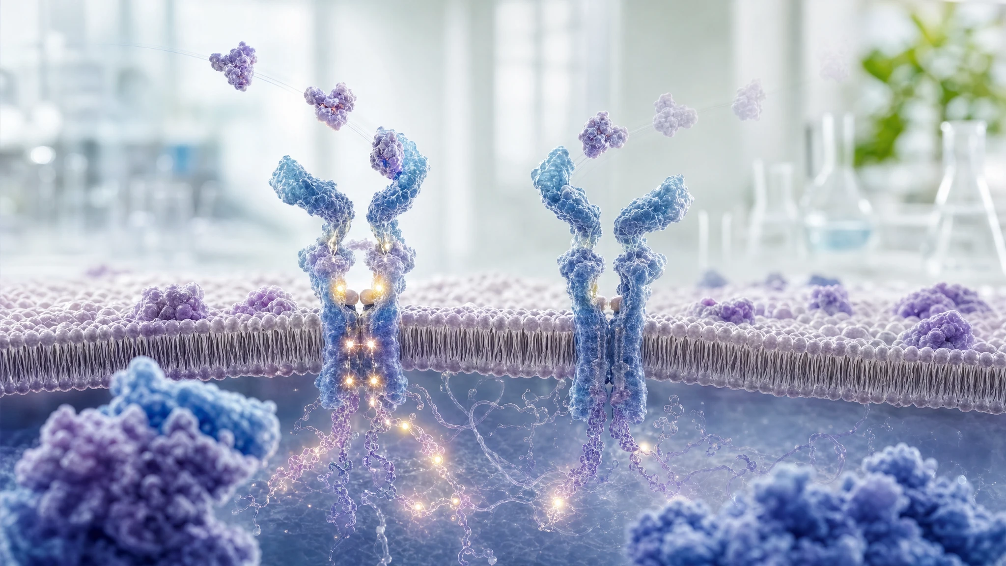
ABSTRACT
The analyzed study evaluated whether prolonged exposure to insulin can reduce the insulin receptor's ability to respond to new stimulation. In rat liver cells and hamster ovary cells modified to express the human receptor, continuous exposure to the hormone reduced receptor autophosphorylation, which is the initial internal signal that allows the cell to respond to insulin. This reduction was not explained by a lower overall amount of receptor or its disappearance from the cell membrane.
The research contributes to the section of "The Choice to Live in Health or Illness" by Dr. A. Colonnese and R. Panzironi dedicated to the idea that prolonged hyperinsulinemia may promote progressive insulin desensitization. Independent evidence in humans, muscle cells, and cellular models confirms that persistent excess insulin can attenuate the insulin response and modify initial components of the signaling pathway. However, the specific "molecular memory" of the receptor described in the original study has not been directly demonstrated in humans, and responses remain dependent on the tissue and the signal considered.
Analyzed study
Insulin resistance induced by hyperinsulinemia coincides with a persistent alteration at the insulin receptor tyrosine kinase domain
DOI: 10.1371/journal.pone.0108693
PMID: 25259572
PMCID: PMC4178179
The research question was whether prolonged exposure to high concentrations of insulin could make the insulin receptor less capable of responding to subsequent exposure to the hormone. The insulin receptor is a protein located on the cell surface: when it binds to insulin, it activates a sequence of signals that contributes, among other things, to the entry and utilization of glucose.
The hypothesis evaluated was that chronic hyperinsulinemia could induce a form of refractory state, i.e., a weakened response to new stimulation, by acting directly on the receptor or in its immediate molecular environment. This is the reason for its relevance to section 3.5 of “The Choice to Live in Health or Illness” by Dr. A. Colonnese and R. Panzironi: the publication examines the initial segment of the chain proposed in the book, namely prolonged exposure to insulin → reduced receptor activation → reduced ability to transmit the signal.
The study can demonstrate an experimental effect in cell cultures, under the conditions and at the concentrations used by the authors. However, it cannot establish that the same structural alteration occurs in human muscle, liver, or adipose tissue, nor can it quantify its contribution to the onset of diabetes in the body.
Materials and methods of the original study
The authors primarily used rat hepatoma cells modified to express the human insulin receptor and Chinese hamster ovary cells. The cells were serum-starved for four hours and then exposed to recombinant insulin at different times and concentrations. The main concentrations were 5 nM, 17 nM, and 170 nM; nM stands for nanomolar, a unit expressing a very low concentration of a substance.
Receptor activation was measured by assessing its phosphorylation, which is the addition of phosphate groups that acts as an activation signal. The authors used western blotting and ELISA (a laboratory test based on the recognition of proteins by antibodies). They specifically analyzed the tyrosine sites Y972 and Y1162/1163 of the receptor, which are important for initiating the signaling cascade.
To verify whether the receptor moved from the membrane into the cell, fluorescence microscopy was used on cells with the receptor tagged with YFP, a fluorescent protein. To explore possible changes near the tyrosine kinase domain, the part of the receptor that transfers signals through chemical reactions, FRET (a technique that measures the relative proximity between two fluorescent markers and can signal a conformational or molecular environment change) was employed.
Data were reported as mean ± standard deviation. Comparisons were performed using one-way analysis of variance and Tukey's post-hoc test; the authors considered a P-value of less than 0.05 to be statistically significant. No inclusion or exclusion criteria are reported, as this is not a study involving human participants.
Results
Under short-term stimulation conditions, insulin rapidly increased receptor autophosphorylation at all concentrations examined. After longer exposures, phosphorylation at the Y972 and Y1162/1163 sites decreased compared to the initial peak, with differences reported as statistically significant. In simple terms, the receptor initially activated, but during prolonged contact with the hormone, it lost some of its ability to maintain that activation.
The total amount of receptor did not change in the western blots. Furthermore, in fluorescently labeled cells, the amount of receptor localized to the membrane was not reduced after prolonged exposure to 17 nM of insulin. This data indicates that, in this model, the reduction in signal was not primarily explained by the loss of the receptor from the cell surface.
After 120 minutes of exposure to 17 or 170 nM of insulin, a ten-minute wash rapidly reduced residual phosphorylation. However, a second short stimulation returned Y972 and Y1162/1163 only to the reduced level typical of the refractory state, rather than reproducing the intense response observed during the first short stimulation. This phenomenon did not appear after an initial exposure of only two minutes.
FRET did not change after ten minutes of insulin, but it increased after 120 minutes. Since an increase in FRET signals that the fluorescent markers have changed their relative proximity, the result is consistent with a change in the conformation of the tyrosine kinase domain or in the environment of the proteins near it. After a short wash, this FRET alteration persisted; after 180 minutes of washing, it returned to baseline levels, but was rapidly re-established with a new exposure to insulin.
Downstream pathways did not all respond in the same way. Phosphorylation of ERK, a protein linked to growth signals and cellular response, was intense and transient, then decreased rapidly. In contrast, phosphorylation of AKT and GSK3 did not decrease within 120 minutes. This shows that desensitization was not uniform across all measured intracellular pathways.
Interpretation
The most immediate biological significance is that continuous insulin stimulation can make the receptor's initial response to a new exposure to the hormone less prompt. The result most relevant to the book is not the demonstration of systemic insulin resistance, but the cellular segment showing that persistent excess insulin can weaken the autophosphorylation of its own receptor without necessarily requiring a reduction in the number of receptors on the membrane.
The publication suggests a possible molecular memory of chronic exposure: after washing, some measurements returned toward baseline, but the reduced response reappeared immediately upon new stimulation. FRET makes a change in the tyrosine kinase domain or its interactions biologically plausible. However, FRET does not identify which molecular modification is responsible, nor does it prove that the detected change is the direct cause of the refractory state.
The experimental model has an important limitation: it uses genetically modified cell lines and insulin concentrations that do not fully replicate the natural alternation of human insulin secretion. Furthermore, the absence of receptor reduction on the membrane observed here does not contradict studies that, in other tissues or after longer exposures, have detected a decrease in receptor expression. Rather, it indicates that the mechanisms of desensitization can vary depending on the context.
Independent supporting evidence
The evidence most closely supporting the book's general narrative comes from a controlled clinical study of 15 healthy young adults. Physiological hyperinsulinemia maintained for 72–96 hours, with normal or slightly elevated glycemia, reduced insulin sensitivity. The defect primarily concerned the non-oxidative use of glucose—that is, its transformation and storage as glycogen—while the suppression of hepatic glucose production remained intact. This result supports the idea in humans that prolonged hyperinsulinemia may directly contribute to peripheral insulin resistance; however, it does not identify a molecular memory of the receptor [1].
A second study cultured human myoblasts for three weeks with elevated insulin. After subsequent brief stimulation, cells chronically exposed to insulin showed no further increase in the phosphorylation of the receptor, IRS-1, PI3K, and AKT, nor in glucose transport, whereas these signals increased in control cells. IRS-1 is a protein that transmits the signal from the receptor; PI3K and AKT participate in subsequent steps. The research therefore confirms, in human muscle-derived cells, an attenuated response to new insulin exposure, consistent with the refractory state observed in the original study [2].
An independent transcriptomic analysis integrated three datasets of human skeletal muscle totaling 488 samples and an experimental model of murine myotubes. In human samples, higher fasting insulin levels were associated with lower expression of INSR, the insulin receptor gene. In myotubes exposed to insulin for 16 hours, Insr RNA, total protein, and surface receptor protein decreased; after acute stimulation, AKT and ERK signals and glucose uptake were also attenuated. The study expands on the book's chain of evidence by showing that, in other models, hyperinsulinemia can decrease not only the functionality but also the availability of the receptor [3].
Conflicting evidence
No directly conflicting evidence of sufficient relevance has been identified regarding the possibility that prolonged insulin exposure induces a reduced insulin response. However, there are findings that require caution in describing the phenomenon as a uniform loss of all insulin signaling.
In adipocytes chronically exposed to insulin, GLUT4-dependent glucose transport was reduced, while the nuclear exclusion of FoxO1, a regulator of gene expression, remained largely preserved. The authors defined this scenario as selective insulin resistance: some cell functions become less responsive, while others remain sensitive because they require different levels of PI3K/AKT signaling. The result does not refute the desensitization described in the book, but shows that its extent depends on the tissue and the specific branch of the cascade being analyzed [4].
Summary table of evidence
The table distinguishes the publication under review from the independent research used to confirm or contextualize the passage in the book.
| Reference | Study type | Population or model | Main result | Direction of evidence | Strength of evidence |
|---|---|---|---|---|---|
| Original study | Experimental cellular | Rat hepatoma cells and hamster ovary cells with human insulin receptor | Prolonged insulin exposure reduced receptor reactivation without necessarily reducing its total or membrane presence. | direct confirmation | preliminary |
| [1] | Experimental clinical study | 15 healthy young adults subjected to 72–96 hours of hyperinsulinemia | Prolonged physiological hyperinsulinemia reduced insulin sensitivity and non-oxidative glycogen synthesis. | indirect confirmation | moderate |
| [2] | Cellular | Human myoblasts cultured for three weeks with high insulin | After restimulation, the response of IR, IRS-1, PI3K, AKT, and glucose transport decreased. | indirect confirmation | moderate |
| [3] | Observational and cellular | 488 human muscle samples and murine myotubes | High insulin was associated with lower human INSR; chronic insulin reduced the receptor and acute response in myotubes. | biologically consistent evidence | moderate |
| [4] | Cellular | Adipocytes in a chronic hyperinsulinemia model | GLUT4 was less responsive, while nuclear exclusion of FoxO1 remained largely preserved. | discordant evidence | low |
Confirmation of the evidence
Claim in the book
In section 3.5, "The Choice to Live in Health or Illness" by Dr. A. Colonnese and R. Panzironi, it is stated that hyperinsulinemia is not only a possible compensatory response to insulin resistance, but can also contribute to tissue desensitization. Regarding the analyzed reference, the specific point is that chronic exposure to insulin can reduce receptor autophosphorylation following a new stimulation, without necessarily decreasing its presence on the membrane, leaving a predisposition for an inadequate response.
Prolonged insulin exposure → reduced receptor autophosphorylation → decreased initial signal transmission → attenuated cellular insulin response
The publication subject to the Evidence Review was used in the book to support the following passage: chronic exposure to insulin can induce a refractory state in the receptor, in which a new brief stimulation produces reduced autophosphorylation even without a necessary reduction in the amount of receptor present on the cell membrane.
The original study directly documents this step in cell cultures and, based on FRET, proposes that a persistent change near the tyrosine kinase domain may accompany the refractory state. However, independent confirmation of this specific conformational alteration of the receptor is limited.
Independent literature confirms the subsequent and more general segments of the narrative. In humans, hyperinsulinemia maintained for several days reduced peripheral insulin sensitivity [1]. In human myoblasts, prolonged exposure attenuated the response of multiple nodes in the cascade, including the downstream response of the receptor and glucose transport [2]. In human muscle and myotubes, elevated insulin is associated with or induces a reduction in receptor expression and a lower acute response [3].
These studies support a convergent chain: prolonged excess of insulin can be followed by lower efficacy of the insulin signal. However, they also indicate that the mechanism is not unique: in some contexts, the receptor remains on the membrane but responds less, in others its quantity decreases, and in others still, the resistance is selective for specific cellular functions [4].
Overall level of confirmation
INDIRECT CONFIRMATION
The original publication directly demonstrates the receptor step for which it is cited in cells. Independent studies confirm that prolonged hyperinsulinemia can reduce insulin sensitivity and alter initial signaling, including in humans. Conversely, independent validation of the specific molecular memory of the tyrosine kinase domain proposed by the authors remains limited.
Conclusions
The analyzed study supports the idea at a cellular level that prolonged exposure to insulin can reduce subsequent insulin receptor activation without necessarily causing a loss of the receptor from the membrane. This observation is consistent with the book's narrative regarding a possible feedback loop between hyperinsulinemia and insulin resistance.
Independent evidence moderately confirms that persistent excess insulin can attenuate the insulin response in peripheral tissues and muscle cells. However, the hypothesis that a specific conformational change in the receptor constitutes a stable and clinically relevant molecular memory in humans remains preliminary. The response appears to be dependent on the cell type, the duration of exposure, and the specific signaling pathway studied.
Overall assessment
The narrative that prolonged hyperinsulinemia may contribute to insulin resistance is fairly well supported by human and cellular data. The specific mechanism proposed in the study—namely, a memory associated with the tyrosine kinase domain of the receptor—remains confirmed primarily at a mechanistic level and requires independent validation in humans.
References
[1] Effect of sustained physiologic hyperinsulinaemia and hyperglycaemia on insulin secretion and insulin sensitivity in man
[2] Continually high insulin levels impair Akt phosphorylation and glucose transport in human myoblasts
[3] Human and mouse muscle transcriptomic analyses identify insulin receptor mRNA downregulation in hyperinsulinemia‐associated insulin resistance
[4] Hyperinsulinemia leads to uncoupled insulin regulation of the GLUT4 glucose transporter and the FoxO1 transcription factor
Editorial note
Artificial Intelligence was used solely as a tool to support editorial and documentary work. All content, interpretations, bibliographic citations and conclusions have been verified, reviewed and approved by the author, who assumes full scientific responsibility for them.
The manuscript was also subject to independent peer review prior to publication.
To ensure maximum transparency, the article's complete history — including the dates of submission, review, acceptance and publication, as well as information on the peer review process — is available on the dedicated Article History page.