


Characterization of insulin resistance induced by hyperinsulinemia in human skeletal muscle cells - Review of the evidence
The study shows that three days of elevated insulin reduce glucose uptake and the activation of IRS-1 and AKT in human muscle cells. Independent research, including human studies, supports the contribution of hyperinsulinemia to insulin resistance; however, significant differences between models, dosages, duration, and tissues remain, requiring further clinical confirmation.
- Published 16/11/2026
- Updated 16/11/2026
- Article history
ABSTRACT
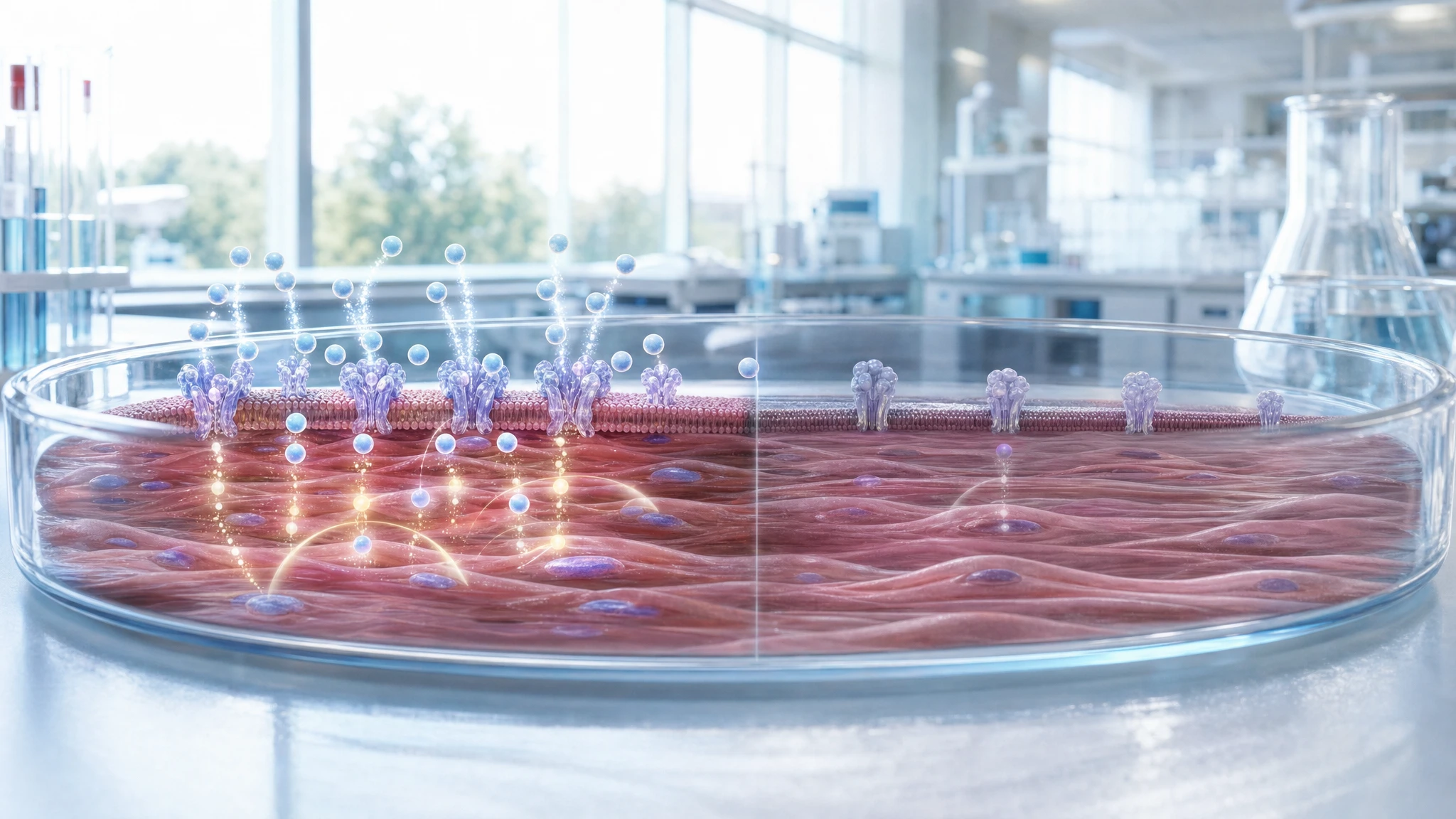
The analyzed study examined whether prolonged exposure to insulin can make human muscle cells cultured in the laboratory less responsive to the hormone. Differentiated muscle cells obtained from healthy donors were exposed to insulin for three days and then subjected to a new acute stimulation. Compared to non-pretreated cells, they showed lower insulin-induced glucose uptake and reduced activation of IRS-1 and AKT, two key nodes in insulin signaling transmission.
In section 3.5 of "The Choice to Live in Health or Illness" by Dr. A. Colonnese and R. Panzironi, the publication contributes to supporting the idea that prolonged hyperinsulinemia may directly participate in muscle desensitization, without necessarily reproducing all the inflammatory and lipid alterations of diabetes. Experimental studies in humans, human cells, and transcriptomic analyses converge on this possibility. However, the strength of the responses varies according to the cell model, duration, and insulin concentration: the framework is well-supported on a mechanistic level and moderately supported in humans, but it does not, by itself, demonstrate a universal causal sequence in people.
Analyzed study
Characterising hyperinsulinemia-induced insulin resistance in human skeletal muscle cells
DOI: 10.1530/jme-19-0169
PMID: 31990657
The research question was whether continuous exposure to high insulin concentrations could reduce the subsequent insulin response in human skeletal muscle cells. The hypothesis evaluated was therefore specific: persistent excess of the hormone could make the cell less capable of taking up glucose when it receives a new stimulation.
This question is relevant to section 3.5 of “The Choice to Live in Health or Illness” by Dr. A. Colonnese and R. Panzironi, because it concerns the muscular segment of the chain proposed in the book: prolonged exposure to insulin → reduced insulin signaling transmission → reduced glucose entry into the cell.
The study can demonstrate a direct effect of insulin in the cell model used, as the cells were compared with and without insulin pretreatment. However, it cannot establish how often this mechanism occurs in the human body, what the most relevant insulin concentration is in daily life, nor prove that it is the sole cause of insulin resistance in diabetes.
Materials and methods of the original study
This is an in vitro experimental study, meaning it was conducted on cells rather than on people undergoing treatment. The authors used human skeletal muscle cells derived from healthy donors, which were differentiated into mature muscle cells. The number of donors and the relevant inclusion and exclusion criteria are not reported in the available material, which consists only of the abstract.
The cells were cultured for three days with or without insulin at a concentration of 100 nanomolar. After this period, they received acute insulin stimulation. The measured outcomes included glucose uptake, i.e., the entry of sugar into the cells; the phosphorylation of IRS-1 at Tyr612 and AKT at Ser473, which are chemical modifications that signal the activation of steps in the insulin pathway; and the expression of genes and proteins involved in glucose transport, metabolism, inflammation, lipid metabolism, and mitochondrial function.
Detailed laboratory procedures, the number of replicates, the statistical method, significance values, confidence intervals, and complete quantitative outcomes are not reported in the available material.
Results
In simple terms, cells exposed to insulin for three days responded less effectively to a subsequent acute dose of the same hormone. Insulin-stimulated glucose uptake was reduced compared to cells not subjected to prolonged exposure. The abstract does not report the numerical magnitude of the reduction nor the P-value.
The lower functional response was accompanied by a weakening of cellular signaling. After acute stimulation, the phosphorylation of IRS-1 at Tyr612 and that of AKT at Ser473 were attenuated. IRS-1 is a protein that receives and transmits the signal from the insulin receptor; AKT is a downstream step that contributes, among other things, to promoting glucose utilization. The result therefore indicates a reduction in the response along this part of the cascade, rather than just an isolated change in a gene.
The GLUT1 transporter, a protein that can facilitate basal glucose entry, increased both as messenger RNA and as a protein. GLUT4, the transporter most involved in the muscle's insulin response, was not modified in the reported material. The messenger RNA of PDK4, a regulator of energy metabolism, and of TNF-a, a molecule associated with the inflammatory response, also decreased. No changes were observed in the lipid markers SREBP1 and CD36, nor in the mitochondrial marker UCP3.
The most relevant point for the book is therefore directional and consistent: prolonged exposure to insulin reduced insulin-induced glucose uptake and attenuated IRS-1 and AKT, without being accompanied, in the model studied, by an increase in TNF-a or changes in the selected lipid and mitochondrial markers.
Interpretation
The biological significance of the result is that a muscle cell subjected to continuous insulin stimulation may become less responsive to a new signal. To simplify, it is as if a system kept constantly switched on reduces its ability to further increase activity when it receives another request.
The segment documented by the study is: prolonged experimental hyperinsulinemia → reduced phosphorylation of IRS-1 and AKT after acute stimulation → reduced insulin-stimulated glucose uptake. This step corresponds directly to the sentence in the book regarding human muscle cells cultured in vitro.
The reduction of TNF-a does not prove that inflammation is unimportant in human insulin resistance. Instead, it indicates that, in this model and under the observed conditions, the reduced insulin response occurred without an increase in that inflammatory marker and without detected changes in certain lipid and mitochondrial genes. The interpretation that insulin alone was sufficient to induce a cellular form of insulin resistance is reasonable in the experimental model, but it does not equate to a demonstration of the entire pathophysiology of diabetes.
Independent confirming evidence
The most direct confirmation in humans comes from an experimental study on 15 healthy young people. A physiological and prolonged increase in insulin, while maintaining normal glycemia for 72–96 hours, reduced insulin sensitivity. The defect primarily affected non-oxidative glucose utilization, which largely reflects glycogen synthesis in the muscle. This finding confirms the general narrative that hyperinsulinemia can directly contribute to insulin resistance even before overt alterations in glycemia occur [1]. The limitation is the small sample size and the short duration of the experimental exposure.
A second study on 20 healthy adults investigated the same phenomenon using a euglycemic clamp—a technique that keeps glycemia stable while measuring the response to insulin—and muscle biopsies. After 24 or 72 hours of physiological hyperinsulinemia, insulin was less effective at increasing total glucose uptake, the portion destined for glycogen synthesis, and the activity of glycogen synthase, the enzyme that builds muscle glucose reserves. The reduction in the non-oxidative portion was 34% after 24 hours and 58% after 72 hours. This evidence links the systemic effect observed in humans to muscle function, but it does not directly measure IRS-1 or AKT [2].
An independent experiment on human myoblasts, which are muscle precursor cells, found a functionally consistent picture. After three weeks of high insulin, a new acute stimulation increased glucose transport only in cells cultured with low insulin. In cells exposed to high insulin, the new stimulation did not significantly increase IRS-1 phosphorylation, PI3K activity, or AKT phosphorylation. PI3K is an enzyme that links IRS-1 to AKT in the pathway that promotes glucose utilization. The study therefore confirms desensitization downstream of insulin stimulation in human cells, albeit using different durations and concentrations than those in the analyzed publication [3].
A study that combined human data, cellular experiments, and mouse models added a possible upstream step. In a transcriptomic meta-analysis of over 450 human skeletal muscle samples, higher fasting insulin was associated with lower messenger RNA expression of INSR, the insulin receptor gene. The human association does not prove causality; however, in mouse myotubes, prolonged exposure to insulin reduced the Insr gene, the receptor on the cell surface, and the total amount of receptor, attenuating acute AKT signaling. The result completes the narrative by showing a possible additional site of desensitization, but the direct causal link is derived primarily from the cells and animals [4].
Conflicting evidence
A relevant but not entirely overlapping result comes from an experiment on murine C2C12 cells. Chronic exposure to insulin during differentiation reduced acute AKT phosphorylation and insulin-stimulated glucose uptake. However, in already differentiated myotubes, the same chronic exposure did not alter AKT phosphorylation, although it did reduce the expression of certain metabolic genes, including GLUT4 and hexokinase II [5].
This evidence does not negate the result of the analyzed study, but it shows that the effect of persistent insulin depends on the cell type, maturation stage, species, and experimental conditions. In particular, a reduction in AKT signaling cannot be considered inevitable in every muscle model exposed to high insulin. No directly conflicting evidence sufficiently relevant to the human studies selected here was identified; this does not prove the absence of contrary results.
Summary table of evidence
The table distinguishes the publication that is the subject of the review from independent research and summarizes the level at which each contributes to the book's narrative.
| Reference | Study type | Population or model | Main result | Direction of evidence | Strength of evidence |
|---|---|---|---|---|---|
| Original study | In vitro cellular | Human skeletal muscle cells from healthy donors | Three days of insulin reduced glucose uptake and acute phosphorylation of IRS-1 and AKT. | direct confirmation | moderate |
| [1] | Human experimental | 15 healthy young individuals | 72–96 hours of physiological hyperinsulinemia reduced insulin sensitivity, especially glycogen synthesis. | direct confirmation | moderate |
| [2] | Human experimental | 20 healthy adults | 24–72 hours of hyperinsulinemia reduced glycogen synthase activation and non-oxidative glucose metabolism. | direct confirmation | moderate |
| [3] | Cellular | Human myoblasts cultured for three weeks | High insulin prevented the acute increase in IRS-1/PI3K/AKT signaling and glucose transport. | direct confirmation | moderate |
| [4] | Transcriptomic, cellular, and animal meta-analysis | Over 450 human muscle samples; myotubes and mice | Higher fasting insulin was associated with lower INSR; prolonged insulin reduced the receptor and attenuated AKT in experimental models. | indirect confirmation | moderate |
| [5] | Cellular | Differentiating murine C2C12 cells or myotubes | The effect on AKT was present during differentiation but not in already mature myotubes. | conflicting evidence | preliminary |
Confirmation of the evidence
Claim in the book
In section 3.5 of "The Choice to Live in Health or Illness" by Dr. A. Colonnese and R. Panzironi, the relevant statement is that human muscle cells exposed to insulin for three days show a reduced subsequent response: glucose uptake and the phosphorylation of IRS-1 and AKT decrease. The concomitant reduction of TNF-a and the absence of significant changes in selected lipid and mitochondrial markers indicate that the model does not reproduce the entire inflammatory and lipotoxic environment of diabetes.
Prolonged insulin exposure → attenuation of IRS-1 and AKT → reduced glucose uptake/utilization in muscle → insulin desensitization
The publication subject to the Evidence Review was used in the book to support the following passage: in human muscle cells, prolonged exposure to insulin can reduce the response to a new insulin stimulation, attenuating IRS-1/AKT signaling and glucose uptake, without requiring in the model an increase in TNF-a or changes in the evaluated lipid and mitochondrial markers.
The correspondence between the study and the paragraph is direct for the cellular segment of the chain. The analyzed publication observes a coordinated reduction in signaling and glucose uptake in human muscle cells after prolonged exposure to insulin. Independent studies in humans show that a physiological and sustained increase in insulin can reduce insulin-stimulated glucose utilization, with a defect concentrated primarily in muscle glycogen synthesis [1,2].
The mechanistic convergence is reinforced by human myoblasts in which high insulin attenuates the ability of a new stimulation to activate IRS-1, PI3K, and AKT and to increase glucose transport [3]. A further study suggests that prolonged exposure may also reduce insulin receptor expression: this part is supported by associations in human samples and by causal experiments in cells and mice [4].
The book's sentence regarding the lack of the entire inflammatory and lipotoxic environment must be read precisely. The original study only shows that TNF-a decreases and that some selected markers do not change in its own cellular system; it does not demonstrate that inflammation and lipotoxicity are irrelevant in diabetes. Furthermore, the absence of an effect on AKT in mature murine myotubes reminds us that desensitization depends on the experimental context [5].
Overall level of confirmation
STRONG CONFIRMATION
The central step is documented directly in the human cellular study and is supported by independent experiments in humans showing a reduction in the insulin response during sustained hyperinsulinemia. Molecular evidence regarding IRS-1 and AKT remains primarily cellular, and variability between models requires caution in generalization.
Conclusions
The analyzed study directly supports the idea that prolonged exposure to insulin can induce a reduced insulin response in human muscle cells, with lower activation of IRS-1 and AKT and decreased glucose uptake. This observation is consistent with human experiments showing reduced glucose utilization and lower glycogen synthesis following sustained physiological hyperinsulinemia.
The book's narrative is therefore well-supported regarding the concept of a possible self-amplifying loop between hyperinsulinemia and insulin resistance. The evidence is strongest for the general muscular and metabolic response, while certain molecular details and the absence of an inflammatory or lipotoxic environment remain specific to the cellular model. This does not provide a single explanation for diabetes, nor does it offer direct therapeutic guidance.
Overall assessment
The narrative is well-supported: prolonged hyperinsulinemia can reduce the insulin response in muscle, with experimental confirmation in both humans and cells. The IRS-1/AKT mechanism is confirmed primarily at the cellular level. Differences between models and exposure conditions make it necessary to be cautious when translating every detail to human pathophysiology as a whole.
References
[1] Effect of sustained physiologic hyperinsulinaemia and hyperglycaemia on insulin secretion and insulin sensitivity in man
[2] Physiological hyperinsulinemia impairs insulin-stimulated glycogen synthase activity and glycogen synthesis
[3] Continually high insulin levels impair Akt phosphorylation and glucose transport in human myoblasts
[4] Human and mouse muscle transcriptomic analyses identify insulin receptor mRNA downregulation in hyperinsulinemia‐associated insulin resistance
Editorial note
Artificial Intelligence was used solely as a tool to support editorial and documentary work. All content, interpretations, bibliographic citations and conclusions have been verified, reviewed and approved by the author, who assumes full scientific responsibility for them.
The manuscript was also subject to independent peer review prior to publication.
To ensure maximum transparency, the article's complete history — including the dates of submission, review, acceptance and publication, as well as information on the peer review process — is available on the dedicated Article History page.