


Eugenol derivatives and PPARγ: in vitro anti-inflammatory effects
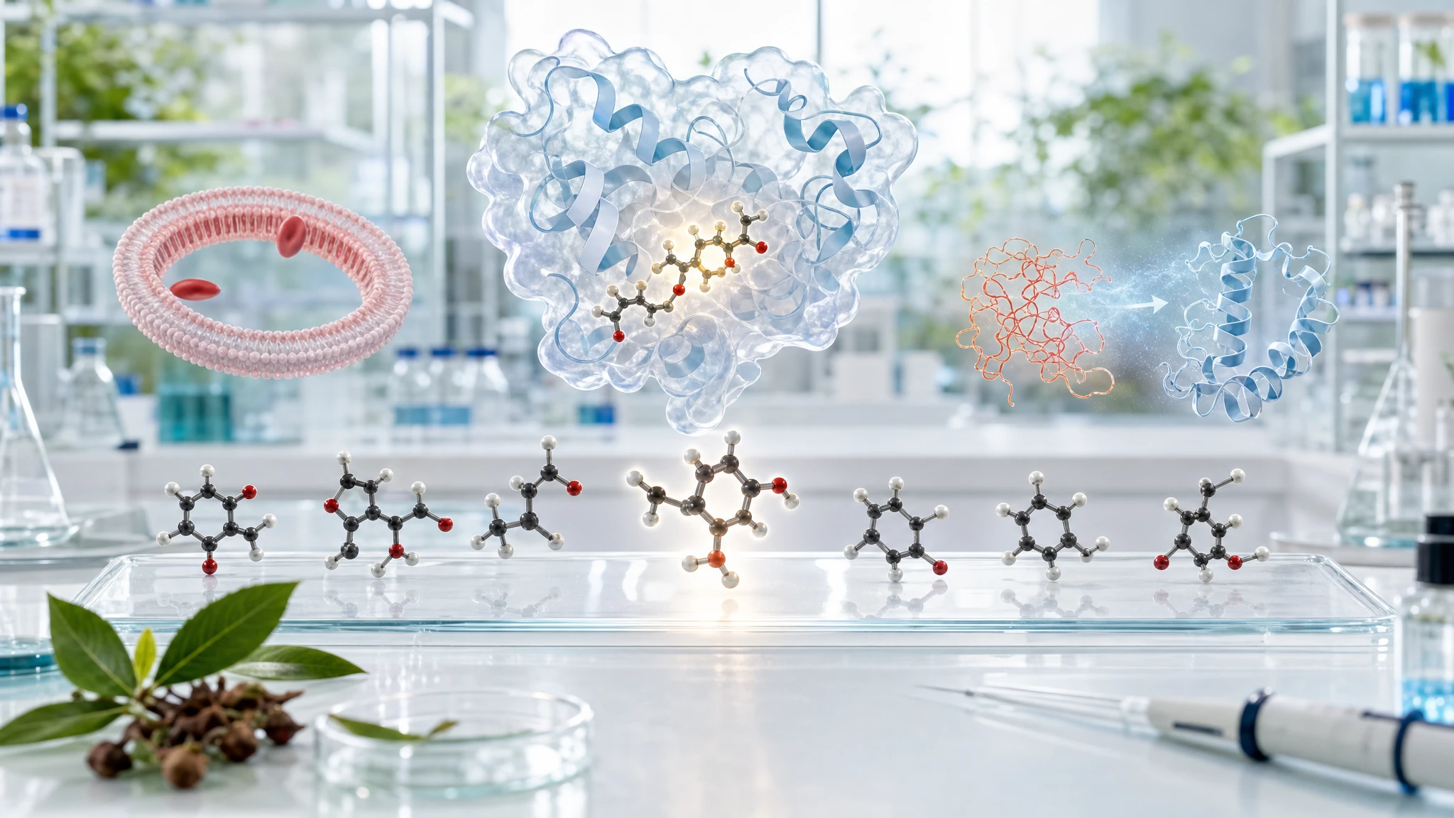
In 2023, Indian researcher Noor Fathima Anjum and colleagues studied six synthetic eugenol derivatives in the journal Molecules, designed as potential molecules capable of binding to PPARγ and exhibiting anti-inflammatory signals in laboratory tests. The work compared computer simulations, protein binding tests, and simplified biological assays, without involving human participants.
- Published 27/10/2026
- Updated 28/10/2026
- Reviewed 28/10/2026
- Article history
ABSTRACT : WHAT THE SCIENCE SAYS
A laboratory study designed six molecules derived from eugenol to verify whether they could bind to PPARγ, a cellular receptor involved in the regulation of metabolism and inflammation. The compound designated as 1f achieved the most favorable results in the series, but it has not been tested in humans.
The authors' summary describes medicinal chemistry research, which is oriented toward creating and selecting new candidate molecules. Anjum and colleagues modified the structure of eugenol, an aromatic compound also found in cinnamon but particularly abundant in cloves, obtaining six derivatives named 1a–1f. They then verified their ability to compete for binding with PPARγ (a receptor activated by molecules that can modify the activity of certain genes) using TR-FRET, a fluorescent technique that detects whether a molecule displaces a tracer substance from the protein's binding site. Derivative 1f showed the lowest value among the six molecules, with an IC50 of 5.15 µM; a lower IC50 indicates that less of it is needed to produce the signal in the test. However, the value remained higher than that of pioglitazone, the reference drug, which was 1.77 µM. In cell-free tests, 1f reduced heat-induced albumin denaturation and protected human red blood cell membranes from lysis. These outcomes suggest experimental activities consistent with further studies, but they do not demonstrate a therapeutic action, nor do they confirm that PPARγ is effectively activated in cells or in the organism.
What this research analyzed
The main focus was to understand whether minor chemical modifications of eugenol could generate molecules with characteristics useful for pharmacological studies. The authors therefore synthesized six new compounds and confirmed their structure using spectroscopy, which are methods that identify the atoms and bonds present in a molecule. First, they used molecular docking, a simulation that evaluates whether the shape of a molecule can fit into a specific area of a protein. Then, they performed molecular dynamics simulations to observe the predicted stability of the complexes over time. These analyses generate hypotheses; they do not prove that the binding occurs in a living organism.
The primary experimental verification was the competitive LanthaScreen TR-FRET assay. In simple terms, the method measures whether the compound under investigation is able to displace a fluorescent tracer from the binding domain of PPARγ. The latter is a nuclear receptor, which is a protein that, after interacting with specific molecules, can help regulate gene expression. The authors also applied two cell-free assays: heat-induced albumin denaturation and the stabilization of human red blood cell membranes. These are useful models for initial screening, but they do not reproduce the inflammation of a tissue or a disease.
Research data
Novel Derivatives of Eugenol as a New Class of PPARγ Agonists in Treating Inflammation: Design, Synthesis, SAR Analysis and In Vitro Anti-Inflammatory Activity
- Six eugenol derivatives, designated 1a–1f, were synthesized with reported yields between 70% and 87%.
- In the TR-FRET competitive assay on PPARγ, 1f showed an IC50 of 5.15 µM and 1d of 6.47 µM; pioglitazone showed 1.77 µM.
- In the erythrocyte membrane stabilization assay, 1f achieved 85.79% protection at 400 µM, compared to 90.77% for diclofenac.
What results emerge from the study
Among the six molecules, 1f emerged as the most consistent in the tests used. In the PPARγ binding assay, its IC50 was 5.15 µM, while for 1d it was 6.47 µM. IC50 refers to the concentration capable of reducing the measured signal by 50%: in this case, a lower value suggests a greater competitive capacity in the analytical system. Pioglitazone, used as a reference, however, showed an IC50 of 1.77 µM. Therefore, 1f bound in the test, but did not outperform the reference drug.
In assays related to inflammation, 1f achieved an IC50 of 67.02 µM in protecting albumin from denaturation and 54.97 µM in stabilizing the erythrocyte membrane. At 400 µM, it protected 85.79% of membranes from lysis, compared to 90.77% for diclofenac. This data indicates that the compound limited artificially induced alterations in a test tube. However, this is not equivalent to demonstrating that it reduces cytokines, pain, tissue damage, or symptoms in people with an inflammatory condition. The next step requires appropriate cellular and animal models.
Why this study is relevant
The main value of the study is methodological: it shows how a natural molecule can become a starting point for creating chemical analogs and selecting the most promising ones. Eugenol, found in cinnamon, is a different molecule from 1f and the other derivatives studied. Specifically, 1f contains structural modifications designed in the laboratory; it is therefore neither a component identified in the spice nor a product equivalent to dietary eugenol.
The research is also useful because it separates three levels that are often confused: computer prediction, binding in a biochemical test, and response in a simplified laboratory model. The proposed biological pathway is PPARγ → reduction of signals regulated by NF-kB (a cellular factor that promotes the production of inflammation mediators) → reduced inflammatory response. However, in the study by Anjum and colleagues, NF-kB, cytokines, or the transcriptional activity of PPARγ were not directly measured. The mechanism therefore remains plausible, but incomplete.
Study limitations and what it does NOT demonstrate
This research is not a clinical trial and does not include animals, patients, oral dosages, real absorption data, or safety assessments in the body. ADMET predictions—that is, absorption, distribution, metabolism, elimination, and toxicity—are computer-generated simulations. Although useful for deciding which molecules to investigate further, they do not replace experimental pharmacokinetic and toxicological studies.
Furthermore, the TR-FRET indicates competition for the binding site, but it does not, on its own, prove that 1f is a functional PPARγ agonist, meaning it does not prove that it activates the receptor to produce a measurable biological response in cells. To demonstrate this, for example, gene transcription tests, comparisons with selective antagonists, and measurements of inflammatory mediators would be required. Assays involving albumin and erythrocyte membranes are indirect: they do not allow for the conclusion that the compound treats clinical inflammation. Finally, no results allow for the attribution of the effects observed for synthetic derivatives to cinnamon, its powder, or its normal dietary consumption.
Independent confirmatory evidence
The first confirmation concerns the possibility that some structures related to eugenol actually interact with PPARγ. Fakhrudin and colleagues studied dieugenol and tetrahydrodieugenol, two neolignans structurally linked to eugenol, using binding assays and cellular tests. In cell models, the compounds selectively activated PPARγ, without activating PPARα or PPARβ/δ, and acted as partial agonists. They also induced the differentiation of 3T3-L1 preadipocytes in a PPARγ-dependent manner. This supports the derived molecule → receptor pathway, with moderate-level experimental confirmation; the limitation is that it does not evaluate inflammation and does not study the 1a–1f compounds from Anjum [1].
A second piece of the puzzle concerns the cellular inflammatory response. Ghasemi Gojani and colleagues treated human THP-1 macrophages stimulated with lipopolysaccharide and ATP using eugenol at 5, 10, and 15 µM. In three biological replicates, the higher concentrations reduced certain signals such as interleukin-6, cyclooxygenase-2, and NF-kB phosphorylation, in addition to modifying components of the NLRP3 inflammasome. The confirmation is indirect compared to the original work because it concerns unmodified eugenol and does not measure PPARγ, but it supports the NF-kB → inflammatory mediators pathway in macrophages [2].
The PPARγ → reduced inflammation chain was explored more directly by Huang and colleagues in a mouse model of chemically induced colitis. Eugenol showed binding to PPARγ in a surface plasmon resonance test; the benefits observed in the model were attenuated by the antagonist GW9662. The work also included cellular tests and measurements of NF-kB and Nrf2, a regulator of the response to oxidative stress. This is a mechanistic confirmation closer to Anjum's proposal, but it remains limited to mice and non-derivatized eugenol; it does not demonstrate clinical efficacy [3].
Conflicting evidence
The responses to eugenol analogues are not uniform. Li and colleagues compared eugenol and isoeugenol in RAW264.7 murine macrophages stimulated with lipopolysaccharide. Both reduced nitric oxide production and the expression of iNOS and COX-2, but isoeugenol was found to be more effective than eugenol. This finding does not refute the activity observed by Anjum and colleagues, but it indicates that a small structural change can substantially alter potency and that the mechanism may involve mediators other than PPARγ. The contradictory confirmation is indirect and limited to murine cells [4].
Further caution comes from Gu and colleagues, who studied eugenol in 3T3-L1 preadipocytes and in mice fed a high-fat diet. In their system, eugenol increased ATF3, a regulator of gene expression, and reduced adipocyte differentiation, lipid accumulation, and PPARγ protein levels. The result differs from the PPARγ activation hypothesis, but it concerns a different tissue, a different biological purpose, and the unmodified compound. It therefore shows a dependence on the cellular context, not a direct refutation of the binding observed for 1f [5].
No directly discordant evidence sufficiently relevant to the six specific derivatives of Anjum has been identified; this does not prove the absence of contrary results.
Comparative overview
The following publications help place the work of Anjum and colleagues along the pathway from molecular binding to biological response, while also highlighting the differences between models.
| Publication | Model and main result | Relation to Anjum's study | Cautious interpretation |
|---|---|---|---|
| Fakhrudin et al., 2010 [1] | Biochemical tests and 3T3-L1 cells; dieugenol and tetrahydrodieugenol acted as partial PPARγ agonists. | Confirms the possible link between eugenol analogs and PPARγ. | Moderate mechanistic evidence; does not study inflammation or compounds 1a–1f. |
| Ghasemi Gojani et al., 2023 [2] | Human THP-1 macrophages; eugenol reduced various pro-inflammatory signals, with concentration-dependent responses. | Confirms the cellular aspect of NF-kB and inflammatory mediators. | Indirect evidence; PPARγ was not measured and the model does not represent a disease. |
| Huang et al., 2025 [3] | Cells and mice with experimental colitis; blocking PPARγ attenuated the effects of eugenol. | Supports the eugenol → PPARγ → inflammatory signals chain. | Relatively strong preclinical confirmation, but not automatically transferable to humans. |
| Li et al., 2006 [4] | Murine RAW264.7 macrophages; isoeugenol more effective than eugenol in reducing nitric oxide and COX-2. | Shows that chemical structure strongly influences anti-inflammatory activity. | Indirect discordant data; does not evaluate PPARγ or the derivatives studied in the main paper. |
| Gu et al., 2025 [5] | 3T3-L1 preadipocytes and mice; eugenol reduced PPARγ expression during adipogenesis. | Indicates that effects on PPARγ may depend on the tissue and biological context. | Contextual discrepancy; does not measure direct binding or inflammation. |
Editorial conclusion
References
[1] Computer-Aided Discovery, Validation, and Mechanistic Characterization of Novel Neolignan Activators of Peroxisome Proliferator-Activated Receptor γ
[2] Anti-Inflammatory Properties of Eugenol in Lipopolysaccharide-Induced Macrophages and Its Role in Preventing b-Cell Dedifferentiation and Loss Induced by High Glucose-High Lipid Conditions
[3] Eugenol Exerted Significant Anti-Oxidant and Anti-Inflammatory Effects on Experimental Colitis via Directly Acting on PPAR-γ
[4] Inhibitory action of eugenol compounds on the production of nitric oxide in RAW264.7 macrophages
[5] Eugenol inhibits preadipocyte differentiation and lipid accumulation via upregulating ATF3
Editorial note
Artificial Intelligence was used solely as a tool to support editorial and documentary work. All content, interpretations, bibliographic citations and conclusions have been verified, reviewed and approved by the author, who assumes full scientific responsibility for them.
The manuscript was also subject to independent peer review prior to publication.
To ensure maximum transparency, the article's complete history — including the dates of submission, review, acceptance and publication, as well as information on the peer review process — is available on the dedicated Article History page.