
Scientific Book

Vitamin C: between tissue integrity and cellular response

Vitamin C is an essential micronutrient involved in collagen synthesis, antioxidant protection, and the normal functioning of the immune system. Through its role in cellular reactions, it contributes to defense against oxidative stress and the maintenance of tissue integrity. In the book "The Choice to Live in Health or Disease," Dr. A. Colonnese and R. Panzironi explore the properties of ascorbic acid, its relationship with inflammation, and the scientific evidence regarding dietary intake and supplementation.

Cite this article
Colonnese, A. & Panzironi, R. (2026). Vitamin C: between tissue integrity and cellular response. The Choice to Live in Health or in Disease. https://lifesciencehub.info/en/la-scelta-di-vivere-in-salute-o-in-malattia/vitamin-c-between-tissue-integrity-and-cellular-response
The Choice to Live in Health or in Disease · ISBN 979-12-82692-03-8

DOI lsh.en.libro.26

  • Published 05/11/2026
  • Updated 05/11/2026
  • VITAMIN C
  • OXIDATIVE STRESS
  • IMMUNOMODULATION
  • COLLAGEN
  • HIF-1Α
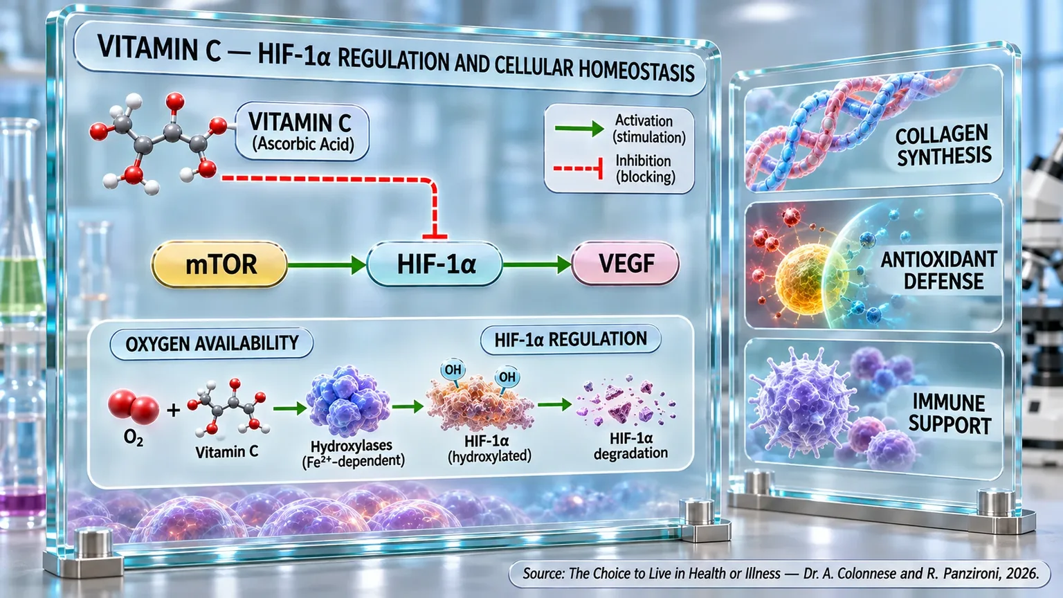
Laboratory infographic with molecules, activation and inhibition arrows, the mTOR–HIF-1α–VEGF pathway, and panels on collagen, antioxidants, and immunity.
The diagram links vitamin C to the regulation of HIF-1α: with oxygen and Fe²⁺-dependent hydroxylases, it promotes its hydroxylation and degradation, while indicating the mTOR–HIF-1α–VEGF axis. On the side, it summarizes the main areas of the chapter: collagen synthesis, antioxidant defense, and immune support.

ABSTRACT

Vitamin C is an essential micronutrient that humans must obtain through their diet. In the book "La scelta di Vivere in Salute o in Malattia" (The Choice to Live in Health or Disease), Dr. A. Colonnese and R. Panzironi explore its antioxidant, immune, and structural functions, along with its involvement in epigenetic regulation and cellular responses to oxygen. Ascorbate also contributes to iron absorption, neuronal communication, vascular function, and the synthesis of molecules necessary for the physiological functioning of tissues.

Vitamin C supports the integrity of skin and mucosal barriers, the migration and phagocytosis of neutrophils, and the resolution of inflammation; effects on Natural Killer cells and T-lymphocyte maturation are also described. As an enzymatic cofactor, it promotes the hydroxylation required for the stability of the collagen triple helix, which is fundamental for connective tissues and vascular walls. Experimental studies document its involvement in collagen deposition and tissue repair, with relevance for skin, cartilage, tendons, ligaments, and bones.

A further mechanism involves HIF-1α, a regulator of responses to hypoxia. In the presence of oxygen, ascorbate supports the hydroxylases that promote its degradation. Cellular and animal studies, as well as tumor observations, link higher vitamin availability to lower activity of HIF-1α and targets such as VEGF and GLUT1. This pathway participates in angiogenesis, glucose metabolism, lactate production, and immunosuppressive modifications of the tumor microenvironment. High concentrations achieved intravenously also show experimental effects on tumor oxidative balance, without automatically demonstrating clinical efficacy.

Observational associations between vitamin C availability and lower cardiovascular, oncological, or cognitive risk do not establish causality. In the context of infections, supplementation shows limited benefits, mainly regarding the duration of the common cold and under conditions of intense physical stress, without generally preventing it. Overall, an adequate intake supports interdependent biological functions; any therapeutic applications require specific clinical confirmation and a rigorous distinction between experimental results and documented benefits in humans.

Its properties

Vitamin C, also known as ascorbic acid, is one of the most important molecules for the survival and proper functioning of the human body. Unlike most mammals, humans have lost the ability to synthesize it independently over the course of evolution and therefore depend entirely on diet to ensure an adequate intake. For a long time, its reputation was linked almost exclusively to the prevention of scurvy, a disease characterized by tissue fragility, bleeding, and impaired wound healing. Today, however, scientific research has shown that this vitamin performs much broader and more profound functions, participating in an extraordinary variety of biological processes essential for maintaining health (Alberts et al., 2025) [1].

To understand the value of vitamin C, one can imagine the body as a large, bustling city. Every second, billions of chemical reactions produce energy, build new cellular structures, and allow different organs to communicate with each other. This incessant work inevitably generates "biological sparks," represented by reactive oxygen species, commonly known as free radicals. In controlled amounts, these molecules perform useful physiological functions, but when they accumulate beyond a certain threshold, they can damage proteins, cell membranes, lipids, and genetic material. In this context, vitamin C acts as one of the body's primary defense systems, neutralizing these reactive molecules before they can cause significant damage (Meščić Macan et al., 2019) [2].

Its antioxidant activity is particularly important because it occurs in the aqueous environments of the body, such as the blood, intracellular fluid, and extracellular spaces. Here, vitamin C donates electrons to oxidizing species, transforming them into less reactive compounds and interrupting chain reactions that could compromise cellular integrity. This mechanism does not merely represent a form of passive protection, but actively contributes to maintaining redox homeostasis, one of the fundamental parameters for cellular survival (Meščić Macan et al., 2019) [2].

Over the years, it has emerged that vitamin C does not only play the role of a "free radical scavenger." It actually participates as an enzymatic cofactor in numerous essential biochemical reactions. Among these, the synthesis of collagen stands out—the protein that forms the scaffolding of skin, tendons, ligaments, bones, and vascular walls. Without adequate amounts of vitamin C, collagen fibers lose stability and strength, which explains why its deficiency manifests as tissue fragility and bleeding. At the same time, vitamin C participates in the production of catecholamines (substances produced by the nervous system and adrenal glands, such as adrenaline, noradrenaline, and dopamine), peptide hormones, and numerous other molecules involved in the regulation of physiological functions (Alberts et al., 2025) [1].

Its properties also extend to iron metabolism. Vitamin C promotes the intestinal absorption of non-heme iron by transforming it into a more easily assimilable form. This effect helps support hemoglobin production and oxygen transport to tissues, demonstrating how a single vitamin can simultaneously influence structural, metabolic, and energetic processes (Alberts et al., 2025) [1].

In recent years, an even more sophisticated view of its biological functions has emerged. In addition to acting as an antioxidant and metabolic cofactor, vitamin C participates in the regulation of gene expression through epigenetic mechanisms (processes that regulate gene activity by activating or deactivating them without altering the DNA sequence). Specifically, it acts as a cofactor for the TET family of enzymes, which are involved in DNA demethylation processes. In other words, it indirectly helps regulate which genes can be activated or silenced within cells, influencing processes ranging from cell differentiation to the response to environmental stimuli (Meščić Macan et al., 2019) [2].

The central nervous system also depends significantly on the presence of vitamin C. The brain contains concentrations of ascorbic acid higher than those of many other tissues in the body, a fact that reflects its biological importance. During neuronal activity, vitamin C is continuously released, used, and recycled through a complex collaboration between neurons and glial cells. In this highly metabolic environment, it not only protects nerve cells from oxidative stress but also contributes to the regulation of synaptic transmission and neuronal energy metabolism (Covarrubias-Pinto et al., 2015) [3].

The versatility of vitamin C is also clearly evident at the cardiovascular level. The most recent evidence shows that this molecule helps preserve the functionality of the endothelium, the thin inner lining of blood vessels, by promoting the bioavailability of nitric oxide and supporting normal vasodilation processes. At the same time, it modulates biological pathways involved in the inflammatory response and protection against oxidative stress, helping to maintain the integrity of the vascular wall and the extracellular matrix (Xu et al., 2025) [4].

A particularly interesting aspect concerns the ability of vitamin C to collaborate with other bioactive compounds present in plant-based foods. Bioflavonoids, for example, seem to establish a relationship of mutual enhancement with it. Recent studies have shown that the combination of vitamin C and flavonoids can amplify antioxidant, anti-inflammatory, and antithrombotic properties compared to the use of individual isolated components. This phenomenon suggests that many of the effects observed in foods rich in vitamin C may depend not only on the vitamin itself but also on the complex network of interactions it establishes with other natural molecules present in the food matrix (Chrysikopoulou et al., 2025) [5].

The image that emerges from modern literature is therefore very different from the traditional one. Vitamin C is not simply a substance useful for preventing a deficiency disease, but a multifunctional molecule involved in cellular protection, tissue synthesis, metabolic regulation, neuronal communication, cardiovascular health, and even the control of gene expression. Its activity spans virtually every level of biological organization, from individual enzymatic reactions to the integrated functioning of entire organs and systems.

Supplementation

If in the previous paragraph we saw how vitamin C participates in an extraordinary variety of biological processes, a question naturally arises: do these mechanisms actually translate into observable benefits for human health when intake is increased through supplementation? Over the last few decades, this question has stimulated thousands of epidemiological, clinical, and experimental studies, generating a body of data that has progressively expanded our understanding of the role of vitamin C well beyond the simple prevention of nutritional deficiencies.

One of the most studied areas concerns cancer prevention. By analyzing dozens of meta-analyses and systematic reviews, an inverse relationship has emerged between vitamin C intake and the risk of developing numerous forms of cancer. The most consistent associations have been observed for breast, stomach, esophageal, prostate, pancreatic, bladder, kidney, and lung cancer, as well as for certain gynecological neoplasms. Furthermore, in subjects already affected by breast cancer, higher vitamin C intake has been associated with a reduced risk of recurrence, cancer-specific mortality, and overall mortality (Chen et al., 2022) [6].

These observations do not seem to depend solely on its ability to neutralize free radicals. Cancer cells, in fact, live in a metabolic condition that is profoundly different from that of healthy cells. At low concentrations, vitamin C acts primarily as an antioxidant, but when it reaches high concentrations, it can take on pro-oxidant characteristics, increasing the production of reactive oxygen species within neoplastic cells. Since many cancer cells already possess a precarious redox balance, this additional oxidative load can exceed their capacity for adaptation and promote cell death. Alongside its effects on oxidative stress, vitamin C also appears to influence some of the mechanisms that regulate the expression of genes involved in tumor transformation. By acting as a cofactor for TET proteins, it contributes to DNA demethylation processes and the maintenance of genomic stability. In parallel, it can modulate the activity of HIF-1α, one of the main regulators of cellular adaptation to hypoxia, a process closely involved in tumor growth, angiogenesis, and resistance to therapies (Chen et al., 2022) [6].

Interest in vitamin C in oncology has further expanded with the development of high-dose intravenous formulations. The most recent evidence suggests that vitamin C does not act only on the tumor cell, but can influence the entire microenvironment that surrounds it. This biological ecosystem includes immune cells, fibroblasts, blood vessels, the extracellular matrix, and numerous molecular mediators that contribute to disease progression. In this context, vitamin C appears to exert immunomodulatory, metabolic, and epigenetic effects capable of modifying the conditions that favor tumor growth (Li et al., 2022) [7].

Particularly interesting are the observations regarding the use of vitamin C in combination with modern oncological immunotherapies. Experimental studies have shown that high concentrations of vitamin C can increase the activity of immune cells involved in recognizing and eliminating tumor cells, enhancing the efficacy of immune checkpoint inhibitors and reducing the probability of recurrence in experimental models. The combination with epigenetic drugs, chemotherapeutic agents, and targeted therapies has also shown promising results, suggesting that vitamin C may act as an amplifier of therapeutic efficacy in specific clinical contexts (Li et al., 2022) [7].

However, the possible associations between supplementation and health are not limited to the oncological field. The cardiovascular system also appears to benefit from adequate levels of vitamin C. In a large prospective cohort of over eighty thousand women followed for sixteen years, total vitamin C intake was associated with a significant reduction in the risk of coronary heart disease. A particularly interesting aspect is that this association was more evident when vitamin C came from supplements rather than from diet alone, suggesting that dosages higher than those normally obtainable through diet may exert additional biological effects (Osganian et al., 2003) [8].

Another important observation comes from studies conducted in elderly populations. In a cohort followed for twenty years, higher plasma concentrations of vitamin C and greater dietary intake were associated with a reduction in stroke mortality. This relationship was observed both using dietary data and by directly measuring blood concentrations of ascorbic acid, reinforcing the biological plausibility of the observed link (Gale et al., 1995) [9].

The brain also appears to be sensitive to the nutritional status of vitamin C. In a population of elderly people living in rural communities, vitamin C supplementation was associated with better cognitive performance in several domains, including immediate memory, language, attention, visuospatial skills, and global cognitive function (Chaudhari et al., 2016) [10].

Taken together, this evidence outlines an extremely interesting picture. Vitamin C supplementation does not appear to be associated with a single isolated benefit, but with a plurality of effects involving different biological systems: cardiovascular protection, neurological function, immune response, and control of tumor growth. The dosage used, the method of administration, the initial nutritional status, genetic characteristics, and the clinical context represent determining variables that can profoundly influence the biological response to supplementation. Vitamin C therefore emerges not as a universal solution, but as a complex biological tool, whose potential seems to depend on the ability to use it in the right context, at the appropriate dose, and in the most suitable subject.

Vitamin C and the immune system

The immune system can be compared to a complex defense network composed of sentinels, mobile patrols, command centers, and specialized units. Every day, this structure is called upon to recognize and neutralize billions of potentially dangerous microorganisms, eliminate damaged cells, and maintain the body's balance. For this biological machine to function efficiently, all its components must receive adequate metabolic support. Among the micronutrients involved in this process, vitamin C occupies a particularly relevant position, so much so that it is now considered one of the main nutritional regulators of immune function (Carr and Maggini, 2017) [11].

For many years, vitamin C was associated almost exclusively with its antioxidant activity. Although this function is indeed fundamental, current knowledge reveals a much more complex reality. Vitamin C actually participates directly in the development, maturation, activation, and regulation of numerous immune cells, influencing both innate immunity, which represents the first line of defense against pathogens, and adaptive immunity, which is responsible for more specific and sophisticated responses (Carr and Maggini, 2017) [11].

The body's first protective barrier consists of the skin and mucous membranes. These structures are not merely a passive covering, but true defensive systems that prevent the entry of bacteria, viruses, and other harmful substances. Vitamin C contributes to maintaining the integrity of these barriers through its involvement in collagen synthesis and the protection of cells from oxidative stress. In this way, it strengthens the body's outer walls even before the immune system is called upon to intervene (Carr and Maggini, 2017) [11].

When a pathogen manages to overcome these initial defenses, the cells of the innate immune system go into action. Among these, neutrophils represent one of the most abundant and important populations. Curiously, these cells accumulate concentrations of vitamin C much higher than those present in blood plasma. This accumulation is not accidental: vitamin C improves the ability of neutrophils to migrate rapidly to the site of infection, a process known as chemotaxis, increases their efficiency in phagocytosis (the process by which certain immune system cells engulf and destroy microbes), and supports the controlled production of reactive oxygen species necessary to destroy invading microorganisms (Carr and Maggini, 2017) [11].

However, the function of vitamin C does not end with the offensive phase of the immune response. After completing their task, neutrophils must be eliminated in an orderly manner to avoid collateral damage to surrounding tissues. Vitamin C facilitates this process by promoting the programmed apoptosis of exhausted immune cells and favoring their subsequent removal by macrophages. In this way, it helps to turn off inflammation when it is no longer needed, preventing a useful defensive response from turning into a source of damage to the body itself (Carr and Maggini, 2017) [11].

Another central element of immune surveillance is represented by Natural Killer cells, often abbreviated as NK cells. These cells act as specialized patrols tasked with identifying and destroying cells infected by viruses or cells that have undergone tumor transformations. A study conducted on healthy volunteers showed that the administration of ascorbic acid is able to significantly increase the cytotoxic capacity of NK cells. The particularly interesting aspect is that this effect did not stem from an increase in the number of NK cells, but from an improvement in their functionality. In other words, vitamin C seemed to make these cells more efficient at performing their job, enhancing immune surveillance without altering the cellular composition of the immune system (Vojdani and Ghoneum, 1993) [12].

The observations on NK cells were subsequently confirmed by numerous scientific reviews that highlighted how vitamin C can influence both their proliferation and functional activity. Although some experimental results show differences related to the concentrations used and the study models employed, the overall picture suggests that adequate levels of vitamin C are necessary to ensure optimal function of these important immune cells (van Gorkom et al., 2018) [13].

Vitamin C also exerts relevant effects on adaptive immunity, the system that allows the body to develop highly specific responses and create immunological memory. In this context, T lymphocytes represent one of the most studied components. The available evidence indicates that the development, maturation, and proliferation of these cells depend on the presence of adequate intracellular concentrations of vitamin C. This effect takes on particular importance in subjects undergoing treatments that compromise the immune system, such as certain oncological therapies, in which the recovery of the lymphocyte population can be slow and incomplete (van Gorkom et al., 2018) [13].

Current knowledge also suggests that vitamin C participates in the regulation of gene expression in immune cells through epigenetic mechanisms. By acting as a cofactor for enzymes involved in DNA demethylation, it helps to modulate the activation of genes that regulate the differentiation and function of lymphocytes. This means that vitamin C does not just provide energy or antioxidant protection to immune cells, but also intervenes in the biological programs that determine their behavior (van Gorkom et al., 2018) [13].

The importance of vitamin C emerges even more clearly during infections. Numerous studies have observed that plasma and leukocyte concentrations of vitamin C decrease rapidly in the presence of acute infections and inflammatory states. This phenomenon suggests that the immune system consumes large amounts of vitamin C precisely at the moments when it is most engaged in fighting pathogens. In other words, vitamin C does not simply represent a passive reserve, but a true metabolic fuel used during the immune response (Wintergerst et al., 2006) [14].

The clinical consequences of this phenomenon have been observed in numerous studies. An adequate intake of vitamin C has been associated with a reduction in the incidence, severity, and duration of respiratory infections, especially in more vulnerable populations, such as the elderly and children. In particular, vitamin C supplementation has shown favorable effects on the course of the common cold and other respiratory infections, helping to reduce their clinical impact (Wintergerst et al., 2006) [14].

However, more recent reviews have highlighted an important aspect. In the general population, regular vitamin C supplementation does not appear to significantly reduce the risk of developing a common cold. However, in subjects subjected to intense physical stress, such as endurance athletes, military personnel, and people exposed to extreme environmental conditions, the risk of developing respiratory infections is significantly lower in those who take vitamin C. Furthermore, when an infection occurs, high dosages seem to help reduce the severity of symptoms and the duration of the illness; another element of interest concerns pneumonia. Although the available evidence is still limited and does not allow for definitive conclusions, some studies have reported favorable preventive and therapeutic effects of vitamin C against this pathology, suggesting the need for further clinical investigation (Hemilä and Chalker, 2025) [15].

Considering the available evidence as a whole, a vision emerges that is very different from the traditional one. Vitamin C does not appear simply as an antioxidant that protects the body from free radicals, but as a multifunctional regulator of the immune response. It contributes to the physical defense of epithelial barriers, supports neutrophil function, improves the activity of Natural Killer cells, promotes the proliferation of T lymphocytes, and participates in the genetic regulation of immune responses. This network of coordinated actions helps explain why a vitamin C deficiency is frequently associated with greater susceptibility to infections and why maintaining adequate levels represents one of the fundamental elements for preserving the efficiency of the immune system throughout life.

Vitamin C and collagen

If there is one biological function that identifies vitamin C in human physiology more than any other, it is certainly its participation in collagen synthesis. Although vitamin C is studied today for its effects on the immune system, cellular metabolism, cardiovascular health, and even epigenetic regulation, its scientific discovery is historically linked precisely to the understanding of its role in the formation of connective tissue. The clinical manifestations of scurvy—gum bleeding, vascular fragility, tooth loss, and difficulty in wound healing—are, in fact, the direct consequence of a defect in collagen production (Pullar et al., 2017) [16].

To understand the importance of this process, it is useful to remember that collagen is the most abundant protein in the entire human body. It forms the structural scaffolding of skin, tendons, ligaments, cartilage, bones, vascular walls, and numerous other tissues. If one were to imagine the human body as a building, collagen would represent the load-bearing pillars, the support beams, and the reinforcement network that holds the various structures together simultaneously. Without properly synthesized and organized collagen, the mechanical strength of tissues would be progressively compromised (Chugaeva et al., 2023) [17].

Collagen production is an extremely sophisticated process that requires a precise sequence of biochemical events. Among these, one of the most important steps is the transformation of the amino acid proline into hydroxyproline. This modification may appear insignificant at first glance, but it actually represents one of the fundamental mechanisms that allow the collagen molecule to acquire its structural stability. Hydroxyproline acts as a sort of molecular reinforcement that stabilizes the characteristic triple-helix structure of collagen, making it resistant to daily mechanical stress. Vitamin C plays an essential role in this very phase. It acts as a cofactor for prolyl hydroxylase, the enzyme responsible for converting proline into hydroxyproline. In the absence of vitamin C, this reaction cannot be completed correctly, and the formation of the collagen triple helix is compromised. Recent molecular simulation studies have allowed this phenomenon to be observed at a level of detail never reached before. Computational analyses show that vitamin C does not merely participate passively in the reaction but actively contributes to correctly orienting the amino acids involved, favoring the formation of the most structurally stable collagen sequence and reducing the energy required to complete the synthesis process (Chugaeva et al., 2023) [17].

The importance of this mechanism emerges clearly in tissue healing and repair processes. After a muscle, tendon, or ligament injury, the body activates a complex cascade of biological events aimed at rebuilding the damaged structures. In this phase, the synthesis of new collagen represents one of the central events of tissue repair. A systematic review of the available evidence has shown that vitamin C supplementation is associated, in experimental models, with an increase in the deposition of type I collagen, the main form of collagen present in tendons and ligaments. Animal studies have also highlighted an acceleration of bone healing processes and a reduction in the oxidative stress that accompanies tissue damage (DePhillipo et al., 2018) [18].

Particularly interesting is the fact that these benefits seem to manifest especially in the initial phases of healing. During the first few weeks following an injury, the damaged tissue undergoes a period characterized by intense inflammation, high production of free radicals, and massive synthesis of new collagen fibers. In this context, vitamin C seems to provide two biological advantages simultaneously: on one hand, it directly supports collagen synthesis; on the other, it limits the oxidative damage that could interfere with repair processes (DePhillipo et al., 2018) [18].

The influence of vitamin C on collagen production does not only concern tendons and ligaments but also extends to articular cartilage. The latter is composed of a complex extracellular matrix in which type II collagen represents one of the main components. Studies conducted on articular cartilage explants have shown that biologically active forms of vitamin C significantly increase the gene expression of type II collagen and stimulate its subsequent protein synthesis. In parallel, an increase in the production of aggrecan, another molecule fundamental for ensuring elasticity and resistance to cartilage, has been observed (Clark et al., 2002) [19].

These results suggest that vitamin C does not act exclusively as an enzymatic cofactor but can directly influence the genetic activity of chondrocytes, the cells responsible for maintaining cartilage. In other words, vitamin C seems to intervene both in the construction of collagen fibers and in the regulation of the cellular programs that control their production (Clark et al., 2002) [19].

The skin also represents a particularly evident example of the importance of vitamin C in collagen synthesis. The dermis, the deep layer of the skin, consists mainly of collagen fibers that provide elasticity, strength, and mechanical recovery capacity. It is therefore not surprising that healthy skin has particularly high concentrations of vitamin C compared to other tissues in the body; as the years pass, collagen production tends to physiologically decrease, contributing to the appearance of wrinkles, loss of elasticity, and structural changes in the skin. The available evidence suggests that an adequate nutritional status of vitamin C can help support dermal collagen synthesis and slow down some of the processes associated with skin aging. However, the benefits seem to be particularly evident in subjects who have suboptimal levels of vitamin C or increased metabolic needs, such as smokers (Pullar et al., 2017) [16].

The relationship between vitamin C and collagen emerges with even greater clarity during wound healing. Following a skin injury, fibroblasts migrate to the damaged area and begin to produce large amounts of collagen to replace the initial clot with a new, stable extracellular matrix. This process requires high amounts of vitamin C, so much so that slowed healing represents one of the first clinical signs of a deficiency. During tissue repair, local consumption of vitamin C increases significantly, reflecting the intense biosynthetic activity necessary for the reconstruction of damaged tissue (Pullar et al., 2017) [16].

Considering the available evidence as a whole, it emerges how the relationship between vitamin C and collagen represents one of the best examples of how a micronutrient can directly influence the physical architecture of the body. From the molecular stabilization of the triple helix to wound healing, from joint health to tendon strength, from skin quality to bone repair, vitamin C accompanies every phase of the construction and maintenance of connective tissue. It is not simply an accessory metabolic support, but one of the indispensable elements for the body to literally build and maintain its own structure.

Specific inhibition of HIF-1α

Among the numerous biological functions attributed to vitamin C, one of the most fascinating and least known concerns its ability to modulate the hypoxia-inducible factor 1-alpha (commonly known as HIF-1α), which we already encountered in Chapter 4, dedicated to the insulin pathway. This mechanism has attracted increasing attention over the last two decades because it directly links the nutritional status of vitamin C with some of the main processes that regulate cell survival, angiogenesis, energy metabolism, and tumor progression. To understand the importance of this relationship, it is first necessary to understand what HIF-1α is and why its activity is considered so relevant in human physiology and pathology.

Every cell in our body requires oxygen to produce energy efficiently. When oxygen availability decreases, cells must adapt rapidly to survive. HIF-1α represents the main biological sensor of this condition. In the presence of adequate oxygen concentrations, the protein is continuously produced and almost immediately degraded. When oxygen levels drop, however, HIF-1α escapes degradation, accumulates within the cell, and activates hundreds of genes that allow for adaptation to hypoxia (a condition in which the body's cells and tissues receive less oxygen than is necessary to function correctly). Among these are genes involved in angiogenesis, glucose utilization, cell survival, and the formation of new blood vessels (Knowles et al., 2003) [20].

This system is essential for survival under physiological conditions. However, in solid tumors, the chronic activation of HIF-1α can turn into a powerful biological advantage for neoplastic cells. As the tumor grows, in fact, some areas become poorly supplied with blood and develop zones of hypoxia. In these regions, HIF-1α allows tumor cells to adapt, continue to proliferate, produce new blood vessels, and develop greater resistance to treatments. It is therefore not surprising that high levels of HIF-1α activity are frequently associated with greater tumor aggressiveness and less favorable prognoses (Campbell et al., 2015) [21].

For many years, it was believed that the control of HIF-1α depended almost exclusively on oxygen availability. More recent research, however, has shown that the system is much more complex. The degradation of HIF-1α actually requires the intervention of specific enzymes, known as prolyl hydroxylases and FIH (Factor Inhibiting HIF), which use oxygen, iron, and vitamin C as essential cofactors for their function. In the absence of adequate concentrations of vitamin C, these enzymes lose part of their efficiency, reducing the cell's ability to degrade HIF-1α (Knowles et al., 2003) [20].

To understand this phenomenon, one can imagine HIF-1α as an emergency switch designed to activate only when truly necessary. The hydroxylases represent the system that continuously monitors this switch, turning it off when oxygen is sufficient. Vitamin C, in this context, acts as an indispensable element for the proper functioning of the control mechanism. When vitamin C is present in adequate amounts, the hydroxylases work efficiently and prevent unnecessary accumulation of HIF-1α. When, on the other hand, the availability of vitamin C decreases, the control system becomes less effective, and HIF-1α can accumulate even in conditions that would not justify a full hypoxic response (Vissers et al., 2007) [22].

The first experimental demonstrations of this mechanism came from studies conducted on tumor cell lines. It was observed that physiological concentrations of ascorbate (the ionized form of vitamin C) significantly reduce the accumulation of HIF-1α and the expression of its target genes. The effect was particularly evident in cells characterized by oncogenic activation of HIF-1α, suggesting that vitamin C may exert a normalizing action precisely in biological contexts where this pathway tends to be excessively activated (Knowles et al., 2003) [20].

Subsequently, studies on human primary cells confirmed these results. Under standard culture conditions, cells exhibited extremely low intracellular concentrations of vitamin C and showed significant activation of HIF-1α. Simply restoring intracellular levels comparable to physiological ones was sufficient to drastically reduce protein stabilization and decrease the expression of genes involved in angiogenesis and glucose metabolism, such as VEGF and GLUT-1. These observations led to a particularly interesting conclusion: vitamin C does not act as a direct pharmacological inhibitor of HIF-1α, but rather promotes the proper functioning of the physiological mechanisms that regulate its degradation. In other words, it does not artificially force the system, but contributes to restoring a biological regulation that may be altered in conditions of vitamin deficiency or high metabolic demand (Vissers et al., 2007) [22].

The importance of this mechanism was subsequently confirmed in animal models incapable of synthesizing vitamin C, a condition that faithfully reproduces human physiology. In these studies, the restoration of physiological and optimal levels of ascorbate resulted in a significant reduction in tumor growth, accompanied by a dose-dependent decrease in the activity of the HIF-1α pathway. In parallel, a reduction in the expression of key genes regulated by HIF-1α was observed, including VEGF and GLUT-1, all of which are closely involved in the metabolic and angiogenic adaptation of tumor cells. A particularly interesting finding that emerged from these experiments is that even moderate reductions in vitamin C levels, while compatible with an apparently normal state of health, were associated with greater HIF-1α activity and more rapid tumor growth. This suggests that the regulation of HIF-1α could be sensitive not only to severe vitamin C deficiencies but also to more subtle variations in vitamin status (Campbell et al., 2015) [21].

Experimental observations have also been confirmed in human studies. In patients with colorectal carcinoma, an inverse relationship has been documented between the ascorbate content in the tumor tissue and the activation of the HIF-1 pathway. Tumors that had higher concentrations of vitamin C showed lower expression of HIF-1α and its target genes, including VEGF. Even more interesting is the fact that these patients had a significantly longer disease-free survival compared to subjects with low tumor levels of ascorbate (Kuiper et al., 2014) [23].

Further evidence has come from studies that used pharmacological doses of ascorbate. In tumor-bearing mouse models, the repeated administration of high amounts of vitamin C allowed for a significant increase in intratumoral ascorbate concentrations. This accumulation was associated with a reduction in HIF-1α activity, lower VEGF expression, a decrease in microvascular density, and a reduction in tumor growth. The effect appeared to derive primarily from the modulation of the hypoxic response and the normalization of the tumor microenvironment rather than from direct cytotoxic activity on neoplastic cells (Campbell et al., 2016) [24].

Taken together, these findings have profoundly changed the biological understanding of vitamin C. In addition to its traditional role in collagen synthesis, antioxidant protection, and immune function, vitamin C now emerges as one of the main cofactors involved in the regulation of cellular oxygen-sensing mechanisms. By supporting the hydroxylases that control HIF-1α, it helps maintain an appropriate hypoxic response, avoiding excessive activations that could favor pathological processes such as uncontrolled angiogenesis, aberrant metabolic adaptation, and tumor progression. This function likely represents one of the most sophisticated and biologically relevant manifestations of vitamin C activity in modern cellular physiology.

HIF-1α and tumors

In the previous paragraph, we saw how vitamin C can contribute to the regulation of HIF-1α by promoting its physiological degradation. To fully understand the biological relevance of this mechanism, it is necessary to delve into the role that HIF-1α plays within tumor biology. Over the last twenty years, in fact, HIF-1α has evolved from being considered a simple cellular oxygen sensor to being recognized as one of the main orchestrators of neoplastic progression. Today, it is described as a true conductor capable of simultaneously coordinating metabolism, angiogenesis, immunity, cell survival, and metastasis (LaGory and Giaccia, 2016) [25].

To understand its importance, one must remember that solid tumors often grow faster than the vascular system can supply oxygen and nutrients. This leads to the formation of vast hypoxic regions where tumor cells suddenly find themselves immersed in a hostile environment. In theory, such a condition should limit their survival. In practice, the opposite happens. Through the stabilization of HIF-1α, neoplastic cells manage to turn this difficulty into an evolutionary advantage, activating genetic programs that allow them to adapt and continue to proliferate (Acuña-Pilarte and Koh, 2025) [26].

One of the first effects of HIF-1α activation concerns angiogenesis. To continue growing, the tumor needs new blood vessels to provide it with oxygen and nutrients. HIF-1α therefore stimulates the production of pro-angiogenic factors, particularly VEGF, inducing the formation of new vascular networks. However, the vessels that develop within tumors are often disorganized, tortuous, and functionally inefficient. This phenomenon creates a biological paradox: while increasing blood supply, such vessels contribute to maintaining zones of chronic hypoxia, further perpetuating the activation of HIF-1α and creating a vicious cycle that favors tumor progression (Acuña-Pilarte and Koh, 2025) [26]; (LaGory and Giaccia, 2016) [25].

In parallel, HIF-1α profoundly modifies the metabolism of tumor cells. Under normal conditions, cells derive most of their energy through mitochondrial oxidative phosphorylation. Hypoxic tumor cells, however, are progressively reprogrammed toward anaerobic glycolysis. This phenomenon, known as the Warburg effect, allows for energy production even in the presence of low oxygen concentrations. HIF-1α increases the expression of glucose transporters such as GLUT1 and GLUT3 and stimulates numerous glycolytic enzymes, favoring accelerated glucose consumption and increased lactate production; this metabolic transformation produces consequences that go well beyond simple energy production. The lactate accumulated in the tumor microenvironment contributes, in fact, to the acidification of surrounding tissues, creating conditions that hinder the activity of immune cells and favor the survival of neoplastic cells. In this way, HIF-1α does not only modify the tumor's metabolism, but actively contributes to building an environment more favorable to its growth (Acuña-Pilarte and Koh, 2025) [26].

In recent years, it has emerged that one of the most important effects of HIF-1α concerns the modulation of the immune system. Tumors are not made up exclusively of neoplastic cells, but of a complex ecosystem defined as the tumor microenvironment, in which fibroblasts, endothelial cells, immune cells, and the extracellular matrix coexist. HIF-1α exerts a profound influence on all these components, progressively favoring the creation of an immunosuppressive environment; hypoxic tumor cells produce numerous chemokines and cytokines that attract immunosuppressive cell populations. Among these are tumor-associated macrophages (TAMs), myeloid-derived suppressor cells (MDSCs), and regulatory T cells (Tregs). Once recruited, these cells contribute to reducing the efficacy of the anti-tumor immune response and creating conditions favorable to neoplastic growth (Triner and Shah, 2016) [27].

Particularly relevant is the role of regulatory T cells. Under hypoxic conditions, HIF-1α favors the expression of the FoxP3 factor and promotes the differentiation of Tregs, increasing the production of immunosuppressive molecules such as IL-10 and TGF-β. These mediators reduce the activity of cytotoxic T cells, the main ones responsible for eliminating tumor cells. In other words, HIF-1α contributes to "disarming" some of the body's most important immune defenses; macrophages are also profoundly influenced by hypoxia. Normally, these cells can assume different functional programs. In the hypoxic tumor microenvironment, they tend to progressively polarize toward the M2 phenotype, characterized by pro-angiogenic, immunosuppressive, and pro-metastatic activity. These macrophages produce high amounts of VEGF, favor the formation of new blood vessels, and support tumor invasion (LaGory and Giaccia, 2016) [25] (Triner and Shah, 2016) [27].

A further mechanism of immune evasion concerns the expression of immune checkpoints. HIF-1α directly stimulates the production of PD-L1 (a protein present on the surface of some cells that, by binding to the PD-1 receptor of T lymphocytes, reduces their activity) both in tumor cells and in infiltrating immune cells. PD-L1 acts as a true "biological brake" that prevents T lymphocytes from correctly performing their cytotoxic function. Numerous studies have shown that tumors characterized by high HIF-1α activity often show lower sensitivity to anti-PD-1 and anti-PD-L1 immunotherapies (Varkiani et al., 2026) [28].

The importance of HIF-1α also emerges in the regulation of programmed cell death. Many oncological therapies act by inducing apoptosis in tumor cells. HIF-1α, however, favors the expression of anti-apoptotic proteins, simultaneously reducing the activity of caspases (enzymes that regulate programmed cell death). This makes tumor cells more resistant to stimuli that would normally cause their death; in addition to apoptosis, HIF-1α modulates numerous other forms of regulated cell death, including autophagy, ferroptosis, necroptosis, and pyroptosis. In many cases, these effects contribute to further increasing the adaptive capacity of tumor cells, allowing them to survive in environments characterized by hypoxia, nutrient deficiency, and high metabolic stress (Varkiani et al., 2026) [28].

The clinical importance of HIF-1α is confirmed by observations made in numerous human neoplasms. High levels of activity of this factor are frequently associated with greater tumor aggressiveness, increased metastasis, reduced therapeutic response, and a poorer prognosis. In ovarian cancer, for example, recent evidence has shown how the stabilization of HIF-1α promotes cell proliferation, angiogenesis, and metabolic reprogramming through the activation of genes such as GLUT1 and VEGFA, favoring disease progression (Jiang et al., 2025) [29].

Taken together, these observations allow us to understand why HIF-1α is now considered one of the main biological drivers of tumor progression. It does not act on a single process, but simultaneously coordinates angiogenesis, metabolism, cell survival, immunosuppression, and invasiveness. In other words, HIF-1α represents one of the main tools through which the tumor manages to adapt to an apparently hostile environment and transform it into an ecosystem favorable to its own survival. Understanding the mechanisms that regulate its activity, including those involving vitamin C described in the previous paragraph, therefore allows us to understand one of the central nodes of modern tumor biology.

Conclusions

The journey covered in this chapter has allowed us to understand how important vitamin C is for the body's balance. Unlike many mammals, humans cannot produce it independently and must obtain it through their diet. Research shows that its functions go far beyond the prevention of scurvy. Vitamin C protects cells from oxidative stress, participates in the synthesis of numerous molecules, and promotes iron absorption. Furthermore, it supports neuronal communication, vascular function, and certain epigenetic mechanisms that regulate gene activity.

These properties have prompted researchers to evaluate the potential benefits of supplementation. Observational studies have identified associations between higher vitamin C availability and a lower risk of certain neoplasms, cardiovascular diseases, and cognitive impairment. In oncology, there is particular interest in the high concentrations achievable through intravenous administration. In experimental models, these can alter the oxidative balance of tumor cells and influence the surrounding microenvironment.

The connection with the immune system further expands this picture. Vitamin C contributes to the integrity of the skin and mucous membranes, the primary barriers against pathogens. It also supports the migration and phagocytosis of neutrophils, subsequently promoting the resolution of inflammation. Research also describes effects on the activity of Natural Killer cells and the maturation of T lymphocytes. During infections, vitamin C consumption can increase. Supplementation shows limited benefits, primarily regarding the duration of the common cold and in people subjected to intense physical stress, without generally preventing infection.

However, protecting the body does not depend solely on immune defenses. A resistant structure capable of supporting organs and repairing damaged tissues is also necessary. Here, the fundamental role of vitamin C in collagen synthesis emerges. Through its function as an enzymatic cofactor, it promotes the formation of hydroxyproline, which is essential for stabilizing the collagen triple helix. Experimental studies document favorable effects on collagen deposition, cartilage, and repair processes. This function affects the skin, tendons, ligaments, bones, and vascular walls, explaining the consequences of vitamin deficiency.

Beyond tissue construction, research has highlighted an important role for vitamin C in regulating biological mechanisms linked to various conditions of tissue oxygenation. Research, in fact, shows that vitamin C supports the activity of hydroxylases involved in the control of HIF-1α. When oxygen is available, these enzymes promote the degradation of the protein, limiting the activation of the hypoxic response. Reduced ascorbate availability can instead compromise this regulation. Cellular studies, animal models, and observations on tumor tissues link higher vitamin C concentrations to lower activity of HIF-1α and some of its targets, including VEGF and GLUT1, which are primarily expressed in tumor progression; in fact, HIF-1α, under hypoxic conditions, promotes angiogenesis, increases glucose utilization, and contributes to lactate production. In parallel, it can modify the tumor microenvironment, supporting immunosuppressive cells, immune checkpoints, and mechanisms of resistance to cell death. These processes help explain the association between high HIF-1α activity and greater aggressiveness in various neoplasms.

Taken together, this evidence shows how vitamin C represents a fundamental element for maintaining cellular balance and the health of the organism. Its importance does not depend on a single property, but on its ability to support closely linked biological processes. From antioxidant protection to the immune response, from collagen synthesis to the regulation of HIF-1α, a common principle emerges: an adequate intake of vitamin C contributes to proper functioning

References

[1] Vitamin C: A Comprehensive Review of Its Role in Health, Disease Prevention, and Therapeutic Potential

Alberts et al., 2025

Molecules. 2025;30(3):748.

[2] Therapeutic Perspective of Vitamin C and Its Derivatives

Meščić Macan et al., 2019

Antioxidants (Basel). 2019;8(8):247.

[3] Old Things New View: Ascorbic Acid Protects the Brain in Neurodegenerative Disorders

Covarrubias-Pinto et al., 2015

International Journal of Molecular Sciences. 2015;16(12):28194-28217.

[4] Vitamin C in Cardiovascular Diseases: From Molecular Mechanisms to Clinical Evidence and Therapeutic Applications

Xu et al., 2025

Antioxidants (Basel). 2025;14(5):506.

[5] Anti-Inflammatory, Antithrombotic and Antioxidant Efficacy and Synergy of a High-Dose Vitamin C Supplement Enriched with a Low Dose of Bioflavonoids: In Vitro and In Vivo Evaluation through a Clinical Study in Healthy Subjects

Chrysikopoulou et al., 2025

Nutrients. 2025;17(16):2643.

[6] Vitamin C Intake and Cancers: An Umbrella Review

Chen et al., 2022

Frontiers in Nutrition. 2022;8:812394.

[7] Repurposing Vitamin C for Cancer Therapy: Attention to the Tumor Microenvironment

Li et al., 2022

Cancers (Basel). 2022;14(11):2608.

[8] Vitamin C and the Risk of Coronary Heart Disease in Women

Osganian et al., 2003

Journal of the American College of Cardiology. 2003;42(2):246-252.

[9] Vitamin C Status and Risk of Stroke and Coronary Heart Disease in Elderly People

Gale et al., 1995

BMJ. 1995;310(6994):1563-1566.

[10] Vitamin C Supplementation, APOE4 Genotype, and Cognitive Function in a Rural-Dwelling Cohort

Chaudhari et al., 2016

Journal of Nutrition Health and Aging. 2016;20(8):841-844.

[11] Vitamin C and Immune Function

Carr e Maggini, 2017

Nutrients. 2017;9(11):1211.

[12] In Vivo Effect of Ascorbic Acid on Enhancement of Human Natural Killer Cell Activity

Vojdani e Ghoneum, 1993

Nutrition Research. 1993;13(7):753-764.

[13] Influence of Vitamin C on Lymphocytes: An Overview

van Gorkom et al., 2018

Antioxidants (Basel). 2018;7(3):41.

[14] Immune-Enhancing Role of Vitamin C and Zinc and Effect on Clinical Conditions

Wintergerst et al., 2006

Annals of Nutrition and Metabolism. 2006;50(2):85-94.

[15] Vitamin C Against the Common Cold and Pneumonia

Hemilä e Chalker, 2025

Polish Archives of Internal Medicine. 2025;135(1):16926.

[16] The Roles of Vitamin C in Skin Health

Pullar et al., 2017

Nutrients. 2017;9(8):866.

[17] Effects of L-Ascorbic Acid (C6H8O6: Vit-C) on Collagen Amino Acids: DFT Study

Chugaeva et al., 2023

Amino Acids. 2023;55(11):1655-1664.

[18] Efficacy of Vitamin C Supplementation on Collagen Synthesis and Oxidative Stress After Musculoskeletal Injuries: A Systematic Review

DePhillipo et al., 2018

Orthopaedic Journal of Sports Medicine. 2018;6(10):2325967118804544.

[19] The Effects of Ascorbic Acid on Cartilage Metabolism in Guinea Pig Articular Cartilage Explants

Clark et al., 2002

Matrix Biology. 2002;21(2):175-184.

[20] Effect of Ascorbate on the Activity of Hypoxia-Inducible Factor in Cancer Cells

Knowles et al., 2003

Cancer Research. 2003;63(8):1764-1768. PMID: 12702559.

[21] Restoring physiological levels of ascorbate slows tumor growth and moderates HIF-1 pathway activity in Gulo(-/-) mice

Campbell et al., 2015

Cancer Medicine. 2015;4(2):303-314.

[22] Modulation of Hypoxia-Inducible Factor-1 Alpha in Cultured Primary Cells by Intracellular Ascorbate

Vissers et al., 2007

Free Radical Biology and Medicine. 2007;42(6):765-772.

[23] Increased Tumor Ascorbate Is Associated with Extended Disease-Free Survival and Decreased Hypoxia-Inducible Factor-1 Activation in Human Colorectal Cancer

Kuiper et al., 2014

Frontiers in Oncology. 2014;4:10.

[24] High-Dose Ascorbate Administration Increases Tumor Ascorbate Levels and Decreases Hypoxia-Inducible Factor-1 Activity and Tumor Growth in Mice

Campbell et al., 2016

Free Radical Biology and Medicine. 2016;100(Suppl):S132.

[25] The Ever-Expanding Role of HIF in Tumour and Stromal Biology

LaGory e Giaccia, 2016

Nature Cell Biology. 2016;18(4):356-365.

[26] The HIF axes in cancer: angiogenesis, metabolism, and immune-modulation

Acuña-Pilarte e Koh, 2025

Trends in Biochemical Sciences. 2025;50(8):677-694.

[27] Hypoxia-Inducible Factors: A Central Link Between Inflammation and Cancer

Triner e Shah, 2016

Journal of Clinical Investigation. 2016;126(10):3689-3698.

[28] Interaction of HIF-1a with various cell death pathways in tumor immune microenvironment (TIME)

Varkiani et al., 2026

Apoptosis. 2026;31:96.

[29] UHRF1-Mediated HIF-1α Stabilization Promotes Ovarian Cancer Through Metabolic Reprogramming and Angiogenesis

Jiang et al., 2025

Cell Death & Disease. 2025;16:751.