Scientific Book

The Choice to Live in Health or in Disease

“The Choice to Live in Health or in Disease” is the book by A. Colonnese and R. Panzironi that explores the relationship between nutrition, metabolism, insulin, inflammation and the immune system. Drawing on scientific evidence, it examines the biological mechanisms involved in the development of chronic diseases and the role of nutrition, physical activity, stress and micronutrients. A journey in science communication to understand how the body works, recognise risk factors and make informed everyday choices aimed at prevention, health and well-being.

Cite this article
Colonnese, A. & Panzironi, R. (2026). The Choice to Live in Health or in Disease. The Choice to Live in Health or in Disease. https://lifesciencehub.info/en/la-scelta-di-vivere-in-salute-o-in-malattia
The Choice to Live in Health or in Disease · ISBN 979-12-82692-03-8

DOI lsh.en.libro

  • Published 05/11/2026
  • Updated 05/11/2026
  • LIFE PATHWAY
  • INSULIN SIGNALLING
  • INSULIN RESISTANCE
  • PI3K/AKT/MTOR
  • AMPK
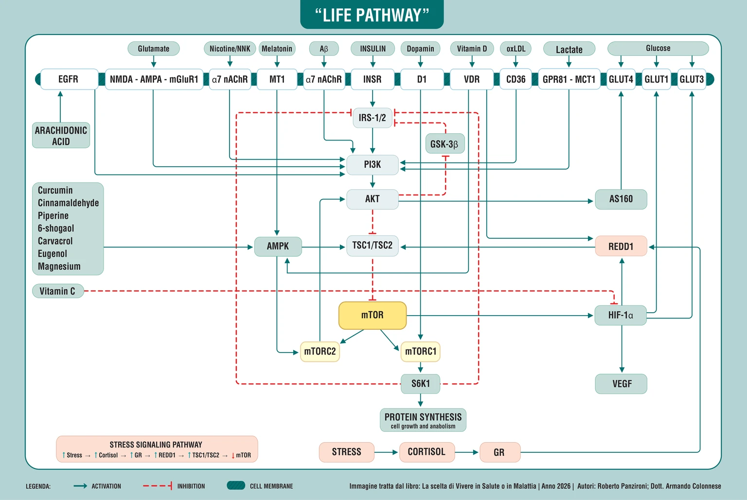
Life Pathway: diagram of membrane receptors and the IRS, PI3K, AKT, AMPK, TSC1/TSC2, mTOR, REDD1, and HIF-1α pathways that link nutrition, stress, nutrients, and cellular metabolism
The Life Pathway integrates membrane receptors and signals from glutamate, insulin, dopamine, vitamin D, lactate, and glucose with the IRS-1/2–PI3K–AKT, AMPK–TSC1/TSC2–mTOR, and REDD1–HIF-1α pathways, connecting stress, nutrients, and metabolism to protein synthesis and cellular homeostasis, the cornerstone of the choice between health and disease.

ABSTRACT

The book “The Choice to Live in Health or in Disease”, by Dr A. Colonnese and R. Panzironi, explores the relationships between nutrition, metabolic regulation, immunity and chronic disease. Health emerges as the dynamic result of the interaction between cellular processes, environmental conditions and behaviour, without individual choices being able to fully determine the risk of disease.

The response to a meal involves glycaemic variability, pancreatic secretion, hepatic clearance and insulin sensitivity. From the insulin receptor, the signal passes through IRS-1/2, PI3K and AKT, integrating with mTORC1, mTORC2, AMPK, HIF-1α, REDD1 and TSC1/TSC2. These regulators coordinate energy availability, protein synthesis, autophagy and adaptation to stress. The links with lipid metabolism, prostaglandins and immunometabolism help to interpret inflammation and immune differentiation, including the Th17/Treg balance. The function of each pathway depends on the context: signals needed for repair can sustain pathological processes under different conditions.

The evidence considered covers diabetes, autoimmunity, neurodegeneration, cancer and atherosclerosis, showing how shared circuits take on different roles depending on tissues and conditions. Epidemiological associations, clinical studies and experimental models call for distinct interpretations. Diet, physical activity, smoking, social relationships, stress and sleep rhythms interact with these processes. Vitamins, magnesium, omega-3s, melatonin and the bioactive compounds of spices offer further examples of cellular regulation.

The concept of the Life Pathway proposes an integrated reading of metabolic, energy and environmental networks, without identifying a new molecular pathway. This interpretive framework links biological mechanisms and everyday behaviour, supporting realistic, individualised preventive choices that are compatible with appropriate care. It remains necessary to clarify the clinical relevance of experimental mechanisms, avoiding single explanations for multifactorial diseases, distinguishing biological plausibility from evidence of efficacy and recognising the contribution of genetic, environmental and social factors as well.

We have reached the end of this book. Before we part ways, we would like to retrace the journey that has taken us from dietary habits to cellular signals, and from chronic diseases to the possibilities offered by prevention. These conclusions gather the meaning of each individual stage and reconnect them, so that what we have encountered can become knowledge applicable to daily life.

Our intention, as authors, has been to guide the reader through the biological mechanisms that contribute to health. Knowing why a behavior matters can make the desire to change it more concrete. A meal, a walk, rest, and a meaningful relationship take on a different value when we understand that they can influence how the body uses energy, copes with stress, and maintains its functions.

The title The Choice to Live in Health or Disease expresses this possibility of participation. Health also depends on genetic, environmental, and social factors that we do not choose, and no one voluntarily decides to become ill. However, there are habits we can intervene on, with different possibilities for each person. The awareness we wish to promote concerns precisely this space: recognizing what we can improve, without turning illness into a fault or prevention into a promise of invulnerability.

By following the research, we have encountered clinical results, epidemiological associations, and mechanisms studied in the laboratory. Each level contributes to our understanding, but answers different questions. Maintaining this distinction allows us to recognize opportunities without attributing the value of certainty to hypotheses. It is also the prerequisite for combining a conscious lifestyle with appropriate medical care, while respecting the complexity of the individual.

We will therefore resume the journey in the order in which it developed, allowing what unites apparently distant topics to emerge. At the end, we will give a name to the perspective that runs through them: Life Pathway, the editorial proposal with which we bring together the communication between metabolism, environment, and cellular adaptation. To understand its meaning, let us return to the point where it all began: our relationship with food.

Nutrition and changes in our habits

The first chapter invited us to view nutrition as part of our biological and daily history. Eating is a habitual act, often performed without questioning what happens afterward. Yet, the choice of foods, their processing, and the way they are combined help determine the body's responses. From this observation, the question that guided this book was born: how much of the metabolic conditions we encounter today is intertwined with habits repeated over time?

The reference to the Mediterranean dietary tradition brought us back to the quality of the meal and its overall composition. Its value, from this perspective, lies in the attention paid to food and the variety of nutritional sources. To recover this meaning, we must look beyond the name given to a diet and consider what is actually consumed, in the quantities and combinations that characterize real life.

We thus began to shift the focus from calories alone to the metabolic response. Two meals can provide energy and produce different effects on the rate of nutrient absorption, glycemia, and insulin secretion. This difference does not exhaust the nutritional value of foods, but it helps us understand why our relationship with food deserves a broader assessment than simple energy counting.

The first result of this reflection is greater attention to the consistency of habits. Metabolic health is built through behaviors that are repeated, within personal and environmental conditions that can make them easier or more difficult. For this reason, a useful dietary choice must be able to fit into daily life, maintaining nutritional adequacy and sustainability. Knowledge serves to make such choices understandable, preventing them from depending solely on prohibitions or momentary enthusiasm.

With this perspective, our journey found its concrete starting point. Food introduces substances that the body must recognize, distribute, and utilize; the response to this intake tells us something about its capacity for adaptation. Following the fate of glucose allowed us to enter into this process and understand why a value measured in the blood is only part of the story.

Glycemia and insulin in the meal response

We have moved from food to the response that a meal produces in the body. Absorbed glucose reaches the blood, while the pancreas and tissues coordinate its use. Glycemia describes one result of this work: to interpret it, one must consider the processes that determined it. The quantity of carbohydrates, the structure of the food, fiber, processing, and the composition of the meal all contribute to the speed at which glucose becomes available.

In this framework, we encountered the glycemic index and glycemic load. The former helps to compare the response to different sources of carbohydrates; the latter also introduces the quantity consumed. These are analytical tools to be placed within the context of the meal and individual characteristics. Proteins, fats, and fiber, in fact, modify digestion, absorption, and hormonal response, making the evaluation of a food through a single number insufficient.

Insulin also requires the same attention. Its secretion responds to glucose, but it integrates additional signals, including amino acids and intestinal hormones. The Food Insulin Index has helped us recognize that an insulin response does not automatically coincide with a glycemic one. Furthermore, before fully reaching peripheral circulation, insulin passes through the liver, which removes a portion of it. The measured concentration therefore depends on both pancreatic production and clearance, that is, the removal of the hormone.

This distinction has also expanded the significance of tests. Fasting glycemia captures a baseline condition; glycated hemoglobin provides information on average glucose exposure; the glucose tolerance test observes the response to a defined stimulus. In the chapter, the focus on one-hour glycemia showed us how some signs of difficulty can emerge before more evident alterations. However, none of these data, when considered in isolation, tells the whole story of metabolic function.

The conclusion is an invitation to view metabolism over time. An apparently preserved glycemia can be sustained by an increasing burden on regulatory systems. Understanding this possibility allows us to value prevention and the overall assessment of the individual. The next step was therefore to observe what happens when fluctuations increase and the collaboration between the pancreas, liver, and tissues begins to lose efficiency.

Glycemic variability and alterations preceding diabetes

Tracking glucose over time has allowed us to recognize a dimension that a single measurement leaves hidden: glycemic variability. Glucose fluctuates physiologically, but the amplitude and frequency of these excursions can change along the path toward prediabetes and diabetes. Continuous monitoring and its indicators have made these trends observable, showing how a similar average can be accompanied by different daily profiles.

The research examined links greater glycemic instability to oxidative stress, inflammation, and vascular alterations. Experimental models also show how intermittent exposure to high glucose concentrations can damage pancreatic β-cells. These observations have helped us understand a possible circuit: the difficulty in regulating glucose can increase the burden on the very cells that participate in its regulation.

Pancreatic function must therefore be interpreted in relation to the needs of the tissues. A high amount of insulin does not guarantee an adequate response if sensitivity to the hormone is reduced. For the same reason, the circulating concentration alone does not describe how much the pancreas is producing. The liver, through insulin clearance, modifies the body's exposure to the hormone; a reduction in this process can contribute to hyperinsulinemia even without an equivalent increase in secretion.

It is here that we delved into one of the central themes of the work: the bidirectional relationship between hyperinsulinemia and insulin resistance. Resistance can induce a compensatory response from the pancreas; in some contexts, high and persistent insulin exposure can, in turn, contribute to tissue desensitization. Secretion, removal, and cellular response influence each other, with a weight that changes between individuals and phases of metabolic dysfunction.

This interpretation makes the gradual nature of the deterioration more understandable. The appearance of overt hyperglycemia can be preceded by years of adaptations, during which the system maintains certain results at the cost of a growing imbalance. The interest in these transitions concerns the possibility of recognizing an unfavorable trajectory early on, through an appropriate assessment. To understand how a signal can progressively lose effectiveness, we then entered the cell, following the proteins that translate the insulin message into action.

Insulin signaling and the cell's ability to adapt

Entering the insulin pathway meant observing what happens after the hormone binds to its receptor. IRS1 and IRS2 participate in transmitting the message, PI3K supports its propagation, and AKT helps transform it into metabolic and survival responses. Nutrient availability thus becomes information capable of guiding glucose utilization, synthesis, and resource conservation.

The pathway then reaches the TSC1/TSC2 system and mTOR, one of the main integration points between energy, nutrients, and growth signals. We have distinguished between mTORC1 and mTORC2, complexes with different and interconnected functions. The former participates in the control of synthesis and autophagy; the latter contributes, among other functions, to the regulation of AKT. This distinction is necessary to understand why modifying mTOR can have different consequences depending on the context.

When mTORC1 is persistently stimulated, the circuit involving S6K1 can attenuate the upstream signal, interfering with IRS. A physiological control system can thus participate in reducing the insulin response. On the other hand, compromising necessary mTOR functions can also alter metabolism. The message that has emerged is therefore the need to preserve appropriate regulation: growth, repair, and turnover require different times and conditions.

In this network, we encountered HIF-1α and REDD1. The former connects metabolic signals to cellular adaptation, especially in relation to oxygenation; the latter participates in the stress response and can contain mTORC1 through the TSC system. These regulators show how the cell integrates information arriving from multiple directions. Food is one of them, along with energy status and tissue conditions.

The value of this stage does not lie in memorizing all the acronyms. It lies in recognizing that the insulin response depends on a distributed and regulated communication, the effectiveness of which can change over time. Insulin maintains an indispensable role; understanding its excesses and alterations means protecting the quality of its signal. From here, we moved on to a broader consequence: the same circuits that guide metabolism can interact with those that support inflammation.

The dialogue between metabolism and inflammation

The link with inflammation has taken us beyond energy distribution. We have observed how persistent metabolic stimulation can influence the availability of substances used to produce inflammatory mediators. From this perspective, hyperinsulinemia represents one of the possible elements of an altered biological environment, capable of intertwining with the processes through which cells communicate damage or a state of activation.

The path described starts from lipid metabolism. Through SREBP-1c and desaturase enzymes, insulin signaling can contribute to modifying the production of arachidonic acid. This fatty acid belongs to normal membrane physiology and constitutes the precursor of various mediators. Its presence becomes relevant to the inflammatory cascade when other signals allow for its release and utilization.

We followed this second step through MAPK/ERK, intracellular calcium, and cPLA2α, the enzyme that can make arachidonic acid available from membrane phospholipids. Subsequently, COX-2 participates in the production of prostaglandin E2. In the contexts described, the prostaglandin signal, mediated by the EP4 receptor, can communicate with EGFR and return to involve PI3K, AKT, and mTOR. Lipid metabolism and growth signaling can therefore enter into circuits of mutual influence.

This sequence has allowed us to attribute a concrete meaning to the expression metabolic inflammation. A change in the use of nutrients can contribute to changing the messages received by neighboring cells, with effects on the tissue environment. The reconstruction of a mechanism shows a biological possibility; its actual relevance depends on the intensity of the stimulus, the duration, and the other conditions present in the organism.

Attention has therefore shifted from the mediators to the cells that produce and interpret them. The macrophage can recruit other defenses, release cytokines, and influence repair. An intracellular phenomenon thus assumes a collective dimension, involving tissues and organs. To understand how a response is initiated, amplified, and finally contained, it has become necessary to understand the immune system and the energy mechanisms that support its various functions.

The immune system and its metabolic balance

With the immune system, we have encountered a network capable of recognizing threats, organizing responses, and limiting tissue damage. Innate immunity offers a first line of intervention, while adaptive immunity develops more specific responses through T and B lymphocytes. Collaboration between these systems requires communication, energy, and control: activation is necessary, but so is terminating a response once it has fulfilled its function.

Immune cells adapt their metabolism to the task at hand. Inflammatory programs are often accompanied by an increased reliance on glycolysis, while certain regulatory and repair functions depend more heavily on oxidative metabolism. Describing macrophages as M1 and M2 has helped us navigate this distinction, although these represent simplifications of more variable cellular behaviors. From this relationship between function and energy, immunometabolism is born.

mTOR and AMPK participate in coordinating these programs. mTORC1 supports processes necessary for the activation and growth of effector cells; AMPK contributes to energy adaptation and can limit costly anabolic activities. HIF-1α links part of this signaling to glycolysis and immune differentiation. The same proteins we have already encountered in metabolism thus acquire further significance: they help guide how the body's defenses utilize their resources.

The relationship between Th17 and Treg cells has made this connection particularly evident. The former participate in inflammatory responses; the latter help maintain tolerance toward the body. FOXP3 is essential for the identity and regulatory function of Tregs. Research on rapamycin, glucocorticoids, and vitamin D has shown that intervening in metabolic signals can modify these balances, with effects dependent on the cells and conditions studied.

We have therefore understood that an effective defense requires proportion, duration, and the capacity for regulation. Indiscriminately boosting immunity does not describe a sufficient biological goal; it is necessary to preserve the response appropriate to the context. This awareness has prepared us to examine autoimmune diseases, in which tolerance can deteriorate and immune activation can contribute to damage. The first step of this transition has led us to the joints, where rheumatoid arthritis makes the dialogue between inflammation and metabolism tangible.

Rheumatoid arthritis and joint response metabolism

In rheumatoid arthritis, the molecular pathway has encountered a disease that can profoundly affect movement, pain, and daily life. Joint inflammation involves immune cells and synovial tissues within a complex autoimmune process. Observing it through the lens of immunometabolism has allowed us to broaden our understanding of what sustains the inflammatory response, while keeping the person and the concrete consequences of the disease at the center.

The first element that emerged concerns the balance between lymphocyte populations. Th17 cells and their mediators can contribute to inflammation, while Treg cells participate in its containment. The relationships between Tfh, Tfr, and B lymphocytes also help us understand the production of autoantibodies. When these systems do not coordinate properly, the response can persist over time and progressively involve the joint environment.

The research considered has also documented alterations in insulin sensitivity, present in some patients even in the early stages and not solely explained by glucocorticoid treatments. This finding suggests a continuous interaction between inflammatory activity and metabolism.

mTOR represents one of the points where these aspects can converge. Its activity participates in effector cell programs, synovial proliferation, and angiogenesis. Through HIF-1α and glycolysis, growth signals can support the energy requirements of the cells involved. Studies on mTOR inhibitors show the interest in this connection as a research target. Vitamin D has also been examined for its relationship with immune tolerance and disease activity.

The lesson we take away concerns the breadth of our perspective. Controlling the disease requires specific treatments, while metabolic health constitutes a relevant dimension of overall patient care. Understanding these relationships helps explain why seemingly distant organs and systems can influence one another. From the joint, we then moved on to the skin: in psoriasis, an immediately visible manifestation has shown us once again processes that extend beyond the tissue in which they appear.

Psoriasis and signals crossing the skin

Psoriasis has taught us to look beyond the surface. Skin manifestations make visible a condition in which keratinocyte proliferation, immune activity, and inflammatory mediators influence one another. The skin thus becomes the place where we can observe the effects of altered biological communication, without the size of a lesion alone describing the complexity of the person presenting it.

Focusing on Th17 cells, IL-17, and other mediators has brought us back to the relationship between effector response and regulatory control. Treg cells may become less effective or lose stability in an inflammatory environment. Meanwhile, keratinocytes actively participate in the process, modifying their growth and communication with the body's defenses. The persistence of inflammation therefore depends on multiple players, which contribute to maintaining the altered skin microenvironment.

This dimension is accompanied by a metabolic one. In the studies examined, hyperinsulinemia and reduced insulin sensitivity were observed even in people without overt diabetes and, in some cases, without obesity. This finding invites us to consider conditions that may remain inconspicuous if one only looks at glycemia or weight. Cardiovascular and metabolic associations reinforce the importance of a comprehensive assessment, beyond the treatment of skin manifestations.

In psoriatic tissue, we have identified PI3K/AKT/mTOR and HIF-1α. These signals can support the glycolytic metabolism necessary for epidermal proliferation and the activity of certain immune cells. Research into their modulation shows how the control of growth and the control of inflammation can share targets. Vitamin D adds another link, through the regulation of skin differentiation and immunity.

This stage has brought us closer to a concrete awareness: taking care of health means also considering what is not immediately visible. The disease does not define the person and does not authorize judgments about their habits. Instead, it offers a reason to integrate knowledge and identify diverse needs. This same perspective becomes even more necessary when autoimmunity can affect different organs, as we encountered in systemic lupus erythematosus.

Lupus and the complexity of immune tolerance

In systemic lupus erythematosus, we have encountered a loss of tolerance capable of involving different organs and manifesting with highly variable clinical pictures. This diversity has reminded us how important it is to maintain an overall perspective. Autoantibodies and B-cell activation represent part of the process, embedded in a network of relationships between defenses, tissues, and metabolic signals.

The balances between Tfh and Tfr, as well as those between Th17 and Treg, have shown the importance of the systems that coordinate and contain the response. When certain effector programs prevail and regulation becomes insufficient, the ability to maintain inflammation can increase. Metabolism accompanies these changes: glycolysis, glutamine utilization, and mitochondrial activity contribute to providing the resources necessary for the different cell populations.

The insulin alterations described in studies have further expanded the picture. Reduced insulin sensitivity can be present even with normal glycemia and during clinically inactive phases. Its observation in different populations suggests that metabolic interaction deserves attention, without automatically attributing it to a single drug or a single characteristic of the disease. Here too, the direction of these relationships requires a cautious and contextual interpretation.

mTOR and HIF-1α have offered us a link between these levels. Their activity can support glycolytic programs and effector responses, while alterations in autophagy and mitochondria contribute to the cellular picture. Studies on sirolimus have shown how some of these connections can become the subject of therapeutic research. Vitamin D has been considered for its relationship with immune status and the balance between regulatory and effector populations.

The value of this stage lies in having brought us closer to a complex disease while maintaining the distinction between shared mechanisms and individual characteristics. A common network helps to formulate questions and interpret observations; the choice of treatments requires specific evidence and evaluations. With this focus, we have moved on to another environment where tolerance is essential every day: the gut, which must reconcile contact with the outside world, nutrient absorption, and protection of the organism.

The gut and maintenance of the immune barrier

In chronic inflammatory bowel diseases, the relationship between defense and tolerance takes on immediate significance. The intestinal mucosa participates in absorption while simultaneously serving as an interface with the external environment. Crohn's disease and ulcerative colitis present significant differences, but they share a persistent or recurrent inflammatory response that can profoundly affect quality of life and nutritional status.

The journey has highlighted the role of Th17 and Treg populations, along with that of macrophages and epithelial cells. The issue concerns the ability to maintain a proportionate response, protecting the tissue while addressing stimuli. Immune activity requires energy, and metabolic programs help guide it. For this reason, the barrier and defenses cannot be understood by separating them from the energy conditions of the cells.

Associations between metabolic indicators, such as the TyG index, and disease risk have opened up a further perspective. These data suggest possible links between metabolic dysfunction and intestinal inflammation, helping us recognize that gut health is part of the framework of the entire organism, with influences that can proceed in both directions.

We have found mTOR and HIF-1α in the programs that support glycolysis and inflammatory activity. In the models examined, the modulation of these pathways can modify immune populations, autophagy, and barrier integrity. Studies with mTOR inhibitors and natural compounds have provided research insights, while observations on vitamin D have drawn attention to nutritional status and immune regulation. The function of each signal remains linked to the cell type and the context.

From this stage, the value of care that considers inflammation, nutrition, and general conditions together emerges. Biological understanding makes it clearer why an intestinal problem can have consequences that extend beyond the digestive system. The same dialogue between immunity and metabolism is found in tissues with very different needs. In the next chapter, we followed it to the nervous system, where multiple sclerosis brings the protection and repair of myelin to the forefront.

Multiple sclerosis and nervous system protection

With multiple sclerosis, the focus has shifted toward a tissue where communication requires particularly delicate organization. The autoimmune involvement of myelin is accompanied by inflammation processes that can alter neurological function. Understanding the metabolic component has allowed us to add a further question to the description of the damage: what resources and signals support the cells that promote it and those that attempt to repair it?

Research on Treg cells has highlighted the role of tolerance, while Th1, Th17, and the relationships between T and B lymphocytes have shown different contributions to the autoimmune response. Microglia, part of the nervous system's defenses, also participate in building the inflammatory environment. The metabolism of individual populations thus becomes relevant for understanding how they are activated and how they maintain their function within the tissue.

In the studies examined, alterations in insulin sensitivity and increased insulin responses were also observed in the early stages of the disease. The connection with inflammatory markers and disability suggests an interplay that deserves attention. The presence of these relationships expands the assessment of the individual, without allowing multiple sclerosis to be reduced to a single metabolic alteration or automatically deducing a definitive intervention.

The most important distinction that emerged concerns mTOR. In some immune compartments, its activity can support pathogenic programs; in oligodendrocytes, however, it contributes to functions necessary for myelination. HIF-1α and glycolysis complete the link with effector responses. Experiments on pathway modulation must therefore be interpreted while keeping in mind different cells, timings, and objectives. Vitamin D also enters this network, with associations and results that must be read according to the level of evidence.

This stage makes a principle of the entire book particularly clear: the same protein can participate in both dysfunction and recovery, depending on the context in which it operates. Health requires coordination between defense and repair. After observing this need in the nervous system, we followed the relationship between autoimmunity and metabolism in a gland that helps regulate the body's activity: the thyroid, the protagonist of the subsequent reflection on Hashimoto's disease.

Hashimoto and the interplay between immunity and thyroid function

Hashimoto's thyroiditis has allowed us to observe the intersection between the immune system and endocrine regulation. The loss of tolerance toward thyroid components can be accompanied by progressive damage to the gland, with consequences that depend on residual function and individual progression. Following this process has helped us understand why the assessment of an endocrine autoimmune disease must consider multiple levels simultaneously.

We have identified the imbalance between Th17 and Treg cells and the role of regulatory populations in containing the response. Within the tissue, oxidative stress, autophagy, and growth signals can influence the ability of cells to remain functional. Autoimmunity therefore involves a dialogue between the defenses and the conditions of the target cells, which in turn participate in the environment in which the response develops.

Observations on insulin resistance have added a metabolic dimension. In the studies considered, reduced insulin sensitivity was documented even in people with preserved thyroid function. This association suggests that autoimmunity and metabolism can interact beyond the effects of hormonal variations. The weight of the different factors remains individual and requires a comprehensive reading, in which visceral adiposity and other concomitant conditions play a role.

At the molecular level, PI3K/AKT/mTOR and HIF-1α link growth, autophagy, and the energy programs of immune cells. Experimental results with rapamycin and other compounds show how the modulation of these signals can reduce some aspects of damage in the models studied. Vitamin D has been analyzed for its effects on autoantibodies and immune regulation. These observations enrich our biological understanding, while keeping preclinical results distinct from established therapies.

The takeaway message concerns the possibility of recognizing complementary needs: preserving endocrine function, monitoring the autoimmune process, and considering metabolic and nutritional status. Knowledge of these connections helps to avoid a fragmented interpretation of tests and symptoms. From this gland, we have moved on to the pancreas, where the loss of tolerance specifically affects the cells that produce the hormone that has accompanied us from the beginning: insulin, which is essential for understanding type 1 diabetes.

Type 1 diabetes and pancreatic cell loss

In type 1 diabetes, the link between immunity and metabolism becomes particularly evident. The autoimmune response damages pancreatic β-cells, progressively reducing the ability to produce insulin. The hormone we have followed as a metabolic signal takes on the concrete meaning here of a function that must be replaced when it fails. This characteristic distinguishes the disease and makes it essential to preserve its specificity throughout this discussion.

The journey has shown that tolerance depends on the function of Tregs, as well as their number. Insufficient regulation can leave room for autoreactive populations, including those that participate in the response against pancreatic cells. Research on the expansion or transfer of Tregs has illustrated the interest in strategies capable of recovering more selective control of the immune response.

Insulin sensitivity can also modify the clinical picture. In some at-risk subjects and in people with established disease, reduced efficacy of available insulin has been observed. This element can increase metabolic complexity without replacing the central role of the autoimmune loss of β-cells. The availability of the hormone and the response of the tissues are different dimensions, which can influence each other in determining glycemic control.

mTOR, HIF-1α, and glycolysis participate in the energy programs of effector cells, while AMPK and autophagy have also been examined in relation to the resilience of pancreatic cells. Studies on rapamycin have highlighted the opportunities and limitations of a modulation that must respect different functions. Vitamin D has been considered for its possible relationships with tolerance and metabolism, without such results modifying the need for insulin therapy when indicated.

The lesson of this chapter is therefore twofold: to recognize shared mechanisms and to protect clinical differences. Metabolic care accompanies a disease whose autoimmune origin can also be attributed to a dietary choice. Understanding this step allows us to address the next one with greater precision. In type 2 diabetes, in fact, the loss of pancreatic function is part of a different history, in which tissue sensitivity, secretion, and insulin clearance take on characteristic relationships.

Type 2 diabetes and progressive metabolic deterioration

Type 2 diabetes has brought us back to the early signals encountered at the beginning of this work, showing how they can fit into a long and complex process. Increased glycemia is a central element of the diagnosis, but the disease involves the interaction between the pancreas, liver, muscle, and adipose tissue. Over time, it can be accompanied by complications affecting numerous organs, making the systemic dimension of the metabolic imbalance evident.

Postprandial excursions and glycemic variability can increase before more stable hyperglycemia occurs. Meal composition influences these responses and offers one of the areas for intervention. Studies on low carb dietary strategies have shown potential benefits for glycemic control, which should be interpreted in conjunction with food quality, adherence, and individual needs. The value of a strategy is also measured by the ability to maintain it adequately over time.

The deterioration of β-cells includes alterations in the early insulin response and the ability to compensate for the body's demands. In parallel, a reduction in insulin clearance can increase exposure to insulin. Hyperinsulinemia and insulin resistance can thus contribute to mutually reinforcing cycles. There is, therefore, a dynamic relationship between the production, removal, and efficacy of the hormone.

Inside the cell, the feedback of mTORC1 and S6K1 on IRS helps us understand one of the mechanisms that can reduce signal quality. AMPK, on the other hand, highlights the ability to adapt to energy demand and support resource utilization. When the imbalance persists, oxidative stress, glucotoxicity, lipotoxicity, and mitochondrial difficulties can converge on the pancreas, reducing its functional capacity.

The preventive significance of this reconstruction is recognizing that the metabolic course develops over time and can be addressed through appropriate nutritional interventions at different stages. A diagnosis requires treatment; the signals that precede it deserve attention and evaluation. This vision has then led us beyond peripheral metabolism, toward an organ that continuously depends on the availability of energy: the brain. In Alzheimer's, we have observed how insulin signaling, glucose utilization, and cellular maintenance can intertwine with neurological deterioration.

Alzheimer's and brain metabolism fragility

In the chapter dedicated to Alzheimer's, we broadened the perspective on neurodegeneration, adding the issue of the brain's energy balance to the accumulation of beta-amyloid and tau. Neurons must maintain communication, integrity, and the turnover of their components. When these functions lose coordination, the alterations can influence each other, contributing to the progressive vulnerability of the networks that support memory and cognitive abilities.

Reduced brain response to insulin was examined as part of this framework and can be observed even in the absence of diabetes. The described models link insulin signaling to beta-amyloid management, protein processing, and autophagy. The IDE enzyme, involved in the degradation of both insulin and beta-amyloid, introduces another possible point of interaction, the relevance of which depends on the biological context.

mTOR participates in functions necessary for synaptic plasticity and memory. However, its altered regulation can be accompanied by a reduced capacity for turnover and the accumulation of harmful components. Feedback on IRS and the regulation of AKT and GSK3β also help to reconstruct possible links with tau modifications. Understanding these mechanisms requires preserving the physiological role of the very proteins involved in the dysfunction.

We also followed glutamate, whose communication must be controlled to avoid excessive excitation. Alterations in its release and removal can aggravate neuronal stress. Meanwhile, mitochondrial difficulties and modifications of the GLUT1 and GLUT3 transporters can reduce the effective availability of energy. HIF-1α enters this network as a regulator of metabolic responses, once again showing effects linked to the tissue and conditions considered.

The chapter has therefore provided us with an interpretation in which proteins, signals, and metabolism participate in the same biological problem. Metabolic associations do not equate to a single explanation for Alzheimer's, but they contribute to understanding its complexity. From a tissue struggling to maintain its functions, we have moved to cells capable of reorganizing resources to support pathological growth. It is this change in perspective that introduced us to the metabolism of tumors.

Tumors and the organization of the metabolic microenvironment

In tumors, we have observed how a cell can modify its use of resources and its communication with the surrounding environment. Genetic alterations maintain an essential role, while metabolism, growth signals, and interactions with other cells contribute to the complexity of the process. The perspective of this work has led us to follow these connections, distinguishing what supports a biological possibility from what demonstrates a therapeutic benefit.

Insulin also transmits growth and survival signals. Epidemiological associations between hyperinsulinemia, insulin resistance, and certain oncological conditions have raised questions about the metabolic context in which neoplasms develop. Through PI3K, AKT, and mTOR, these signals can interact with synthesis and proliferation programs. However, their significance depends on the characteristics of the tumor and the alterations present in the cells.

Growth requires materials as well as energy. The Warburg effect describes the increased reliance on glycolysis, even when oxygen is available, observed in many neoplastic cells. HIF-1α contributes to adaptation to oxygenation conditions and to the regulation of transporters and enzymes. Glucose thus enters metabolic programs that can support the construction of new cellular components and the production of lactate.

Lactate itself has expanded our understanding. Through MCT1 and MCT4, it can be exchanged between cells, becoming a resource in certain compartments of the tumor. Its signaling activity and the processes of lactylation link metabolism and cellular regulation. Tumor-associated fibroblasts participate in these exchanges: in the reverse Warburg effect, some stromal cells provide metabolites that can be used by neoplastic cells. The tumor tissue thus appears as a cooperative and heterogeneous environment.

This organization interacts with the systems that control damage and proliferation. p53 contributes to cellular surveillance; the relationship between AKT, MDM2, and p53 shows how survival signals can interfere with this protection. AMPK and TSC1/TSC2 add connections to the control of mTOR. Tumor development therefore also depends on the ability of altered cells to overcome the limits normally imposed on growth.

The microenvironment also includes immune defenses. Hypoxia, lactate, and adenosine can foster conditions that reduce the effectiveness of the antitumor response. CD39 and CD73 participate in the production of adenosine, while its receptors transmit its effects. Macrophages and Tregs can take on functions that, in this context, favor tolerance toward the tumor. Finally, VEGF links metabolic adaptation to the formation of new blood vessels.

This stage has helped us understand how the same mechanism can have different effects depending on the context: curbing the immune response can protect against the damage of autoimmunity, but it can also weaken defenses against a tumor. Studying metabolism therefore allows us to better understand these balances and opens up new research perspectives. Following the thread that connects metabolism, lipids, and inflammation, we then shifted our focus from the blood vessels that feed the tumor to the arterial wall, to understand how these factors contribute to the development of atherosclerosis.

Atherosclerosis and vascular wall protection

In atherosclerosis, we encounter a process that can develop slowly before a cardiovascular event makes it evident. The arterial wall actively participates in flow regulation, immune communication, and lipid management. The endothelium, the inner lining of blood vessels, provides a privileged vantage point for understanding how the quality of metabolic signals can influence vascular health.

Under physiological conditions, the IRS/PI3K/AKT pathway contributes to activating eNOS and producing nitric oxide. This molecule promotes vasodilation and supports the protective functions of the endothelium. When insulin signaling loses efficacy, this contribution may be reduced. This chapter has thus reminded us that insulin retains important actions for vascular physiology and that its concentration must be interpreted in conjunction with tissue response.

The reduction in endothelial protection is intertwined with inflammation and the adhesion of immune cells. Signals such as NF-κB and VCAM-1 participate in leukocyte recruitment, while lipoproteins present in the wall can undergo oxidative modifications. Oxidized LDL are recognized by receptors such as CD36, which facilitate their entry into macrophages. Lipid accumulation contributes to the formation of foam cells, which are characteristic components of atherosclerotic lesions.

CD36 has shown us how uptake and signaling can proceed together. In macrophages, it contributes to inflammatory responses and lipid accumulation; on platelets, it can participate in increasing reactivity to stimuli. PI3K/AKT/mTOR and autophagy add links to the cells' ability to manage lipids. These processes belong to a network in which insulin metabolism, oxidative stress, and the vascular environment can influence one another.

This reconstruction has made the concept of long-term prevention more concrete. Vascular health depends on the combination of risk factors and appropriate care, while the metabolic dimension helps us understand some less visible connections. After following these relationships in pathologies, it was necessary to return to daily life. Nutrition, introduced at the beginning as a starting point, has reappeared with a broader meaning: as one of the repeated exposures through which we can help modify the biological context of the organism.

Dietary quality in daily prevention

Returning to nutrition after examining various pathologies has allowed us to better understand its role as a risk factor. Every meal triggers glycemic and insulin responses which, when repeated over time, can contribute to altering metabolic balance. Attention has focused primarily on the quantity and quality of carbohydrates: rapidly digestible foods and portions with a high glycemic load can produce more marked increases in glucose and a greater demand for insulin.

The research examined has associated a high glycemic index, a high glycemic load, and a greater insulinemic potential of the diet with an increased risk of type 2 diabetes and, in various populations, of general, cardiovascular, and oncological mortality. These results suggest that dietary quality deserves attention beyond just weight control. In prediabetes, in particular, a high glycemic load has been associated with a greater probability of progression toward the disease.

Cardiovascular involvement has shown us how the consequences can extend to blood vessels. Repeated glycemic excursions can promote oxidative stress, inflammation, and endothelial dysfunction, processes that contribute to the vulnerability of the arterial wall. Associations with coronary heart disease, atherosclerosis, and stroke fit into this framework. Studies on dementia and Alzheimer's have also drawn attention to dietary glycemic exposure, without demonstrating that it directly causes neurodegeneration.

In the oncological field, hyperinsulinemia and signaling of the PI3K/AKT/mTOR axis offer possible links between nutrition, cell growth, and survival. In autoimmunity, however, alterations in insulin sensitivity suggest an interaction with inflammation, but do not prove that reducing glycemic load prevents these diseases.

Intervention studies have therefore indicated some concrete possibilities. Diets with a lower glycemic index and load can improve insulinemia, HOMA-IR, and certain inflammatory markers; low carb strategies can promote glycemic control and improve triglycerides and hepatic steatosis. However, the effects depend on the overall quality of the diet, the substitute foods, and the continuity of the intervention.

The concluding message therefore concerns the ability to reduce unfavorable metabolic stress through appropriate portions, quality food sources, and meals tailored to individual needs. Understanding these relationships makes daily choices more conscious. Their effect then intertwines with the use of nutrients: hence the transition to movement, which increases the energy demand of muscles and contributes to glucose regulation.

Movement and alternating between work and recovery

After observing how nutrition influences nutrient availability, we explored how movement conditions their utilization. Prolonged sedentary behavior reduces the energy demand of muscles and can promote alterations in insulin sensitivity. The studies examined associate the accumulation of hours spent sitting with an increase in mortality and cardiovascular and metabolic risk, also drawing attention to the duration of consecutive periods of immobility.

This aspect allowed us to distinguish between exercise and movement distributed throughout the day. Engaging in physical activity is important, but spending many consecutive hours sitting can maintain a component of risk even in people who exercise. Breaking up immobility and replacing part of it with light activity therefore represents a concrete opportunity. Research on daily steps shows benefits even in the transition from inactivity to relatively modest amounts of movement, which is particularly relevant for the elderly and frail individuals.

The link with insulin resistance makes the biological significance of these observations clearer. Even short periods of marked reduction in activity can worsen the tissue response to insulin. Conversely, aerobic exercise, resistance training, and combined programs can improve glucose uptake, mitochondrial function, and indicators of insulin sensitivity. These effects can appear even without significant weight loss: the benefit directly concerns muscle function.

During contraction, the consumption of ATP, the main cellular energy resource, increases. AMPK helps recognize this increased demand and promotes the processes necessary to satisfy it, supporting the use of fatty acids and glucose uptake through GLUT4. Movement thus modifies the ability of cells to utilize available nutrients.

This response is coordinated with mTOR, which is involved in protein synthesis, repair, and muscle adaptation. The relationship between the two pathways varies according to the type of exercise, its intensity, and the recovery phase. Autophagy and mitochondrial turnover also participate in these adaptations, contributing to the maintenance of cellular efficiency.

The conclusion is therefore concrete: reducing sedentary time, breaking up long periods of sitting, and practicing physical activity appropriate to one's condition are complementary behaviors. Their consistency supports glucose regulation and muscle functionality, offering benefits that go beyond caloric expenditure. Alongside these favorable habits, we then examined an exposure capable of compromising metabolism and vascular health: smoking, the impact of which also involves the circuits that make it difficult to stop consumption.

Smoking and the circuits that maintain addiction

After observing how physical activity supports metabolic health, we examined an exposure capable of compromising it: smoking. Its effects reach the lungs, blood vessels, and tissues, also involving the brain's addiction circuits. The research analyzed shows that even a few cigarettes a day increase cardiovascular risk and mortality. Reducing consumption, therefore, does not eliminate the risk associated with exposure.

The damage develops through processes that can reinforce each other. Oxidative stress and inflammation compromise endothelial function, while increased platelet reactivity promotes thrombus formation. In parallel, the carcinogenic substances in tobacco can damage DNA. Quitting progressively alters this trajectory, offering cardiovascular, respiratory, and oncological benefits even after years of consumption.

The metabolic involvement brought us back to insulin signaling. In smokers, lower insulin sensitivity and alterations associated with the quantity and duration of exposure have been documented. Nicotine can contribute to these effects even independently of combustion. In experimental models, the modulation of mTOR and alterations in IRS-1 help explain how the effectiveness of the insulin response can be reduced, increasing the body's need for compensation.

These same signals take on further significance in tumor processes. Nicotine and NNK, a carcinogenic tobacco nitrosamine, can involve nicotinic receptors and activate PI3K/AKT/mTOR. In the models studied, this pathway supports proliferation, angiogenesis, and resistance to cell death. NNK can also cause DNA alterations: genetic damage and survival signals can thus converge, favoring the persistence of altered cells.

Electronic cigarettes also require attention. The absence of combustion reduces exposure to some toxins, but it does not make aerosols harmless, as they can contain nicotine, aldehydes, ultrafine particles, and metals. Different products present different profiles, and combined use with traditional cigarettes maintains exposure to the harms of smoking.

The difficulty of stopping consumption is ultimately explained by addiction circuits. Nicotine increases dopamine release in reward systems; synaptic plasticity processes, in which mTOR also participates, help consolidate the associations between the substance, gratification, and habits.

Understanding these mechanisms means recognizing both the value of cessation and the need to support those who undertake it. Knowledge of the risks can be motivating, while an appropriate program helps maintain the change. From here, we moved on to the role of social relationships, which can offer concrete support for health care and managing difficulties.

Social relationships and biological responses to loneliness

After examining smoking and the difficulties associated with addiction, we explored another factor that can influence health: the lack of meaningful relationships. The studies analyzed associate social isolation and persistent loneliness with an increase in mortality and cardiovascular risk. The quality of our bonds therefore takes on concrete value, as it can influence both daily behaviors and the biological systems through which we cope with stress.

An evolutionary perspective has helped us understand this relationship. Cooperation, resource sharing, and social learning have supported human survival. Even today, being able to count on others makes it easier to manage difficulties and practice self-care. Observations on isolation during childhood and adolescence also suggest that vulnerability can begin early, associating with greater inflammation and cardiometabolic alterations in later years.

Part of the risk is mediated through habits. Isolation can be accompanied by less adequate nutrition, nutritional deficiencies, increased tobacco use, less physical activity, and difficulty in adhering to medical treatments. These conditions can accumulate and reinforce one another. Social support, on the other hand, can make lifestyle changes more achievable and foster their consistency. The quality of relationships is what matters most: having numerous contacts does not guarantee feeling heard, understood, or supported.

The connection also extends to metabolism. The research examined associates loneliness and poor social support with insulin resistance and alterations in glycemic regulation. A possible explanation involves the hypothalamic-pituitary-adrenal axis: persistent loneliness can be accompanied by changes in the stress response and cortisol rhythms, influencing the availability and utilization of energy.

At the cellular level, in fact, glucocorticoids can induce REDD1, which, through TSC1/TSC2, contributes to containing mTORC1 activity. This response temporarily limits energy-intensive processes, such as protein synthesis, but prolonged activation of stress signals can promote catabolism and metabolic dysfunction. Other experimental evidence shows that the impairment of mTORC2 can interfere with AKT, AS160, and GLUT4, reducing glucose uptake; these mechanisms help interpret the link between stress and metabolism.

The conclusion is that health also requires attention to relational conditions. Recognizing loneliness and fostering reliable bonds can support daily self-care and reduce the burden of difficulties. To better understand how these experiences reach our tissues, the journey continued with stress and the hormonal signals that connect personal experience to metabolic regulation.

Chronic stress and metabolic regulation

After exploring the role of social relationships, we followed the primary link between daily experiences and metabolic regulation: the stress response. When faced with a threat, whether real or perceived, the body activates systems that make the necessary resources available to cope with it. The risk arises when this pressure persists and recovery becomes insufficient, promoting alterations in hormonal regulation and energy utilization.

At the center of this response, we find the hypothalamic-pituitary-adrenal axis and cortisol. The research examined describes changes in its daily rhythm and the awakening response, along with associations with hypertension and metabolic alterations. Chronic stress can therefore affect health through a dysregulation of the neuroendocrine response, which does not necessarily coincide with a constant increase in cortisol in every person.

Metabolically, cortisol increases hepatic glucose production and can reduce its utilization in peripheral tissues. Interference with IRS-1, PI3K/AKT, and the mechanisms that mobilize GLUT4 can make insulin action less effective. Initially, the pancreas may compensate by producing more insulin and maintaining an apparently normal glycemia. This compensation helps explain why an alteration in insulin sensitivity can develop before an evident increase in glucose, even independently of obesity.

The connection to cellular regulation also involves REDD1, a protein whose expression can be induced by glucocorticoids. Through TSC1/TSC2, REDD1 contributes to reducing mTORC1 activity and protein synthesis. A temporary response can support adaptation; if prolonged, it can contribute to catabolism and muscle mass loss. Evidence on the impairment of mTORC2 also shows possible consequences on AKT activation, but this mechanism must be distinguished from the mere reduction of mTORC1.

The pathway finally reaches the AKT–AS160–GLUT4 system. The phosphorylation of AS160 by AKT promotes the transfer of vesicles containing GLUT4 to the membrane, increasing glucose entry, especially in muscle and adipose tissue. Alterations in signaling or vesicle trafficking can hinder this process and contribute to insulin resistance.

We have thus understood why persistent stress represents a risk factor to be considered alongside diet, physical activity, and other personal conditions. Recognizing sources of overload and enabling recovery means paying attention to metabolic health as well. To complete this understanding, we have therefore traced back to the origin of the response: the brain circuits that transform the perception of a threat into hormonal signals capable of reaching the entire organism.

From nerve signal to hormonal response

After observing the effects of cortisol on metabolism, we traced the pathway back to understand how the stress response originates. Before hormones reach the tissues, the brain must recognize a threat—whether real or perceived—and coordinate the signals necessary to address it. The risk arises when this activation is prolonged and the systems meant to contain it fail to facilitate adequate recovery.

Glutamate, the primary excitatory neurotransmitter, participates in the communication between the involved circuits. Its activity is intertwined with mTOR, which contributes to the function and plasticity of synapses. Astrocytes maintain the balance of this communication by removing glutamate from the extracellular space and participating in its recycling. In the experimental models described, altered regulation of mTOR can compromise these functions, promoting glutamate accumulation, increased excitability, and neuroinflammatory signals.

This connection has shown us how the metabolic conditions of cells can influence the activity of neural networks. Glutamatergic signals also reach the neurons that produce orexin, located in the lateral hypothalamus. Orexin supports wakefulness and helps activate alertness systems, creating connections through which the response can be maintained and amplified.

Among the structures involved is the locus coeruleus, a brain nucleus that produces noradrenaline. The convergence of orexinergic and glutamatergic signals promotes its activation, preparing the body for attention and action. This response is functional when it accompanies a temporary need; its persistence, however, can hinder the return to conditions compatible with rest and recovery.

The transition to the endocrine response involves the paraventricular nucleus of the hypothalamus. Noradrenergic signals help regulate the neurons that produce CRH, the hormone that stimulates the anterior pituitary to release ACTH. The latter reaches the adrenal cortex and induces the production of cortisol. Through this network, brain activation takes on a systemic dimension, modifying energy availability, cardiovascular function, and immune system activity.

Circadian rhythms and negative feedback normally help regulate the response. When stress persists, hormonal rhythmicity, sensitivity to glucocorticoids, and the control of inflammation can be altered. The circuits described are therefore important links between the perception of a threat and biological consequences, within a broader and more complex regulation.

The conclusion concerns the ability to recover after activation: protecting health also means recognizing the conditions that keep the body on alert. The functioning of these networks also depends on the resources available to the cells. From here, the path has brought us back to nutrition, delving into the role of vitamins and minerals in supporting metabolic homeostasis.

Micronutrients and nutritional adequacy

After exploring the stress response, we examined another condition capable of compromising the body's balance: micronutrient deficiency. Vitamins, minerals, and trace elements are necessary to produce energy, support immune defenses, repair DNA, and maintain antioxidant protection. Insufficient availability can therefore weaken multiple functions simultaneously, even when the diet provides sufficient or excessive calories.

This is the meaning of so-called hidden hunger: eating enough does not guarantee that you are consuming everything your cells need. Overweight and obesity can coexist with nutritional deficiencies, making an assessment based solely on weight insufficient. The research examined associates a better micronutritional quality of the diet with lower overall mortality and, in various populations, lower cardiovascular and oncological mortality. These observations highlight the importance of food composition, beyond its energy content.

The link with insulin resistance makes this risk more concrete. Magnesium, zinc, calcium, potassium, and vitamin D participate, with different roles, in insulin secretion, ATP production, mitochondrial function, and the transmission of signals that allow for glucose utilization. Insufficient availability can interfere with these processes and be accompanied by oxidative stress and inflammation. In profiles characterized by multiple deficiencies, higher HOMA-IR values and poorer glycemic control have been observed.

However, the relationship can proceed in both directions. The deficiencies found in people with type 2 diabetes, including those of vitamin D, magnesium, and vitamin B12, do not necessarily represent the initial cause of the metabolic dysfunction, but can accompany or promote it. Understanding this dynamic helps to assess individual needs without attributing every alteration to a single nutrient.

Vulnerability can also begin before birth. The maternal nutritional environment contributes to pancreatic development and the metabolic regulation of the offspring. Experimental studies show that deficiencies during pregnancy and breastfeeding can modify energy and epigenetic programs, leaving effects that emerge later. These results underscore the value of nutritional adequacy in the early stages of life.

Prevention therefore requires attention to the quality and variety of the diet, along with the recognition of actual insufficiencies. Supplementation can be useful when it corrects a deficiency or meets specific needs; in people who are already adequately nourished, indiscriminately increasing vitamins and minerals does not guarantee further benefits. From this distinction, we started to delve into individual nutrients, beginning with vitamin D and its contribution to immune and metabolic regulation.

Vitamin D and the regulation of cellular functions

After recognizing the consequences of micronutrient deficiencies, we explored vitamin D, encountering functions that extend beyond the maintenance of bone health. Through the VDR receptor, present in numerous tissues, its active form helps regulate cellular programs involved in immunity, metabolism, and the stress response. This distribution helps explain why its role has been studied in different organs and conditions.

On an immune level, vitamin D participates in the coordination between defense and tolerance. It can influence antigen-presenting cells, contain certain pro-inflammatory responses, and promote the activity of regulatory T cells. It also contributes to the maintenance of the intestinal and respiratory barriers, which represent a fundamental interface between the organism and the environment. These functions offer a biological basis for interpreting the associations between vitamin deficiency, inflammation, and autoimmunity.

The link with metabolism primarily concerns pancreatic function and the response to insulin. The research examined suggests that vitamin D may act on β-cells, the inflammatory environment, and the sensitivity of peripheral tissues. Some supplementation studies have documented improvements in glycemic and insulin parameters, especially in people with insufficient initial levels. However, the extent of the benefits remains variable and depends on the characteristics of the participants and the intervention.

To understand these effects, we delved into intracellular mechanisms. In experimental models, VDR activation can increase the expression of REDD1, which, through TSC1/TSC2, helps contain mTORC1. This regulation can limit proliferative signals and promote autophagy, the process by which cells degrade and recycle part of their own components. The significance of the effect depends on the tissue and the conditions studied.

Alongside this pathway, we encountered AMPK, which is involved in energy adaptation. Research on hepatic steatosis, the gut, and osteoarthritis suggests links with glucose utilization, fat oxidation, and the management of cellular stress. Other studies have explored possible effects in diabetic nephropathy and neurological conditions, while many applications remain to be clinically confirmed.

In oncology, too, it is necessary to distinguish the results: some meta-analyses on supplementation have detected a reduction in cancer mortality, without demonstrating a significant decrease in new cases. Molecular and clinical observations therefore answer different questions.

The value of vitamin D emerges from its participation in essential functions and the possibility of correcting an insufficiency when present. Supporting its adequacy means contributing to the normal functioning of the organism, as part of comprehensive health care. From this regulation of cellular programs, we moved on to vitamin C, exploring its contribution to oxidative protection, tissue integrity, and the control of oxygenation responses.

Vitamin C: between tissue integrity and cellular response

After exploring vitamin D, we encountered another essential element for maintaining health in vitamin C. Humans cannot synthesize it and must obtain it through their diet. Its availability supports antioxidant protection, tissue formation, iron absorption, and various immune system functions. It also contributes to the synthesis of molecules involved in nerve signaling and to the activity of enzymes that participate in the regulation of gene expression.

One of its fundamental functions concerns collagen, a structural protein present in the skin, tendons, ligaments, bones, and vascular walls. Vitamin C acts as a cofactor for the enzymes necessary for its proper formation, promoting modifications that stabilize its structure. This contribution helps us understand its role in tissue integrity and repair processes, as well as the consequences of a prolonged deficiency.

Structural protection is directly linked to immune defenses. The skin and mucous membranes constitute the first barriers against external agents, while vitamin C also supports the activity of cells that intervene when these barriers are breached. It promotes neutrophil migration and phagocytosis—the ability to engulf and eliminate microorganisms—and participates in the processes that accompany the resolution of inflammation. The research examined also describes an involvement in the functions of Natural Killer cells and the maturation of T lymphocytes.

During infections, the consumption of vitamin C may increase, but its physiological functions must be distinguished from the results of supplementation. Studies on the common cold show limited benefits, particularly regarding the duration of symptoms under certain conditions, without demonstrating general prevention of the infection.

A further connection concerns HIF-1α, a protein that coordinates part of the cellular response to oxygen scarcity. Vitamin C supports the activity of hydroxylases which, when oxygen and other cofactors are available, help limit its stability and activity. This regulation affects programs related to glucose utilization, lactate production, and the formation of new blood vessels.

In oncological models, the relationship with HIF-1α has opened research perspectives on metabolism and the tumor microenvironment. Even high concentrations achievable via intravenous administration have been studied for their effects on the oxidative balance of neoplastic cells. However, these results belong to conditions different from dietary intake and do not demonstrate an anti-tumor efficacy of ordinary supplementation.

Vitamin C therefore emerges as a nutrient necessary to support complementary functions: protecting, building, repairing, and regulating. Ensuring an adequate intake contributes to the normal functioning of the body. From tissue integrity, we then moved on to the composition of cell membranes, exploring the relationship between structure, communication, and inflammation control with omega-3.

Omega-3 and cell membrane composition

After exploring the contribution of vitamin C to tissue integrity, we turned our attention to cell membranes. Here, EPA and DHA, two important omega-3 fatty acids, participate in the structure through which cells receive and transmit signals. Their incorporation modifies the lipid composition of the membranes and can influence the functioning of systems involved in communication, immunity, and metabolism.

The studies examined have documented this process in the heart, red blood cells, platelets, and immune cells. Incorporation occurs progressively and, in various compartments, is accompanied by a reduction in the proportion of arachidonic acid. This changes the availability of the precursors used to produce lipid mediators, helping to modulate the inflammatory response. This relationship should be interpreted by considering the physiological functions of both families of fatty acids, without attributing an exclusively unfavorable role to omega-6s.

These modifications lead to links with various areas of health. Research has reported favorable effects on certain cardiovascular, inflammatory, and metabolic parameters, while studies on autoimmune diseases have investigated the regulation of immune responses. In the nervous system, interest primarily concerns neuroinflammation and the maintenance of neuronal function. However, the applications explored in Alzheimer's, Parkinson's, and oncology present different levels of evidence and do not demonstrate generalizable therapeutic efficacy.

A key aspect concerns insulin sensitivity. EPA and DHA can help modulate adipose tissue inflammation, creating conditions potentially more favorable to the insulin response. Some studies conducted on gestational diabetes, polycystic ovary syndrome, and pediatric obesity have observed improvements in metabolic indicators. The results remain heterogeneous and depend on the characteristics of the individuals and the interventions.

To understand these effects, we also followed intracellular signaling. In specific experimental models, arachidonic acid can involve PI3K/AKT/mTOR; modifying its availability in membranes could therefore influence part of these signals. Other research shows that EPA and DHA can contain mTOR activity and, under certain conditions, promote autophagy, contributing to the turnover of cellular components.

AMPK completes this connection with energy regulation. Its activation, observed in various models, can participate in the effects of omega-3 on metabolism, inflammation, and the control of mTOR. These mechanisms help interpret the observations, while keeping biological plausibility distinct from proven clinical benefits.

The nutritional significance of omega-3 therefore lies in their contribution to cellular structure and regulation. An adequate intake is part of the overall quality of one's diet, while supplementation requires an assessment of individual needs. From the composition of the membranes, we then moved on to the timing of biological responses: with melatonin, we explored how daily rhythms, rest, and cellular maintenance participate in the same balance.

Melatonin and the coordination of biological rhythms

After exploring the role of omega-3 in cell membranes, we observed how the body's functioning also depends on the regularity of its rhythms. Melatonin, produced by the pineal gland and also in various peripheral tissues, participates in circadian signaling and the coordination between activity and rest. The research examined also describes its antioxidant, anti-inflammatory, and immunomodulatory actions, with possible effects on mitochondrial functionality.

The most well-known connection concerns sleep. Melatonin helps synchronize the processes that accompany the transition from wakefulness to rest. Clinical studies have documented, under specific conditions, a reduction in the time required to fall asleep and improvements in certain sleep parameters. However, the magnitude of the effects varies according to the disorder, individual characteristics, and the method of administration.

To understand its action, we followed the MT1 receptor, through which some signals are transmitted to nerve cells. In the experimental models described, this receptor participates in the regulation of the circuits that organize sleep phases. In the locus coeruleus, the reduction in the activity of noradrenergic neurons accompanies the transition to states of lower alertness and is particularly marked during REM sleep.

These circuits communicate with the orexinergic neurons of the lateral hypothalamus, which support wakefulness and stimulate alertness systems. Experimental research suggests that melatonin may also modulate their activity through MT1, contributing to the transition toward rest. We have thus encountered structures already seen in stress, understanding how much activation and recovery depend on coordinated regulation.

The presence of receptors in the pancreas, intestine, blood vessels, and immune cells extends this communication beyond the brain. Melatonin can influence insulin secretion, glucose metabolism, vascular tone, and inflammatory responses. These connections show how the circadian signal participates in the organization of the entire organism's functions.

A further aspect concerns cellular maintenance. In the models studied, melatonin modulates autophagy, through which cellular components are degraded and recycled, and mitophagy, dedicated to the removal of compromised mitochondria. AMPK and mTORC1 participate in this regulation, linking energy availability, synthesis, and turnover. The effects depend on the tissue and conditions: maintaining these processes efficiently requires appropriate modulation.

Melatonin has therefore helped us understand the relationship between biological rhythms, rest, and cellular balance. Its physiological functions offer a broad perspective, while therapeutic applications require specific evidence. From this temporal organization, we have moved on to magnesium, which is necessary for numerous reactions that support energy production and signal transmission.

Magnesium and the functioning of metabolic reactions

After exploring the relationship between melatonin, rest, and cellular maintenance, we encountered magnesium as an element necessary for the functioning of numerous biological processes. This mineral participates in hundreds of enzymatic reactions involved in energy production and utilization, nerve transmission, and muscle activity. It also contributes to vitamin D metabolism, demonstrating how the effectiveness of nutrients also depends on the availability of the cofactors necessary for their use.

Its role extends to cardiovascular regulation. The clinical studies examined have documented moderate reductions in blood pressure with supplementation, especially in people with hypertension or insufficient availability of the mineral. Epidemiological research also associates a higher dietary intake of magnesium with a lower risk of stroke, type 2 diabetes, and other cardiometabolic outcomes. These observations should be interpreted in the context of overall diet, because foods that provide magnesium also supply other useful substances.

Its participation in nerve and muscle function has suggested further areas of research. Some studies report benefits for recovery after exercise, certain sleep parameters, and symptoms of anxiety or depression. A particularly important connection concerns insulin sensitivity. Magnesium contributes to the reactions that support insulin receptor activity and the transmission of signals necessary for glucose utilization. Insufficient availability can interfere with these processes and be accompanied by inflammation and oxidative stress. Some meta-analysis and clinical trials have observed improvements in HOMA-IR and glycemic parameters, especially in people with metabolic alterations or a deficiency of the mineral.

The relationship can become reciprocal: metabolic dysfunction can promote the loss of magnesium, while insufficiency can make the insulin response less efficient. Correcting a deficiency therefore takes on concrete significance in supporting normal cellular functions.

In experimental models, we also followed the connections with AMPK, mTOR, and autophagy. Under certain conditions, magnesium can influence these systems, contributing to energy regulation and the recycling of cellular components; the effects described in bone tissue, the liver, and tumor models depend on the context.

The concluding message therefore concerns adequacy: ensuring magnesium intake means supporting reactions that are essential for health, while supplementation must respond to individual needs. From this nutritional foundation, we moved on to spices, exploring how their bioactive compounds can interact with some of the same metabolic signals.

Spices and their metabolic links

The last six chapters have brought our attention back to familiar ingredients, showing the biological complexity that can hide behind an aroma. Cinnamon, turmeric, cloves, oregano, black pepper, and ginger contain compounds capable of interacting with inflammatory and metabolic signals. Considering them together allows us to recognize the common thread that links them, while simultaneously acknowledging the differences in the evidence available for each.

Cinnamon introduced us to cinnamaldehyde, cinnamic acid, and polyphenols. Clinical studies have reported results on certain glycemic and lipid parameters, with variability also linked to the formulations used. In experimental models, the relationship with GLUT4 and AMPK offers possible explanations for the metabolic effects. Observations regarding PI3K/AKT/mTOR, HIF-1α, and autophagy, however, belong to specific contexts, including tumor models, and require an interpretation distinct from the use of the spice in a meal.

Turmeric expanded this theme through curcumin and other curcuminoids. The modulation of NF-κB, oxidative stress, and cytokines has established a link with inflammation. Clinical results on joint pain and metabolic indicators have shown promise, but also differences between preparations. Bioavailability has become an essential part of the interpretation: the amount ingested does not necessarily coincide with the amount that reaches the tissues or produces the effects observed in the laboratory.

In tumor models, curcumin can influence proliferation, apoptosis, autophagy, and ferroptosis. In other tissues, AMPK and mTOR participate in protection and adaptation responses. This diversity confirms that a molecule does not possess a single effect independent of the context. The goal of research is to understand where, how, and under what conditions a property can translate into a useful result.

With cloves, we primarily encountered eugenol. The antioxidant, anti-inflammatory, and antimicrobial properties described experimentally have opened perspectives on different tissues. The first clinical observations on extracts and glycemic parameters represent an interesting level of evidence, but one that is still limited compared to the breadth of the mechanisms explored. The involvement of AMPK, GLUT4, and autophagy makes the metabolic interest understandable, without allowing us to attribute the effects of every studied preparation to culinary use.

Oregano added carvacrol and thymol to this network. In the models examined, the modulation of oxidative stress and inflammation is accompanied by effects on insulin signaling and liver metabolism. IRS, AKT, AS160, and GLUT4 reappear in the pathway that facilitates glucose utilization. In other contexts, the reduction of proliferative signals involves PI3K/AKT/mTOR. The presence of common targets helps in forming hypotheses, but it does not make a compound equivalent to a drug that acts on part of the same network.

Black pepper led us to piperine and an additional aspect: the ability to modify the bioavailability of other substances. Associations with curcumin have been studied under various conditions, making it necessary to distinguish the effects of the combination from those of the individual components. In preclinical models, piperine, AMPK, and GLUT4 are linked to glucose uptake, while autophagy and growth signals appear in neurological and oncological studies. The modification of absorption and metabolism also explains the potential for interactions with drugs and supplements.

Finally, ginger brought many of these connections together through gingerols and shogaols. Clinical results on glycemia, glycated hemoglobin, and other metabolic indicators are encouraging, albeit with outcomes that are not always uniform. In muscle models, 6-gingerol can involve AMPK, AS160, and GLUT4; 6-shogaol has also been studied for the modulation of proliferative signals and autophagy. Observations on the intestinal barrier have brought the path back to the communication between inflammation and tissue integrity.

Viewed together, the six spices show a common point: diet introduces substances that can have biological activity beyond their energy contribution. However, the strength of the conclusions varies according to what has been studied. A cellular experiment, a standardized extract, and a culinary quantity do not represent the same exposure. Doses, absorption, and the individual's conditions determine the distance between a molecular property and a possible benefit.

Their culinary use can contribute to the variety and pleasure of a meal, while extracts require their own evaluation. No spice alone summarizes the value of an adequate diet or replaces medical treatment. The significance of these chapters has been to show, through different examples, the return of the same biological nodes: insulin, AMPK, mTOR, inflammation, and cell turnover. From this convergence, we can now take the final step and name the perspective that has connected the entire work.

Life Pathway as a unified editorial proposal

At this point, we can recognize more clearly the thread that connects the book. We have followed glucose from the meal to the tissues, observed insulin as a message, and delved into the proteins that translate it. We then encountered those same networks in immunity, in pathologies, and in responses to daily behaviors. The concept of the “Life Pathway” stems from the desire to make this continuity readable and constitutes the editorial proposal with which we wish to conclude the work.

With this name, we define the integrated functional network through which metabolic, energetic, and environmental signals converge into cellular responses. At the center of our reading, we find the pathway that runs from the insulin receptor through IRS-1/2, PI3K, and AKT, along with the regulators and effectors that expand its meaning. mTOR represents one of the main nodes of this integration; AMPK, HIF-1α, REDD1, and TSC1/TSC2 contribute to connecting energy, oxygenation, nutrient availability, and stress conditions.

The novelty we propose is both editorial and interpretive: to bring these connections together into a recognizable perspective, capable of guiding the reader through topics often addressed separately. The “Life Pathway” does not designate the discovery of a new molecular pathway distinct from those already known. It is the name assigned in this work to an integrated reading of their relationships. Its value is measured by its ability to clarify connections while preserving the complexity and limitations of available knowledge.

This clarification also allows us to understand the role of insulin. The hormone signals the availability of resources and contributes to using, storing, and allocating them for synthesis. The quality of the response depends on secretion, clearance, and tissue sensitivity. Persistent hyperinsulinemia can become part of dysfunction circuits, but the network receives many other signals, and not all diseases stem from a single insulin alteration. The “Life Pathway” preserves these relationships without flattening them into a universal sequence.

The principle that emerges concerns the capacity for modulation. A cell must be able to grow when needed, use energy during a demand, repair structures, and recycle compromised components. mTOR and AMPK participate in this coordination, with functions that change according to tissue and timing. HIF-1α contributes to metabolic adaptation; REDD1 and the TSC system introduce control signals. Health also depends on the ability to alternate appropriate programs, keeping the response proportional to conditions.

This is why we have encountered different effects of the same pathway. In muscle, synthesis signals support recovery; in some tumor cells, they can favor pathological growth. Tregs help contain autoimmunity, but in the tumor microenvironment, they can hinder the antitumor response. Autophagy contributes to turnover, while its role varies according to the condition. The unified perspective gains value precisely when it helps us interpret such differences, avoiding absolute labels for individual molecules.

The “Life Pathway” connects this complexity to daily life. Nutrition modifies nutrient availability and hormonal responses; movement increases energy demand and supports adaptations; stress, sleep, and relationships interact with neuroendocrine systems. Smoking introduces harmful exposures and involves addiction circuits. Micronutrients and lipids participate in the functioning and structure of cells. These factors meet in the organism and contribute, with different weights, to the context in which it must remain efficient.

The awareness we wish to leave the reader with stems from this vision. A habit gains meaning when we understand which functions it can support and why its continuity matters. Changing may mean making nutrition more adequate, finding opportunities for movement, protecting rest, and addressing smoking with appropriate support. Every path must consider the person's health, possibilities, and life conditions, so as to transform knowledge into realistic actions.

The choice referred to in the book's title finds its most responsible meaning here. We can participate in the care of our health, even without controlling every factor that determines it. Getting sick does not prove that one has made poor choices, just as favorable behavior does not guarantee the absence of disease. However, knowing the mechanisms allows us to recognize a space for action and to attribute value to the changes we can sustain, alongside the necessary treatments and evaluations.

With the “Life Pathway,” we therefore offer the reader a tool to hold together what they have learned. The journey ends on the narrative level, but the questions that guided it can accompany everyday decisions: what conditions am I offering my organism, what needs am I neglecting, what change can I make stable? The purpose of this work is this greater conscious participation. Understanding what happens inside us can help us take care of it with more continuity, competence, and respect for the biological complexity of life.

References

Chapter 1 · Nutrition and changes in our habits (29)

[1] Stone agers in the fast lane: chronic degenerative diseases in evolutionary perspective

Eaton et al., 1988

Am J Med. 1988 Apr;84(4):739-49.

[2] Plant-animal subsistence ratios and macronutrient energy estimations in worldwide hunter-gatherer diets

Cordain et al., 2000

Am J Clin Nutr. 2000 Mar;71(3):682-92.

[3] Evolution of the human diet: linking our ancestral diet to modern functional foods as a means of chronic disease prevention

Jew et al., 2009

J Med Food. 2009 Oct;12(5):925-34.

[4] Paleolithic nutrition: twenty-five years later

Konner et al., 2010

Nutr Clin Pract. 2010 Dec;25(6):594-602.

[5] Por que comemos o que comemos?

Zucoloto, 2008

Editora Mauad. Rio de Janeiro. FS (2008)

[6] Australian aboriginal plant foods: a consideration of their nutritional composition and health implications

Brand-Miller et al., 1998

Nutr Res Rev. 1998 Jun;11(1):5-23.

[7] Origins and evolution of the Western diet: health implications for the 21st century

Cordain et al., 2005

Am J Clin Nutr. 2005 Feb;81(2):341-54.

[8] Evolution of the human feeding behavior

Zucoloto, 2011

Psychology & Neuroscience, 2011 4(1), 131–141.

[9] Starvation in humans: evolutionary background and contemporary implications

Prentice, 2005

Mech Ageing Dev. 2005 Sep;126(9):976-81.

[10] Animal source foods and human health during evolution

Larsen, 2003

J Nutr. 2003 Nov;133(11 Suppl 2):3893S-3897S.

[11] The paleo diet: lose weight and get healthy by eating the food you were designed to eat.

Cordain, 2002

New York: Wiley. 2002.

[12] Sedentary lifestyle a disease from XXI century

Rodulfo, 2019

Clin Investig Arterioscler. 2019 Sep-Oct;31(5):233-240.

[13] Amitriptyline at an Environmentally Relevant Concentration Alters the Profile of Metabolites Beyond Monoamines in Gilt-Head Bream

Ziarrusta et al., 2019

Environ Toxicol Chem. 2019 May;38(5):965-977. Epub 2019 Apr 2.

[14] Climate change and overfishing increase neurotoxicant in marine predators

Schartup et al., 2019

Nature. 2019 Aug;572(7771):648-650. Epub 2019 Aug 7.

[15] International table of glycemic index and glycemic load values: 2002

Foster-Powell et al., 2002

Am J Clin Nutr. 2002 Jul;76(1):5-56.

[16] Assessing the evolution of wheat grain traits during the last 166 years using archived samples

Ben Mariem et al., 2020

Sci Rep. 2020 Dec 11;10(1):21828.

[17] Domestication and the storage starch biosynthesis pathway: signatures of selection from a whole sorghum genome sequencing strategy

Campbell et al., 2016

Plant Biotechnol J. 2016 Dec;14(12):2240-2253.

[18] Kernel Carbohydrates Concentration in Sugary-1, Sugary Enhanced and Shrunken Sweet Corn Kernels

Szymanek et al., 2015

Agriculture and Agricultural Science Procedia Volume 7, 2015, Pages 260-264.

[19] CChanges in USDA food composition data for 43 garden crops, 1950 to 1999

Davis et al., 2004

J Am Coll Nutr. 2004 Dec;23(6):669-82.

[20] Colorless tetrapyrrolic chlorophyll catabolites found in ripening fruit are effective antioxidants

Müller et al., 2007

Angew Chem Int Ed Engl. 2007;46(45):8699-702.

[21] Assessment of Morphological Traits and Fruit Metabolites in Eleven Fig Varieties (Ficus Carica L.)

Hssaini et al., 2020

International Journal of Fruit Science Volume 20, 2020 - Issue sup2.

[22] Gains and Losses of Agricultural Food Production: Implications for the Twenty-First Century

Komarnytsky et al., 2022

Annu Rev Food Sci Technol. 2022 Mar 25:13:239-261.

[23] Nitrates/Nitrites in Food-Risk for Nitrosative Stress and Benefits

Karwowska et al., 2020

Antioxidants (Basel). 2020 Mar 16;9(3):241.

[24] A Tonoplast Sugar Transporter Underlies a Sugar Accumulation QTL in Watermelon

Ren et al., 2018

Plant Physiol. 2018 Jan;176(1):836-850.

[25] Serge Renaud: from French paradox to Cretan miracle

Simini, 2000

Lancet. 2000 Jan 1;355(9197):48.

[26] Origin, components and mechanisms of action of the Mediterranean diet

Urquiaga et al., 2017

Rev Med Chil. 2017 Jan;145(1):85-95.

[27] Dietary carbohydrate restriction as the first approach in diabetes management: critical review and evidence base

Feinman et al., 2015

Nutrition. 2015 Jan;31(1):1-13.

[28] The loss of postprandial glycemic control precedes stepwise deterioration of fasting with worsening diabetes

Monnier et al., 2007

Diabetes Care. 2007 Feb;30(2):263-9.

[29] European guidelines on cardiovascular disease prevention in clinical practice: executive summary: Fourth Joint Task Force of the European Society of Cardiology and Other Societies on Cardiovascular Disease Prevention in Clinical Practice (Constituted by representatives of nine societies and by invited experts)

Graham et al., 2007

Eur Heart J. 2007 Oct;28(19):2375-414.

Chapter 2 · Glycemia and insulin in the meal response (56)

[1] Glycemic Index: Physiological Significance

Esfahani et al., 2009

Journal of the American College of Nutrition, vol. 28, suppl. 4, 2009, pp. 439S–445S.

[2] Glycemic Index, Glycemic Load and Glycemic Response: An International Scientific Consensus Summit from the International Carbohydrate Quality Consortium (ICQC).

Augustin et al., 2015

Nutrition, Metabolism and Cardiovascular Diseases, vol. 25, no. 9, 2015, pp. 795–815.

[3] Acute Effect of Meal Glycemic Index and Glycemic Load on Blood Glucose and Insulin Responses in Humans.

Galgani et al., 2006

Nutrition Journal, vol. 5, 2006, article 22.

[4] Prediction of Postprandial Glycemia and Insulinemia in Lean, Young, Healthy Adults: Glycemic Load Compared with Carbohydrate Content Alone.

Bao et al., 2011

Am J Clin Nutr. 2011 May;93(5):984-96.

[5] Glycemic Impact, Glycemic Glucose Equivalents, Glycemic Index, and Glycemic Load: Definitions, Distinctions, and Implications.

Monro et al., 2008

The American Journal of Clinical Nutrition, vol. 87, no. 1, 2008, pp. 237S–243S.

[6] Effects of Diet, Lifestyle, Chrononutrition and Alternative Dietary Interventions on Postprandial Glycemia and Insulin Resistance

Papakonstantinou et al., 2022

Nutrients. 2022 Feb 16;14(4):823.

[7] Glycemic Index, Postprandial Glycemia, and the Shape of the Curve in Healthy Subjects: Analysis of a Database of More than 1000 Foods.

Brand-Miller et al., 2009

The American Journal of Clinical Nutrition, vol. 89, no. 1, 2009, pp. 97–105.

[8] Glycemic Index (GI) or Glycemic Load (GL) and Dietary Interventions for Optimizing Postprandial Hyperglycemia in Patients with Type 2 Diabetes: A Review

Vlachos et al., 2020

Nutrients, vol. 12, no. 6, 2020, article 1561.

[9] Evaluation of Glycemic Response to Model Breakfasts with Different Glycemic Index (GI) in 5–7-Year-Old School Children.

Sünram-Lea et al., 2021

Nutrients, vol. 13, no. 12, 2021, article 4246.

[10] Consumption of a Low Glycemic Index Asian Diet over 24 Hours Improves Glycemic Response and Promotes Fat Oxidation: A Randomized Crossover Trial.

Camps et al., 2017

Nutrition Journal, vol. 16, no. 1, 2017, article 43.

[11] Nutrient and non-nutrient regulators of insulin secretion.

Kolic et al., 2026

Nature Reviews Endocrinology, 2026.

[12] Algorithms to Improve the Prediction of Postprandial Insulinaemia in Response to Common Foods.

Bell et al., 2016

Nutrients, vol. 8, no. 4, 2016, article 210.

[13] Impact of Overweight and Glucose Tolerance on Postprandial Responses to High- and Low-Glycaemic Index Meals.

Perälä et al., 2011

British Journal of Nutrition, vol. 105, no. 11, 2011, pp. 1627–1634.

[14] High-Glycemic Index, Low-Fiber Breakfast Affects Postprandial Glucose, Insulin and Ghrelin Responses in Patients with Type 2 Diabetes

Silva et al., 2015

The Journal of Nutrition, vol. 145, no. 4, 2015, pp. 736–741.

[15] A Low-Glycemic Index Diet Does Not Affect Postprandial Energy Metabolism but Decreases Postprandial Insulinaemia and Increases Satiety in Healthy Women.

Krog-Mikkelsen et al., 2011

The Journal of Nutrition, vol. 141, no. 9, 2011, pp. 1679–1684.

[16] Reducing the Glycemic Index or Carbohydrate Content of Mixed Meals Reduces Postprandial Glycemia and Insulinemia throughout the Day but Does Not Affect Satiety.

Liu et al., 2012

Diabetes Care, vol. 35, no. 8, 2012, pp. 1633–1637.

[17] Cellular Biology of Systemic Insulin Function.

Tokarz et al., 2018

Journal of Cell Biology, vol. 217, no. 7, 2018, pp. 2273–2289.

[18] Hepatic Insulin Clearance: Mechanism and Physiology.

Najjar et al., 2019

Physiology, vol. 34, no. 3, 2019, pp.198–215.

[19] Adaptation to Insulin Resistance in Obesity: Role of Insulin Secretion and Clearance.

Jung et al., 2018

Diabetologia, vol. 61, no. 3, 2018, pp.681–687.

[20] Hepatic insulin clearance is the primary determinant of insulin sensitivity in the normal dog

Ader et al., 2014

Obesity, vol. 22, no. 5, 2014, pp.1238–1245.

[21] Adaptation of Insulin Clearance to Metabolic Demand Is a Determinant of Glucose Tolerance.

Gastaldelli et al., 2021

Diabetes, vol. 70, no. 2, 2021, pp. 377–385.

[22] Glucose Homeostatic Law: Insulin Clearance Predicts the Progression of Glucose Intolerance in Humans.

Ohashi et al., 2015

PLoS ONE, vol. 10, no. 12, 2015, article e0143880.

[23] Prediction of glucose and insulin responses of normal subjects after consuming mixed meals varying in energy, protein, fat, carbohydrate and glycemic index

Wolever e Bolognesi, 1996

The Journal of Nutrition, vol. 126, no. 11, 1996, pp. 2807–2812.

[24] Exponential Increase in Postprandial Blood Glucose Exposure with Increasing Dietary Carbohydrate Load Using a Linear Carbohydrate-to-Insulin Ratio.

Marran et al., 2013

South African Medical Journal, vol. 103, no. 7, 2013, pp. 461–463.

[25] The Effects of Fat and Protein on Glycemic Responses in Nondiabetic Humans Vary with Waist Circumference, Fasting Plasma Insulin, and Dietary Fiber Intake.

Moghaddam et al., 2006

The Journal of Nutrition, vol. 136, no. 10, 2006, pp. 2506–2511.

[26] Effect of Chicken, Fat and Vegetable on Glycaemia and Insulinaemia to a White Rice-Based Meal in Healthy Adults.

Sun et al., 2014

European Journal of Nutrition, vol. 53, no. 8, 2014, pp. 1719–1726.

[27] Ingestion of a Mixed Meal Decreases Glucose Excursions in Comparison with Glucose Ingestion via Several Adaptive Mechanisms in People with and without Type 2 Diabetes.

Alsalim et al., 2016

Diabetes, Obesity and Metabolism, vol. 18, no. 1, 2016, pp. 24–33.

[28] Acute Effects of Adding Intact and Homogenised Vegetables to Rice Meals on Postprandial Glycaemia and in Vitro Carbohydrate Digestion.

Zhu et al., 2018

British Journal of Nutrition, vol. 120, no. 9, 2018, pp. 1023–1033.

[29] Glycaemic and Insulinaemic Responses to Mashed Potato Alone or with Broccoli, Broccoli Fibre or Cellulose in Healthy Adults.

Ballance et al., 2018

European Journal of Nutrition, vol. 57, no. 1, 2018, pp. 199–207.

[30] Effects of Carbohydrate Restriction on Postprandial Glucose Metabolism, β-Cell Function, Gut Hormone Secretion and Satiety in Patients with Type 2 Diabetes.

Skytte et al., 2021

American Journal of Physiology-Endocrinology and Metabolism, vol. 320, no. 1, 2021, pp. E7–E18.

[31] A Modified Mediterranean Diet Improves Fasting and Postprandial Glycaemia in Overweight and Obese Adults: A Pilot Study.

Gioxari et al., 2022

International Journal of Environmental Research and Public Health, vol. 19, no. 22, 2022, article 15347.

[32] Carbohydrate Restriction for Diabetes: Rediscovering Centuries-Old Wisdom.

Lennerz et al., 2021

The Journal of Clinical Investigation, vol. 131, no. 1, 2021, article e142246.

[33] Diagnosis and Classification of Diabetes: Standards of Care in Diabetes—2026

American Diabetes Association, 2026

Diabetes Care. 2026;49(Suppl. 1):S27–S49.

[34] Fasting Blood Glucose Trajectories and Risk of Atherosclerosis: A Longitudinal Cohort Study with Threshold Analysis in Chongqing, China

Li et al., 2026

Journal of Health, Population and Nutrition. 2026;45(1):103.

[35] The Effect of Changes in Fasting Glucose on the Risk of Myocardial Infarction, Stroke, and All-Cause Mortality: A Nationwide Cohort Study

Lee et al., 2018

Cardiovascular Diabetology. 2018;17(1):51.

[36] Performance of Fasting Plasma Glucose for Community Screening of Undiagnosed Diabetes and Prediabetes in Sub-Saharan Africa

Baye et al., 2025

Frontiers in Endocrinology. 2025;16:1501383.

[37] Evaluation of Glycated Hemoglobin (HbA1c) for Diagnosing Type 2 Diabetes and Prediabetes among Palestinian Arab Population

Kharroubi et al., 2014

PLoS ONE. 2014;9(2):e88123.

[38] Diagnostic Accuracy of Tests for Type 2 Diabetes and Prediabetes: A Systematic Review and Meta-Analysis

Kaur et al., 2020

PLoS ONE. 2020;15(11):e0242415.

[39] Prevalence of Prediabetes and Undiagnosed Diabetes in Canada (2007–2011) According to Fasting Plasma Glucose and HbA1c Screening Criteria

Rosella et al., 2015

Diabetes Care. 2015;38(7):1299–1305.

[40] Risk of Progression to Diabetes from Prediabetes Defined by HbA1c or Fasting Plasma Glucose Criteria in Koreans

Kim et al., 2016

Diabetes Research and Clinical Practice. 2016;118:105–111.

[41] Predictive Performance of Glycated Hemoglobin for Incident Diabetes Compared with Glucose Tolerance Test According to Central Obesity

Yoo et al., 2020

Endocrinology and Metabolism. 2020;35(4):873–881.

[42] Associations between Three Diagnostic Glycemic Measures with All-Cause and Cardiovascular Mortality among Individuals without Antidiabetic Medication: A Prospective Cohort Study

Zhang et al., 2025

Journal of Diabetes and Its Complications. 2025;39(3):108970.

[43] Hemoglobin A1c versus Oral Glucose Tolerance Test in Identifying Prediabetes among the Qatari Population

Iskandar et al., 2019

BMC Endocrine Disorders. 2019;19(1):87.

[44] Performance of HbA1c Compared with Oral Glucose Tolerance Test as a Screening Tool to Diagnose Dysglycemia in High-Risk Thai Patients

Thewjitcharoen et al., 2019

BMC Endocrine Disorders. 2019;19(1):23.

[45] Screening for Diabetes with HbA1c: Test Performance of HbA1c Compared to Fasting Plasma Glucose among Chinese, Malay and Indian Community Residents in Singapore

Lim et al., 2018

Scientific Reports. 2018;8:12419.

[46] International Diabetes Federation Position Statement on the 1-Hour Plasma Glucose for the Diagnosis of Intermediate Hyperglycaemia and Type 2 Diabetes

Bergman et al., 2024

Diabetes Research and Clinical Practice. 2024;209:111589.

[47] Accuracy of 1-Hour Plasma Glucose during the Oral Glucose Tolerance Test in Diagnosing Type 2 Diabetes in Adults: A Meta-Analysis

Ahuja et al., 2021

Diabetes Care. 2021;44(4):1062–1069.

[48] One-Hour Plasma Glucose Is the Best Predictor of Future Dysglycemia among Individuals with Normal Glucose Tolerance

Sai Prasanna et al., 2017

Journal of Diabetes and Its Complications. 2017;31(11):1592–1596.

[49] The 1-hour post-load glucose level is more effective than HbA1c for screening dysglycemia

Jagannathan et al., 2016

Acta Diabetologica. 2016;53(4):543–550.

[50] Diagnostic Performance of One-Hour Plasma Glucose and Glycemic Curve Shape during Oral Glucose Tolerance Testing: A Cross-Sectional Study in a Brazilian Cohort

Castro et al., 2025

Diabetology & Metabolic Syndrome. 2025;17(1):428.

[51] Normal Fasting Plasma Glucose and Risk of Type 2 Diabetes Diagnosis

Nichols et al., 2008

The American Journal of Medicine. 2008;121(6):519–524.

[52] Normal Fasting Plasma Glucose and Risk of Type 2 Diabetes

Brambilla et al., 2011

Diabetes Care. 2011;34(6):1372– 1374.

[53] Fasting Plasma Glucose Levels Between 90 and 99 mg/dL and the Risk of Type 2 Diabetes: The Panasonic Cohort Study Based on the General Population

Munekawa et al., 2022

Journal of Diabetes Investigation. 2022;13(3):453–459.

[54] J-Shaped Relationship Between Normal Fasting Plasma Glucose and Risk of Type 2 Diabetes in the General Population: Results from Two Cohort Studies

He et al., 2023

Journal of Translational Medicine. 2023;21(1):175.

[55] Glucose Levels Are Associated with Cardiovascular Disease and Mortality in a Prospective Study of Men and Women from 21 Countries: The EpiDREAM Cohort Study

Anand et al., 2012

European Journal of Preventive Cardiology. 2012;19(4):755–764.

[56] Is the Current Definition for Diabetes Relevant to Mortality Risk from All Causes and Cardiovascular and Noncardiovascular Diseases?

DECODE Study Group, 2003

Diabetes Care. 2003;26(3):688–696.

Chapter 3 · Glycemic variability and alterations preceding diabetes (54)

[1] Contributions of Preprandial and Postprandial Glucose Exposures, Glycemic Variability, and Non-Glycemic Factors to HbA1c in People with and without Diabetes

Færch et al., 2018

Nutrition & Diabetes, 2018; 8:38.

[2] Glucose Fluctuations in Subjects with Normal Glucose Tolerance, Impaired Glucose Regulation and Newly Diagnosed Type 2 Diabetes Mellitus

Wang et al., 2012

Clinical Endocrinology, 2012; 76(6):810–815.

[3] The Rationale for Pre- and Postprandial Blood Glucose Monitoring: The Role of Glycemic Variability in Micro- and Macrovascular Risk

Gerich, 2007

Current Medical Research and Opinion, 2007; 23(8):1791–1798.

[4] Dietary Modification with Food Order and Split Carbohydrate Intake Improves Glycemic Excursions in Healthy Young Women

Higuchi et al., 2025

Nutrients, 2025; 17(20):3194.

[5] Optimizing Glycemic Variability in Type 2 Diabetes through Simple Dietary and Culinary Recommendations to Modulate Starch Digestibility: A Randomized Controlled Trial

Chisbert et al., 2025

The American Journal of Clinical Nutrition, 2025; 122(6):1591–1601.

[6] Continuous Glucose Monitoring Glycemic Variability Parameters as Predictors of Diabetes: A Prospective Evaluation in a General Non-Diabetic Population

Rodríguez García et al., 2025

Advances in Laboratory Medicine, 2025; 6(1):46–51.

[7] Glycemic Variability: How Do We Measure It and Why Is It Important?

Suh e Kim, 2015

Diabetes & Metabolism Journal, 2015; 39(4):273–282.

[8] Glycemic Variability: Adverse Clinical Outcomes and How to Improve It?

Zhou et al., 2020

Cardiovascular Diabetology, 2020;19(1):102.

[9] Glycemic Variability in Relation to Oral Disposition Index in Subjects with Different Stages of Glucose Tolerance

Chen et al., 2013

Diabetology & Metabolic Syndrome, 2013; 5:38.

[10] Glucose Variability: A Review of Clinical Applications and Research Developments

Rodbard, 2018

Diabetes Technology & Therapeutics, 2018; 20(Suppl. 2):S25–S215.

[11] One-Hour Plasma Glucose Concentration During the OGTT: What Does It Tell About β-Cell Function Relative to Insulin Sensitivity in Overweight/Obese Children?

Tfayli et al., 2011

Pediatric Diabetes, 2011; 12(6):572–579.

[12] Normal Glucose Tolerance with a High 1-Hour Postload Plasma Glucose Level Exhibits Decreased β-Cell Function Similar to Impaired Glucose Tolerance

Oh et al., 2015

Diabetes & Metabolism Journal, 2015; 39(2):147–153.

[13] Elevated 1-Hour Plasma Glucose Levels Are Associated with Dysglycemia, Impaired β-Cell Function and Insulin Sensitivity: Pilot Study from a Real-World Health Care Setting

Jagannathan et al., 2016

Endocrine, 2016; 52(1):172–175.

[14] Relationship Between Glycemic Variability, Hemoglobin A1c, and Mean Glucose in Phase 3 Clinical Trials Among Patients with Type 1 and Type 2 Diabetes

Luo et al., 2018

Journal of Diabetes Science and Technology, 2018; 12(2):325–332.

[15] Insulin Resistance and β-Cell Dysfunction in Individuals with Normal Glucose Tolerance but Elevated 1-Hour Plasma Glucose After Oral Glucose Load

Nie et al., 2025

Frontiers in Endocrinology, 2025; 16:1507107.

[16] Postprandial Glycemic Profiles Can Reflect Heterogeneity in Insulin Secretion and Insulin Sensitivity in Type 2 Diabetes

Giosuè et al., 2026

Metabolism, 2026; 181:156626.

[17] Beta-Cell Dysfunction and Glucose Intolerance: Results from the San Antonio Metabolism (SAM) Study

Gastaldelli et al., 2004

Diabetologia, 2004; 47(1):31–39.

[18] The Normal Glucose Tolerance Continuum in Obese Youth: Evidence for Impairment in β-Cell Function Independent of Insulin Resistance

Yeckel et al., 2005

The Journal of Clinical Endocrinology & Metabolism, 2005; 90(2):747–754.

[19] The Relationship Between Fasting Hyperglycemia and Insulin Secretion in Subjects with Normal or Impaired Glucose Tolerance

Abdul-Ghani et al., 2008

American Journal of Physiology-Endocrinology and Metabolism, 2008; 295(2)–E406.

[20] Pancreatic β-Cell Dysfunction in Normoglycemic Patients and Risk Factors

Vilchis-Flores et al., 2019

Acta Diabetologica, 2019; 56(12):1305–1314.

[21] Evidence of Reduced β-Cell Function in Asian Indians with Mild Dysglycemia

Staimez et al., 2013

Diabetes Care, 2013; 36(9):2772–2778.

[22] Associations of β-Cell Function and Insulin Resistance with Youth-Onset Type 2 Diabetes and Prediabetes Among Asian Indians

Mohan et al., 2013

Diabetes Technology & Therapeutics, 2013; 15(4):315–322.

[23] Deterioration of Beta Cell Function Is the Dominant Determinant of Progression from Normal Glucose Tolerance to Prediabetes/Diabetes in Young Women After Pregnancy

Retnakaran et al., 2023

Diabetologia, 2023; 66(11):2154–2163.

[24] Early Beta Cell Dysfunction Versus Insulin Hypersecretion as the Primary Event in the Pathogenesis of Dysglycaemia

Esser et al., 2020

Diabetologia, 2020; 63(10):2007–2021.

[25] Association Between Glucose Fluctuation During 2-h Oral Glucose Tolerance Test, Inflammatory Markers, Oxidative Stress and β-Cell Function in a Chinese Population with Normal Glucose Tolerance

Ma et al., 2021

Annals of Translational Medicine, 2021; 9(4):327.

[26] Effect of Glucose Fluctuation on Pancreatic Beta-Cell Apoptosis and Function

Kim et al., 2010

Korean Diabetes Journal, 2010; 34(1):47–54.

[27] Intermittent High Glucose Enhances Apoptosis in INS-1 Cells

Shi et al., 2011

Experimental Diabetes Research, 2011; 2011:754673.

[28] Enhanced Apoptosis of INS-1 Cells Under Glucose Fluctuation and the Involvement of FOXO-SIRT Pathway

Kim et al., 2012

Diabetes Research and Clinical Practice, 2012; 98(1):132–139.

[29] Systematic Investigation of the Role of Intermittent Hyperglycemia in Pancreatic Beta Cells

Shao et al., 2015

International Journal of Clinical and Experimental Medicine, 2015; 8(4):5462–5469. PMID: 26131124.

[30] Cytotoxic Effect of Intermittent High Glucose on Apoptosis and Cell Viability in Pancreatic Beta Cells

Zhang et al., 2014

Journal of Diabetes Research, 2014; 2014:712781.

[31] Reduction of Insulin Clearance During Hyperglycemia

Tillil et al., 1988

Diabetes, 1988; 37(10):1351–1357.

[32] Pulsatile Insulin Secretion Dictates Systemic Insulin Delivery by Regulating Hepatic Insulin Extraction in Humans

Meier et al., 2005

Diabetes, 2005; 54(6):1649–1656.

[33] Impaired Insulin Clearance as a Cause Rather than a Consequence of Insulin Resistance

Watada et al., 2017

Journal of Diabetes Investigation, 2017; 8(6):723–725.

[34] Hypothesis: Role of Reduced Hepatic Insulin Clearance in the Pathogenesis of Type 2 Diabetes

Bergman et al., 2019

Diabetes. 2019 Sep;68(9):1709-1716.

[35] Hepatic Insulin Clearance Is the Primary Determinant of Insulin Sensitivity in the Normal Dog

Ader et al., 2014

Obesity (Silver Spring), 2014; 22(5):1238–1245.

[36] Reduced Insulin Clearance Is Associated with Increased Risk of Type 2 Diabetes in Native Americans

Shah et al., 2021

Diabetologia, 2021; 64(4):914–922.

[37] A Systematic Review of Beta Cell Function in Adults of Black African Ethnicity

Ladwa et al., 2019

Journal of Diabetes Research, 2019; 2019:7891359.

[38] Pathophysiology of type 2 diabetes in sub-Saharan Africans

Goedecke e Mendham, 2022

Diabetologia. 2022 Dec;65(12):1967-1980.

[39] Hyperinsulinism and polycystic ovary syndrome (PCOS): role of insulin clearance

Amato et al., 2015

J Endocrinol Invest. 2015 Dec;38(12):1319-26.

[40] Fasting Insulin Reflects Heterogeneity in Predominant Pathophysiological Processes Underlying Glucose Intolerance: The Insulin Resistance Atherosclerosis Study (IRAS)

Goodarzi et al., 2011

Diabetes Care, 2011; 34(8):1824–1829.

[41] Variability in Directly Measured First-Pass Hepatic Insulin Extraction and Its Association with Insulin Sensitivity and Plasma Insulin

Asare-Bediako et al., 2018

Diabetes, 2018; 67(8):1495– 1503.

[42] Development of hyperinsulinemia and insulin resistance during the early stage of weight gain

Erdmann et al., 2008

Am J Physiol Endocrinol Metab. 2008 Mar;294(3):E568-75.

[43] Weight-dependent differential contribution of insulin secretion and clearance to hyperinsulinemia of obesity

Erdmann et al., 2009

Regul Pept. 2009 Jan 8;152(1-3):1-7.

[44] Impaired Insulin Clearance as the Initial Regulator of Obesity-Associated Hyperinsulinemia: Novel Insight Into the Underlying Mechanism Based on Serum Bile Acid Profiles

Fu et al., 2022

Diabetes Care. 2022 Feb 1;45(2):425-435.

[45] Disturbances of basal and postprandial insulin secretion and clearance in obese patients with type 2 diabetes mellitus

Erdmann et al., 2012

Horm Metab Res. 2012 Jan;44(1):60-9.

[46] Relationship between disease severity, hyperinsulinemia, and impaired insulin clearance in patients with nonalcoholic steatohepatitis

Bril et al., 2014

Hepatology. 2014 Jun;59(6):2178-87.

[47] Insights into circulating CEACAM1 in insulin clearance and disease progression: Evidence from the Portuguese PREVADIAB2 study

Patarrão et al., 2024

Eur J Clin Invest. 2024 Dec 14;54(Suppl 2):e14344.

[48] Hyperinsulinemia drives hepatic insulin resistance in male mice with liver-specific Ceacam1 deletion independently of lipolysis

Ghadieh et al., 2019

Metabolism. 2019 Apr:93:33-43.

[49] Effect of sustained physiologic hyperinsulinaemia and hyperglycaemia on insulin secretion and insulin sensitivity in man

Del Prato et al., 1994

Diabetologia. 1994 Oct;37(10):1025-35.

[50] Characterising hyperinsulinemia-induced insulin resistance in human skeletal muscle cells

Turner et al., 2020

J Mol Endocrinol. 2020 Apr;64(3):125-132.

[51] Insulin resistance induced by hyperinsulinemia coincides with a persistent alteration at the insulin receptor tyrosine kinase domain

Catalano et al., 2014

PLoS One. 2014 Sep 26;9(9):e108693.

[52] Human and mouse muscle transcriptomic analyses identify insulin receptor mRNA downregulation in hyperinsulinemia-associated insulin resistance

Cen et al., 2022

FASEB J. 2022 Jan;36(1):e22088.

[53] Chronic hyperinsulinism induced down-regulation of insulin post-receptor signaling transduction in Hep G2 cells

Yuan et al., 2002

J Huazhong Univ Sci Technolog Med Sci. 2002;22(4):313-6.

[54] Hyperinsulinemia alters insulin receptor presentation and internalization in brain microvascular endothelial cells

Watson et al., 2022

Diab Vasc Dis Res. 2022 Jul-Aug; 19 (4):14791641221118626.

Chapter 4 · Insulin signaling and the cell's ability to adapt (91)

[1] Human insulin receptor and its relationship to the tyrosine kinase family of oncogenes.

Ullrich et al., 1985

Nature. 1985;313(6005):756–761.

[2] An in vivo and in vitro study on the mechanism of prednisone-induced insulin resistance in healthy individuals.

Pagano et al., 1983

Institute of Internal Medicine, University of Turin, 10126 Torino-Corso Polonia 14, Italy; Lepetit Group spA, Milan, Italy Test clinico J Clin Investire.1983 novembre;72(5):1814-20.

[3] Mechanisms of stress, energy homeostasis and insulin resistance in European adolescents--the HELENA study

Huybrechts et al., 2014

Nutr Metab Cardiovasc Dis. 2014 Oct;24(10):1082-9.

[4] Reduced Tyrosine and Serine-632 Phosphorylation of Insulin Receptor Substrate-1 in the Gastrocnemius Muscle of Obese Zucker Rat

Ola, 2022

Curr Issues Mol Biol 2022 Nov 29;44(12):6015-6027.

[5] Distinct Signalling Properties of Insulin Receptor Substrate (IRS)-1 and IRS-2 in Mediating Insulin/IGF-1 Action

Rabiee et al., 2018

Cell Signal. 2018 Mar 14;47:1–15.

[6] PI3K/PTEN Signaling in Angiogenesis and Tumorigenesis

Jiang & Liu, 2009

Adv Cancer Res. 2009:102:19-65.

[7] The PTEN/PI3K pathway governs normal vascular development and tumor angiogenesis

Hamada et al., 2005

Genes Dev. 2005 Sep 1;19(17):2054–2065.

[8] AKT ISOFORMS-AS160-GLUT4: The defining axis of insulin resistance

Sharma e Dey, 2021

Rev Endocr Metab Disord. 2021 Dec;22(4):973-986.

[9] Preferential effect of montelukast on dapagliflozin: Modulation of IRS-1/AKT/GLUT4 signaling and endoplasmic reticulum stress response elements improves insulin sensitivity in soleus muscle of type 2 diabetic rat model

Fleifel et al., 2022

Life Sciences. 2022;307:120865.

[10] Luteolin extracted from Lonicera japonica Thunb. restores GLUT4-mediated glucose uptake through the PI3K/Akt signaling pathway in insulin resistance

Park et al., 2026

Biomedicine & Pharmacotherapy. 2026;201:119616.

[11] Neuregulin-1β increases glucose uptake and promotes GLUT4 translocation in palmitate-treated C2C12 myotubes by activating the PI3K/AKT signaling pathway

Yu et al., 2023

Frontiers in Pharmacology. 2023;13:1066279.

[12] Trilobatin ameliorates insulin resistance through the IRS-AKT-GLUT4 signaling pathway in C2C12 myotubes and ob/ob mice

Liu et al., 2020

Chinese Medicine. 2020;15:110.

[13] Stevioside attenuates insulin resistance in skeletal muscle by facilitating IR/IRS-1/Akt/GLUT4 signaling pathways: An in vivo and in silico approach

Deenadayalan et al., 2021

Molecules. 2021;26(24):7689.

[14] Insulin signaling pathway activation and GLUT4 membrane translocation in human induced pluripotent stem cell-derived cardiomyocytes

Querio et al., 2024

International Journal of Molecular Sciences. 2024;25(15):8197.

[15] Rheb GTPase is a direct target of TSC2 GAP activity and regulates mTOR signaling

Inoki et al., 2003

Genes Dev. 2003 Aug 1;17(15):1829-34.

[16] Mammalian Target of Rapamycin (mTOR) Inhibition as a Potential Antiepileptogenic Therapy: From Tuberous Sclerosis to Common Acquired Epilepsies

Wong, 2009

Epilepsia. 2009 Oct 8;51(1):27–36.

[17] Regulation of mTOR function in response to hypoxia by REDD1 and the TSC1/TSC2 tumor suppressor complex

Brugarolas et al., 2004

Genes Dev. 2004 Dec 1;18(23):2893-904.

[18] The hypoxia-induced paralogs Scylla and Charybdis inhibit growth by down-regulating S6K activity upstream of TSC in Drosophila

Reiling & Hafen, 2004

Genes Dev. 2004 Dec 1;18(23):2879-92.

[19] Role of mTOR in Glucose and Lipid Metabolism

Mao & Zhang, 2018

Int. J. Mol. Sci. 2018, 19, 2043.

[20] Role of mTORC1-S6K1 Signaling in Regulation of Hematopoietic Stem Cells and Acute Myeloid Leukemia

Ghosh & Kapur, 2017

Experimental Hematology. 2017;50:13–21.

[21] Nuclear S6K1 Regulates cAMP Response Element-Dependent Gene Transcription through Activation of the mTOR Signaling Pathway

Jeon et al., 2022

Biochemical and Biophysical Research Communications. 2022;594:101–108.

[22] Direct Visualization of S6K1 Recruitment and Phosphorylation in the mTORC1 Signaling Pathway in Living Cells

Ahmed et al., 2019

Scientific Reports. 2019;9:3408.

[23] Hyperbaric oxygen treatment increases intestinal stem cell proliferation through the mTORC1/S6K1 signaling pathway in Mus musculus

Casanova-Maldonado et al., 2023

Biol Res. 2023 Jul 13;56(1):41.

[24] Dysregulation of m6A RNA Methylation Impairs Adaptive Hypertrophic Response and Induces Maladaptation through Hyperactivation of mTORC1-S6K1 and Impaired Autophagy

Annamalai et al., 2025

Cell Communication and Signaling. 2025;23(1):522.

[25] Arginine Deficiency Induced by Acute Liver Failure Impairs the Blood-Brain Barrier by Inhibiting the mTORC1-S6K1/4EBP1 Pathway and Inducing Autophagy

Yang et al., 2025

Cell Death & Disease. 2025;16(1):842.

[26] Effect of the lysosomotropic agent chloroquine on mTORC1 activation and protein synthesis in human skeletal muscle

Borack et al., 2021

Nutr Metab (Lond). 2021 Jun 12;18(1):61.

[27] Nutrient Overload, Insulin Resistance, and Ribosomal Protein S6 Kinase 1, S6K1

Um et al., 2006

Cell Metabolism. 2006;3(6):393–402.

[28] Nutrients suppress phosphatidylinositol 3-kinase/Akt signaling via raptor-dependent mTOR-mediated insulin receptor substrate 1 phosphorylation

Tzatsos & Kandror, 2006

Molecular and Cellular Biology. 2006;26(1):63–76.

[29] The TSC1-2 tumor suppressor controls insulin-PI3K signaling via regulation of IRS proteins

Harrington et al., 2004

J Cell Biol. 2004 Jul 19;166(2):213-23.

[30] Inappropriate Activation of the TSC/Rheb/mTOR/S6K Cascade Induces IRS1/2 Depletion, Insulin Resistance, and Cell Survival Deficiencies

Shah et al., 2004

Current Biology. 2004;14(18):1650–1656.

[31] Saturated fatty acids induce insulin resistance in podocytes through inhibition of IRS1 via activation of both IKKβ and mTORC1

Denhez et al., 2020

Sci Rep. 2020 Dec 10;10(1):21628.

[32] Chronic Increase in Physical Activity Inhibits mTOR/S6K1 Signaling and Reduces IRS-1 Serine Phosphorylation in Rat Skeletal Muscle in the Fed State

Glynn et al., 2008

Applied Physiology, Nutrition, and Metabolism. 2008;33(1):93–101.

[33] Targeting HIF-1 for cancer therapy

Semenza, 2003

Nat Rev Cancer. 2003 Oct;3(10):721-32.

[34] Insulin activates hypoxia-inducible factor-1alpha in human and rat vascular smooth muscle cells via phosphatidylinositol-3 kinase and mitogen-activated protein kinase pathways: impairment in insulin resistance owing to defects in insulin signalling

Doronzo et al., 2006

Diabetologia. 2006 May;49(5):1049-63.

[35] Insulin stimulates hypoxia-inducible factor 1 through a phosphatidylinositol 3-kinase/target of rapamycin-dependent signaling pathway

Treins et al., 2002

J Biol Chem. 2002 Aug 2;277(31):27975-81.

[36] Insulin induces REDD1 expression through hypoxia-inducible factor 1 activation in adipocytes

Regazzetti et al., 2010

J Biol Chem. 2010 Feb 19;285(8):5157-64.

[37] REDD1 enhances protein phosphatase 2A-mediated dephosphorylation of Akt to repress mTORC1 signaling

Dennis et al., 2014

Sci Signal. 2014 Jul 22;7(335):ra68.

[38] Hypoxia regulates TSC1/2-mTOR signaling and tumor suppression through REDD1-mediated 14-3-3 shuttling

DeYoung et al., 2008

Genes Dev. 2008 Jan 15;22(2):239-51.

[39] Aberrant REDD1-mTORC1 responses to insulin in skeletal muscle from Type2 diabetics

Williamson et al., 2015

Am J Physiol Regul Integr Comp Physiol. 2015 Oct 15;309(8):R855-63. Epub 2015 Aug 12.

[40] mTOR complex 2 signaling and functions

Oh & Jacinto, 2011

Cell Cycle. 2011;10(14):2305–2316.

[41] Association of mSin1 with mTORC2, Ras, and Akt Reveals a Crucial Domain on mSin1 Involved in Akt Phosphorylation

Yao et al., 2017

Oncotarget. 2017;8(38):63392–63404.

[42] mTORC2 Associates with Ribosomes to Promote Cotranslational Phosphorylation and Stability of Nascent Akt Polypeptide

Oh et al., 2010

EMBO Journal. 2010;29(23):3939–3951.

[43] mTORC2 Controls Folding and Stability of Akt and Protein Kinase C

Facchinetti et al., 2008

EMBO Journal. 2008;27(14):1932–1943.

[44] Autoregulation of the Mechanistic Target of Rapamycin (mTOR) Complex 2 Integrity Is Controlled by an ATP-Dependent Mechanism

Chen et al., 2013

Journal of Biological Chemistry. 2013;288(38):27019–27030.

[45] Hepatic mTORC2 Activates Glycolysis and Lipogenesis Through Akt, Glucokinase, and SREBP1c

Hagiwara et al., 2012

Cell Metabolism. 2012;15(5):725–738.

[46] Prolonged Rapamycin Treatment Inhibits mTORC2 Assembly and Akt/PKB

Sarbassov et al., 2006

Molecular Cell. 2006;22(2):159–168.

[47] Inhibition of histone deacetylase overcomes rapamycin-mediated resistance in diffuse large B-cell lymphoma by inhibiting Akt signaling through mTORC2

Gupta et al., 2009

Blood. 2009;114(14):2926–2935.

[48] Role of Glycogen Synthase Kinase-3 in the Phosphatidylinositol 3-Kinase/Akt Cell Survival Pathway

Pap & Cooper, 1998

Journal of Biological Chemistry. 1998;273(32):19929–19932.

[49] Essential Role for Protein Kinase B (PKB) in Insulin-induced Glycogen Synthase Kinase-3 Inactivation. Characterization of Dominant-negative Mutant of PKB

Van Weeren et al., 1998

Journal of Biological Chemistry. 1998;273(21):13150–13156.

[50] Akt Signalling Through GSK-3β, mTOR and Foxo1 Is Involved in Human Skeletal Muscle Hypertrophy and Atrophy

Léger et al., 2006

The Journal of Physiology. 2006;576(Pt 3):923–933.

[51] Insulin Regulates AKT/GSK-3β Signaling, Tau Phosphorylation and Redox Homeostasis in SH-SY5Y Neuroblastoma Cells

Jorda et al., 2026

International Journal of Molecular Sciences. 2026;27(12):5565.

[52] Targeting of the PI3 K/AKT/GSK3β Pathway in Parkinson's Disease: A Therapeutic Blueprint

AlRuwaili et al., 2025

Molecular Neurobiology. 2025;62(11):15108–15131.

[53] Nup358 restricts ER-mitochondria connectivity by modulating mTORC2/Akt/GSK3β signalling

Kalarikkal et al., 2024

EMBO Reports. 2024;25(10):4226–4251.

[54] AKT/GSK3β Signaling in Glioblastoma

Majewska & Szeliga, 2017

Neurochemical Research. 2017;42(3):918–924.

[55] Salidroside Inhibits Platelet Function and Thrombus Formation Through AKT/GSK3β Signaling Pathway

Wei et al., 2020

Aging (Albany NY). 2020;12(9):8151–8166.

[56] Phosphorylation of IRS-1 by Glycogen Synthase Kinase 3 Impairs Insulin Action

Eldar-Finkelman & Krebs, 1997

Proceedings of the National Academy of Sciences of the United States of America. 1997;94(18):9660–9664.

[57] Constitutive activity of glycogen synthase kinase-3β: Positive regulation of steady-state levels of insulin receptor substrates-1 and -2 in adrenal chromaffin cells

Nemoto et al., 2006

Brain Res. 2006 Sep 19;1110(1):1-12.

[58] Modulation of Muscle Insulin Resistance by Selective Inhibition of Glycogen Synthase Kinase-3 in Zucker Diabetic Fatty Rats

Henriksen et al., 2003

American Journal of Physiology – Endocrinology and Metabolism. 2003;284(5):E892– E900.

[59] Short-term in vitro inhibition of glycogen synthase kinase 3 potentiates insulin signaling in type I skeletal muscle of Zucker Diabetic Fatty rats

Henriksen & Teachey, 2007

Metabolism, Volume 56, Issue 7, July 2007, Pages 931-938.

[60] Acute selective glycogen synthase kinase-3 inhibition enhances insulin signaling in prediabetic insulin-resistant rat skeletal muscle

Dokken et al., 2005

American Journal of Physiology – Endocrinology and Metabolism. 2005;288(6):E1188–E1194.

[61] Role of Glycogen Synthase Kinase-3 in Insulin Resistance and Type 2 Diabetes

Henriksen & Dokken, 2006

Current Drug Targets. 2006;7(11):1435–1441.

[62] Hyperinsulinemia drives glomerular podocyte injury and albuminuria via a self-perpetuating GSK3β-IRS1 insulin desensitization circuit

Chen et al., 2026

Metabolism. 2026 Jun:179:156571.

[63] Dexamethasone impairs insulin signalling and glucose transport by depletion of insulin receptor substrate-1, phosphatidylinositol 3-kinase and protein kinase B in primary cultured rat adipocytes

Buren et al., 2002

Eur J Endocrinol. 2002 Mar;146(3):419-29.

[64] REDD1 deletion prevents dexamethasone-induced skeletal muscle atrophy

Britto et al., 2014

Am J Physiol Endocrinol Metab. 2014 Dec 1;307(11):E983-93.

[65] Glucocorticoid-Mediated Skeletal Muscle Atrophy: Molecular Mechanisms and Potential Therapeutic Targets

Permpoon et al., 2025

Int. J. Mol. Sci. 2025, 26(15), 7616.

[66] Effect of dexamethasone on insulin binding, glucose transport, and glucose oxidation of isolated rat adipocytes

Olefsky, 1975

J Clin Invest. 1975 Dec;56(6):1499-1508.

[67] Glucocorticoid-dependent REDD1 expression reduces muscle metabolism to enable adaptation under energetic stress

Britto et al., 2018

BMC Biology (2018) 16:65.

[68] Investigation of the Relationship Between Chronic Stress and Insulin Resistance in a Chinese Population

Yan et al., 2016

J Epidemiol. 2016 Jul 5;26(7):355-60.

[69] Molecular Mechanisms of Glucocorticoid-Induced Insulin Resistance

Beaupere et al., 2021

Int. Journal of Molecular. Science. 2021, 22, 623.

[70] Association of hair glucocorticoid levels with sleep quality indicators: a pilot study in apparently healthy perimenopausal and menopausal women

Mazgelytė et al., 2023

Front Endocrinol (Lausanne). 2023 Jul 17:14:1186014.

[71] A single night of partial sleep deprivation induces insulin resistance in multiple metabolic pathways in healthy subjects

Donga et al., 2010

J Clin Endocrinol Metab. 2010 Jun;95(6):2963-8.

[72] Effects of Sleep Fragmentation on Glucose Metabolism in Normal Subjects

Stamatakis & Punjabi, 2010

Chest. 2010 Jan;137(1):95-101.

[73] Sleep loss results in an elevation of cortisol levels the next evening

Leproult et al., 1997

Sleep, Volume 20, Issue 10, October 1997, Pages 865–870.

[74] Impact of Sleep and Its Disturbances on Hypothalamo-Pituitary-Adrenal Axis Activity

Balbo et al., 2010

Int J Endocrinol. 2010:2010:759234.

[75] Skeletal muscle insulin signaling and whole-body glucose metabolism following acute sleep restriction in healthy males

Sweeney et al., 2017

Physiol Rep. 2017; Dec 12;5(23):e13498.

[76] Sleep loss: a novel risk factor for insulin resistance and Type 2 diabetes

Spiegel et al., 2008

J Appl Physiol 99: 2008 –2019, 2005.

[77] Associations between sleep loss and increased risk of obesity and diabetes

Knutson & Eve Van Cauter, 2008

Ann N Y Acad Sci. 2008; 1129: 287–304.

[78] Chronic Insufficient Sleep in Women Impairs Insulin Sensitivity Independent of Adiposity Changes: Results of a Randomized Trial

Zuraikat et al., 2024

Diabetes Care. 2024 Jan 1;47(1):117-125.

[79] Sleep influences on obesity, insulin resistance, and risk of type 2 diabetes

Reutrakul & Eve Van Cauter, 2018

Metabolism. 2018 Jul:84:56-66.

[80] Caffeine Stimulation of Cortisol Secretion Across the Waking Hours in Relation to Caffeine Intake Levels

Lovallo et al., 2005

Psychosom Med. 2005 Sep-Oct;67(5):734-9.

[81] Caffeinated coffee consumption impairs blood glucose homeostasis in response to high and low glycemic index meals in healthy men

Moisey et al., 2008

Am J Clin Nutr. 2008 May;87(5):1254-61.

[82] Effects of intravenous caffeine administered to healthy males during sleep

Lin et al., 1997

Depress Anxiety. 1997;5(1):21-8. <21::AID-DA4>3.0.CO;2-8.

[83] Inhibitory mechanism of caffeine on insulin-stimulated glucose uptake in adipose cells

Akiba et al., 2004

Biochem Pharmacol. 2004 Nov 15;68(10):1929-37.

[84] Caffeine Can Decrease Insulin Sensitivity in Humans

Keijzers et al., 2002

Diabetes Care 25:364–369, 2002.

[85] Consumption of caffeinated coffee and a high carbohydrate meal affects postprandial metabolism of a subsequent oral glucose tolerance test in young, healthy males

Moisey et al., 2010

Br J Nutr. 2010 Mar;103(6):833-41.

[86] Acute caffeine ingestion reduces insulin sensitivity in healthy subjects: a systematic review and meta-analysis

Shi et al., 2016

Nutr J. 2016 Dec 28;15(1):103.

[87] Caffeine Ingestion Is Associated With Reductions in Glucose Uptake Independent of Obesity and Type 2 Diabetes Before and After Exercise Training

Lee et al., 2005

Diabetes Care. 2005 Mar;28(3):566-72.

[88] Simultaneous coffee caffeine intake and sleep deprivation alter glucose homeostasis in Iranian men: a randomized crossover trial

Rasaei et al., 2016

Asia Pac J Clin Nutr. 2016 Dec;25(4):729-739.

[89] Dexamethasone represses signaling through the mammalian target of rapamycin in muscle cells by enhancing expression of REDD1

Wang et al., 2006

J Biol Chem. 2006 Dec 22;281(51):39128-34.

[90] Glucocorticoid elevation of dexamethasone-induced gene 2 (Dig2/RTP801/REDD1) protein mediates autophagy in lymphocytes

Molitoris et al., 2011

J Biol Chem. 2011 Aug 26;286(34):30181-9.

[91] REDD1 (regulated in development and DNA damage response 1) expression in skeletal muscle as a surrogate biomarker of the efficiency of glucocorticoid receptor blockade

Kumari et al., 2011

Biochem Biophys Res Commun. 2011 Sep 9;412(4):644-7.

Chapter 5 · The dialogue between metabolism and inflammation (48)

[1] Hyperinsulinemia in Obesity, Inflammation, and Cancer

Zhang et al., 2021

Diabetes Metab J. 2021 May;45(3):285-311.

[2] Chronic hyperinsulinemia promotes meta-inflammation and extracellular matrix deposition in adipose tissue: Implications of nitric oxide

Kumar et al., 2018

Mol Cell Endocrinol. 2018 Dec 5:477:15-28.

[3] Hyperinsulinemia-induced microglial mitochondrial dynamic and metabolic alterations lead to neuroinflammation in vivo and in vitro

Yang et al., 2022

Front Neurosci. 2022 Nov 16:16:1036872.

[4] Macrophages, Low-Grade Inflammation, Insulin Resistance and Hyperinsulinemia: A Mutual Ambiguous Relationship in the Development of Metabolic Diseases

Püschel et al., 2022

J Clin Med. 2022 Jul 27;11(15):4358.

[5] Dual regulation of mouse Delta(5)- and Delta(6)-desaturase gene expression by SREBP-1 and PPARalpha

Matsuzaka et al., 2002

J Lipid Res. 2002 Jan;43(1):107-14.

[6] Cytosolic phospholipase A2 is required for macrophage arachidonic acid release by agonists that Do and Do not mobilize calcium. Novel role of mitogen-activated protein kinase pathways in cytosolic phospholipase A2 regulation

Gijón et al., 2000

J Biol Chem. 2000 Jun30;275(26):20146-56.

[7] Activation of epidermal growth factor receptor signaling by the prostaglandin E(2) receptor EP4 pathway during gastric tumorigenesis

Oshima et al., 2011

Cancer Sci. 2011 Apr;102(4):713-9.

[8] Bifurcation of insulin signaling pathway in rat liver: mTORC1 required for stimulation of lipogenesis, but not inhibition of gluconeogenesis

Li et al., 2010

Proc Natl Acad Sci U S A. 2010 Feb 23;107(8):3441-6.

[9] Insulin stimulation of SREBP-1c processing in transgenic rat hepatocytes requires p70 S6-kinase

Owen et al., 2012

Proc Natl Acad Sci U S A. 2012 Oct 2;109(40):16184-9.

[10] SREBP activity is regulated by mTORC1 and contributes to Akt-dependent cell growth

Porstmann et al., 2008

Cell Metab. 2008 Sep;8(3):224-36.

[11] Insulin induces fatty acid desaturase expression in human monocytes

Arbo et al., 2011

Scand J Clin Lab Invest. 2011 Jul;71(4):330-9.

[12] Essential fatty acid synthesis and its regulation in mammals

Nakamura e Nara, 2003

Prostaglandins Leukot Essent Fatty Acids. 2003 Feb;68(2):145-50.

[13] Delta5 desaturase mRNA levels are increased by simvastatin via SREBP-1 at early stages, not via PPARalpha, in THP-1 cells

Patrizia Risé, 2007

Silvia Ghezzi, Romina Carissimi, Francesca Mastromauro, Anna Petroni, Claudio Galli Eur J Pharmacol. 2007 Oct 1;571(2-3):97-105.

[14] Gene regulation of mammalian desaturases

Nakamura e Nara, 2002

Biochem Soc Trans. 2002 Nov;30(Pt 6):1076-9.

[15] Direct in vivo characterization of delta 5 desaturase activity in humans by deuterium labeling: effect of insulin

El Boustani et al., 1989

Metabolism. 1989 Apr;38(4):315-21.

[16] A Novel Orally Available Delta-5 Desaturase Inhibitor Prevents Atherosclerotic Lesions Accompanied by Changes in Fatty Acid Composition and Eicosanoid Production in ApoE Knockout Mice

Takagahara et al., 2019

J Pharmacol Exp Ther. 2019 Nov;371(2):290-298.

[17] A Novel Selective Inhibitor of Delta-5 Desaturase Lowers Insulin Resistance and Reduces Body Weight in Diet-Induced Obese C57BL/6J Mice

Yashiro et al., 2016

PLoS One. 2016 Nov 10;11(11):e0166198.

[18] Dynamic Modeling and Analysis of the Cross-Talk between Insulin/AKT and MAPK/ERK Signaling Pathways

Yaman Arkun, 2016

PLoS One. 2016 Mar 1;11(3):e0149684.

[19] Mitotic regulators and the SHP2-MAPK pathway promote IR endocytosis and feedback regulation of insulin signaling

Choi et al., 2019

Nat Commun. 2019 Apr 1;10(1):1473.

[20] MAPK signalling in cellular metabolism: stress or wellness?

Gehart et al., 2010

EMBO Rep. 2010 Nov;11(11):834-40.

[21] MAPK/ERK signaling regulates insulin sensitivity to control glucose metabolism in Drosophila

Zhang et al., 2011

PLoS Genet. 2011 Dec;7(12):e1002429.

[22] cPLA2 is phosphorylated and activated by MAP kinase

Lin et al., 1993

Cell. 1993 Jan 29;72(2):269-78.

[23] Regulation of arachidonic acid release and cytosolic phospholipase A2 activation

Gijón e Leslie, 1999

J Leukoc Biol. 1999 Mar;65(3):330-6.

[24] Calcium influx: a possible role for insulin modulation of intracellular distribution and activity of 6-phosphofructo-1-kinase in human erythrocytes

Zancan e Sola-Penna, 2005

Mol Genet Metab. 2005 Nov;86(3):392-400.

[25] IGF-I and insulin induce different intracellular calcium signals in skeletal muscle cells

Espinosa et al., 2004

J Endocrinol. 2004 Aug;182(2):339-52.

[26] Calcium signaling in insulin action on striated muscle

Contreras-Ferrat et al., 2014

Cell Calcium. 2014 Nov;56(5):390-6.

[27] Insulin elicits a ROS-activated and an IP2-dependent Ca2+ release, which both impinge on GLUT4 translocation

Contreras-Ferrat et al., 2014

J Cell Sci. 2014 May 1;127(Pt 9):1911-23.

[28] Properties and regulation of cytosolic phospholipase A2

Leslie, 1997

J Biol Chem. 1997 Jul 4;272(27):16709-12.

[29] Localization and regulation of cytosolic phospholipase A(2)

Hirabayashi e Shimizu, 2000

Biochim Biophys Acta. 2000 Oct 31;1488(1-2):124-38.

[30] Induction of cyclooxygenase-2 by the activated MEKK1 --> SEK1/MKK4 --> p38 mitogen-activated protein kinase pathway

Guan et al., 1998

J Biol Chem. 1998 May 22;273(21):12901-8.

[31] Rottlerin enhances IL-1β-induced COX-2 expression through sustained p38 MAPK activation in MDA-MB-231 human breast cancer cells

Park e Kwon, 2011

Exp Mol Med. 2011 Dec 31;43(12):669-75.

[32] Insulin activates microglia and increases COX-2/IL-1β expression in young but not in aged hippocampus

Haas et al., 2020

Brain Res. 2020 Aug 15:1741:146884.

[33] Signal transduction pathways (MAPKs, NF-κB, and C/EBP) regulating COX-2 expression in nasal fibroblasts from asthma patients with aspirin intolerance

Garcia-Garcia et al., 2012

PLoS One. 2012;7(12):e51281.

[34] Induction of prostaglandin endoperoxide synthase 2 by mitogen-activated protein kinase cascades

McGinty et al., 2000

Biochem J. 2000 Dec 1;352 Pt 2(Pt 2):419-24.

[35] TGF-beta1 induces COX-2 expression and PGE2 synthesis through MAPK and PI3K pathways in human mesangial cells

Rodríguez-Barbero et al., 2006

Kidney Int. 2006 Sep;70(5):901-9.

[36] Effect of selective cyclooxygenase-2 (COX-2) inhibitor treatment on glucose-stimulated insulin secretion in C57BL/6 mice

Fujita et al., 2007

Biochem Biophys Res Commun. 2007 Nov 9;363(1):37-43.

[37] Induction of COX-2-PGE2 synthesis by activation of the MAPK/ERK pathway contributes to neuronal death triggered by TDP-43-depleted microglia

Xia et al., 2015

Cell Death Dis. 2015 Mar 26;6(3):e1702.

[38] Prostaglandin E2 regulates cell migration via the intracellular activation of the epidermal growth factor receptor

Buchanan et al., 2003

J Biol Chem. 2003 Sep 12;278(37):35451-7.

[39] PGE2-induced hypertrophy of cardiac myocytes involves EP4 receptor-dependent activation of p42/44 MAPK and EGFR transactivation

Mendez e LaPointe, 2005

Am J Physiol Heart Circ Physiol. 2005 May;288(5):H2111-7.

[40] Role of beta-arrestin 1 in the metastatic progression of colorectal cancer

Buchanan et al., 2006

Proc Natl Acad Sci U S A. 2006 Jan 31;103(5):1492-7. Epub 2006 Jan 23.

[41] Prostaglandin E2 induced functional expression of early growth response factor-1 by EP4, but not EP2, prostanoid receptors via the phosphatidylinositol 3-kinase and extracellular signal-regulated kinases

Fujino et al., 2003

J Biol Chem. 2003 Apr 4;278(14):12151-6.

[42] Colon carcinoma cell growth is associated with prostaglandin E2/EP4 receptor-evoked ERK activation

Pozzi et al., 2004

J Biol Chem. 2004 Jul 9;279(28):29797-804.

[43] Prostaglandin E receptor 4 (EP4) promotes colonic tumorigenesis

Chang et al., 2015

Oncotarget. 2015 Oct 20;6(32):33500-11.

[44] Eicosanoids in Cancer: Prostaglandin E2 Receptor 4 in Cancer Therapeutics and Immunotherapy

Ching et al., 2020

Front Pharmacol. 2020 May 29:11:819.

[45] Regulation of innate immune cell function by mTOR

Weichhart et al., 2015

Review Nat Rev Immunol. 2015 Oct;15(10):599-614.

[46] Regulation of endothelial cell adhesion molecule expression by mast cells, macrophages, and neutrophils

Zhang et al., 2011

PLoS One. 2011 Jan 14;6(1):e14525.

[47] Macrophage cytokines: involvement in immunity and infectious diseases

Duque e Descoteaux, 2014

Front Immunol. 2014 Oct 7:5:491.

[48] mTOR-Mediated Regulation of Dendritic Cell Differentiation and Function

Sukhbaatar et al., 2016

Trends Immunol. 2016 Nov;37(11):778-789.

Chapter 6 · Immune defenses and their metabolic balance (40)

[1] Alternative activation of macrophages: mechanism and functions

Gordon e Martinez, 2010

Immunity. 2010 May 28;32(5):593-604.

[2] Janeway’s Immunobiology

Murphy e Weaver, 2016

Garland Science. (2016)

[3] A guide to immunometabolism for immunologists

O'Neill et al., 2016

Nat Rev Immunol. 2016 Sep;16(9):553-65.

[4] Inflammation, metaflammation and immunometabolic disorders

Hotamisligil, 2017

Nature. 2017 Feb 8;542(7640):177-185.

[5] HIF-1αlpha-dependent glycolytic pathway orchestrates a metabolic checkpoint for the differentiation of TH17 and Treg cells

Shi et al., 2011

J Exp Med. 2011 Jul 4;208(7):1367-76.

[6] Hypoxia-inducible factor-1α: a promising therapeutic target for autoimmune diseases

Guan et al., 2017

Expert Opin Ther Targets. 2017 Jul;21(7):715-723.

[7] Control of T(H)17/T(reg) balance by hypoxia-inducible factor 1

Dang et al., 2011

Cell. 2011 Sep 2;146(5):772-84.

[8] mTOR promotes an inflammatory response through the HIF1 signaling pathway in ulcerative colitis

Wang et al., 2024

Int Immunopharmacol. 2024 Jun 15:134:112217.

[9] Anti-arthritis effect of berberine associated with regulating energy metabolism of macrophages through AMPK/ HIF-1α pathway

Yu et al., 2020

Int Immunopharmacol. 2020 Oct:87:106830.

[10] Costunolide ameliorates colitis via specific inhibition of HIF1α/glycolysis-mediated Th17 differentiation

Lv et al., 2021

Int Immunopharmacol. 2021 Aug:97:107688.

[11] RNAi Silencing of HIF-1α Ameliorates Lupus Development in MRL/lpr Mice

Zhao et al., 2018

Inflammation. 2018 Oct;41(5):1717-1730.

[12] Cassiaside C Inhibits M1 Polarization of Macrophages by Downregulating Glycolysis

Kim et al., 2022

Int J Mol Sci. 2022 Feb 1;23(3):1696.

[13] Dexmedetomidine inhibits LPS-induced proinflammatory responses via suppressing HIF1α-dependent glycolysis in macrophages

Meng et al., 2020

Aging (Albany NY). 2020 May 20;12(10):9534-9548.

[14] The mammalian target of rapamycin: linking T cell differentiation, function, and metabolism

Powell e Delgoffe, 2010

Immunity. 2010 Sep 24;33(3):301-11.

[15] Metabolism Controls the Balance of Th17/T-Regulatory Cells

Sun et al., 2017

Front Immunol. 2017 Nov 27:8:1632.

[16] Cutting edge: distinct glycolytic and lipid oxidative metabolic programs are essential for effector and regulatory CD4+ T cell subsets

Michalek et al., 2011

J Immunol. 2011 Mar 15;186(6):3299-303.

[17] Regulation and function of mTOR signalling in T cell fate decisions

Chi, 2012

Nat Rev Immunol. 2012 Apr 20;12(5):325-38.

[18] mTOR, metabolism, and the regulation of T-cell differentiation and function

Waickman e Powell, 2012

Immunol Rev. 2012 Sep;249(1):43-58.

[19] Hyperglycemia exacerbates acetaminophen-induced acute liver injury by promoting liver-resident macrophage proinflammatory response via AMPK/PI3K/AKT-mediated oxidative stress

Wang et al., 2019

Cell Death Discov. 2019 Jul 19:5:119.

[20] Metabolic pathways in T cell fate and function Gerriets e Rathmell, Trends Immunol.

2012

2012 Apr;33(4):168-73.

[21] Cryptotanshinone activates AMPK-TSC2 axis leading to inhibition of mTORC1 signaling in cancer cells

Chen et al., 2017

BMC Cancer. 2017 Jan 7;17(1):34.

[22] Adiponectin attenuates the osteoblastic differentiation of vascular smooth muscle cells through the AMPK/mTOR pathway

Zhan et al., 2014

Exp Cell Res. 2014 May 1;323(2):352-8.

[23] Role of AMP-activated protein kinase in cancer therapy

Rehman et al., 2014

Arch Pharm (Weinheim). 2014 Jul;347(7):457-68.

[24] Rapamycin selectively expands CD4+CD25+FoxP3+ regulatory T cells

Battaglia et al., 2005

Blood. 2005 Jun 15;105(12):4743-8.

[25] Rapamycin inhibits relapsing experimental autoimmune encephalomyelitis by both effector and regulatory T cells modulation

Esposito et al., 2010

J Neuroimmunol. 2010 Mar 30;220(1-2):52-63.

[26] Arctigenin ameliorates inflammation in vitro and in vivo by inhibiting the PI3K/AKT pathway and polarizing M1 macrophages to M2-like macrophages

Hyam et al., 2013

Eur J Pharmacol. 2013 May 15;708(1-3):21-9.

[27] LZ205, a newly synthesized flavonoid compound, exerts anti-inflammatory effect by inhibiting M1 macrophage polarization through regulating PI3K/AKT/mTOR signaling pathway

Bai et al., 2018

Exp Cell Res. 2018 Mar 1;364(1):84-94.

[28] Role of regulatory T cells and FOXP3 in human diseases

Bacchetta et al., 2007

J Allergy Clin Immunol. 2007 Aug;120(2):227-35; quiz 236-7.

[29] Foxp3+ CD25+ CD4+ natural regulatory T cells in dominant self-tolerance and autoimmune disease

Sakaguchi et al., 2006

Immunol Rev. 2006 Aug:212:8-27.

[30] T cell receptor signaling controls Foxp3 expression via PI3K, Akt, and mTOR

Sauer et al., 2008

Proc Natl Acad Sci U S A. 2008 Jun 3;105(22):7797-802.

[31] The AKT-mTOR axis regulates de novo differentiation of CD4+Foxp3+ cells

Haxhinasto et al., 2008

J Exp Med. 2008 Mar 17;205(3):565-74.

[32] Glucocorticoid Hormone Treatment Enhances the Cytokine Production of Regulatory T Cells by Upregulation of Foxp3 Expression

Ugor et al. Immunobiology, 2018

vol. 223, nos. 4–5, 2018, pp. 422–431.

[33] Anti-inflammatory Roles of Glucocorticoids Are Mediated by Foxp3+ Regulatory T Cells via a miR-342-Dependent Mechanism

Kim et al., 2020

Immunity, vol. 53, no. 3, 2020, pp. 581– 596.e5.

[34] Comparative Study of Immunomodulatory Agents to Induce Human T Regulatory (Treg) Cells: Preferential Treg-Stimulatory Effect of Prednisolone and Rapamycin

Janyst et al., 2020

Archivum Immunologiae et Therapiae Experimentalis, vol. 68, 2020, article 20.

[35] Cutting Edge: Steroid Responsiveness in Foxp3+ Regulatory T Cells Determines Steroid Sensitivity during Allergic Airway Inflammation in Mice

Nguyen et al., 2021

The Journal of Immunology, vol. 207, no. 3, 2021, pp. 765–770.

[36] Restoration of regulatory and effector T cell balance and B cell homeostasis in systemic lupus erythematosus patients through vitamin D supplementation

Terrier et al., 2012

Arthritis Res Ther. 2012 Oct 17;14(5):R221.

[37] Vitamin D3 Priming of Dendritic Cells Shifts Human Neutrophil-Dependent Th17 Cell Development to Regulatory T Cells

Hafkamp et al., 2022

Front Immunol. 2022 Jul 7:13:872665.

[38] Vitamin D Supplementation and T Cell Regulation in Preterm Infants: A Randomized Controlled Trial

Aly et al., 2019

J Pediatr Gastroenterol Nutr. 2019 Nov;69(5):607-610.

[39] Vitamin D: a new player in the world of mTOR signaling

Lisse e Hewison, 2011

Cell Cycle. 2011 Jun 15;10(12):1888-9.

[40] Exploring vitamin D signalling within skin cancer

Vishlaghi e Lisse, 2020

Clin Endocrinol (Oxf). 2020 Apr;92(4):273-281.

Chapter 7 · Rheumatoid arthritis and joint response metabolism (36)

[1] The Th17/Treg imbalance and cytokine environment in peripheral blood of patients with rheumatoid arthritis

Wang et al., 2012

Rheumatol Int. 2012 Apr;32(4):887-93.

[2] Mechanisms of impaired regulation by CD4+CD25+FOXP3+ regulatory T cells in human autoimmune diseases

Buckner, 2011

Nature Rev Immunol. 2011 Jul 1.2010 Dec; 10(12): 849–859.

[3] Augmenting regulatory T cells: new therapeutic strategy for rheumatoid arthritis

Zhang et al., 2024

Front Immunol. 2024 Jan 23:15:1312919. eCollection 2024.

[4] Regulatory T cells in patients with early untreated rheumatoid arthritis: Phenotypic changes in the course of methotrexate treatment

Avdeeva et al., 2020

Biochimie. 2020 Jul:174:9-17.

[5] Increased Circulating Follicular Treg Cells Are Associated With Lower Levels of Autoantibodies in Patients With Rheumatoid Arthritis in Stable Remission

Liu et al., 2018

Arthritis Rheumatol. 2018 May; 70(5):711-721.

[6] Imbalance of circulating Tfr/Tfh ratio in patients with rheumatoid arthritis

Wang et al., 2019

Clin Exp Med. 2019 Feb;19(1):55-64.

[7] An imbalance between blood CD4+CXCR5+Foxp3+ Tfr cells and CD4+CXCR5+Tfh cells may contribute to the immunopathogenesis of rheumatoid arthritis

Cao et al., 2020

Mol Immunol. 2020 Sep:125:1-8.

[8] Insulin Resistance in Early Rheumatoid Arthritis Is Associated with Low Appendicular Lean Mass

Müller et al., 2017

Biomed Res Int. 2017 Aug 28;2017:9584720.

[9] Insulin resistance and adverse lipid profile in untreated very early rheumatoid arthritis patients: A single-center, cross-sectional study in China

Ye et al., 2022

Arch Rheumatol. 2022 Jun 6;37(4):593-602.

[10] Relationship Between Insulin Sensitivity and β-Cell Secretion in Nondiabetic Subjects with Rheumatoid Arthritis

Tejera-Segura et al., 2019

J Rheumatol. 2019 Mar;46(3):229-236.

[11] Glucose tolerance, insulin sensitivity and β-cell function in patients with rheumatoid arthritis treated with or without low-to-medium dose glucocorticoids

Hoes et al., 2011

Ann Rheum Dis. 2011 Nov;70(11):1887-94.

[12] Activation of mTOR (mechanistic target of rapamycin) in rheumatic diseases

Perl, 2016

Nat Rev Rheumatol. 2016 Mar;12(3):169-82.

[13] Mechanistic target of rapamycin (mTOR): a potential new therapeutic target for rheumatoid arthritis

Zhang et al., 2023

Arthritis Res Ther. 2023 Oct 2;25(1):187.

[14] Sirolimus selectively increases circulating Treg cell numbers and restores the Th17/Treg balance in rheumatoid arthritis patients with low disease activity or in DAS28 remission who previously received conventional disease-modifying anti-rheumatic drugs

Niu et al., 2020

Clin Exp Rheumatol. 2020 Jan-Feb;38(1):58-66. Epub 2019 Apr 29.

[15] Expanding human T regulatory cells with the mTOR-inhibitor rapamycin

Battaglia et al., 2012

Methods Mol Biol. 2012:821:279-93.

[16] The mTOR kinase differentially regulates effector and regulatory T cell lineage commitment

Delgoffe et al., 2009

Immunity. 2009 Jun 19;30(6):832-44.

[17] WTD Attenuating Rheumatoid Arthritis via Suppressing Angiogenesis and Modulating the PI3K/AKT/mTOR/HIF-1α Pathway

Ba et al., 2021

Front Pharmacol. 2021 Sep 27:12:696802.

[18] Low-Dose Sirolimus Immunoregulation Therapy in Patients with Active Rheumatoid Arthritis: A 24-Week Follow-Up of the Randomized, Open-Label, Parallel-Controlled Trial

Wen et al. Journal of Immunology Research, 2019

vol. 2019, 2019, article 7684352.

[19] Sustained codelivery of heat shock protein peptide and rapamycin via nano-in-hydrogel induces immune tolerance in rheumatoid arthritis

Cao et al., 2025

J Control Release. 2025 Jul 10:383:113842.

[20] Mesoporous Silica-Based Nanomotors Loaded with Rapamycin for Synergistic Treatment of Rheumatoid Arthritis

Ma et al., 2025

ACS Nano. 2025 Jul 1;19(25):22914-22930.

[21] Modulation of the immune response in rheumatoid arthritis with strategically released rapamycin

Shao et al., 2017

Mol Med Rep. 2017 Oct;16(4):5257-5262.

[22] Iguratimod Restrains Circulating Follicular Helper T Cell Function by Inhibiting Glucose Metabolism via Hif1α-HK2 Axis in Rheumatoid Arthritis

Bai et al., 2022

Front Immunol. 2022 Jun 1:13:757616.

[23] Berberine Ameliorates Collagen-Induced Arthritis in Mice by Restoring Macrophage Polarization via AMPK/mTORC1 Pathway Switching Glycolytic Reprogramming.

Cheng et al. International Immunopharmacology, 2023

vol. 124, part B, 2023, article 111024.

[24] Ermiao San Attenuating Rheumatoid Arthritis via PI3K/AKT/mTOR Signaling Activate HIF-1α Induced Glycolysis.

Zhang et al. Journal of Ethnopharmacology, 2025

vol. 345, 2025, article 119615.

[25] The Glycolysis-HIF-1α Axis Induces IL-1β of Macrophages in Rheumatoid Arthritis.

Jia et al. Arthritis Research & Therapy, 2025

vol. 27, 2025, article 180.

[26] Triptolide Alleviates Rheumatoid Arthritis by Inhibiting Lactate-Mediated Activation of mTORC1/HIF-1α to Restrain Th17 Cell Differentiation.

Zhang et al. International Immunopharmacology, 2026

vol. 180, 2026, article 116676.

[27] Rheumatoid Arthritis Synovial Fibroblasts Promote the Glycolysis of Myeloid-Derived Suppressor Cells via TREM1/mTOR Axis.

Deng et al. International Immunopharmacology, 2026

vol. 184, 2026, article 116961.

[28] Association Between Rheumatoid Arthritis and Serum Vitamin D Levels

Sukharani et al., 2021

Cureus. 2021 Sep 24;13(9):e18255.

[29] Vitamin D level in rheumatoid arthritis and its correlation with the disease activity: a meta-analysis

Lee e Bae, 2016

Clin Exp Rheumatol. 2016 Sep-Oct;34(5):827-833. Epub 2016 Apr 6.

[30] Serum Vitamin D Level and Rheumatoid Arthritis Disease Activity: Review and Meta-Analysis

Lin et al., 2016

PLoS One. 2016 Jan 11;11(1):e0146351.

[31] Vitamin D deficiency in rheumatoid arthritis patients of India - a single-arm meta-analysis

Bose et al., 2023

Afr Health Sci. 2023 Mar;23(1):795-806.

[32] Correlation of Rheumatoid arthritis disease severity with serum vitamin D levels

Tv et al., 2023

Clin Nutr ESPEN. 2023 Oct:57:697-702.

[33] 1,25-Dihydroxyvitamin D3 Ameliorates Collagen-Induced Arthritis via Suppression of Th17 Cells Through miR-124 Mediated Inhibition of IL-6 Signaling

Zhou et al., 2019

Front Immunol. 2019 Feb 7:10:178.

[34] Role of vitamin D supplementation in improving disease activity in rheumatoid arthritis: An exploratory study

Chandrashekara e Patted, 2017

Int J Rheum Dis. 2017 Jul;20(7):825-831.

[35] Vitamin D: does it help Tregs in active rheumatoid arthritis patients

El-Banna e Gado, 2020

Expert Rev Clin Immunol. 2020 Aug;16(8):847-853.

[36] Vitamin D and Muscle Status in Inflammatory and Autoimmune Rheumatic Diseases: An Update

Hysa et al., 2024

Nutrients. 2024 Jul 19;16(14):2329.

Chapter 8 · Psoriasis and signals crossing the skin (36)

[1] The Defect in Regulatory T Cells in Psoriasis and Therapeutic Approaches

Kanda et al., 2021

J Clin Med. 2021 Aug 29;10(17):3880.

[2] Changes in the Immune Profile and Chromatin Accessibility of Peripheral Regulatory T Cells in Psoriasis Patients Before and After Treatment With Biologics

Zhang et al., 2025

Exp Dermatol. 2025 Apr;34(4):e70079.

[3] Alleviating psoriatic skin inflammation through augmentation of Treg cells via CTLA-4 signaling peptide

Lee et al., 2023

Front Immunol. 2023 Sep 25:14:1233514.

[4] Induction of Regulatory T Cells and Correction of Cytokine Disbalance by Short-Chain Fatty Acids: Implications for Psoriasis Therapy

Schwarz et al., 2021

J Invest Dermatol. 2021 Jan;141(1):95-104.e2.

[5] The vitamin D3 analog, maxacalcitol, reduces psoriasiform skin inflammation by inducing regulatory T cells and downregulating IL-23 and IL-17 production

Hau et al., 2018

J Dermatol Sci. 2018 Nov;92(2):117-126.

[6] Insulin resistance in psoriasis

Brenelli et al., 1995

Braz J Med Biol Res. 1995 Mar;28(3):297-301.

[7] Patients with psoriasis are insulin resistant

Gyldenløve et al., 2015

J Am Acad Dermatol. 2015 Apr;72(4):599-605.

[8] Association between baseline insulin resistance and psoriasis incidence: the Women's Health Initiative

Chan et al., 2022

Arch Dermatol Res. 2022 Nov;314(9):869-880.

[9] Association of METS-IR index with psoriasis in US adults: a cross-sectional study

Liu et al., 2024

Sci Rep. 2024 Oct 30;14(1):26123.

[10] Positive association between different triglyceride glucose index-related indicators and psoriasis: evidence from NHANES

Huang et al., 2023

Front Immunol. 2023 Dec 20:14:1325557.

[11] The levels of serum pentraxin3, CRP, fetuin-A, and insulin in patients with psoriasis

Uysal et al., 2014

Eur Rev Med Pharmacol Sci. 2014 Nov;18(22):3453-8.

[12] Comparison of various insulin sensitivity indices in psoriatic patients and their relationship with type of psoriasis

Ucak et al., 2006

J Eur Acad Dermatol Venereol. 2006 May;20(5):517-22.

[13] Clinico-Biochemical Correlation Between Psoriasis and Insulin Resistance

Dhara et al., 2015

Indian J Clin Biochem. 2015 Jan;30(1):99–103.

[14] Is psoriasis a pre-atherosclerotic disease? Increased insulin resistance and impaired endothelial function in patients with psoriasis

Karadag et al., 2010

Int J Dermatol. 2010 Jun;49(6):642-6.

[15] High prevalence of metabolic syndrome and of insulin resistance in psoriatic arthritis is associated with the severity of underlying disease

Haroon et al., 2014

J Rheumatol. 2014 Jul;41(7):1357-65.

[16] Recent Updates on the Involvement of PI3K/AKT/mTOR Molecular Cascade in the Pathogenesis of Hyperproliferative Skin Disorders

Mercurio et al., 2021

Front Med (Lausanne). 2021 Apr 30:8:665647.

[17] Psoriasis immunometabolism: progress on metabolic biomarkers and targeted therapy

Sarandi et al., 2023

Front Mol Biosci. 2023 Jun 19:10:1201912.

[18] Phosphoinositide-3 kinase/protein kinase-B/mammalian target of rapamycin pathway in psoriasis pathogenesis. A potential therapeutic target?

Huang et al., 2014

Acta Derm Venereol. 2014 Jul;94(4):371-9.

[19] Mammalian target of rapamycin and its downstream signalling components are activated in psoriatic skin

Buerger et al., 2013

Br J Dermatol. 2013 Jul;169(1):156-9.

[20] Pivotal Role of mTOR in Non-Skin Manifestations of Psoriasis

Joo et al., 2024

Int J Mol Sci. 2024 Jun 20;25(12):6778.

[21] mTOR Signaling Cascade in Psoriatic Disease: Double Kinase mTOR Inhibitor a Novel Therapeutic Target

Raychaudhuri et al., 2014

Indian J Dermatol. 2014 Jan;59(1):67-70.

[22] Epidermal mTORC1 Signaling Contributes to the Pathogenesis of Psoriasis and Could Serve as a Therapeutic Target

Buerger, 2018

Front Immunol. 2018 Nov 30:9:2786.

[23] Predominant Role of mTOR Signaling in Skin Diseases with Therapeutic Potential

Karagianni et al., 2022

Int J Mol Sci. 2022 Feb 1;23(3):1693.

[24] Blocking mTOR Signalling with Rapamycin Ameliorates Imiquimod-induced Psoriasis in Mice

Bürger et al., 2017

Acta Derm Venereol. 2017 Oct 2;97(9):1087-1094.

[25] Treatment of psoriasis with topical sirolimus: preclinical development and a randomized, double-blind trial

Ormerod et al., 2005

Br J Dermatol. 2005 Apr;152(4):758-64.

[26] Efficacy of sirolimus (rapamycin) administered concomitantly with a subtherapeutic dose of cyclosporin in the treatment of severe psoriasis: a randomized controlled trial

Reitamo et al., 2001

Br J Dermatol. 2001 Sep;145(3):438-45.

[27] Liangxue Jiedu Formula Improves Psoriasis and Dyslipidemia Comorbidity via PI3K/Akt/mTOR Pathway

Xie et al., 2021

Front Pharmacol. 2021 Mar 3:12:591608.

[28] Methotrexate restores the function of peripheral blood regulatory T cells in psoriasis vulgaris via the CD73/AMPK/mTOR pathway

Yan et al., 2018

Br J Dermatol. 2018 Oct;179(4):896-905.

[29] LPA/LPAR1 Signaling Induces PGAM1 Expression via AKT/mTOR/HIF-1α Pathway and Increases Aerobic Glycolysis, Contributing to Keratinocyte Proliferation

Kim et al., 2022

Life Sciences, vol. 311, pt. B, 2022, article 121201.

[30] Cross Talk between Proliferative, Angiogenic, and Cellular Mechanisms Orchestred by HIF-1α in Psoriasis

Torales-Cardeña et al., 2015

Mediators of Inflammation, vol. 2015, 2015, article 607363.

[31] Deciphering the Mechanism of PSORI-CM02 in Suppressing Keratinocyte Proliferation through the mTOR/HK2/Glycolysis Axis

Wang et al., 2023

Frontiers in Pharmacology, vol. 14, 2023, article 1152347.

[32] Association between circulating 25-hydroxyvitamin D levels and psoriasis, and correlation with disease severity: a meta-analysis

Lee e Song, 2018

Clin Exp Dermatol. 2018 Jul;43(5):529-535.

[33] Deficiency of serum concentration of 25-hydroxyvitamin D in psoriatic patients: a case-control study

Orgaz-Molina et al., 2012

J Am Acad Dermatol. 2012 Nov;67(5):931-8.

[34] The role of vitamin D in psoriasis: a review

Soleymani et al., 2015

Int J Dermatol. 2015 Apr;54(4):383-92.

[35] Oral vitamin D, still a viable treatment option for psoriasis

Kamangar et al., 2013

J Dermatolog Treat. 2013 Aug;24(4):261-7.

[36] A pilot study assessing the effect of prolonged administration of high daily doses of vitamin D on the clinical course of vitiligo and psoriasis

Finamor et al., 2013

Dermatoendocrinol. 2013 Jan 1;5(1):222-34.

Chapter 9 · Lupus and the complexity of immune tolerance (31)

[1] The ratio of circulating follicular T helper cell to follicular T regulatory cell is correlated with disease activity in systemic lupus erythematosus

Xu et al., 2017

Clin Immunol. 2017 Oct:183:46-53.

[2] T Cell Metabolism: A New Perspective on Th17/Treg Cell Imbalance in Systemic Lupus Erythematosus

Shan et al., 2020

Front Immunol. 2020 May 22:11:1027.

[3] T cell metabolism: new insights in systemic lupus erythematosus pathogenesis and therapy

2020

Sharabi e Tsokos. Nat Rev Rheumatol. 2020 Feb;16(2):100-112.

[4] Mechanisms of impaired regulation by CD4(+) CD25(+)FOXP3(+) regulatory T cells in human autoimmune diseases

Buckner, 2010

Nat Rev Immunol. 2010 Dec;10(12):849-59.

[5] Hyperinsulinemia, insulin resistance, and circulating oxidized low density lipoprotein in women with systemic lupus erythematosus

Magadmi et al., 2006

J Rheumatol. 2006 Jan;33(1):50-6.

[6] Clinical evaluation of insulin resistance and beta-cell function by the homeostasis model assessment in patients with systemic lupus erythematosus

Tso et al., 2004

Clin Rheumatol. 2004 Oct;23(5):416-20.

[7] Insulin resistance in children with juvenile systemic lupus erythematosus and ınvestigation of the possibly responsible factors

Tarçın et al., 2022

Clin Rheumatol. 2022 Mar;41(3):795-801.

[8] The mTOR kinase differentially regulates effector and regulatory T cell lineage commitment

Delgoffe et al., 2009

Immunity. 2009 Jun 19;30(6):832-44.

[9] mTOR signaling: A pivotal player in Treg cell dysfunction in systemic lupus erythematosus

Zhao et al., 2022

Clin Immunol. 2022 Dec:245:109153.

[10] The Role of Immunometabolism in the Pathogenesis of Systemic Lupus Erythematosus

Robinson et al., 2022

Front Immunol. 2022 Jan 26:12:806560.

[11] Role of T cells in the pathogenesis of systemic lupus erythematous: Focus on immunometabolism dysfunctions

Saadh et al., 2023

Int Immunopharmacol. 2023 Jun:119:110246.

[12] Advances in systemic lupus erythematosus pathogenesis via mTOR signaling pathway

He et al., 2020

Semin Arthritis Rheum. 2020 Apr;50(2):314-320.

[13] mTOR signaling: a central pathway to pathogenesis in systemic lupus erythematosus?

Fernandez e Perl, 2010

Discov Med. 2010 Mar;9(46):173-8.

[14] Expanding human T regulatory cells with the mTOR-inhibitor rapamycin

Battaglia et al., 2012

Methods Mol Biol. 2012:821:279-93.

[15] Rapamycin prolongs survival and arrests pathophysiologic changes in murine systemic lupus erythematosus

Warner et al., 1994

Arthritis Rheum. 1994 Feb;37(2):289-97.

[16] Rapamycin reduces disease activity and normalizes T cell activation-induced calcium fluxing in patients with systemic lupus erythematosus

Fernandez et al., 2006

Arthritis Rheum. 2006 Sep;54(9):2983-8.

[17] Clinical efficacy and safety of sirolimus in systemic lupus erythematosus: a real-world study and meta-analysis

Peng et al., 2020

Ther Adv Musculoskelet Dis. 2020 Sep 14:12:1759720X20953336.

[18] Efficacy and safety of sirolimus in patients with systemic lupus erythematosus: A systematic review and meta-analysis

Ji et al., 2020

Semin Arthritis Rheum. 2020 Oct;50(5):1073-1080.

[19] Clinical efficacy and safety of sirolimus in childhood-onset systemic lupus erythematosus in real world

Zhang et al., 2022

Medicine (Baltimore). 2022 Nov 18;101(46):e31551.

[20] Rapamycin for refractory discoid lupus erythematosus

Herold et al., 2018

Dermatol Ther. 2018 Sep;31(5):e12631.

[21] Sirolimus versus mycophenolate mofetil for the treatment of lupus nephritis: Results from a real-world CSTAR cohort study

Bai et al., 2025

Rheumatol Immunol Res. 2025 Jul 1;6(2):80-89.

[22] The Deacetylase SIRT2 Contributes to Autoimmune Disease Pathogenesis by Modulating IL-17A and IL-2 Transcription.

Hisada et al. Cellular & Molecular Immunology, 2022

vol. 19, no. 6, 2022, pp. 738–750.

[23] Increased HIF-1α Expression in T Cells and Associated with Enhanced Th17 Pathway in Systemic Lupus Erythematosus.

Liao et al., 2022

Journal of the Formosan Medical Association, vol. 121, no. 12, 2022, pp. 2446–2456.

[24] Glutaminase 1 Inhibition Reduces Glycolysis and Ameliorates Lupus-like Disease in MRL/lpr Mice and Experimental Autoimmune Encephalomyelitis.

Kono et al. Arthritis & Rheumatology, 2019

vol. 71, no. 11, 2019, pp. 1869–1878.

[25] HIF-1 Regulates Pathogenic Cytotoxic T Cells in Lupus Skin Disease.

Little et al. JCI Insight, 2023

vol. 8, no. 16, 2023, e166076.

[26] Association between circulating 25-hydroxyvitamin D and systemic lupus erythematosus: A systematic review and meta-analysis

Guan et al., 2019

Int J Rheum Dis. 2019 Oct;22(10):1803-1813.

[27] Vitamin D status in patients with systemic lupus erythematosus (SLE): A systematic review and meta-analysis

Islam et al., 2019

Autoimmun Rev. 2019 Nov;18(11):102392.

[28] Serum concentrations of 25-OH vitamin D in patients with systemic lupus erythematosus (SLE) are inversely related to disease activity: is it time to routinely supplement patients with SLE with vitamin D?

Amital et al., 2010

Ann Rheum Dis. 2010 Jun;69(6):1155-7.

[29] Seasonal vitamin D levels and lupus low disease activity state in systemic lupus erythematosus

Kim et al., 2024

Eur J Clin Invest. 2024 Jan;54(1):e14092.

[30] The changes of Treg and Th17 cells relate to serum 25(OH)D in patients with initial-onset childhood systemic lupus erythematosus

Jiang et al., 2023

Front Pediatr. 2023 Aug 23:11:1228112.

[31] Restoration of regulatory and effector T cell balance and B cell homeostasis in systemic lupus erythematosus patients through vitamin D supplementation

Terrier et al., 2012

Arthritis Res Ther. 2012 Oct 17;14(5):R221.

Chapter 10 · The gut and maintenance of the immune barrier (36)

[1] The Function and Role of the Th17/Treg Cell Balance in Inflammatory Bowel Disease

Yan et al., 2020

J Immunol Res. 2020 Dec 15:2020:8813558.

[2] Peripheral and intestinal regulatory CD4+ CD25(high) T cells in inflammatory bowel disease

Maul et al., 2005

Gastroenterology. 2005 Jun;128(7):1868-78.

[3] Metabolic regulation of the Th17/Treg balance in inflammatory bowel disease

Zhang et al., 2024

Review Pharmacol Res. 2024 May:203:107184. Epub 2024 Apr 12.

[4] mTOR inhibitor INK128 attenuates dextran sodium sulfate-induced colitis by promotion of MDSCs on Treg cell expansion

Shi et al., 2019

J Cell Physiol. 2019 Feb;234(2):1618-1629.

[5] mTOR Inhibition Attenuates Dextran Sulfate Sodium-Induced Colitis by Suppressing T Cell Proliferation and Balancing TH1/TH17/Treg Profile

Hu et al., 2016

PLoS One. 2016 Apr 29;11(4):e0154564.

[6] Increased insulin resistance and beta cell activity in patients with Crohn's disease

Bregenzer et al., 2006

Inflamm Bowel Dis. 2006 Jan;12(1):53-6.

[7] Increased pulse wave velocity and relationship with inflammation, insulin, and insulin resistance in inflammatory bowel disease

Korkmaz et al., 2014

Eur J Gastroenterol Hepatol. 2014 Jul;26(7):725-32.

[8] Prolonged QT dispersion in inflammatory bowel disease

Yorulmaz et al., 2013

World J Gastroenterol. 2013 Jan 7;19(1):65-71.

[9] Evaluating the triglyceride-glucose index as a biomarker for inflammatory bowel disease risk: insights from a UK Biobank prospective study

Zhang et al., 2025

Front Immunol. 2025 Aug 27:16:1548444.

[10] Activation of PI3K/Akt/mTOR signaling pathway triggered by PTEN downregulation in the pathogenesis of Crohn's disease

Long et al., 2013

J Dig Dis. 2013 Dec;14(12):662-9.

[11] The mTOR kinase differentially regulates effector and regulatory T cell lineage commitment

Delgoffe et al., 2009

Immunity. 2009 Jun 19;30(6):832-44.

[12] Expanding human T regulatory cells with the mTOR-inhibitor rapamycin

Battaglia et al., 2012

Methods Mol Biol. 2012:821:279-93.

[13] The colonic epithelium plays an active role in promoting colitis by shaping the tissue cytokine profile

Lyons et al., 2018

PLoS Biol. 2018 Mar 29;16(3):e2002417.

[14] Targeting Mammalian Target of Rapamycin: Prospects for the Treatment of Inflammatory Bowel Diseases

Lashgari et al., 2021

Curr Med Chem. 2021;28(8):1605-1624.

[15] mTOR as a Potential Target for the Treatment of Microbial Infections, Inflammatory Bowel Diseases, and Colorectal Cancer

Afzal et al., 2022

Int J Mol Sci. 2022 Oct 18;23(20):12470.

[16] Therapeutic effect of a new immunosuppressive agent, everolimus, on interleukin-10 gene-deficient mice with colitis

Matsuda et al., 2007

Clin Exp Immunol. 2007 May;148(2):348-59.

[17] Sirolimus ameliorates inflammatory responses by switching the regulatory T/T helper type 17 profile in murine colitis

Yin et al., 2013

Immunology. 2013 Aug;139(4):494-502.

[18] The use of sirolimus (rapamycin) in the management of refractory inflammatory bowel disease in children

Mutalib et al., 2014

J Crohns Colitis. 2014 Dec;8(12):1730-4.

[19] Anti-inflammatory effect of Rhein on ulcerative colitis via inhibiting PI3K/Akt/mTOR signaling pathway and regulating gut microbiota

Dong et al., 2022

Phytother Res. 2022 May;36(5):2081-2094.

[20] Glaucocalyxin A alleviates ulcerative colitis by inhibiting PI3K/AKT/mTOR signaling

Zhou et al., 2025

Sci Rep. 2025 Feb 24;15(1):6556.

[21] Moringin alleviates DSS-induced ulcerative colitis in mice by regulating Nrf2/NF-κB pathway and PI3K/AKT/mTOR pathway

Zhang et al., 2024

Int Immunopharmacol. 2024 Jun 15:134:112241.

[22] Triptolide attenuates LPS-induced activation of RAW 264.7 macrophages by inducing M1-to-M2 repolarization via the mTOR/STAT3 signaling

Zhu et al., 2022

Immunopharmacol Immunotoxicol. 2022 Dec;44(6):894-901.

[23] Targeting Immune Cell Metabolism in the Treatment of Inflammatory Bowel Disease

Bittencourt et al., 2021

Inflamm Bowel Dis. 2021 Oct 18;27(10):1684-1693.

[24] mTOR Promotes an Inflammatory Response through the HIF1 Signaling Pathway in Ulcerative Colitis.

Wang et al. International Immunopharmacology, 2024

vol. 134, 2024, article 112217.

[25] Dioscin Ameliorates Murine Ulcerative Colitis by Regulating Macrophage Polarization.

Wu et al. Pharmacological Research, 2021

vol. 172, 2021, article 105796.

[26] Mechanisms of Astragalus Polysaccharide Alleviated Experimental Colitis Involved mTreg Cells and the mTOR/HIF-1α Pathway.

Liu et al. The Journal of Nutritional Biochemistry, 2025

vol. 145, 2025, article 110010.

[27] Artesunate Alleviated DSS Induced Colitis via Inhibiting M1 Polarization through Directly Targeting INSR/mTORC1/HIF-1α Pathway.

Huang et al. Phytomedicine, 2026

vol. 150, 2026, article 157752.

[28] Wenyang Decoction Ameliorates DSS-Induced Ulcerative Colitis by Regulating Macrophage Polarization and the PI3K/AKT/mTOR/HIF-1α Signaling Pathway.

Li et al. Mediators of Inflammation, 2026

vol. 2026, 2026, article 7737168.

[29] Systematic review with meta-analysis: association of vitamin D status with clinical outcomes in adult patients with inflammatory bowel disease

Gubatan et al., 2019

Aliment Pharmacol Ther. 2019 Dec;50(11-12):1146-1158.

[30] Vitamin D Deficiency is Associated with Increased Disease Activity in Patients with Inflammatory Bowel Disease

Hausmann et al., 2019

J Clin Med. 2019 Aug 27;8(9):1319.

[31] The Association of Vitamin D Status with Disease Activity in a Cohort of Crohn's Disease Patients in Canada

Alrefai et al., 2017

Nutrients. 2017 Oct 12;9(10):1112.

[32] Vitamin D status in relation to Crohn's disease: Meta-analysis of observational studies

Sadeghian et al., 2016

Nutrition. 2016 May;32(5):505-14.

[33] Efficacy of vitamin D in treatment of inflammatory bowel disease: A meta-analysis

Li et al., 2018

Medicine (Baltimore). 2018 Nov;97(46):e12662.

[34] The Role of Vitamin D in Immune System and Inflammatory Bowel Disease

Wu et al., 2022

J Inflamm Res. 2022 May 28;15:3167–3185.

[35] Therapeutic effect of vitamin d supplementation in a pilot study of Crohn's patients

Yang et al., 2013

Clin Transl Gastroenterol. 2013 Apr 18;4(4):e33.

[36] The association between serum vitamin D and inflammatory bowel disease

Zhao et al., 2019

Medicine (Baltimore). 2019 May;98(18):e15233.

Chapter 11 · Multiple sclerosis and nervous system protection (34)

[1] Loss of functional suppression by CD4+CD25+ regulatory T cells in patients with multiple sclerosis

Viglietta et al., 2004

J Exp Med. 2004 Apr 5;199(7):971-9.

[2] Abnormal effector and regulatory T cell subsets in paediatric-onset multiple sclerosis

Mexhitaj et al., 2019

Brain. 2019 Mar 1;142(3):617-632.

[3] Alterations of Thymus-Derived Tregs in Multiple Sclerosis

Lorenzini et al., 2024

Neurol Neuroimmunol Neuroinflamm. 2024 Jul;11(4):e200251.

[4] Peripheral imbalanced TFH/TFR ratio correlates with intrathecal IgG synthesis in multiple sclerosis at clinical onset

Puthenparampil et al., 2019

Mult Scler. 2019 Jun;25(7):918-926.

[5] Sorafenib inhibits multiple sclerosis by regulating T cell differentiation

Wang et al., 2025

Cell Signal. 2025 Sep:133:111872.

[6] Adoptive Transfer of Regulatory T Cells as a Promising Immunotherapy for the Treatment of Multiple Sclerosis

Duffy et al., 2019

Front Neurosci. 2019 Oct 15:13:1107.

[7] Mechanisms of impaired regulation by CD4(+) CD25(+)FOXP3(+) regulatory T cells in human autoimmune diseases

Buckner, 2010

Nat Rev Immunol. 2010 Dec;10(12):849-59.

[8] Regulatory T cells in autoimmune neuroinflammation

Kleinewietfeld e Hafler, 2014

Immunol Rev. 2014 May;259(1):231-44.

[9] Hyperinsulinemia in newly diagnosed patients with multiple sclerosis

Penesova et al., 2015

Metab Brain Dis. 2015 Aug;30(4):895-901.

[10] Association between insulin resistance and multiple sclerosis: a systematic review and meta-analysis

Sepidarkish et al., 2024

Metab Brain Dis. 2024 Jun;39(5):1015-1026.

[11] Disability in patients with multiple sclerosis: influence of insulin resistance, adiposity, and oxidative stress

Oliveira et al., 2014

Nutrition. 2014 Mar;30(3):268-73.

[12] Metabolomic profile of insulin resistance in patients with multiple sclerosis is associated to the severity of the disease

Ruiz-Argüelles et al., 2018

Mult Scler Relat Disord. 2018 Oct:25:316-321.

[13] Could the performance of oral glucose tolerance test contribute to the brain health-focused care in multiple sclerosis?

Maric et al., 2020

Mult Scler Relat Disord. 2020 Nov:46:102536.

[14] Insulin resistance, atherogenicity, and iron metabolism in multiple sclerosis with and without depression: Associations with inflammatory and oxidative stress biomarkers and uric acid

Oliveira et al., 2017

Psychiatry Res. 2017 Apr:250:113-120.

[15] The mTOR Signaling Pathway in Multiple Sclerosis; from Animal Models to Human Data

Vakrakou et al., 2022

Int J Mol Sci. 2022 Jul 22;23(15):8077.

[16] Preclinical evaluation of the PI3K/Akt/mTOR pathway in animal models of multiple sclerosis

Mammana et al., 2018

Oncotarget. 2018 Jan 3;9(9):8263-8277.

[17] mTOR kinase, a key player in the regulation of glial functions: relevance for the therapy of multiple sclerosis

Dello Russo et al., 2013

Glia. 2013 Mar;61(3):301-11.

[18] The mitochondriogenic but not the immunosuppressant effects of mTOR inhibitors prompt neuroprotection and delay disease evolution in a mouse model of progressive multiple sclerosis

Buonvicino et al., 2024

Neurobiol Dis. 2024 Feb:191:106387.

[19] Rapamycin Ameliorates Experimental Autoimmune Encephalomyelitis by Suppressing the mTOR-STAT3 Pathway

Hou et al., 2017

Neurochem Res. 2017 Oct;42(10):2831-2840.

[20] Rapamycin rescues loss of function in blood-brain barrier-interacting Tregs

Baeten et al., 2024

JCI Insight. 2024 Apr 8;9(7):e167457.

[21] Tip-concentrated microneedle patch delivering everolimus for therapy of multiple sclerosis

Yang et al., 2022

Biomater Adv. 2022 Apr:135:212729.

[22] Fingolimod ameliorates the development of experimental autoimmune encephalomyelitis by inhibiting Akt-mTOR axis in mice

Hou et al., 2016

Int Immunopharmacol. 2016 Jan:30:171-178.

[23] Discovery of Novel Azaindoles as Potent and Selective PI3Kδ Inhibitors for Treatment of Multiple Sclerosis

Yu et al., 2024

J Med Chem. 2024 Jun 13;67(11):9628-9644.

[24] HIF-1αlpha-Dependent Glycolytic Pathway Orchestrates a Metabolic Checkpoint for the Differentiation of TH17 and Treg Cells.

Shi et al. Journal of Experimental Medicine, 2011

vol. 208, no. 7, 2011, pp. 1367–1376.

[25] Dynamic Regulation of Th17 Differentiation by Oxygen Concentrations.

Ikejiri et al. International Immunology, 2012

vol. 24, no. 3, 2012, pp. 137–146.

[26] Metformin Ameliorates the Development of Experimental Autoimmune Encephalomyelitis by Regulating T Helper 17 and Regulatory T Cells in Mice.

Sun et al. Journal of Neuroimmunology, 2016

vol. 292, 2016, pp. 58–67.

[27] Intranuclear Delivery of HIF-1α-TMD Alleviates EAE via Functional Conversion of TH17 Cells.

Shin et al. Frontiers in Immunology, 2021

vol. 12, 2021, article 741938.

[28] Functional genomics analysis of vitamin D effects on CD4+ T cells in vivo in experimental autoimmune encephalomyelitis ‬

Zeitelhofer et al., 2017

Proc Natl Acad Sci U S A. 2017 Feb 28;114(9):E1678-E1687.

[29] Association of seasonal serum 25-hydroxyvitamin D levels with disability and relapses in relapsing-remitting multiple sclerosis

Brola et al., 2016

Eur J Clin Nutr. 2016 Sep;70(9):995-9.

[30] Vitamin D intake and incidence of multiple sclerosis

Munger et al., 2004

Neurology. 2004 Jan 13;62(1):60-5.

[31] The association between vitamin D deficiency and multiple sclerosis: an updated systematic review and meta-analysis

Balasooriya et al., 2024

Mult Scler Relat Disord. 2024 Oct:90:105804.

[32] Vitamin D status and disability among patients with multiple sclerosis: a systematic review and meta-analysis

Moosazadeh et al., 2021

AIMS Neurosci. 2021 Feb 5;8(2):239-253.

[33] Radiological Benefits of Vitamin D Status and Supplementation in Patients with MS-A Two-Year Prospective Observational Cohort Study

Galus et al., 2023

Observational Study Nutrients. 2023 Mar 17;15(6):1465.

[34] Vitamin D increases glucocorticoid efficacy via inhibition of mTORC1 in experimental models of multiple sclerosis

Hoepner et al., 2019

Acta Neuropathol. 2019 Sep;138(3):443-456.

Chapter 12 · Hashimoto and the interplay between immunity and thyroid function (28)

[1] The possible role of CD4+CD25(high)Foxp3+/CD4+IL-17A+ cell imbalance in the autoimmunity of patients with Hashimoto thyroiditis

Xue et al., 2015

Endocrine. 2015 Dec;50(3):665-73.

[2] Imbalances in T Cell-Related Transcription Factors Among Patients with Hashimoto's Thyroiditis

Safdari et al., 2017

Sultan Qaboos Univ Med J. 2017 May;17(2):e174-e180.

[3] The changes in peripheral blood Th17 and Treg ratios in Hashimoto's thyroiditis are accompanied by differential PD-1/PD-L1 expression

Fang et al., 2022

Front Endocrinol (Lausanne). 2022 Aug 25:13:959477.

[4] T regulatory (Treg) and T helper 17 (Th17) lymphocytes in thyroid autoimmunity

González-Amaro e Marazuela, 2016

Review Endocrine. 2016 Apr;52(1):30-8.

[5] Selenium regulates T cell differentiation in experimental autoimmune thyroiditis in mice

Wang et al., 2023

Int Immunopharmacol. 2023 Nov;124(Pt B):110993.

[6] Protective effects of Jiayan Kangtai granules on autoimmune thyroiditis in a rat model by modulating Th17/Treg cell balance

Hou et al., 2018

J Tradit Chin Med. 2018 Jun;38(3):380-390.

[7] Treg cells as a protective factor for Hashimoto`s thyroiditis: a Mendelian randomization study

Guo et al., 2024

Front Endocrinol (Lausanne). 2024 Mar 8:15:1347695.

[8] The association of subclinical insulin resistance with thyroid autoimmunity in euthyroid individuals

Blaslov et al., 2020

Acta Clin Croat. 2020 Dec;59(4):696-702.

[9] Insulin resistance attenuates the impact of levothyroxine on thyroid autoimmunity and hypothalamic-pituitary-thyroid axis activity in women with autoimmune subclinical hypothyroidism

Krysiak et al., 2021

Clin Exp Pharmacol Physiol. 2021 Sep;48(9):1215-1223. Epub 2021 Jun 21.

[10] Endoplasmic reticulum stress contributes to insulin resistance in Hashimoto's thyroiditis

Zhou et al., 2025

Endocr Connect. 2025 Aug 11;14(8):e250158.

[11] Glutamic Acid Decarboxylase Antibody Positivity Is Associated with an Impaired Insulin Response to Glucose and Arginine in Nondiabetic Patients with Autoimmune Thyroiditis

Lethagen et al., 2002

J Clinic Endocr & Metab, Volume 87, Issue 3, 1 March 2002, Pages 1177–1183.

[12] Mediating role of triglyceride-glucose index and its derivatives in the relationship between central obesity and Hashimoto thyroiditis in type 2 diabetes

Zhai et al., 2024

Lipids Health Dis. 2024 Dec 19;23(1):411.

[13] A Comparative Study on Insulin Secretion, Insulin Resistance and Thyroid Function in Patients with Polycystic Ovary Syndrome with and without Hashimoto's Thyroiditis

Zhao et al., 2021

Diabetes Metab Syndr Obes. 2021 Apr 28:14:1817-1821.

[14] DINP aggravates autoimmune thyroid disease through activation of the Akt/mTOR pathway and suppression of autophagy in Wistar rats

Duan et al., 2019

Environ Pollut. 2019 Feb:245:316-324.

[15] Increased Interleukin-23 in Hashimoto's Thyroiditis Disease Induces Autophagy Suppression and Reactive Oxygen Species Accumulation

Zheng et al., 2018

Front Immunol. 2018 Jan 29:9:96.

[16] The immune mechanism of the mTOR/ACC1/CPT1A fatty acid oxidation signaling pathway in Hashimoto's thyroiditis

Zhang et al., 2025

J Endocrinol Invest. 2025 Apr;48(4):845-859.

[17] Reduced serum sestrin 2 levels in Hashimoto's disease: a cross-sectional study on a potential pathophysiological and diagnostic role

Ates et al., 2025

Endocr Connect. 2025 Sep 29;14(9):e250546.

[18] Dysregulated tryptophan metabolism: driving T cell subsets and PI3K-Akt pathway alterations in Hashimoto's thyroiditis

Zhang et al., 2025

Front Immunol. 2025 Sep 2:16:1605739.

[19] Reversal of Abnormal CD4+ T Cell Metabolism Alleviates Thyroiditis by Deactivating the mTOR/HIF1a/Glycolysis Pathway

Zhao et al., 2021

Front Endocrinol (Lausanne). 2021 Jun 4:12:659738.

[20] Dioscin Ameliorates Experimental Autoimmune Thyroiditis via the mTOR and TLR4/NF-κB Signaling.

Zhang et al. Drug Design, 2023

Development and Therapy, vol. 17, 2023, pp. 2273–2285.

[21] Notch Signaling Regulates Th17 Cells Differentiation through PI3K/AKT/mTORC1 Pathway and Involves in the Thyroid Injury of Autoimmune Thyroiditis.

He et al. Journal of Endocrinological Investigation, 2024

vol. 47, no. 8, 2024, pp. 1971–1986.

[22] Is vitamin D related to pathogenesis and treatment of Hashimoto's thyroiditis?

Mazokopakis et al., 2015

Clinical Trial Hell J Nucl Med. 2015 Sep-Dec;18(3):222-7.

[23] Physiological serum 25-hydroxyvitamin D concentrations are associated with improved thyroid function-observations from a community-based program

Mirhosseini et al., 2017

Endocrine. 2017 Dec;58(3):563-573.

[24] Impact of Vitamin D on Immunopathology of Hashimoto's Thyroiditis: From Theory to Practice

Lebiedziński e Lisowska, 2023

Review Nutrients. 2023 Jul 17;15(14):3174.

[25] Vitamin D supplementation reduces thyroid peroxidase antibody levels in patients with autoimmune thyroid disease: An open-labeled randomized controlled trial

Chaudhary et al., 2016

Indian J Endocrinol Metab. 2016 May-Jun;20(3):391-8.

[26] Effects of vitamin D supplements on frequency of CD4+ T-cell subsets in women with Hashimoto's thyroiditis: a double-blind placebo-controlled study

Nodehi et al., 2019

Eur J Clin Nutr. 2019 Sep;73(9):1236-1243.

[27] Effects of vitamin D supplementation on autoantibodies and thyroid function in patients with Hashimoto's thyroiditis: A systematic review and meta-analysis

Tang et al., 2023

Medicine (Baltimore). 2023 Dec 29;102(52):e36759.

[28] Effects of vitamin D on thyroid autoimmunity markers in Hashimoto's thyroiditis: systematic review and meta-analysis

Zhang et al., 2021

J Int Med Res. 2021 Dec;49(12):3000605211060675.

Chapter 13 · Type 1 diabetes and pancreatic cell loss (30)

[1] CD4+ CD45RA+ FoxP3high activated regulatory T cells are functionally impaired and related to residual insulin-secreting capacity in patients with type 1 diabetes

Haseda et al., 2013

Clin Exp Immunol. 2013 Aug;173(2):207-16.

[2] Expansion of Th17 cells and functional defects in T regulatory cells are key features of the pancreatic lymph nodes in patients with type 1 diabetes

Ferraro et al., 2011

Diabetes. 2011 Nov;60(11):2903-13.

[3] Type 1 diabetes in BioBreeding rats is critically linked to an imbalance between Th17 and regulatory T cells and an altered TCR repertoire

Van den Brandt et al., 2010

J Immunol. 2010 Aug 15;185(4):2285-94.

[4] Rapamycin selectively expands CD4+CD25+FoxP3+ regulatory T cells

Battaglia et al., 2005

Blood. 2005 Jun 15;105(12):4743-8.

[5] Administration of CD4+CD25highCD127- regulatory T cells preserves β-cell function in type 1 diabetes in children

Marek-Trzonkowska et al., 2012

Clinical Trial Diabetes Care. 2012 Sep;35(9):1817-20.

[6] Clinical application of regulatory T cells in type 1 diabetes

Marek-Trzonkowska et al., 2013

Pediatr Diabetes. 2013 Aug;14(5):322-32.

[7] Therapy of type 1 diabetes with CD4(+)CD25(high) CD127-regulatory T cells prolongs survival of pancreatic islets - results of one year follow-up

Marek-Trzonkowska et al., 2014

Clin Immunol. 2014 Jul;153(1):23-30.

[8] Mechanisms of impaired regulation by CD4+CD25+FOXP3+ regulatory T cells in human autoimmune diseases

Buckner, 2010

Nat Rev Immunol. 2010 Dec;10(12):849–859.

[9] Role of insulin resistance in predicting progression to type 1 diabetes

Xu et al., 2007

Diabetes Care. 2007 Sep;30(9):2314-20.

[10] Insulin resistance is a risk factor for progression to type 1 diabetes

Fourlanos et al., 2004

Diabetologia. 2004 Oct;47(10):1661-7.

[11] HOMA-IR and the Matsuda Index as predictors of progression to type 1 diabetes in autoantibody-positive relatives

Petrelli et al., 2024

Diabetologia. 2024 Feb;67(2):290-300.

[12] Insulin resistance in patients with type 1 diabetes assessed by glucose clamp studies: systematic review and meta-analysis

Donga et al., 2015

Eur J Endocrinol. 2015 Jul;173(1):101-9.

[13] Inverse association of insulin antibody levels with insulin sensitivity in adults with Type 1 diabetes

Burkart et al., 2018

Diabet Med. 2018 May;35(5):595-601.

[14] The mTOR kinase differentially regulates effector and regulatory T cell lineage commitment

Delgoffe et al., 2009

Immunity. 2009 Jun 19;30(6):832-44.

[15] Metformin prevents diabetes development in type 1 diabetes models via suppression of mTOR and STAT3 signaling in immune cells

Suzuki et al., 2025

Scientific Reports volume 15, Article number: 10641 (2025)

[16] Cai's herbal tea enhances mitochondrial autophagy of type 1 diabetic mellitus β cells through the AMPK/mTOR pathway and alleviates inflammatory response

Li et al., 2024

Acta Diabetol. 2024 Dec;61(12):1553-1567.

[17] Rapamycin Prevents the Onset of Insulin-Dependent Diabetes Mellitus (IDDM) in NOD Mice.

Baeder et al., 1992

Clinical and Experimental Immunology, vol. 89, no. 2, 1992, pp. 174–178.

[18] Modification of Adverse Inflammation Is Required to Cure New-Onset Type 1 Diabetic Hosts.

Koulmanda et al., 2007

Proc. Natl. Acad. Sci. U.S.A., vol. 104, no. 32, 2007, pp. 13074–13079.

[19] Mitochondrial-Encoded MOTS-c Prevents Pancreatic Islet Destruction in Autoimmune Diabetes.

Kong et al., 2021

Cell Reports, vol. 36, no. 4, 2021, article 109447.

[20] Rapamycin Nanoparticles Increase the Therapeutic Window of Engineered Interleukin-2 and Drive Expansion of Antigen-Specific Regulatory T Cells for Protection against Autoimmune Disease.

Kishimoto et al., 2023

Journal of Autoimmunity, vol. 140, 2023, article 103125.

[21] Metformin Mitigates Autoimmune Insulitis by Inhibiting Th1 and Th17 Responses while Promoting Treg Production.

Duan et al., 2019

American Journal of Translational Research, vol. 11, no. 4, 2019, pp. 2393–2402. PMID: 31105845; PMCID: PMC6511786.

[22] Activation of the HIF1α/PFKFB3 Stress Response Pathway in Beta Cells in Type 1 Diabetes.

Nomoto et al., 2020

Diabetologia, vol. 63, no. 1, 2020, pp. 149–161.

[23] Intake of vitamin D and risk of type 1 diabetes: a birth-cohort study

Hyppönen et al., 2001

Lancet. 2001 Nov 3;358(9292):1500-3.

[24] Lower serum 25 (OH) D concentrations in type 1 diabetes: A meta-analysis

Feng et al., 2015

Diabetes Res Clin Pract. 2015 Jun;108(3):e71-5.

[25] Vitamin D Deficiency as a Possible Cause of Type 1 Diabetes in Children and Adolescents up to 15 Years Old: A Systematic Review

Daskalopoulou et al., 2022

Rev Diabet Stud. 2022 Jun 30;18(2):58-67.

[26] Immunomodulatory Effect of Vitamin D and Its Potential Role in the Prevention and Treatment of Type 1 Diabetes Mellitus-A Narrative Review

Rak e Bronkowska, 2018

Molecules. 2018 Dec 24;24(1):53.

[27] Dietary supplementation with high doses of regular vitamin D3 safely reduces diabetes incidence in NOD mice when given early and long term

Takiishi et al., 2014

Diabetes. 2014 Jun;63(6):2026-36.

[28] Effect of vitamin D on HbA1c levels of children and adolescents with diabetes mellitus type 1

Ordooei et al., 2017

Minerva Pediatr. 2017 Oct;69(5):391-395.

[29] T-lymphocyte and glycemic status after vitamin D treatment in type 1 diabetes: A randomized controlled trial with sequential crossover

Bogdanou et al., 2017

Diabetes Metab Res Rev. 2017 Mar;33(3)

[30] Treating vitamin D deficiency in children with type I diabetes could improve their glycaemic control

Giri et al., 2017

BMC Res Notes. 2017 Sep 7;10:465.

Chapter 14 · Type 2 diabetes and progressive metabolic deterioration (96)

[1] Life expectancy associated with different ages at diagnosis of type 2 diabetes in high-income countries: 23 million person-years of observation

Emerging Risk Factors Collaboration, 2023

Lancet Diabetes & Endocrinology. 2023;11(10):731-742.

[2] Editorial: New insights into comorbidities and mortality in patients with diabetes

Li et al., 2024

Frontiers in Endocrinology. 2024;15:1406131.

[3] The burden of multiple long-term conditions associated with diabetes on years of life lived and lost

Gregg et al., 2024

Nature Medicine. 2024;30:2830-2837.

[4] Global prevalence of multimorbidity among people with type 2 diabetes: a systematic review and meta-analysis

Ferede et al., 2025

BMC Public Health. 2025;26(1):193.

[5] Variation in comorbidity burden in people with type 2 diabetes over the disease duration: a population-based real-world data analysis

Pearson-Stuttard et al., 2022

eClinicalMedicine. 2022;52.

[6] The cardio-renal-metabolic connection: a review of the evidence

Marassi e Fadini, 2023

Cardiovascular Diabetology. 2023;22:195.

[7] Prevalence of cardiovascular and chronic kidney diseases in individuals with type 2 diabetes mellitus: a global umbrella review

Hassen et al., 2026

Diabetes & Metabolic Syndrome. 2026;20(2):103381.

[8] Emerging trends and disparities in cardiovascular, renal and diabetes-related mortality: a retrospective analysis of a large online epidemiological database

Goyal et al., 2025

PLoS One. 2025;20(5):e0320670.

[9] The burden and risks of emerging complications of diabetes mellitus

Tomic et al., 2022

Nature Reviews Endocrinology. 2022;18(9):525-539.

[10] Evaluation of the impact of type 2 diabetes on mortality and life expectancy according to the number of risk factor targets achieved: an observational study

Wang et al., 2024

BMC Medicine. 2024;22:114.

[11] Classifying diabetes and prediabetes through continuous glucose monitoring glycemic variability indices

Acciaroli et al., 2018

Journal of Diabetes Science and Technology. 2018;12(1):105-113.

[12] From Stability to Variability: Classification of Healthy Individuals, Prediabetes, and Type 2 Diabetes Using Glycemic Variability Indices Derived from Continuous Glucose Monitoring Data

Cichosz et al., 2025

Diabetes Technology & Therapeutics. 2025;27(1):34-44.

[13] Differences in Continuous Glucose Monitoring Profiles in Prediabetes Compared with Normoglycemic Individuals: A Matched-Pair Analysis

Rizos et al., 2024

Journal of Diabetes Science and Technology. 2024;18(2):414-422.

[14] Continuous glucose monitoring reveals high intraindividual variability in fasting glucose

Shilo et al., 2024

Nature Medicine. 2024;30:1424-1431.

[15] Continuous Glucose Monitoring for Prediabetes: What Are the Best Metrics?

Zahalka et al., 2024

Journal of Diabetes Science and Technology. 2024;18(4):835-846.

[16] Glycemic Variability Parameters in Continuous Glucose Monitoring as Predictors of Diabetes: A Prospective Assessment in a General Non-Diabetic Population

Rodríguez García et al., 2025

Advances in Laboratory Medicine. 2025;6(1):46-51.

[17] A Mixed Meal Tolerance Test Predicts Type 2 Diabetes in Southwest Native American Adults

Mitchell et al., 2024

Nutrition & Diabetes. 2024;14(1):50.

[18] Individual Variations in Glycemic Responses to Carbohydrates and the Underlying Metabolic Physiology

Wu et al., 2025

Nature Medicine. 2025;31:2232-2243.

[19] Continuous Glucose Monitoring for Prediabetes: Roles, Evidence, and Gaps

Zahalka et al., 2025

Endocrine Practice. 2025;31(8):1054-1060.

[20] Glycemic Index, Glycemic Load and Glycemic Response: An International Scientific Consensus Summit from the International Carbohydrate Quality Consortium (ICQC)

Augustin et al., 2015

Nutrition, Metabolism and Cardiovascular Diseases. 2015;25(9):795-815.

[21] Associations of Diet Composition and Quality With Continuous Glucose Monitoring-Derived Glycemic Metrics in a Community-Based Cohort

Bakhshi et al., 2025

American Journal of Clinical Nutrition. 2025;122(4):942-953.

[22] Effect of Macronutrients and Fiber on Postprandial Glycemic Responses and Meal Glycemic Index and Glycemic Load Value Determinations

Meng et al., 2017

American Journal of Clinical Nutrition. 2017;105(4):842-853.

[23] Age, Sex, BMI, Meal Timing, and Meal Glycemic Load Shape Postprandial Glycemic Responses

Calvo-Malvar et al., 2025

JAMA Network Open. 2025;8(9):e2533193.

[24] Reducing Breakfast Glycemic Index and Glycemic Load Attenuates Postprandial Glycemic Response: A Systematic Review and Meta-analysis of Randomized Controlled Trials

Toh et al., 2020

Nutrition. 2020;71:110634.

[25] Effects of Different Proportions of Carbohydrates in Breakfast on Postprandial Glucose Excursions in Individuals With Normal Glucose Tolerance and Impaired Glucose Regulation

Kang et al., 2013

Diabetes Technology & Therapeutics. 2013;15(7):569-574.

[26] Restricting Carbohydrates at Breakfast Is Sufficient to Reduce 24-Hour Exposure to Postprandial Hyperglycemia and Improve Glycemic Variability

Chang et al., 2019

American Journal of Clinical Nutrition. 2019;109(5):1302-1309.

[27] Individual Postprandial Glycemic Responses to Different Types of Meals According to Carbohydrate Levels and Their Associations With Glycemic Variability Measured by Continuous Glucose Monitoring

Song et al., 2023

Nutrients. 2023;15(16):3571.

[28] Glycemic Variability and Control Assessed by Continuous Glucose Monitoring in Healthy Young and Older Adults and Their Relationship With Diet

Köhlmoos e Dittmar, 2025

Journal of the Endocrine Society. 2025;9(7):bvaf081.

[29] The Beneficial Impact of a Low-Carbohydrate Diet on Glycemic Variability in Insulin-Deficient Diabetes

Song et al., 2026

Frontiers in Nutrition. 2026;12:1733037.

[30] Effects of Carbohydrate-Restricted Diets on 24-Hour Mean Blood Glucose Levels Measured by Continuous Glucose Monitoring in Type 2 Diabetes: An Exploratory Meta-Analysis

Wang e Pei, 2025

Frontiers in Nutrition. 2025;12:1670022.

[31] The Clinical Effects of a Carbohydrate-Reduced High-Protein Diet on Glycaemic Variability in Patients with Type 2 Diabetes Mellitus Treated with Metformin: A Randomised Controlled Trial

Thomsen et al., 2020

Clinical Nutrition ESPEN. 2020;39:46–52.

[32] Effects of Carbohydrate Restriction on Postprandial Glucose Metabolism, β-Cell Function, Gut Hormone Secretion, and Satiety in Patients with Type 2 Diabetes

Skytte et al., 2021

American Journal of Physiology-Endocrinology and Metabolism. 2021;320(1):E7-E18.

[33] Association Between Low-Carbohydrate Diets and Time in Range Assessed by Continuous Glucose Monitoring in Patients with Type 2 Diabetes

Osugi et al., 2023

Journal of Diabetes Investigation. 2023;14(5):659-668.

[34] Long-Term Effects of a Novel Continuous Remote Care Intervention Including Nutritional Ketosis for the Management of Type 2 Diabetes: A 2-Year Non-randomized Clinical Trial

Athinarayanan et al., 2019

Frontiers in Endocrinology. 2019;10:348.

[35] Effectiveness of Real-Time Continuous Glucose Monitoring to Improve Effects of a Lifestyle Intervention in Type 2 Diabetes: A Pilot Study

Taylor et al., 2019

Diabetes Therapy. 2019;10(2):509-522.

[36] The Effect of Carbohydrate Intake on Glycemic Control in Individuals with Type 1 Diabetes: An Open-Label Randomized Crossover Trial

Sterner Isaksson et al., 2023

The Lancet Regional Health – Europe. 2023;37:100806.

[37] Phasic Insulin Release and Metabolic Regulation in Type 2 Diabetes

Del Prato et al., 2002

Diabetes. 2002;51(Suppl 1):S109-S116.

[38] Loss of Early Insulin Secretion Leads to Postprandial Hyperglycaemia

Del Prato, 2003

Diabetologia. 2003;46(Suppl 1):M2-M8.

[39] Oral disposition index in obese youth from normal to prediabetes to diabetes: relationship to clamp disposition index

Sjaarda et al., 2012

J Pediatr. 2012 Jul;161(1):51-7.

[40] Insulin Resistance and β-Cell Dysfunction in Individuals With Normal Glucose Tolerance but Elevated 1-Hour Plasma Glucose After Oral Glucose Load

Nie et al., 2025

Frontiers in Endocrinology. 2025;16:1507107.

[41] Glycemic Variability Is Associated With Beta-Cell Function in Patients With Different Glucose Tolerance Statuses

Chen et al., 2013

Diabetology & Metabolic Syndrome. 2013;5:38.

[42] Glycemic variability correlates strongly with postprandial beta-cell dysfunction in a segment of type 2 diabetic patients using oral hypoglycemic agents

Kohnert et al., 2009

Diabetes Care. 2009;32(6):1058-1062.

[43] Daily Glycemic Variability Is Associated With Intrahepatic Fat Content, β-Cell Sensitivity, and Glycolytic Biomarkers in Youth With Obesity

Barbieri et al., 2025

Obesity (Silver Spring). 2025;33(1):116-124.

[44] Glycemic Variability in Gestational Diabetes Mellitus and Its Association With β-Cell Function

Su et al., 2013

Endocrine. 2013;43(2):370-375.

[45] Glycemic Variability Assessed by Continuous Glucose Monitoring in Non-Diabetic Individuals and Associations With Cardiometabolic Risk Markers: A Systematic Review and Meta-analysis

Hjort et al., 2024

Clinical Nutrition. 2024;43(4):915-925.

[46] Insulin Clearance in Obesity and Type 2 Diabetes

Koh et al., 2022

Int J Mol Sci. 2022 Jan 6;23(2):596.

[47] The Physiology of Insulin Clearance

Bergman et al., 2022

Int J Mol Sci. 2022 Feb 5;23(3):1826.

[48] Hypothesis: Role of Reduced Hepatic Insulin Clearance in the Pathogenesis of Type 2 Diabetes

Bergman et al., 2019

Diabetes. 2019;68(9):1709-1716.

[49] Longitudinal Compensation for Fat-Induced Insulin Resistance Includes Reduced Insulin Clearance and Enhanced β-Cell Response

Mittelman et al., 2000

Diabetes. 2000;49(12):2116-2125.

[50] Beta-cell "rest" accompanies reduced first-pass hepatic insulin extraction in the insulin-resistant, fat-fed canine model

Kim et al., 2007

Am J Physiol Endocrinol Metab. 2007 Jun;292(6):E1581-9.

[51] Reduced insulin clearance in paediatric metabolic (dysfunction)-associated fatty liver disease and its dual role in beta-cell offload and diabetes risk

Jiang et al., 2024

Diabetes, Obesity and Metabolism. 2024;26(11):5390–5398.

[52] Postprandial Insulin Response and Clearance Among Black and White Women: The Federal Women's Study

Chung et al., 2019

Journal of Clinical Endocrinology & Metabolism. 2019;104(1):181-192.

[53] Three Days of a Eucaloric Low-Carbohydrate High-Fat Diet Increases Insulin Clearance in Healthy Non-Obese Japanese Men

Suzuki et al., 2019

Scientific Reports. 2019;9(1):3857.

[54] Reduced Insulin Clearance Is Associated With Increased Risk of Type 2 Diabetes in American Indians

Shah et al., 2021

Diabetologia. 2021;64(4):914-922.

[55] Reduced Insulin Clearance and Insulin-Degrading Enzyme Activity Contribute to Hyperinsulinemia in African Americans

Fosam et al., 2020

J Clin Endocrinol Metab. 2020 Apr 1;105(4):e1835-e1846.

[56] Fasting insulin reflects heterogeneous physiological processes: role of insulin clearance

Goodarzi et al., 2011

Am J Physiol Endocrinol Metab. 2011 Aug;301(2):E402-8.

[57] β Cell function and plasma insulin clearance in people with obesity and different glycemic status

Mittendorfer et al., 2022

J Clin Invest. 2022 Feb 1;132(3):e154068.

[58] Development of Hyperinsulinemia and Insulin Resistance During the Early Stage of Weight Gain

Erdmann et al., 2008

American Journal of Physiology-Endocrinology and Metabolism. 2008;294(3):E568-E575.

[59] Three Days of Mild Hyperglycemia Impairs β-Cell Function, Insulin Sensitivity, and Insulin Clearance in Healthy Individuals

Merovci et al., 2021

Diabetes. 2021;70(1):204–213.

[60] Insulin Resistance and Associated Compensatory Responses in African-American and Hispanic Children

Goran et al., 2002

Diabetes Care. 2002;25(12):2184-2190.

[61] Lower Insulin Clearance Parallels a Reduced Insulin Sensitivity and Is Associated With a Decline in β-Cell Function Over Time in Obese Youth

Galderisi et al., 2019

Diabetes. 2019;68(11):2074-2084.

[62] Metabolic Clearance Rate of Insulin Across the Glucose Tolerance Spectrum by Race and Ethnicity in Youth With Obesity

Cho et al., 2025

Obesity (Silver Spring). 2025;33(7):1365-1374.

[63] A Low n-6:n-3 Polyunsaturated Fatty Acid Ratio Diet Improves Hyperinsulinemia by Restoring Insulin Clearance in Obese Youth

Tricò et al., 2022

Diabetes, Obesity and Metabolism. 2022;24(7):1267-1276.

[64] Overnutrition, Hyperinsulinemia and Ectopic Fat Accumulation: Time for a Paradigm Shift in the Management of Type 2 Diabetes

Janssen, 2024

International Journal of Molecular Sciences. 2024;25(10):5488.

[65] Pathophysiology of Prediabetes Hyperinsulinemia and Insulin Resistance in the Cardiovascular System

2025

Bkaily et al. Biomedicines. 2025;13(8):1842.

[66] Banting Lecture 2011: Hyperinsulinemia: Cause or Consequence?

Corkey, 2012

Diabetes. 2012;61(1):4-13.

[67] Hyperinsulinemia: An Early Indicator of Metabolic Dysfunction

Thomas et al., 2019

J Endocr Soc. 2019 Jul 24;3(9):1727-1747.

[68] Pancreatic beta-cell specific BAG3 knockout results in chronic hyperinsulinemia inducing insulin resistance

Damiani et al., 2023

Mol Metab. 2023 Jun 10;74:101752.

[69] Diabetes: Have We Got It All Wrong? Hyperinsulinism as the Culprit: Surgery Provides the Evidence

Pories e Dohm, 2012

Diabetes Care. 2012;35(12):2438-2442.

[70] Hyperinsulinemia in Obesity, Inflammation, and Cancer

Zhang et al., 2021

Diabetes Metab J. 2021 May;45(3):285-311.

[71] Hyperinsulinemia and Its Pivotal Role in Aging, Obesity, Type 2 Diabetes, Cardiovascular Disease and Cancer

Janssen, 2021

Int J Mol Sci. 2021 Jul 21;22(15):7797.

[72] Identification of IRS-1 Ser-1101 as a target of S6K1 in nutrient- and obesity-induced insulin resistance

Tremblay et al., 2007

Proc Natl Acad Sci U S A. 2007 Aug 20;104(35):14056–14061.

[73] MAP Kinases and mTOR Mediate Insulin-Induced Phosphorylation of Insulin Receptor Substrate-1 on Serine Residues 307, 612 and 632

Gual et al., 2003

Diabetologia. 2003;46(11):1532-1542.

[74] Human and mouse muscle transcriptomic analyses identify insulin receptor mRNA downregulation in hyperinsulinemia-associated insulin resistance

Cen et al., 2022

FASEB Journal. 2022;36(1):e22088.

[75] Chronic Hyperinsulinemia Drives Human Hepatocyte Senescence

Baboota et al., 2022

Molecular Metabolism. 2022;64:101558.

[76] Hyperinsulinemia Causes Glomerular Podocyte Injury and Albuminuria Through a Self-Perpetuating GSK3β–IRS1 Insulin Desensitization Circuit

Chen et al., 2026

Metabolism. 2026;179:156571.

[77] Nutrient Overload, Insulin Resistance, and Ribosomal Protein S6 Kinase 1 (S6K1)

Um et al., 2006

Cell Metabolism. 2006;3(6):393-402.

[78] Nutrients Suppress Phosphatidylinositol 3-Kinase/Akt Signaling via Raptor-Dependent mTOR-Mediated Phosphorylation of Insulin Receptor Substrate 1

Tzatsos e Kandror, 2006

Molecular and Cellular Biology. 2006;26(1):63-76.

[79] The TSC1-2 Tumor Suppressor Controls Insulin– PI3K Signaling via Regulation of IRS Proteins

Harrington et al., 2004

Journal of Cell Biology. 2004;166(2):213-223.

[80] Increased Activation of the Mammalian Target of Rapamycin Pathway in Liver and Skeletal Muscle of Obese Rats: Possible Involvement in Obesity-Linked Insulin Resistance

Khamzina et al., 2005

Endocrinology. 2005;146(3):1473-1481.

[81] Inappropriate Activation of the TSC/Rheb/mTOR/S6K Cassette Induces IRS1/2 Depletion, Insulin Resistance, and Cell Survival Deficiencies

Shah et al., 2004

Current Biology. 2004;14(18):1650-1656.

[82] Absence of S6K1 Protects Against Age- and Diet-Induced Obesity While Enhancing Insulin Sensitivity

Um et al., 2004

Nature. 2004;431(7005):200-205.

[83] Regulation of Insulin Signalling by Hyperinsulinaemia: Role of IRS-1/2 Serine Phosphorylation and the mTOR/p70 S6K Pathway

Ueno et al., 2005

Diabetologia. 2005;48(3):506-518.

[84] AMPK Exerts Dual Regulatory Effects on the PI3K Pathway

Tao et al., 2010

Journal of Molecular Signaling. 2010;5:1.

[85] Downregulation of AMPK Accompanies Leucine- and Glucose-Induced Increases in Protein Synthesis and Insulin Resistance in Rat Skeletal Muscle.

Saha et al., 2010

Diabetes. 2010;59(10):2426-2434.

[86] Palmitate Activates mTOR/p70S6K through AMPK Inhibition and Hypophosphorylation of Raptor in C2C12 Skeletal Muscle Cells: Reversal by Oleate Is Similar to Metformin.

Kwon e Querfurth, 2015

Biochimie. 2015;118:141-150.

[87] The Role of the AMPK/mTOR/S6K1 Signaling Pathway in Mediating the Physiological Process of Exercise-Induced Insulin Sensitization in Skeletal Muscle of C57BL/6 Mice.

Liu et al., 2012

Biochimica et Biophysica Acta – Molecular Basis of Disease. 2012;1822(11):1716-1726.

[88] AMPD1 Regulates mTORC1-p70 S6 Kinase Axis in the Control of Insulin Sensitivity in Skeletal Muscle.

Tandelilin et al., 2015

BMC Endocrine Disorders. 2015;15:11.

[89] Hyperinsulinemia-Induced Insulin Resistance in Skeletal Muscle Cells Is Associated with Impaired AMPK Activation and Restoration of Insulin Signaling by Resveratrol.

Vlavcheski et al., 2020

Nutrients. 2020;12(4):914.

[90] Evodiamine Improves Insulin Sensitivity by Inhibiting mTOR-S6K Signaling and IRS-1 Serine Phosphorylation in Obese and Diabetic Mice.

Wang et al., 2013

PLoS ONE. 2013;8(12):e83264.

[91] HM-Chromanone, a Bioactive Compound from Portulaca oleracea L., Improves Palmitate-Induced Insulin Resistance through Activation of the LKB1/AMPK Signaling Pathway in L6 Skeletal Muscle Cells.

Park e Han, 2022

Toxicology Research. 2022;11(5):774-783.

[92] Cyanidin-3-O-β-glucoside and protocatechuic acid activate AMPK/mTOR/S6K pathway and improve glucose homeostasis in mice

Talagavadi et al., 2016

Journal of Functional Foods. 2016;21:338-348.

[93] Eugenol Ameliorates Hepatic Steatosis and Fibrosis by Downregulating SREBP-1 through the AMPK/mTOR/p70S6K Signaling Pathway.

Jo et al., 2014

Biological & Pharmaceutical Bulletin. 2014;37(8):1341-1351.

[94] Beta-cell deficit and increased beta-cell apoptosis in humans with type 2 diabetes

Butler et al., 2003

Diabetes. 2003;52(1):102-110.

[95] Pancreatic beta-cell mass in European subjects with type 2 diabetes.

Rahier et al., 2008

Diabetes, Obesity and Metabolism. 2008;10(Suppl 4):32-42.

[96] Relationship between beta-cell mass and diabetes onset.

Matveyenko e Butler, 2008

Diabetes, Obesity and Metabolism. 2008;10(Suppl 4):23-31.

Chapter 15 · Alzheimer and cerebral metabolic fragility (98)

[1] Pathological mechanisms and treatment progress in Alzheimer's disease

Zhang et al., 2025

European Journal of Medical Research. 2025;30:625.

[2] Targeting Metabolic Dysregulation in Alzheimer's Disease: A Potential Therapeutic Strategy

Barnwal et al., 2025

Current Drug Metabolism. 2025;26(7):455–471.

[3] Energy Metabolism Dysregulation in Alzheimer's Disease

Yuan et al., 2025

Journal of Neurology. 2025;272:2.

[4] Association of Glucose Metabolism Parameters and Diabetes Status With Alzheimer's Disease Biomarkers Amyloid and Tau: A Systematic Review and Meta-analysis

Van Gils et al., 2024

Neuroscience & Biobehavioral Reviews. 2024;159:105604.

[5] Relationships Between Patterns of Brain Glucose Metabolism and Altered Glycemic Status: A Systematic Review of FDG-PET Studies With Emphasis on Alzheimer's Disease

Soltani et al., 2025

Human Brain Mapping. 2025;46(4):e70180.

[6] The Role of Tau in Alzheimer's Disease and Related Disorders

Medeiros et al., 2011

CNS Neuroscience & Therapeutics. 2011;17(5):514–524.

[7] Tau pathology and neurodegeneration contribute to cognitive impairment in Alzheimer's disease

Bejanin et al., 2017

Brain. 2017;140(12):3286–3300.

[8] Alterations in insulin signaling in Alzheimer's disease: current knowledge

Bedse et al., 2015

Frontiers in Neuroscience. 2015;9:204.

[9] Long-term treatment with intranasal insulin ameliorates cognitive impairment, tau hyperphosphorylation, and microglial activation in a streptozotocin-induced Alzheimer's rat model

Guo et al., 2017

Scientific Reports. 2017;7:45971.

[10] Alzheimer's Disease as Type 3 Diabetes: Common Pathophysiological Mechanisms between Alzheimer's Disease and Type 2 Diabetes

Michailidis et al., 2022

International Journal of Molecular Sciences. 2022;23(5):2687.

[11] Early-stage Alzheimer's disease is associated with simultaneous systemic and central nervous system dysregulation of insulin-linked metabolic pathways

de la Monte et al., 2019

Journal of Alzheimer's Disease. 2019;68(2):657–668.

[12] Insulin Resistance as a Link between Amyloid-Beta and Tau Pathologies in Alzheimer's Disease

Mullins et al., 2017

Frontiers in Aging Neuroscience. 2017;9:118.

[13] Demonstrated brain insulin resistance in Alzheimer's disease patients is associated with IGF-1 resistance, IRS-1 dysregulation, and cognitive decline

Talbot et al., 2012

Journal of Clinical Investigation. 2012;122(4):1316–1338.

[14] Glucose tolerance and insulin sensitivity are impaired in APP/PS1 transgenic mice prior to amyloid plaque pathogenesis and cognitive decline

Macklin et al., 2017

Experimental Gerontology. 2017;88:9–18.

[15] Brain insulin resistance and deficiency as therapeutic targets in Alzheimer's disease

de la Monte, 2012

Current Alzheimer Research. 2012;9(1):35–66.

[16] Role of insulin receptor substrate-1 in the modulation of insulin signaling PI3K/AKT pathway in Alzheimer's disease

Zheng e Wang, 2021

3 Biotech. 2021;11(4):179.

[17] State of the science on brain insulin resistance and cognitive decline due to Alzheimer's disease

Rhea et al., 2024

Aging and Disease. 2024;15(4):1688–1725.

[18] Contributions of brain insulin resistance and deficiency to amyloid-related neurodegeneration in Alzheimer's disease

de la Monte, 2012

Drugs. 2012;72(1):49–66.

[19] The effects of insulin and insulin-like growth factor I on amyloid precursor protein phosphorylation in in vitro and in vivo models of Alzheimer's disease

Kim et al., 2019

Neurobiology of Disease. 2019;132:104541.

[20] Insulin receptor dysfunction impairs cellular clearance of neurotoxic oligomeric amyloid-beta

Zhao et al., 2009

Journal of Biological Chemistry. 2009;284(28):18742–18753.

[21] Brain insulin resistance accelerates Aβ fibrillogenesis by inducing GM1 ganglioside clustering in the presynaptic membranes

Yamamoto et al., 2012

Journal of Neurochemistry. 2012;121(4):619–628.

[22] Altered processing of amyloid precursor protein in insulin-resistant conditions is mediated by autophagosome accumulation via the inhibition of mammalian target of rapamycin pathway

Son et al., 2012

Diabetes. 2012;61(12):3126–3138.

[23] Mechanism involved in insulin resistance via accumulation of beta-amyloid and neurofibrillary tangles: link between type 2 diabetes and Alzheimer's disease

Rad et al., 2018

Drug Design, Development and Therapy. 2018;12:3999–4021.

[24] Insulin resistance as a mediator of physical activity effects on beta-amyloid accumulation and tau phosphorylation: a scoping review

Slee et al., 2026

Ageing Research Reviews. 2026;114:102956.

[25] Insulin-degrading enzyme regulates the levels of insulin, amyloid β-protein, and the β-amyloid precursor protein intracellular domain in vivo

Farris et al., 2003

Proceedings of the National Academy of Sciences of the United States of America. 2003;100(7):4162–4167.

[26] Insulin-degrading enzyme as a downstream target of insulin receptor signaling cascade: implications for Alzheimer's disease intervention

Zhao et al., 2004

Journal of Neuroscience. 2004;24(49):11120–11126.

[27] Metformin ameliorates Aβ pathology by insulin-degrading enzyme in a transgenic mouse model of Alzheimer's disease

Lu et al., 2020

Oxidative Medicine and Cellular Longevity. 2020;2020:2315106.

[28] Insulin resistance exacerbates Alzheimer disease via multiple mechanisms

Wei et al., 2021

Frontiers in Neuroscience. 2021;15:687157.

[29] Insulin-degrading enzyme: roles and pathways in ameliorating cognitive impairment associated with Alzheimer's disease and diabetes

Tian et al., 2023

Ageing Research Reviews. 2023;90:101999.

[30] Inhibition of insulin-degrading enzyme in human neurons promotes amyloid-β deposition

Rowland et al., 2023

Neuronal Signaling. 2023;7(4):NS20230016.

[31] Activation of mTOR: A culprit of Alzheimer's disease?

Cai et al., 2015

Neuropsychiatric Disease and Treatment. 2015;11:1015–1030.

[32] Differential Activation of mTOR Complex 1 Signaling in Human Brain with Mild to Severe Alzheimer's Disease

Sun et al., 2014

Journal of Alzheimer's Disease. 2014;38(2):437–444.

[33] Activation of mTOR Signaling Pathway Leads to Cognitive Decline after Orthopedic Surgery in Mice through β-Amyloid Accumulation and Tau Hyperphosphorylation

Shen et al., 2016

Molecular Medicine Reports. 2016;14(4):3925–3934.

[34] Inhibition of mTOR by Rapamycin Abolishes Cognitive Deficits and Reduces Amyloid-β Levels in a Mouse Model of Alzheimer's Disease

Spilman et al., 2010

PLoS ONE. 2010;5(4):e9979.

[35] mTOR Signaling in Alzheimer's Disease and Its Early Phases: Links to Oxidative Damage in the Progression of This Dementia

Perluigi et al., 2021

Free Radical Biology and Medicine. 2021;169:382–396.

[36] mTOR in Down Syndrome: Role in Aβ and Tau Neuropathology and Transition to Alzheimer Disease-Like Dementia

Di Domenico et al., 2018

Free Radical Biology and Medicine. 2018;114:94–101.

[37] Targeting Alzheimer's Disease through the Mammalian Target of Rapamycin (mTOR)

Maiese, 2014

Annals of Medicine. 2014;46(8):587–596.

[38] Brain Insulin Resistance in Down Syndrome: Involvement of the PI3K-AKT/mTOR Axis in Early-Onset Alzheimer's Disease and Its Therapeutic Potential

Azimzadeh et al., 2024

Biochemical and Biophysical Research Communications. 2024;733:150713.

[39] PI3-Kinase/AKT/mTOR Signaling: Impaired On/Off Switches in Ageing, Cognitive Decline and Alzheimer's Disease

O'Neill, 2013

Experimental Gerontology. 2013;48(7):647–653.

[40] Physiologic levels of beta-amyloid activate phosphatidylinositol 3-kinase with the involvement of tyrosine phosphorylation

Luo et al., 1996

Journal of Neurochemistry. 1996;67(3):978–987.

[41] The Role of mTOR Signaling in Alzheimer Disease

Oddo, 2012

Frontiers in Bioscience (Scholar Edition). 2012;4:941–952.

[42] Molecular interplay between mammalian target of rapamycin (mTOR), amyloid-beta, and Tau: effects on cognitive impairments

Caccamo et al., 2010

J Biol Chem. 2010 Apr 23;285(17):13107-20.

[43] Amyloid β1-42 (Aβ1-42) Induces the CDK2-Mediated Phosphorylation of Tau through the Activation of the mTORC1 Signaling Pathway While Promoting Neuronal Cell Death

Lee et al., 2017

Front Mol Neurosci. 2017 Jul 24:10:229.

[44] Naturally Secreted Amyloid-β Increases Mammalian Target of Rapamycin (mTOR) Activity via a PRAS40-Mediated Mechanism

Caccamo et al., 2011

Journal of Biological Chemistry. 2011;286(11):8924–8932.

[45] PI3K-AKT-mTOR Signaling Regulates Aβ Oligomer-Induced Neuronal Cell Cycle Events

Bhaskar et al., 2009

Molecular Neurodegeneration. 2009;4:14.

[46] Oligomeric Amyloid β Suppresses Hippocampal Gamma Oscillations through Activation of the mTOR/S6K1 Pathway

Wang et al., 2023

Aging and Disease. 2023;14(4):1390–1406.

[47] Abeta(1-42) modulation of AKT phosphorylation via alpha7 nAChR and NMDA receptors

Abbott et al., 2008

Neurobiology of Aging. 2008;29(7):992–1001.

[48] Aβ1-42 Oligomers Induced a Short-Term Increase of Glutamate Release Prior to Its Depletion As Measured by Amperometry on Single Varicosities

Yang et al., 2019

Analytical Chemistry. 2019;91(23):15123–15129.

[49] Aβ oligomers induce glutamate release from hippocampal neurons

Brito-Moreira et al., 2011

Aguayo, R Panizzutti, S T Ferreira Curr Alzheimer Res. 2011 Aug;8(5):552-62.

[50] Soluble oligomers of amyloid Beta protein facilitate hippocampal long-term depression by disrupting neuronal glutamate uptake

Li et al., 2009

Neuron. 2009 Jun 25;62(6):788-801.

[51] Amyloid-β Peptide Decreases Glutamate Uptake in Cultured Astrocytes: Involvement of Oxidative Stress and Mitogen-Activated Protein Kinase Cascades

Matos et al., 2008

Neuroscience. 2008;156(4):898–910.

[52] Aβ Induces Astrocytic Glutamate Release, Extrasynaptic NMDA Receptor Activation, and Synaptic Loss

Talantova et al., 2013

Proceedings of the National Academy of Sciences of the United States of America. 2013;110(27):E2518–E2527.

[53] Levetiracetam Inhibits Oligomeric Amyloid-β-Induced Glutamate Release from Human Astrocytes

Sanz-Blasco et al., 2016

NeuroReport. 2016;27(9):705–709.

[54] Activation of the Phosphoinositide 3-Kinase-AKT-Mammalian Target of Rapamycin Signaling Pathway Is Required for Metabotropic Glutamate Receptor-Dependent Long-Term Depression

Hou e Klann, 2004

Journal of Neuroscience, 2004, 24(28):6352–6361.

[55] AMPA Receptor–mTOR Activation is Required for the Antidepressant-Like Effects of Sarcosine during the Forced Swim Test in Rats: Insertion of AMPA Receptor may Play a Role

Chen et al., 2015

Frontiers in Behavioral Neuroscience, 2015, 9:162.

[56] Echinacoside Exhibits Antidepressant-Like Effects through AMPAR–AKT/ERK–mTOR Pathway Stimulation and BDNF Expression in Mice

Chuang et al., 2022

Chinese Medicine, 2022, 17(1):9.

[57] Danshensu Interventions Mediate Rapid Antidepressant Effects by Activating the Mammalian Target of Rapamycin Signaling and Brain-Derived Neurotrophic Factor Release

Chuang et al., 2024

Psychiatry Investigation, 2024, 21(11):1286–1298.

[58] Roles of AKT and ERK in mTOR-Dependent Antidepressant Effects of Vanillic Acid

Chuang et al., 2020

ACS Omega, 2020, 5(7):3709–3716.

[59] Potential Protective Roles of Phytochemicals on Glutamate-Induced Neurotoxicity: A Review

Afshari et al., 2020

Iranian Journal of Basic Medical Sciences, 2020, 23(9):1113–1123.

[60] The Neuroprotective Effects of BMSC-Derived Exosomes against Glutamate-Induced HT22 Cell Cytotoxicity

Rong et al., 2024

Neuroscience, 2024, 542:1–10.

[61] Excitotoxic Glutamate Causes Neuronal Insulin Resistance by Inhibiting Insulin Receptor/AKT/mTOR Pathway

Pomytkin et al., 2019

Molecular Brain, 2019, 12(1):112.

[62] Nutrient Overload, Insulin Resistance, and Ribosomal Protein S6 Kinase 1, S6K1

Um et al., 2006

Cell Metabolism, 2006, 3(6):393–402.

[63] Identification of Ser-1101 in IRS-1 as a Target of S6K1 in Nutrient- and Obesity-Induced Insulin Resistance

Tremblay et al., 2007

Proceedings of the National Academy of Sciences of the United States of America, 2007, 104(35):14056–14061.

[64] Inappropriate Activation of the TSC/Rheb/mTOR/S6K Cassette Induces IRS1/2 Depletion, Insulin Resistance, and Cell Survival Deficiencies

Shah et al., 2004

Current Biology, 2004, 14(18):1650–1656.

[65] Saturated Fatty Acids Induce Insulin Resistance in Podocytes through Inhibition of IRS1 via Activation of Both IKKβ and mTORC1

Denhez et al., 2020

Scientific Reports, 2020, 10(1):21628.

[66] AKT signaling pathway: a potential therapy for Alzheimer's disease through glycogen synthase kinase 3 beta inhibition

Limantoro et al., 2023

Egyptian Journal of Neurology, Psychiatry and Neurosurgery, 2023, 59:147.

[67] Dysregulation of insulin signaling, glucose transporters, O-GlcNAcylation, and phosphorylation of tau and neurofilaments in the brain: implication for Alzheimer's disease

Deng et al., 2009

The American Journal of Pathology, 2009, 175(5):2089–2098.

[68] Diabetes mellitus and Alzheimer's disease: GSK-3β as a potential link

Zhang et al., 2018

Behavioural Brain Research, 2018, 339:57–65.

[69] Glycogen synthase kinase-3 signaling in Alzheimer's disease

Lauretti et al., 2020

Biochimica et Biophysica Acta (BBA) – Molecular Cell Research, 2020, 1867(5):118664.

[70] Neuroprotective effects of regulators of the glycogen synthase kinase-3β signaling pathway in a transgenic model of Alzheimer's disease are associated with reduced amyloid precursor protein phosphorylation

Rockenstein et al., 2007

The Journal of Neuroscience, 2007, 27(8):1981–1991.

[71] Gypenoside IX restores AKT/GSK-3β pathway and alleviates Alzheimer's disease-like neuropathology and cognitive deficits

Lei et al., 2023

Aging (Albany NY), 2023, 15(23):14172– 14191.

[72] GSK3: A potential target and pending issues for treatment of Alzheimer's disease

Zhao et al., 2024

CNS Neuroscience & Therapeutics, 2024, 30(7):e14818.

[73] The role of glycogen synthase kinase 3 beta in neurodegenerative diseases

Yu et al., 2023

Frontiers in Molecular Neuroscience, 2023, 16:1209703.

[74] Decoding Alzheimer's disease from perturbed cerebral glucose metabolism: implications for diagnostic and therapeutic strategies

Chen et al., 2013

Progress in Neurobiology, 2013, 108:21–43.

[75] Brain glucose hypometabolism and oxidative stress in preclinical Alzheimer's disease

Mosconi et al., 2008

Annals of the New York Academy of Sciences, 2008, 1147:180–195.

[76] Evidence for brain glucose dysregulation in Alzheimer's disease

An et al., 2018

Alzheimer's & Dementia, 2018, 14(3):318–329.

[77] Reduction of Glycolysis Intermediate Concentrations in the Cerebrospinal Fluid of Alzheimer's Disease Patients

Bergau et al., 2019

Frontiers in Neuroscience, 2019, 13:871.

[78] Brain cells derived from Alzheimer's disease patients have multiple specific innate abnormalities in energy metabolism

Ryu et al., 2021

Molecular Psychiatry, 2021, 26(10):5702–5714.

[79] Hypometabolism, Alzheimer's disease, and possible therapeutic targets: an overview

Raut et al., 2023

Cells, 2023, 12(16):2019.

[80] A cross-sectional comparison of brain glucose and ketone metabolism in cognitively healthy older adults, mild cognitive impairment and early Alzheimer's disease

Croteau et al., 2018

Experimental Gerontology, 2018, 107:18–26.

[81] Glucose transporter 3 in neuronal glucose metabolism: Health and diseases

Peng et al., 2021

Metabolism, 2021, 123:154869.

[82] Decreased glucose transporters correlate to abnormal hyperphosphorylation of tau in Alzheimer disease

Liu et al., 2008

FEBS Letters, 2008, 582(2):359–364.

[83] GLUT1 reductions exacerbate Alzheimer's disease vasculo-neuronal dysfunction and degeneration

Winkler et al., 2015

Zuniga E, De Vivo DC, Zlokovic BV. Nature Neuroscience, 2015, 18(4):521–530.

[84] A Systematic Review of Glucose Transport Alterations in Alzheimer's Disease

Kyrtata et al., 2021

Frontiers in Neuroscience, 2021, 15:626636.

[85] Interplay of Glut1 alteration and Alzheimer's disease: A narrative review on glucose transport and energy metabolism

Fakorede et al., 2025

Journal of Alzheimer's Disease, 2025, 108(2):470–486.

[86] Regular Exercise Enhances Cognitive Function and Intracerebral GLUT Expression in Alzheimer's Disease Model Mice

Pang et al., 2019

Journal of Alzheimer's Disease, 2019, 72(1):83–96.

[87] Regulation of hypoxia-inducible factor 1alpha expression and function by the mammalian target of rapamycin

Hudson et al., 2002

Molecular and Cellular Biology, 2002, 22(20):7004–7014.

[88] Insulin stimulates hypoxia-inducible factor 1 through a phosphatidylinositol 3-kinase/target of rapamycin-dependent signaling pathway

Treins et al., 2002

Journal of Biological Chemistry, 2002, 277(31):27975–27981.

[89] Phosphatidylinositol 3-kinase signaling controls levels of hypoxia-inducible factor 1

Jiang et al., 2001

Cell Growth & Differentiation, 2001, 12(7):363–369. PMID: 11457733.

[90] Insulin activates hypoxia-inducible factor-1alpha in human and rat vascular smooth muscle cells via phosphatidylinositol-3 kinase and mitogen-activated protein kinase pathways: impairment in insulin resistance owing to defects in insulin signalling

Doronzo et al., 2006

Diabetologia, 2006, 49(5):1049–1063.

[91] The dual PI3K/mTOR inhibitor NVP-BEZ235 suppresses hypoxia-inducible factor (HIF)-1α expression by blocking protein translation and increases cell death under hypoxia

Karar et al., 2012

Cancer Biology & Therapy, 2012, 13(11):1102–1111.

[92] Bortezomib represses HIF-1α protein expression and nuclear accumulation by inhibiting both PI3K/AKT/TOR and MAPK pathways in prostate cancer cells

Befani et al., 2012

Journal of Molecular Medicine, 2012, 90(1):45–54.

[93] Regulation of glut1 mRNA by Hypoxia-inducible Factor-1: Interaction between H-ras and Hypoxia

Chen et al., 2001

Journal of Biological Chemistry, 2001, 276(12):9519–9525.

[94] Hypoxia regulation of facilitated glucose transporter-1 and glucose transporter-3 in mouse chondrocytes mediated by HIF-1α

Ren et al., 2008

Joint Bone Spine, 2008, 75(2):176–181.

[95] IGF-1 induces hypoxia-inducible factor 1α-mediated GLUT3 expression through PI3K/AKT/mTOR dependent pathways in PC12 cells

Yu et al., 2012

Brain Research, 2012, 1430:18–24.

[96] Hypoxia preconditioning protects neuronal cells against traumatic brain injury through stimulation of glucose transport mediated by HIF-1α/GLUTs signaling pathway in rat

Wu et al., 2021

Neurosurgical Review, 2021, 44(1):411–422.

[97] Glucose Metabolic Dysfunction in Neurodegenerative Diseases—New Mechanistic Insights and the Potential of Hypoxia as a Prospective Therapy Targeting Metabolic Reprogramming

Han et al., 2021

International Journal of Molecular Sciences, 2021, 22(11):5887.

[98] HIF-1α integrates lipogenic FASN and glycolytic GLUT3 to overcome intratumor oxidative and hypoxic stress for colorectal cancer metastasis

Wang et al., 2026

Oncogene, 2026, 45:2671–2682.

Chapter 16 · Tumors and the organization of the metabolic microenvironment (169)

[1] Global cancer statistics 2022: GLOBOCAN estimates of incidence and mortality worldwide for 36 cancers in 185 countries

Bray et al., 2024

CA: A Cancer Journal for Clinicians, 2024, 74(3):229–263.

[2] Association between hyperinsulinemia and increased risk of cancer death in nonobese and obese people: A population-based observational study

Tsujimoto et al., 2017

International Journal of Cancer, 2017, 141(1):102–111.

[3] Increased cancer mortality among Japanese individuals with hyperinsulinemia

Kira et al., 2020

Metabolism Open, 2020, 7:100048.

[4] Hyperinsulinemia, insulin resistance and colorectal adenomas: A meta-analysis

Yoon et al., 2015

Metabolism, 2015, 64(10):1324–1333.

[5] Association between markers of glucose metabolism and risk of colorectal cancer

Xu et al., 2016

BMJ Open, 2016, 6(6):e011430.

[6] Association of Diet-Induced Hyperinsulinemia With Accelerated Growth of Prostate Cancer (LNCaP) Xenografts

Venkateswaran et al., 2007

Journal of the National Cancer Institute, 2007, 99(23):1793–1800.

[7] Endogenous Hyperinsulinemia Contributes to Pancreatic Cancer Development

Zhang et al., 2019

Cell Metabolism, 2019, 30(3):403–404.

[8] Association of insulinemic and inflammatory dietary potential with risk of liver cancer and chronic liver disease mortality in postmenopausal women: a prospective cohort study

Zhang et al., 2023

The American Journal of Clinical Nutrition, 2023, 118(3):530–537.

[9] Inflammatory and insulinemic dietary and lifestyle patterns and incidence of endometrial cancer: the multiethnic cohort

Habeshian et al., 2025

The American Journal of Clinical Nutrition, 2025, 121(6):1236–1245.

[10] Post-diagnosis dietary insulinemic potential and survival outcomes among colorectal cancer patients

Tabung et al., 2020

BMC Cancer, 2020, 20(1):817.

[11] Risk of malignancies in patients with diabetes treated with human insulin or insulin analogues: a cohort study

Hemkens et al., 2009

Diabetologia, 2009, 52(9):1732–1744.

[12] Recent Advances in Dual PI3K/mTOR Inhibitors for Tumour Treatment

Wu et al., 2022

Frontiers in Pharmacology, 2022, 13:875372.

[13] The Role of the PI3K/Akt/mTOR Axis in Head and Neck Squamous Cell Carcinoma

Jiang et al., 2024

Biomedicines, 2024, 12(7):1610.

[14] PI3K-Akt-mTOR inhibition by GNE-477 inhibits renal cell carcinoma cell growth in vitro and in vivo

Ye et al., 2020

Aging (Albany NY), 2020, 12(10):9489–9499.

[15] PI3K-PKB-mTOR hyperactivation in relation to nasopharyngeal carcinoma progression and prognosis

Yu et al., 2019

Journal of Cellular Biochemistry, 2019, 120(6):10186–10194.

[16] Molecular heterogeneity and individualized therapeutic targets of oral squamous cell carcinoma: A study of PI3K/AKT/mTOR pathway and PD-L1 expression

Gu, 2026

Journal of Stomatology, Oral and Maxillofacial Surgery, 2026, 127(6):102856.

[17] PI3K/AKT pathway as a critical regulator of epithelial-mesenchymal transition in lung cancer cells

Moghbeli, 2024

Cancer Cell International, 2024, 24:165.

[18] Midkine promotes thyroid cancer cell migration and invasion by activating the phosphatidylinositol 3 kinase/protein kinase B/mammalian target of rapamycin pathway

Yuan et al., 2024

CytoJournal, 2024, 21:41.

[19] MicroRNA-195-5p Targets MYB to Regulate Proliferation and Malignant Metastasis in Triple-Negative Breast Cancer via PI3K/AKT/mTOR Signaling

Tang et al., 2025

The Breast Journal, 2025, 2025:7303173.

[20] The PI3K/Akt Pathway and Glucose Metabolism: A Dangerous Liaison in Cancer

Fontana et al., 2024

International Journal of Biological Sciences, 2024, 20(8):3113–3125.

[21] Cancer-associated Fibroblast-like Cells Promote Osteosarcoma Metastasis by Upregulation of Phosphoserine Aminotransferase 1 and Activation of the mTOR/S6K Pathway

Feng et al., 2025

International Journal of Biological Sciences, 2025, 21(9):4153–4171.

[22] Is Akt the “Warburg kinase”?—Akt-energy metabolism interactions and oncogenesis

Robey et al., 2009

Seminars in Cancer Biology, 2009, 19(1):25–31.

[23] Mammalian target of rapamycin up-regulation of pyruvate kinase isoenzyme type M2 is critical for aerobic glycolysis and tumor growth

Sun et al., 2011

Proceedings of the National Academy of Sciences of the United States of America, 2011, 108(10):4129–4134.

[24] ARHGAP4 mediates the Warburg effect in pancreatic cancer through the mTOR and HIF-1α signaling pathways

Shen et al., 2019

OncoTargets and Therapy, 2019, 12:5003–5012.

[25] CRNDE silencing promotes apoptosis and enhances cisplatin sensitivity of colorectal carcinoma cells by inhibiting the Akt/mTORC1-mediated Warburg effect

Yang et al., 2022

Oncology Letters, 2022, 23(2):70.

[26] Everolimus regulates the activity of gemcitabine-resistant pancreatic cancer cells by targeting the Warburg effect via PI3K/AKT/mTOR signaling

Cui et al., 2021

Molecular Medicine, 2021, 27(1):38.

[27] Vitamin D Affects the Warburg Effect and Stemness Maintenance of Non-Small-Cell Lung Cancer Cells by Regulating the PI3K/AKT/mTOR Signaling Pathway

Yiyan et al., 2022

Current Cancer Drug Targets, 2022, 22(1):86–95.

[28] Holothuria scabra Extract Induces Cell Apoptosis and Suppresses Warburg Effect by Down-Regulating Akt/mTOR/HIF-1 Axis in MDA-MB-231 Breast Cancer Cells

Yurasakpong et al., 2021

Nutrition and Cancer, 2021, 73(10):1964–1975.

[29] Berberine exerts its antineoplastic effects by reversing the Warburg effect via downregulation of the Akt/mTOR/GLUT1 signaling pathway

Guo et al., 2021

Oncology Reports, 2021, 46(6):253.

[30] Icariin-Curcumol promotes docetaxel sensitivity in prostate cancer through modulation of the PI3K-Akt signaling pathway and the Warburg effect

Xu et al., 2023

Cancer Cell International, 2023, 23(1):190.

[31] PDGF Promotes the Warburg Effect in Pulmonary Arterial Smooth Muscle Cells via Activation of the PI3K/AKT/mTOR/HIF-1α Signaling Pathway

Xiao et al., 2017

Cellular Physiology and Biochemistry, 2017, 42(4):1603–1613.

[32] The definition of hypoxia in tumours and its implications for treatment response

McKeown, 2014

British Journal of Radiology, 2014, 87(1035):20130676.

[33] Hypoxia-Inducible Factor 1-Alpha (HIF-1α) and Cancer: Mechanisms of Tumor Hypoxia and Targeted Therapeutic Strategies

Basheeruddin e Qausain, 2024

Cureus, 2024, 16(10):e70700.

[34] Targeting Hypoxia-Inducible Factor-1 (HIF-1) in Cancer: Emerging Therapeutic Strategies and Pathway Regulation

Qannita et al., 2024

Pharmaceuticals, 2024, 17(2):195.

[35] Cancer Cell Metabolism in Hypoxia: Role of HIF-1 as Key Regulator and Therapeutic Target

Infantino et al., 2021

International Journal of Molecular Sciences, 2021, 22(11):5703.

[36] Targeting HIF-1α by Natural and Synthetic Compounds: A Promising Approach for Developing Anticancer Therapeutics

Ghosh et al., 2022

Molecules, 2022, 27(16):5192.

[37] Role of hypoxia in the tumor microenvironment and targeted therapy

Chen et al., 2022

Frontiers in Oncology, 2022, 12:961637.

[38] HIF-1α signaling: Essential roles in tumorigenesis and implications in targeted therapies

Zhao et al., 2024

Genes & Diseases, 2024, 11(1):234–251.

[39] Targeting hypoxia-inducible factors for breast cancer therapy: A narrative review

Luo et al., 2022

Frontiers in Pharmacology, 2022, 13:1064661.

[40] A Molecular Perspective on HIF-1α and Angiogenic Stimulatory Networks and Their Role in Solid Tumors: An Update

Magar et al., 2024

International Journal of Molecular Sciences, 2024, 25(6):3313.

[41] Correlation between 18F-FDG uptake and the expression of glucose transporter-1 and hypoxia-inducible factor-1α in transplanted VX2 tumors

Xu et al., 2013

Nuclear Medicine Communications, 2013, 34(10):953–958.

[42] How does hypoxia inducible factor-1α participate in enhancing the glycolysis activity in cervical cancer?

Cheng et al., 2013

Annals of Diagnostic Pathology, 2013, 17(3):305–311.

[43] GLUT1 Expression Is Increased in Hepatocellular Carcinoma and Promotes Tumorigenesis

Amann et al., 2009

The American Journal of Pathology, 2009, 174(4):1544–1552.

[44] Effect of Glut-1 and HIF-1α double knockout by CRISPR/CAS9 on radiosensitivity in laryngeal carcinoma via the PI3K/Akt/mTOR pathway

Bao et al., 2022

Journal of Cellular and Molecular Medicine, 2022, 26(10):2881–2894.

[45] Pseudoginsengenin DQ exerts antitumour activity against hypopharyngeal cancer cells by targeting the HIF-1α-GLUT1 pathway

Wang et al., 2021

Cancer Cell International, 2021, 21(1):382.

[46] Honokiol Inhibits HIF-1α-Mediated Glycolysis to Halt Breast Cancer Growth

Yi et al., 2022

Frontiers in Pharmacology, 2022, 13:796763.

[47] Sodium butyrate blocks the growth of colorectal cancer by inhibiting the aerobic glycolysis mediated by SIRT4/HIF-1α

Zhang et al., 2024

Chemical-Biological Interactions, 2024, 403:111227.

[48] A High-Loading Zn Single-Atom Nanozyme Targets the Zn/HIF-1α/GLUT1 Axis to Disrupt Glucose Metabolic Reprogramming and Remodel the Tumor Immune Microenvironment

Wang et al., 2026

Advanced Science, 2026, online ahead of print:e76780.

[49] Hypoxia stimulates lactate release and modulates monocarboxylate transporter (MCT1, MCT2, and MCT4) expression in human adipocytes

Pérez de Heredia et al., 2010

Pflügers Archiv, 2010, 459(3):509–518.

[50] Metabolic reprogramming and two-compartment tumor metabolism

Chiavarina et al., 2012

Pestell RG, Sotgia F, Lisanti MP. Cell Cycle, 2012, 11(17):3280–3289.

[51] Metabolic markers in relation to hypoxia; staining patterns and colocalization of pimonidazole, HIF-1α, CAIX, LDH-5, GLUT-1, MCT1 and MCT4

Rademakers et al., 2011

BMC Cancer, 2011, 11:167.

[52] Alterations of monocarboxylate transporter densities during hypoxia in brain and breast tumour cells

Cheng et al., 2012

Cellular Oncology, 2012, 35(3):217–227.

[53] Hypoxia-mediated upregulation of MCT1 expression supports the glycolytic phenotype of glioblastomas

Miranda-Gonçalves et al., 2016

Oncotarget, 2016, 7(29):46335–46353.

[54] MCT1 regulates aggressive and metabolic phenotypes in bladder cancer

Zhang et al., 2018

Journal of Cancer, 2018, 9(14):2492– 2501.

[55] Restricting metabolic plasticity enhances stress adaptation through the modulation of PDH and HIF-1Α in TRAP1-depleted colon cancer

Tsai et al., 2025

Cancer Letters, 2025, 632:217977.

[56] Cr (VI) induces lactate utilization through HIF-1α/MCT1 dependent on p53 protein level

Li et al., 2025

Food and Chemical Toxicology, 2025, 202:115505.

[57] Regulation of lactate accumulation in bovine mammary epithelial cells by LPS-induced HIF-1α/MCT1 pathway

Cheng et al., 2025

Microbial Pathogenesis, 2025, 199:107261.

[58] Reexamining cancer metabolism: lactate production for carcinogenesis could be the purpose and explanation of the Warburg Effect

San-Millán e Brooks, 2017

Carcinogenesis, 2017, 38(2):119–133.

[59] Function and mechanism of lactate and lactylation in tumor metabolism and microenvironment

Zhang et al., 2023

Genes & Diseases, 2023, 10(5):2029–2037.

[60] Lactate in the Regulation of Tumor Microenvironment and Therapeutic Approaches

de la Cruz-López et al., 2019

Frontiers in Oncology, 2019, 9:1143.

[61] Targeting lactate-fueled respiration selectively kills hypoxic tumor cells in mice

Sonveaux et al., 2008

Journal of Clinical Investigation, 2008, 118(12):3930–3942.

[62] Metabolic symbiosis in cancer: refocusing the Warburg lens

Nakajima e Van Houten, 2013

Molecular Carcinogenesis, 2013, 52(5):329–337.

[63] Lactic acid induces lactate transport and glycolysis/OXPHOS interconversion in glioblastoma

Duan et al., 2018

Biochemical and Biophysical Research Communications, 2018, 503(2):888–894.

[64] Lactate: a rising star in tumors and inflammation

Liu et al., 2024

Frontiers in Immunology, 2024, 15:1496390.

[65] Lactate metabolism and lactylation in cancer: from pathogenesis to therapeutic advances

Fang et al., 2026

Signal Transduction and Targeted Therapy, 2026, 11(1):190.

[66] Role of lactate and lactylation in tumor immunosuppression and drug resistance

Li et al., 2026

Frontiers in Immunology, 2026, 17:1836534.

[67] Cell surface lactate receptor GPR81 is crucial for cancer cell survival

Roland et al., 2014

Cancer Research, 2014, 74(18):5301– 5310.

[68] Dual roles of lactate in EGFR-TKI-resistant lung cancer by targeting GPR81 and MCT1

Ma et al., 2022

Journal of Oncology, 2022, 2022:3425841.

[69] Lactate enhanced the effect of parathyroid hormone on osteoblast differentiation via GPR81-PKC-Akt signaling

Wu et al., 2018

Biochemical and Biophysical Research Communications, 2018, 503(2):737–743.

[70] L-lactate exerts a pro-proliferative effect on adult hippocampal precursor cells in vitro

Pötzsch et al., 2021

iScience, 2021, 24(2):102126.

[71] Lactate-modulated immunosuppression of myeloid-derived suppressor cells contributes to the radioresistance of pancreatic cancer

Yang et al., 2020

Cancer Immunology Research, 2020, 8(11):1440–1451.

[72] Lactate activates Axl, Tie2, and VEGF receptor 2 tyrosine kinases to activate phosphoinositide 3-kinase/Akt and promote angiogenesis

Ruan e Kazlauskas, 2013

Journal of Biological Chemistry, 2013, 288(29):21161–21172.

[73] The effects of exogenous lactate administration on the IGF1/Akt/mTOR pathway in rat skeletal muscle

Kyun et al., 2020

International Journal of Environmental Research and Public Health, 2020, 17(21):7805.

[74] Lactate administration induces skeletal muscle synthesis by affecting Akt/mTOR and MuRF1 in untrained mice, but not in trained mice

Kyun et al., 2024

Physiological Reports, 2024, 12(4):e15952.

[75] Lactate inhibits osteogenic differentiation of human periodontal ligament stem cells through autophagy via the MCT1-mTOR signaling pathway

Luo et al., 2022

Bone, 2022, 162:116444.

[76] Akt enhances Mdm2-mediated ubiquitination and degradation of p53

Ogawara et al., 2002

Journal of Biological Chemistry, 2002, 277(24):21843–21850.

[77] Antitumor effect of involucrasin A on colorectal cancer cells by modulating the Akt/MDM2/p53 pathway

Wei et al., 2023

Oncology Letters, 2023, 25(6):218.

[78] FOXN3 inhibits cell proliferation and invasion by modulating the AKT/MDM2/p53 axis in human glioma

Wang et al., 2021

Aging (Albany NY), 2021, 13(17):21587–21598.

[79] Licochalcone A alleviates ferroptosis in doxorubicin-induced cardiotoxicity through the PI3K/AKT/MDM2/p53 pathway

Chen et al., 2024

Naunyn-Schmiedeberg's Archives of Pharmacology, 2024, 397(6):4247–4262.

[80] Inhibition of esophageal-carcinoma cell proliferation by genistein via suppression of JAK1/2-STAT3 and AKT/MDM2/p53 signaling pathways

Gao et al., 2020

Aging (Albany NY), 2020, 12(7):6240–6259.

[81] Fibronectin and vitronectin attenuate adipose-derived stem cell senescence during long-term culture through the AKT/MDM2/P53 pathway

Tragoonlugkana et al., 2024

Scientific Reports, 2024, 14(1):14242.

[82] Mdm2 phosphorylation by Akt regulates the p53 response to oxidative stress to promote cell proliferation and tumorigenesis

Chibaya et al., 2021

Proceedings of the National Academy of Sciences of the United States of America, 2021, 118(4):e2003193118.

[83] Lactic acid stimulates ESM1 expression to attenuate DNA damage and CD8+ T-cell infiltration in cancer

Tan et al., 2026

Oncology Research, 2026, 34(4):28.

[84] CRISPR-based kinome screening revealed MINK1 as a druggable factor to reprogram 5-FU resistance in oral squamous cell carcinoma through the AKT/MDM2/p53 axis

Mohanty et al., 2022

Oncogene, 2022, 41(45):4929–4940.

[85] TPI1 promotes p53 ubiquitination in bladder cancer by recruiting AKT to enhance MDM2 phosphorylation

Wang et al., 2025

Pharmacological Research, 2025, 215:107695.

[86] The dual role model for p53 in maintaining genomic integrity

Janus et al., 1999

Cellular and Molecular Life Sciences, 1999, 55(1):12–27.

[87] The Rich World of p53 DNA Binding Targets: The Role of DNA Structure

Brázda e Fojta, 2019

International Journal of Molecular Sciences, 2019, 20(22):5605.

[88] Recognition of Local DNA Structures by p53 Protein

Brázda e Coufal, 2017

International Journal of Molecular Sciences, 2017, 18(2):375.

[89] p53-dependent chromatin relaxation is required for DNA double-strand break repair

Chen et al., 2025

Acta Biochimica et Biophysica Sinica, 2025, 57(5):701–711.

[90] p53 enhances DNA repair and suppresses cytoplasmic chromatin fragmentation and inflammation in senescent cells

Miller et al., 2025

Nature Communications, 2025, 16:2229.

[91] Retrotransposon-derived p53 binding sites enhance telomere maintenance and genome protection

Lieberman, 2016

BioEssays, 2016, 38(10):943–949.

[92] p53 as guardian of the mitochondrial genome

Park et al., 2016

FEBS Letters, 2016, 590(7):924–934.

[93] Genome stability requires p53

Eischen, 2016

Cold Spring Harbor Perspectives in Medicine, 2016, 6(6):a026096.

[94] Regulation of Cellular Metabolism and Hypoxia by p53

Humpton e Vousden, 2016

Cold Spring Harbor Perspectives in Medicine, 2016, 6(7):a026146.

[95] Deterministic evolution and stringent selection during preneoplasia

Karlsson et al., 2023

Nature, 2023, 618(7964):383–393.

[96] Ordered and deterministic cancer genome evolution after p53 loss

Baslan et al., 2022

Nature, 2022, 608(7924):795–802.

[97] p53 protein degradation redefines initiation mechanisms and drives transition mutations in colorectal cancer

Herranz-Montoya et al., 2025

Nature Communications, 2025, 16:3934.

[98] Trp53 deletion promotes colitis exacerbation, facilitates Lgr5+ cancer stem cell expansion and fuels tumorigenesis in AOM/DSS-induced colorectal cancer

Cunha et al., 2024

International Journal of Molecular Sciences, 2024, 25(20):10953.

[99] Diet suppresses glioblastoma initiation in mice by maintaining quiescence of mutation-bearing neural stem cells

Amodeo et al., 2023

Developmental Cell, 2023, 58(10):836–846.e6.

[100] Mutant p53 facilitates somatic cell reprogramming and augments the malignant potential of reprogrammed cells

Sarig et al., 2010

Journal of Experimental Medicine, 2010, 207(10):2127–2140.

[101] Loss-of-function but not gain-of-function properties of mutant TP53 are critical for the proliferation, survival, and metastasis of a broad range of cancer cells

Wang et al., 2024

Cancer Discovery, 2024, 14(2):362–379.

[102] Mice carrying p53 nonsense mutations frequently develop multicentric or metastatic tumors

Strandgren et al., 2025

Cell Death & Disease, 2025, 17(1):85.

[103] The coordinate regulation of the p53 and mTOR pathways in cells

Feng et al., 2005

Proceedings of the National Academy of Sciences. 2005;102(23):8204–8209.

[104] The regulation of energy metabolism and the IGF-1/mTOR pathways by the p53 protein

Feng e Levine, 2010

Trends in Cell Biology, 2010, 20(7):427–434.

[105] The regulation of AMPK beta1, TSC2, and PTEN expression by p53: stress, cell and tissue specificity, and the role of these gene products in modulating the IGF-1-AKT-mTOR pathways

Feng et al., 2007

Cancer Research, 2007, 67(7):3043–3053.

[106] p53 regulation of the IGF-1/AKT/mTOR pathways and the endosomal compartment

Feng, 2010

Cold Spring Harbor Perspectives in Biology, 2010, 2(2):a001057.

[107] Regulation of tumor angiogenesis by p53-induced degradation of hypoxia-inducible factor 1α

Ravi et al., 2000

Genes & Development, 2000, 14(1):34–44.

[108] MARK2 enhances aerobic glycolysis-mediated cell growth in breast cancer through regulating mTOR/HIF-1α and p53 signaling pathways

Ponnusamy e Natarajan, 2022

Journal of Cellular Biochemistry, 2022, 123:759–771.

[109] Alanyl-tRNA synthetase, AARS1, is a lactate sensor and lactyltransferase that lactylates p53 and contributes to tumorigenesis

Zong et al., 2024

Cell, 2024, 187(10):2375–2392.e33.

[110] SLC2A3-mediated lactate metabolism promotes lung cancer bone metastasis by modulating P53 lactylation and immune evasion

Ding et al., 2026

Advanced Science, 2026, 13(22):e16622.

[111] SLC4A4 is a novel determinant of enzalutamide resistance in prostate cancer

Chen et al., 2024

Cancer Letters, 2024, 597:217070.

[112] Integrative analysis of lactylation patterns reveals prognostic biomarkers and therapeutic targets in pancreatic cancer

Zhu et al., 2026

Cancer Letters, 2026, 651:218536.

[113] Metabolic coupling and the Reverse Warburg Effect in cancer: implications for novel biomarker and anticancer agent development

Wilde et al., 2017

Seminars in Oncology, 2017, 44(3):198–203.

[114] Organized metabolic crime in prostate cancer: the coexpression of MCT1 in tumor and MCT4 in stroma is an independent prognosticator for biochemical failure

Andersen et al., 2015

Urologic Oncology, 2015, 33(8):338. e9–338.e17.

[115] The microenvironment of solid tumors and corresponding targeting strategies: a brief review

Wang et al., 2025

Frontiers in Immunology, 2025, 16:1563858.

[116] The tumor microenvironment innately modulates cancer progression

Hinshaw e Shevde, 2019

Cancer Research, 2019, 79(18):4557–4566.

[117] Cancer cells metabolically “fertilize” the tumor microenvironment with hydrogen peroxide, driving the Warburg effect

Martinez-Outschoorn et al., 2011

Cell Cycle, 2011, 10(15):2504–2520.

[118] The reverse Warburg effect is likely to be an Achilles' heel of cancer that can be exploited for cancer therapy

Fu et al., 2017

Oncotarget, 2017, 8(34):57813–57825.

[119] A lactate shuttle system between tumour and stromal cells is associated with poor prognosis in prostate cancer

Pértega-Gomes et al., 2014

BMC Cancer, 2014, 14:352.

[120] Using the “reverse Warburg effect” to identify high-risk breast cancer patients: stromal MCT4 predicts poor clinical outcome in triple-negative breast cancers

Witkiewicz et al., 2012

Cell Cycle, 2012, 11(6):1108–1117.

[121] Cancer-associated fibroblasts reuse tumor-derived lactate to maintain a fibrotic and immunosuppressive microenvironment in pancreatic cancer

Kitamura et al., 2023

JCI Insight, 2023, 8(20):e163022.

[122] The transcription factor POU1F1 induces metabolic reprogramming and breast cancer progression through LDHA regulation

Martínez-Ordoñez et al., 2021

Oncogene, 2021, 40(15):2725–2740.

[123] Lactate-mediated crosstalk between cancer cells and cancer-associated fibroblasts: mechanisms and therapeutic opportunities

Tan et al., 2025

International Journal of Molecular Sciences, 2025, 26(12):5583.

[124] Lactate-mediated epigenetic reprogramming regulates formation of human pancreatic cancer-associated fibroblasts

Bhagat et al., 2019

eLife, 2019, 8:e50663.

[125] Lactate-induced activation of mesenchymal stem cells promotes migration and proliferation of gastric cancer cells

Tao et al., 2023

Experimental Cell Research, 2023, 424(1):113492.

[126] Increased ITGB4 expression promotes hepatocellular carcinoma metastasis by activating hypoxia-mediated glycolysis and cancer-associated fibroblasts

Mai et al., 2025

European Journal of Pharmacology, 2025, 986:177102.

[127] Functional polarization of tumour-associated macrophages by tumour-derived lactic acid

Colegio et al., 2014

Nature, 2014, 513(7519):559–563.

[128] Lactic acid promotes macrophage polarization through MCT-HIF1α signaling in gastric cancer

Zhang e Li, 2020

Experimental Cell Research, 2020, 388(2):111846.

[129] Lactate secreted by esophageal cancer cells induces M2 macrophage polarization through the AKT/ERK pathway

Zhang et al., 2023

Thoracic Cancer, 2023, 14(22):2139– 2148.

[130] Lactate-induced M2 polarization of tumor-associated macrophages promotes the invasion of pituitary adenoma by secreting CCL17

Zhang et al., 2021

Theranostics, 2021, 11(8):3839–3852.

[131] A375 melanoma-derived lactate controls A375 melanoma phenotypes by inducing M2 macrophage polarization through the TCA cycle and TGF-β signaling

Wang et al., 2025

PeerJ, 2025, 13:e18887.

[132] Crizotinib: a novel strategy to reverse immunosuppression in melanoma by targeting lactate transport

Zhou et al., 2025

MedComm, 2025, 6(8):e70286.

[133] Lactate at the crossroads of tumor metabolism and immune evasion: a new frontier in cancer therapy

Dong et al., 2025

Journal of Translational Medicine, 2025, 23(1):1239.

[134] Metabolic profiles of regulatory T cells in the tumor microenvironment

Rao et al., 2021

Cancer Immunology, Immunotherapy, 2021, 70(9):2417–2427.

[135] ACT001 suppresses malignant progression of small cell lung cancer by inhibiting lactate production and promoting antitumor immunity

Ding et al., 2025

Thoracic Cancer, 2025, 16(5):e70028.

[136] Adenosine Blockade in Tumor Microenvironment and Improvement of Cancer Immunotherapy

Arab e Hadjati, 2019

Immune Network, 2019, 19(4):e23.

[137] MicroRNA-223 attenuates hepatocarcinogenesis by blocking hypoxia-driven angiogenesis and immunosuppression

Fu et al., 2023

Gut, 2023, 72(10):1942–1958.

[138] Chemotherapy induces enrichment of CD47+/CD73+/PDL1+ immune evasive triple-negative breast cancer cells

Samanta et al., 2018

Proceedings of the National Academy of Sciences of the United States of America, 2018, 115(6):E1239–E1248.

[139] HIF-1 regulates CD47 expression in breast cancer cells to promote evasion of phagocytosis and maintenance of cancer stem cells

Zhang et al., 2015

Proceedings of the National Academy of Sciences of the United States of America, 2015, 112(45):E6215–E6223.

[140] Upregulation of CD39/CD73 on myeloid-derived suppressor cells via TGF-β-mTOR-HIF-1 signaling in patients with non-small cell lung cancer

Li et al., 2017

OncoImmunology, 2017, 6(6):e1320011.

[141] Systemic oxygenation weakens the hypoxia and hypoxia inducible factor 1α-dependent and extracellular adenosine-mediated tumor protection

Hatfield et al., 2014

Journal of Molecular Medicine, 2014, 92(12):1283–1292.

[142] Targeting hypoxia-A2A adenosinergic immunosuppression in antitumor T cells during cancer immunotherapy

Steingold e Hatfield, 2020

Frontiers in Immunology, 2020, 11:570041.

[143] Oxygenation and A2AR blockade to eliminate hypoxia/HIF-1α-adenosinergic immunosuppressive axis and improve cancer immunotherapy

Halpin-Veszeleiova e Hatfield, 2020

Current Opinion in Pharmacology, 2020, 53:84–90.

[144] Oxygen-carrying nanoemulsions and respiratory hyperoxia eliminate tumor hypoxia-induced immunosuppression

Halpin-Veszeleiova et al., 2025

JCI Insight, 2025, 10(6):e174675.

[145] Suppression of the hypoxia-adenosinergic axis by a tailored nanoreactor for enhanced photothermal immunotherapy

Gu et al., 2024

Small Science, 2024, 4(4):2300242.

[146] Regulatory role of CD39 and CD73 in antitumor immunity

Kaplinsky et al., 2024

Future Oncology, 2024, 20(19):1367-1380.

[147] The Immunomodulatory Role of Adenosine in the Tumor Microenvironment

Xing et al., 2023

International Journal of Molecular Sciences, 2023, 24(19):14928.

[148] Targeting Adenosine in Cancer Immunotherapy to Enhance T-Cell Function

Vigano et al., 2019

Frontiers in Immunology, 2019, 10:925.

[149] Regulatory T Cell-Derived Adenosine Induces CD8+ T Cell Exhaustion in Gastric Cancer through A2aR Pathway

Shi et al., 2019

BioMed Research International, 2019, 2019:4093214.

[150] Ectonucleotidases CD39 and CD73 on OvCA cells are potent adenosine-generating enzymes responsible for adenosine receptor 2A-dependent suppression of T cell function and NK cell cytotoxicity

Häusler et al., 2011

Cancer Immunology, Immunotherapy, 2011, 60(10):1405-1418.

[151] Unveiling the Complex Mechanisms of CD73/Adenosine Signaling: The Lung Immune and Stromal Tumor Microenvironment in Lung Cancer

Saigí et al., 2023

Cancers, 2023, 15(23):5706.

[152] The CD39-CD73-adenosine axis: A key regulator of immune evasion and therapeutic target in pancreatic ductal adenocarcinoma

Liu et al., 2025

Biochimica et Biophysica Acta (BBA) -Reviews on Cancer, 2025, 1880(5):189443.

[153] Preventive Treatment with a Small-Molecule CD73 Inhibitor Improves Immune Surveillance in K-Ras Mutant Pancreatic Intraepithelial Neoplasia

Strickland et al., 2024

Cancer Prevention Research, 2024, 17(10):457-470.

[154] Ectonucleotidases CD39 and CD73 on T cells: The new pillar of hematological malignancy

Jiang et al., 2023

Frontiers in Immunology, 2023, 14:1110325.

[155] Current research perspectives and trends of CD39-CD73-eAdo/A2aR in tumor microenvironment: a bibliometric analysis

Huang et al., 2024

Frontiers in Immunology, 2024, 15:1427380.

[156] Adenosine A2A receptor in bone marrow-derived cells mediates macrophage M2 polarization through PPARγ-P65 pathway in chronic hypoperfusion situations

Mou et al., 2022

Frontiers in Aging Neuroscience, 2022, 13:792733.

[157] Defining the spatial distribution of extracellular adenosine revealed a myeloid-dependent immunosuppressive microenvironment in pancreatic ductal adenocarcinoma

Graziano et al., 2023

Journal for ImmunoTherapy of Cancer, 2023, 11(8):e006457.

[158] Adenosine signaling mediates hypoxic responses in the chronic lymphocytic leukemia microenvironment

Serra et al., 2016

Blood Advances, 2016, 1(1):47–61.

[159] Adenosine-generating ovarian cancer cells attract myeloid cells which differentiate into adenosine-generating tumor associated macrophages – a self-amplifying, CD39- and CD73-dependent mechanism for tumor immune escape

Montalbán del Barrio et al., 2016

Journal for ImmunoTherapy of Cancer, 2016, 4:49.

[160] Therapeutically expanded human regulatory T-cells are super-suppressive due to HIF-1Α induced expression of CD73

Jarvis et al., 2021

Communications Biology, 2021, 4(1):1186.

[161] Regulatory T cell metabolism in cancer

Mortezaee, 2026

Immunology, 2026, 177(4):686–700.

[162] The signaling metabolite succinylacetone activates HIF-1α and promotes angiogenesis in GSTZ1-deficient hepatocellular carcinoma

Luo et al., 2023

JCI Insight, 2023, 8(23):e164968.

[163] ANXA3 Activates HIF-1α/VEGF Signaling via the PI3K/AKT/mTOR Pathway to Promote the Stem Cell-Like Phenotype of Osteosarcoma

Wang et al., 2026

Stem Cell Reviews and Reports, 2026, 22(6):2919–2937.

[164] Epigallocatechin gallate derivative Y6 inhibits human hepatocellular carcinoma by inhibiting angiogenesis in MAPK/ERK1/2 and PI3K/AKT/HIF-1α/VEGF dependent pathways

Liao et al., 2020

Journal of Ethnopharmacology, 2020, 259:112852.

[165] Echinacoside regulates the PI3K/AKT/HIF-1α/VEGF cross-talk signaling axis in breast cancer proliferation and apoptosis

Liang et al., 2024

Analytical Biochemistry, 2024, 684:115360.

[166] JAM3 promotes cervical cancer metastasis by activating the HIF-1α/VEGFA pathway

Peng et al., 2024

BMC Women’s Health, 2024, 24(1):293.

[167] Epithelial-mesenchymal transition and angiogenesis mediated by the HIF-1α/VEGF signaling pathway are critically involved in the anti-metastatic effect of luteolin in melanoma cells

Li et al., 2019

Phytotherapy Research, 2019, 33(3):798–807.

[168] Arsenic sulfide inhibits hepatocellular carcinoma metastasis by suppressing the HIF-1α/VEGF signaling pathway

Lu et al., 2023

Frontiers in Bioscience-Landmark, 2023, 28(7):152.

[169] KIF4A promotes malignant progression of glioblastoma and transmits temozolomide resistance in the tumor microenvironment through the HIF-1Α/VEGFA axis

Shen et al., 2025

CNS Neuroscience & Therapeutics, 2025, 31(10):e70550.

Chapter 17 · Atherosclerosis and protection of the vascular wall (104)

[1] Global cancer statistics 2022: GLOBOCAN 1. The prevalence of carotid subclinical atherosclerosis according to age: A systematic review and meta-analysis of the young to middle-age

Johri et al., 2026

J Clin Lipidol, 2026, 20(2):250-261.

[2] Prevalence of Subclinical Coronary Artery Atherosclerosis in the General Population

Bergström et al., 2021

Circulation, 2021, 144(12):916-929.

[3] Vascular actions of insulin with implications for endothelial dysfunction

Potenza et al., 2009

Am J Physiol Endocrinol Metab, 2009, 297(3)

[4] Role of insulin resistance in endothelial dysfunction

Muniyappa e Sowers, 2013

Rev Endocr Metab Disord, 2013, 14(1):5-12.

[5] Effect of Endothelium-Specific Insulin Resistance on Endothelial Function In Vivo

Duncan et al., 2008

Diabetes, 2008, 57(12):3307-3314.

[6] Obesity/insulin resistance is associated with endothelial dysfunction. Implications for the syndrome of insulin resistance

Steinberg et al., 1996

J Clin Invest, 1996, 97(11):2601-2610.

[7] Insulin Enhances Endothelial Function Throughout the Arterial Tree in Healthy But Not Metabolic Syndrome Subjects

Jahn et al., 2016

J Clin Endocrinol Metab, 2016, 101(3):1198-1206.

[8] Metabolic actions of angiotensin II and insulin: a microvascular endothelial balancing act

Muniyappa e Yavuz, 2013

Mol Cell Endocrinol, 2013, 378(1-2):59-69.

[9] An integrated view of insulin resistance and endothelial dysfunction

Muniyappa et al., 2008

Endocrinol Metab Clin North Am, 2008, 37(3):685-711.

[10] Insulin resistance and endothelial function in children and adolescents

Miniello et al., 2014

Int J Cardiol, 2014, 174(2):343-347.

[11] Increased body fat and reduced insulin sensitivity are associated with impaired endothelial function and subendocardial viability in healthy, non-Hispanic white adolescents

Hoffman et al., 2019

Pediatr Diabetes, 2019, 20(7):842-848.

[12] Insulin therapy improves insulin-stimulated endothelial function in patients with type 2 diabetes and ischemic heart disease

Rask-Madsen et al., 2001

Diabetes, 2001, 50(11):2611-2618.

[13] Association between the homeostasis model assessment of insulin resistance and coronary artery calcification: a meta-analysis of observational studies

Li et al., 2023

Front Endocrinol (Lausanne), 2023, 14:1271857.

[14] Associations between insulin resistance indices and subclinical atherosclerosis: A contemporary review

Scott et al., 2024

Am J Prev Cardiol, 2024, 18:100676.

[15] Association between triglyceride-glucose index and carotid atherosclerosis in Chinese steelworkers: a cross-sectional study

Cao et al., 2025

Sci Rep, 2025, 15:3827.

[16] Triglyceride-glucose index is associated with atherosclerosis in patients with symptomatic coronary artery disease, regardless of diabetes mellitus and hyperlipidaemia

Li et al., 2023

Cardiovasc Diabetol, 2023, 22(1):224.

[17] Association Between Triglyceride-Glucose Index and Carotid Plaque Stability in Different Glycemic Status: A Single-Center Retrospective Study

Huo et al., 2025

J Am Heart Assoc, 2025, 14(3)

[18] Loss of insulin signaling in vascular endothelial cells accelerates atherosclerosis in apolipoprotein E null mice

Rask-Madsen et al., 2010

Cell Metab, 2010, 11(5):379-389.

[19] Insulin decreases atherosclerosis by inducing endothelin receptor B expression

Park et al., 2016

JCI Insight, 2016, 1(6)

[20] Exogenous Insulin Infusion Can Decrease Atherosclerosis in Diabetic Rodents by Improving Lipids, Inflammation, and Endothelial Function

Park et al., 2018

Arterioscler Thromb Vasc Biol, 2018, 38(1):92-101.

[21] Insulin's actions on vascular tissues: Physiological effects and pathophysiological contributions to vascular complications of diabetes

Fu et al., 2021

Mol Metab, 2021, 52:101236.

[22] Endothelial dysfunction due to selective insulin resistance in vascular endothelium: insights from mechanistic modeling

Muniyappa et al., 2020

Am J Physiol Endocrinol Metab, 2020, 319(3)

[23] Inhibition of insulin signaling in endothelial cells by protein kinase C-induced phosphorylation of p85 subunit of phosphatidylinositol 3-kinase (PI3K)

Maeno et al., 2012

J Biol Chem, 2012, 287(7):4518-4530.

[24] The hydroxamic acid derivative YPX-C-05 alleviates hypertension and vascular dysfunction through the PI3K/AKT/eNOS pathway

Pang et al., 2024

Vascul Pharmacol, 2024, 154:107251.

[25] Exogenous hydrogen sulfide activates the PI3K/AKT/eNOS pathway to ameliorate replicative senescence in human umbilical vein endothelial cells

Niu et al., 2023

Cardiol Res Pract, 2023, 2023:7296874.

[26] Curcumae Longae Rhizoma extract suppresses angiogenesis via VEGF-induced PI3K/AKT-eNOS-NO pathway

Guo et al., 2023

J Ethnopharmacol, 2023, 308:116299.

[27] Insulin resistance compromises cellular integrity, mitochondrial function and inflammatory signaling in lymphatic endothelium

Lee et al., 2018

Microcirculation, 2018, 25(7)

[28] The role of the Ca2+/PI3K/AKT/eNOS/NO pathway in astragaloside IV-induced inhibition of angiotensin II-triggered endothelial inflammation

Zhang et al., 2024

Mediators Inflamm, 2024, 2024:3193950.

[29] Nitric oxide regulates vascular cell adhesion molecule 1 gene expression and redox-sensitive transcriptional events in human vascular endothelial cells

Khan et al., 1996

Proc Natl Acad Sci U S A, 1996, 93(17):9114-9119.

[30] The prominent role of NF-κB in the induction of endothelial activation by endogenous nitric oxide inhibition

Lazzerini et al., 2009

Nitric Oxide, 2009, 21(3-4):184-191.

[31] Vasoprotective effects of xanthotoxin and its mechanism of action in rat aorta and human vascular endothelial cells

Cao et al., 2020

J Vasc Res, 2020, 57(6):313-324.

[32] Progranulin protects vascular endothelium against atherosclerotic inflammatory reaction via AKT/eNOS and nuclear factor-κB pathways

Hwang et al., 2013

PLoS One, 2013, 8(9)

[33] Insulin inhibits leukocyte-endothelium adherence via an AKT-NO-dependent mechanism in myocardial ischemia/reperfusion

Li et al., 2009

J Mol Cell Cardiol, 2009, 47(4):512-519.

[34] Celiprolol activates eNOS through the PI3K-AKT pathway and inhibits VCAM-1 via NF-κB induced by oxidative stress

Kobayashi et al., 2003

Hypertension, 2003, 42(5):1004-1013.

[35] CTRP3 attenuates the inflammatory response and endothelial dysfunction induced by ox-LDL in mouse aortic endothelial cells by activating the PI3K/AKT/eNOS pathway

Chen et al., 2019

Inflammation, 2019, 42(4):1350-1359.

[36] Experimental and molecular docking studies demonstrate that 1,25-D3 protects against diabetic brain injury through the GLP-1R/PI3K/AKT signaling pathway

Song et al., 2025

Mediators Inflamm, 2025, 2025:8217035.

[37] Biological effect of licochalcone C on the regulation of PI3K/AKT/eNOS and NF-κB/iNOS/NO signaling pathways in H9c2 cells in response to LPS stimulation

Franceschelli et al., 2017

Int J Mol Sci, 2017, 18(4):690.

[38] Systemic insulin sensitivity, low-density lipoprotein (LDL) particle size, and oxidized LDL in overweight, nondiabetic men

Ho et al., 2002

Metabolism, 2002, 51(11):1478-1483.

[39] Relation of circulating oxidized LDL to obesity and insulin resistance in children

Kelly et al., 2010

Pediatr Diabetes, 2010, 11(8):552-555.

[40] Co-existence of insulin resistance and high concentrations of circulating oxidized LDL lipids

Linna et al., 2015

Ann Med, 2015, 47(5):394-398.

[41] The effect of 75 g of glucose during OGTT on plasma markers of lipid and lipoprotein peroxidation, oxidized LDL and thiobarbituric acid reactive substances, in people with increased body mass

Bielawska et al., 2023

Metabolites, 2023, 13(4):483.

[42] Relationship between serum oxidized lipoprotein levels, circulating myeloperoxidase and paraoxonase 1 levels, and diet in young subjects with insulin resistance

Marchán-Figueroa et al., 2024

Nutrients, 2024, 16(22):3930.

[43] Effects of a lifestyle intervention on cardiometabolic risk factors and oxidized lipoproteins in obese adolescents with prediabetes

Rentería-Mexía et al., 2019

Public Health Nutr, 2019, 22(4):706-713.

[44] Carotid intima-media thickness is lower after sleeve gastrectomy with concomitant changes in oxidized LDL and PAI-1

Hany et al., 2026

Obes Surg, 2026, 36:1414-1426.

[45] Oxidized lipoproteins and risk of type 2 diabetes mellitus: A 5-year cohort study of β-cell dysfunction and prediabetes progression

Gu et al., 2026

Diabetes Obes Metab, 2026, 28(5):3632-3643.

[46] Inhibition of low-density lipoprotein oxidation by nitric oxide. Potential role in atherogenesis

Hogg et al., 1993

FEBS Lett, 1993, 334(2):170-174.

[47] The effect of nitric oxide release rates on the oxidation of human low density lipoprotein

Goss et al., 1997

J Biol Chem, 1997, 272(34):21647-21653.

[48] Autoinhibition of murine macrophage-mediated oxidation of low-density lipoprotein by nitric oxide synthesis

Jessup e Dean, 1993

Atherosclerosis, 1993, 101(2):145-155.

[49] Inhibitory effect of inducible type nitric oxide synthase on oxidative modification of low density lipoprotein by vascular smooth muscle cells

Rikitake et al., 1998

Atherosclerosis, 1998, 136(1):51-57.

[50] Angiopoietin-2 confers atheroprotection in apoE−/− mice by inhibiting LDL oxidation via nitric oxide

Ahmed et al., 2009

Circ Res, 2009, 104(12):1333-1336.

[51] Chronic treatment with nitric oxide-releasing aspirin reduces plasma low-density lipoprotein oxidation and oxidative stress, arterial oxidation-specific epitopes, and atherogenesis in hypercholesterolemic mice

Napoli et al., 2002

Proc Natl Acad Sci U S A, 2002, 99(19):12467-12470.

[52] L-arginine improves endothelial function and reduces LDL oxidation in patients with stable coronary artery disease

Yin et al., 2005

Clin Nutr, 2005, 24(6):988-997.

[53] Role of endothelial dysfunction in atherosclerosis

Davignon e Ganz, 2004

Circulation, 2004, 109(23 Suppl 1)

[54] Oxidized LDL binds to CD36 on human monocyte-derived macrophages and transfected cell lines. Evidence implicating the lipid moiety of the lipoprotein as the binding site

Nicholson et al., 1995

Arterioscler Thromb Vasc Biol, 1995, 15(2):269-275.

[55] Comparative investigation on the sizes and scavenger receptor binding of human native and modified lipoprotein particles with atomic force microscopy

Gan et al., 2018

J Nanobiotechnology, 2018, 16(1):25.

[56] The binding of oxidized low density lipoprotein to mouse CD36 is mediated in part by oxidized phospholipids that are associated with both the lipid and protein moieties of the lipoprotein

Boullier et al., 2000

J Biol Chem, 2000, 275(13):9163-9169.

[57] Structural Insights into Oxidized Phospholipid Recognition and Translocation by CD36 Using Mass Spectrometry, Molecular Docking, Dynamics, and Metadynamics Simulations

Gao et al., 2025

J Am Chem Soc, 2025, 147(51):46871-46885.

[58] Mouse CD36 has opposite effects on LDL and oxidized LDL metabolism in vivo

Luangrath et al., 2008

Arterioscler Thromb Vasc Biol, 2008, 28(7):1290-1295.

[59] A CD36-dependent signaling cascade is necessary for macrophage foam cell formation

Rahaman et al., 2006

Cell Metab, 2006, 4(3):211-221.

[60] Oxidized LDL-bound CD36 recruits an Na+/K+-ATPase-Lyn complex in macrophages that promotes atherosclerosis

Chen et al., 2015

Sci Signal, 2015, 8(393)

[61] Oxidized LDL stimulates PKM2-mediated mtROS production and phagocytosis

Zhang et al., 2025

J Lipid Res, 2025, 66(5):100809.

[62] Contribution of hyperglycemia and oxidized LDL to macrophage inflammation: a role for PRAS40/AKT-dependent soluble CD14 shedding

Sanjurjo et al., 2023

Antioxidants (Basel), 2023, 12(5):1083.

[63] OxLDL primes platelets for enhanced extracellular vesicle formation capable of fine-tuning macrophage gene expression

Maaninka et al., 2023

Eur J Cell Biol, 2023, 102(2):151311.

[64] CD36, a scavenger receptor implicated in atherosclerosis

Park, 2014

Exp Mol Med, 2014, 46(6)

[65] Targeted disruption of the class B scavenger receptor CD36 protects against atherosclerotic lesion development in mice

Febbraio et al., 2000

J Clin Invest, 2000, 105(8):1049-1056.

[66] Absence of CD36 protects against atherosclerosis in ApoE knock-out mice with no additional protection provided by absence of scavenger receptor A I/II

Kuchibhotla et al., 2008

Cardiovasc Res, 2008, 78(1):185-196.

[67] Inhibition of macrophage CD36 expression and cellular oxidized low density lipoprotein (oxLDL) accumulation by tamoxifen: a peroxisome proliferator-activated receptor (PPAR)γ-dependent mechanism

Yu et al., 2016

J Biol Chem, 2016, 291(33):16977-16989.

[68] Salvianolic acid B inhibits macrophage uptake of modified low density lipoprotein (mLDL) in a scavenger receptor CD36-dependent manner

Bao et al., 2012

Atherosclerosis, 2012, 223(1):152-159.

[69] Macrophage PIM1 promotes atherosclerosis by enhancing foam cell formation through CD36

Beg et al., 2026

Arterioscler Thromb Vasc Biol, 2026;46(9):e324938.

[70] Lipid-induced insulin resistance is associated with increased monocyte expression of scavenger receptor CD36 and internalization of oxidized LDL

Kashyap et al., 2009

Obesity (Silver Spring), 2009, 17(12):2142-2148.

[71] Overexpression of CD36: a possible etiopathogenic mechanism of atherosclerosis in patients with prediabetes and diabetes

Lopez-Carmona et al., 2017

Diabetol Metab Syndr, 2017, 9:55.

[72] Nicotine potentiates proatherogenic effects of oxLDL by stimulating and upregulating macrophage CD36 signaling

Zhou et al., 2013

Am J Physiol Heart Circ Physiol, 2013, 305(4)

[73] Oleic acid induces smooth muscle foam cell formation and enhances atherosclerotic lesion development via CD36

Ma et al., 2011

Lipids Health Dis, 2011, 10:53.

[74] Platelet CD36 links hyperlipidemia, oxidant stress and a prothrombotic phenotype

Podrez et al., 2007

Nat Med, 2007, 13(9):1086-1095.

[75] Fc receptor γ-chain mediates CD36-induced platelet activation and thrombosis by oxidized low-density lipoprotein

Wraith et al., 2025

Blood Adv, 2025, 9(22):5774-5786.

[76] Oxidized LDL activates blood platelets through CD36/NOX2-mediated inhibition of the cGMP/protein kinase G signaling cascade

Magwenzi et al., 2015

Blood, 2015, 125(17):2693-2703.

[77] Atherogenic lipid stress induces platelet hyperactivity through CD36-mediated hyposensitivity to prostacyclin: the role of phosphodiesterase 3A

Berger et al., 2020

Haematologica, 2020, 105(3):808-819.

[78] Elevated glycosylation of CD36 in platelets is a risk factor for oxLDL-mediated platelet activation in type 2 diabetes

Agarwal et al., 2024

FEBS J, 2024, 291(2):376-391.

[79] LDL cholesterol and platelets: insights into their interactions in atherosclerosis

Gąsecka et al., 2021

Life (Basel), 2021, 11(1):39.

[80] Effects of antiplatelet agents and tyrosine kinase inhibitors on oxLDL-mediated platelet procoagulant activity

Zheng et al., 2023

Blood Adv, 2023, 7(8):1366-1378.

[81] Co-stimulation of PAFR and CD36 is required for oxLDL-induced human macrophages activation

Rios et al., 2012

PLoS One, 2012, 7(5)

[82] PKCδ signalling regulates SR-A and CD36 expression and foam cell formation

Lin et al., 2012

Cardiovasc Res, 2012, 95(3):346-355.

[83] The fatty acid receptor CD36 promotes HCC progression through activating Src/PI3K/AKT axis-dependent aerobic glycolysis

Luo et al., 2021

Cell Death Dis, 2021, 12(4):328.

[84] Apolipoprotein C-II induces EMT to promote gastric cancer peritoneal metastasis via PI3K/AKT/mTOR pathway

Wang et al., 2021

Clin Transl Med, 2021, 11(8)

[85] Salvianolic acid B attenuates inflammation and prevents pathological fibrosis by inhibiting CD36-mediated PI3K-AKT signaling pathway activation in frozen shoulder

Yan et al., 2023

Front Pharmacol, 2023, 14:1230174.

[86] Oleic acid stimulates HC11 mammary epithelial cells proliferation and mammary gland development in peripubertal mice through the activation of CD36-Ca2+ and PI3K/AKT signaling pathway

Meng et al., 2018

Oncotarget, 2018, 9(16):12982-12994.

[87] Lnc001209 participates in aluminum-induced PC12 cell apoptosis by regulating PI3K-AKT-mTOR signaling pathway

Li et al., 2023

Ecotoxicol Environ Saf, 2023, 259:115062.

[88] Strongylocentrotus nudus egg polysaccharide regulates intestinal epithelial autophagy through CD36/PI3K-AKT signaling pathway, thereby contributing to the amelioration of inflammatory bowel disease

Zhou et al., 2023

Int J Biol Macromol, 2023, 244:125373.

[89] Clarifying the Distinct Roles of Smooth Muscle Cell-Derived and Macrophage-Derived Foam Cells in Atherosclerosis and Their Implications

Miller e Zhang, 2021

Arterioscler Thromb Vasc Biol, 2021, 41(6):2035-2037.

[90] Intermedin inhibits macrophage foam-cell formation via tristetraprolin-mediated decay of CD36 mRNA

Dai et al., 2014

Cardiovasc Res, 2014, 101(2):297-305.

[91] Insulin promotes macrophage foam cell formation: potential implications in diabetes-related atherosclerosis

Park et al., 2012

Lab Invest, 2012, 92(8):1171-1180.

[92] Zdhhc1 deficiency attenuates foam cell formation and atherosclerosis by inhibiting PI3K-AKT-mTOR signaling through facilitating nuclear translocation of p110α

Zhou et al., 2025

Biochim Biophys Acta Mol Basis Dis, 2025, 1871(2):167577.

[93] Rutin inhibits ox-LDL-mediated macrophage inflammation and foam cell formation by inducing autophagy and modulating PI3K/AKT signaling

Li et al., 2022

Molecules, 2022, 27(13):4201.

[94] CXCR4 blockade promotes macrophage autophagy through the PI3K/AKT/mTOR pathway to alleviate coronary heart disease

Li et al., 2023

Int J Cardiol, 2023, 392:131303.

[95] Macrophage CD147 blockade protects against foam cell formation in atherosclerosis

Lv et al., 2021

Front Cell Dev Biol, 2021, 8:609090.

[96] Increased CD36 protein as a response to defective insulin signaling in macrophages

Liang et al., 2004

J Clin Invest, 2004, 113(5):764-773.

[97] Oxidized LDL impair adipocyte response to insulin by activating serine/threonine kinases

Scazzocchio et al., 2009

J Lipid Res, 2009, 50(5):832-845.

[98] A CD36-dependent pathway enhances macrophage and adipose tissue inflammation and impairs insulin signalling

Kennedy et al., 2011

Cardiovasc Res, 2011, 89(3):604-613.

[99] Oxidized LDL modify the human adipocyte phenotype to an insulin resistant, proinflammatory and proapoptotic profile

Santiago-Fernández et al., 2020

Biomolecules, 2020, 10(4):534.

[100] CD36/ANXA1/TLR4/NF-κB axis orchestrates lipid metabolism, chronic inflammation and insulin resistance in obese children

Yao et al., 2026

Immunol Lett, 2026, 280:107184.

[101] Enhanced EC CD36 signaling promotes skeletal muscle insulin resistance in female mice

Dada et al., 2024

Am J Physiol Endocrinol Metab, 2024, 327(4)

[102] CD36 aggravates obesity-related vascular injury by promoting endoplasmic reticulum stress and impairing endothelial insulin signaling

Xu et al., 2026

Cell Signal, 2026, 146:112643.

[103] Regulation of insulin receptor pathway and glucose metabolism by CD36 signaling

Samovski et al., 2018

Diabetes, 2018, 67(7):1272-1284.

[104] Differential roles of CD36 in regulating muscle insulin response depend on palmitic acid load

Sun et al., 2023

Biomedicines, 2023, 11(3):729.

Chapter 18 · Dietary quality in daily prevention (55)

[1] Association of glycemic index and glycemic load with type 2 diabetes, cardiovascular disease, cancer, and all-cause mortality: a meta-analysis of mega cohorts of more than 100,000 participants.

Jenkins et al., 2024

The Lancet Diabetes & Endocrinology. 2024;12(2):107-118.

[2] Dietary glycemic index, glycemic load and all-cause and cause-specific mortality: a meta-analysis of prospective cohort studies.

Kim e Je, 2023

Clinical Nutrition. 2023;42(10):1827-1838.

[3] Dietary glycemic index, glycemic load and cause-specific mortality: two population-based prospective cohort studies.

Zhao et al., 2022

European Journal of Clinical Nutrition. 2022;76(8):1142-1149.

[4] Dietary glycemic index, glycemic load, and mortality: a prospective study in Japan Public Health Center-based cohorts.

Huang et al., 2021

European Journal of Nutrition. 2021;60:4607-4620.

[5] Glycemic Index, Glycemic Load, and Cardiovascular Disease and Mortality.

Jenkins et al., 2021

New England Journal of Medicine. 2021;384(14):1312-1322.

[6] Dietary Insulinemic Potential and Risk of Total and Cause-Specific Mortality in the Nurses' Health Study and Health Professionals Follow-up Study.

Wan et al., 2022

Diabetes Care. 2022;45(2):451-459.

[7] Association between dietary insulin and glycemic indices and the risk of type 2 diabetes.

Teymoori et al., 2021

Clinical Nutrition. 2021;40(4):2138-2144.

[8] Associations of glycaemic index and glycaemic load with type 2 diabetes in 127,594 people from 20 countries (PURE): a prospective cohort study.

Miller et al., 2024

The Lancet Diabetes & Endocrinology. 2024;12(5):330-338.

[9] Dietary Glycemic Index and Glycemic Load and Risk of Type 2 Diabetes: Updated Systematic Review and Dose-Response Meta-analysis of Prospective Cohort Studies.

Livesey et al., 2019

Nutrients. 2019;11(6):1280.

[10] Dietary Glycemic Index and Glycemic Load and the Risk of Type 2 Diabetes: Assessment of Causal Relations.

Livesey et al., 2019

Nutrients. 2019;11(6):1436.

[11] Low glycaemic index diets favourably affect the progression and regression of prediabetes: a prospective cohort study.

He, 2018

Journal of Human Nutrition and Dietetics. 2018;31(3):292-300.

[12] Coronary Heart Disease and Dietary Carbohydrate, Glycemic Index, and Glycemic Load: Dose-Response Meta-analyses of Prospective Cohort Studies.

Livesey e Livesey., 2019

Mayo Clinic Proceedings: Innovations, Quality & Outcomes. 2019;3(1):52-69.

[13] Dietary Glycemic Load, Glycemic Index and Risk of Cerebrovascular Disease in the EPICOR Cohort.

Sieri et al., 2013

PLoS One. 2013;8(5):e62625.

[14] Dietary Glycemic Index and Glycemic Load in Relation to Atherosclerotic Stenosis of the Carotid Arteries and Cardiovascular Risk Factors in Patients with Ischemic Stroke.

Peng et al., 2020

Journal of Atherosclerosis and Thrombosis. 2020;27(11):1152-1159.

[15] Dietary Glycemic Index, Glycemic Load, and Refined Carbohydrates Are Associated with Risk of Stroke: A Prospective Cohort Study in Urban Chinese Women.

Yu et al., 2016

American Journal of Clinical Nutrition. 2016;104(5):1345-1351.

[16] High Dietary Glycemic Load Was Associated with the Presence and Severity of Cerebral Small Vessel Diseases in Acute Ischemic Stroke Patients.

Song et al., 2018

Nutrition Research. 2018;51:93-101.

[17] Glycemic index, glycemic load and dementia risk: a prospective analysis within the UK Biobank cohort

Novau-Ferré et al., 2025

International Journal of Epidemiology, 2025, 54(6):dyaf182.

[18] A high-glycemic diet is associated with cerebral amyloid burden in cognitively normal older adults

Taylor et al., 2017

The American Journal of Clinical Nutrition, 2017, 106(6):1463-1470.

[19] A high-glycemic diet is associated with cerebral amyloid accumulation over one year in the preclinical stage of Alzheimer’s disease

Taylor et al., 2021

Frontiers in Nutrition, 2021, 8:741534.

[20] High glycemic load is associated with cognitive decline in apolipoprotein E ε4 allele carriers

Gentreau et al., 2020

Nutrients, 2020, 12(12):3619.

[21] Association between high dietary carbohydrate-to-fiber ratio and dementia risk in older adults: an analysis from the UK Biobank

Lee et al., 2026

BMC Neurology, 2026, 26:33.

[22] Dietary glycemic load and plasma amyloid-β biomarkers of Alzheimer’s disease

Gentreau et al., 2022

Nutrients, 2022, 14(12):2485.

[23] Unravelling the relationship between glucose, insulin and brain health: a UK Biobank study

Mason et al., 2026

Diabetes, Obesity and Metabolism, 2026, 28(3):1741-1749.

[24] High glycemic index and glycemic load are associated with moderately increased cancer risk: A meta-analysis of observational studies.

Turati et al., 2015

Molecular Nutrition & Food Research. 2015;59(7):1384-1394.

[25] Dietary glycemic index, glycemic load, and cancer risk: results from the EPIC-Italy study.

Sieri et al., 2017

Scientific Reports. 2017;7:9757.

[26] Dietary glycemic index, glycemic load, and cancer risk: results from the prospective NutriNet-Santé cohort.

Debras et al., 2022

International Journal of Epidemiology. 2022;51(1):250-264.

[27] High glycemic diet and breast cancer occurrence in the Italian EPIC cohort.

Sieri et al., 2013

Nutrition, Metabolism and Cardiovascular Diseases. 2013;23(7):628-634.

[28] Postdiagnosis Dietary Glycemic Index, Glycemic Load, Dietary Insulin Index, and Insulin Load and Breast Cancer Survival.

Farvid et al., 2021

Cancer Epidemiology, Biomarkers & Prevention. 2021;30(2):335-343.

[29] Associations between Dietary Glycemic Index, Glycemic Load, Carbohydrate, and the Risk of Cervical Intraepithelial Neoplasia and Cervical Cancer: A Case-Control Study.

Sreeja et al., 2020

Nutrients. 2020;12(12):3742.

[30] Association between dietary carbohydrate intake and multiple sclerosis risk: a large-scale cohort study

Yuan et al., 2025

Frontiers in Nutrition. 2025;12:1654538.

[31] Association between insulin resistance and multiple sclerosis: a systematic review and meta-analysis

Sepidarkish et al., 2024

Metabolic Brain Disease. 2024;39(5):1015–1026.

[32] Hyperinsulinemia in newly diagnosed patients with multiple sclerosis.

Penesova et al., 2015

Metabolic Brain Disease. 2015;30(4):895–901.

[33] Clinical evaluation of insulin resistance and β-cell function by homeostasis model assessment in patients with systemic lupus erythematosus.

Tso et al., 2004

Clinical Rheumatology. 2004;23(5):416-420.

[34] Insulin resistance is a risk factor for progression to type 1 diabetes.

Fourlanos et al., 2004

Diabetologia. 2004; 47(10):1661–1667.

[35] Role of insulin resistance in predicting progression to type 1 diabetes.

Xu et al., 2007

Diabetes Care. 2007;30(9):2314-2320.

[36] Effect of dietary glycemic index on insulin resistance in adults without diabetes mellitus: a systematic review and meta-analysis.

Yu et al., 2025

Frontiers in Nutrition. 2025;12:1458353.

[37] Long-term effects of low glycemic index/load vs. high glycemic index/load diets on parameters of obesity and obesity-associated risks: a systematic review and meta-analysis.

Schwingshackl e Hoffmann, 2013

Nutrition, Metabolism and Cardiovascular Diseases. 2013;23(8):699-706.

[38] Effect of different protein content and glycemic index of ad libitum diets on diabetes risk factors in overweight adults: the DIOGenes study.

Goyenechea et al., 2011

Diabetes/Metabolism Research and Reviews. 2011;27(7):705-716.

[39] Carbohydrate nutrition, insulin resistance, and the prevalence of the metabolic syndrome in the Framingham Offspring Cohort.

McKeown et al., 2004

Diabetes Care. 2004;27(2):538-546.

[40] Dietary total antioxidant capacity, glycemic index and glycemic load are associated with insulin resistance in subjects at cardiometabolic risk.

Galarregui et al., 2018

International Journal of Molecular Sciences. 2018;19(11):3662.

[41] Effects of 12 nutritional interventions on type 2 diabetes: a systematic review and network meta-analysis of randomized controlled trials.

Liu et al., 2025

Nutrition & Metabolism. 2025;22:94.

[42] Low glycaemic index and glycaemic load diets in adults with excess weight: Systematic review and meta-analysis of randomised clinical trials

Perin et al., 2022

Journal of Human Nutrition and Dietetics. 2022;35(6):1124–1135.

[43] Effects of low glycaemic index/load diets on body weight and associated risk factors in overweight/obese children and adolescents: a systematic review and meta-analysis.

Schwingshackl et al., 2015

Nutrition Journal. 2015;14:87.

[44] Metabolic effects of low glycaemic index diets.

Radulian et al., 2009

Nutrition Journal. 2009;8:5.

[45] Effect of a low glycemic load on body composition and Homeostasis Model Assessment (HOMA) in overweight and obese subjects.

Armendáriz-Anguiano et al., 2011

Nutrición Hospitalaria. 2011;26(1):170-175.

[46] Dietary protein and glycemic index manage insulin resistance within a nutritional program for weight loss maintenance: results from the DIOGenes study.

Vidal-Ostos et al., 2022

Nutrition & Metabolism. 2022;19(1):71.

[47] Carbohydrate-restricted diet types and macronutrient replacements for metabolic health in adults: A meta-analysis of randomized trials

Feng et al., 2025

Clinical Nutrition. 2025;54:27–41.

[48] Are low-carbohydrate dietary interventions beneficial for metabolic syndrome and its components? A systematic review and meta-analysis of randomized controlled trials.

Zheng et al., 2025

International Journal of Obesity. 2025;49:1252-1263.

[49] Systematic review and meta-analysis of clinical trials of the effects of low carbohydrate diets on cardiovascular risk factors.

Santos et al., 2012

Obesity Reviews. 2012;13(11):1048-1066.

[50] Low-carbohydrate diets reduce cardiovascular risk factors in patients with metabolic dysfunction-associated steatotic liver disease: a systematic review and meta-analysis of randomized controlled trials.

Pi et al., 2025

Frontiers in Nutrition. 2025;12:1626352.

[51] Efficacy and safety of low-carbohydrate dietary patterns for metabolic dysfunction-associated fatty liver disease: evidence from randomized controlled trials with severity analysis.

Chu et al., 2026

Nutrition & Metabolism. 2026;23(1):10.

[52] Efficacy and safety of low and very low carbohydrate diets for type 2 diabetes remission: systematic review and meta-analysis of published and unpublished randomized trial data.

Goldenberg et al., 2021

The BMJ. 2021;372:m4743.

[53] Low-carbohydrate diets for diabetic cardiomyopathy: a hypothesis.

Kleissl-Muir et al., 2022

Frontiers in Nutrition. 2022;9:865489.

[54] Association Between Moderate Low-Carbohydrate Diets and Mortality Among Adults With Type 2 Diabetes: A Prospective Cohort Study.

Wan et al., 2022

The Journal of Clinical Endocrinology & Metabolism. 2022;107(7):e2702-e2709.

[55] Low-Carbohydrate Diets and Cardiovascular and Total Mortality in Japanese: A 29-Year Follow-Up of NIPPON DATA80.

Nakamura et al., 2014

British Journal of Nutrition. 2014;112(6):916-924.

Chapter 19 · Movement and the balance between work and recovery (34)

[1] Effect of physical inactivity on major non-communicable diseases worldwide: an analysis of burden of disease and life expectancy

Lee et al., 2012

Lancet, 2012, 380(9838):219-229.

[2] Sedentary Behavior and Cardiovascular Disease in Older Women: The Objective Physical Activity and Cardiovascular Health (OPACH) Study

Bellettiere et al., 2019

Circulation, 2019, 139(8):1036-1046.

[3] Accelerometer-Measured Sedentary Behavior and Risk of Future Cardiovascular Disease

Ajufo et al., 2025

Journal of the American College of Cardiology, 2025, 85(5):473-486.

[4] A cut-off of daily sedentary time and all-cause mortality in adults: a meta-regression analysis involving more than 1 million participants

Ku et al., 2018

BMC Medicine, 2018, 16(1):74.

[5] A Threshold of Objectively-Assessed Daily Sedentary Time for All-Cause Mortality in Older Adults: A Meta-Regression of Prospective Cohort Studies

Ku et al., 2019

Journal of Clinical Medicine, 2019, 8(4):564.

[6] Patterns of Sedentary Behavior and Mortality in U.S. Middle-Aged and Older Adults: A National Cohort Study

Diaz et al., 2017

Annals of Internal Medicine, 2017, 167(7):465-475.

[7] Sedentary Behavior and Incident Dementia Among Older Adults

Raichlen et al., 2023

JAMA, 2023, 330(10):934-940.

[8] Association of Daily Step Count With All-Cause and Cardiovascular Mortality: A Meta-Analysis

Banach et al., 2023

European Journal of Preventive Cardiology, 2023, 30(18):1975-1985.

[9] Relationship of Daily Step Counts to All-Cause Mortality and Cardiovascular Events

Stens et al., 2023

Journal of the American College of Cardiology, 2023, 82(15):1483-1494.

[10] Association of Physical Activity Intensity With Mortality: A National Cohort Study of 403 681 US Adults

Wang et al., 2021

JAMA Internal Medicine, 2021, 181(2):203-211.

[11] Device-Measured Light Physical Activity and Mortality: A Meta-Analysis

Ku et al., 2020

Scandinavian Journal of Medicine & Science in Sports, 2020, 30(1):13-24.

[12] Associations Between Physical Activity and Incident Cerebrovascular Disease or All-Cause Mortality in 146,742 Older Adults: A 13-Year Prospective Cohort Study

Yang et al., 2024

Journal of the American Medical Directors Association, 2024, 25(7):105010.

[13] Physical Activity and All-Cause Mortality by Age Across 4 Multinational Mega Cohorts

Martinez-Gomez et al., 2024

JAMA Network Open, 2024, 7(11):e2446802.

[14] Accelerometer-Measured Physical Activity, Frailty, All-Cause Mortality and Life Expectancy in Middle-Aged and Older Adults: A Longitudinal Study of the UK Biobank

Yang et al., 2025

BMC Medicine, 2025, 23(1):125.

[15] Device-Measured Physical Activity, Sedentary Time, and Risk of All-Cause Mortality: An Individual Participant Data Analysis of Four Prospective Cohort Studies

Sagelv et al., 2023

Nordström A, Saint-Maurice PF, Løvsletten O, Wilsgaard T, Ekelund U, Tarp J British Journal of Sports Medicine, 2023, 57(22):1457-1463.

[16] Dose-Response Relationship Between Physical Activity and All-Cause Mortality in Chinese Adults

Wang et al., 2025

Scientific Reports, 2025, 15(1):43359.

[17] Associations Between Sedentary Behavior, Screen Time, Inflammation and Insulin Resistance Biomarkers

Coughlin et al., 2024

Journal of Behavioral Medicine, 2024, 47(5):828-838.

[18] Short-Term Decreased Physical Activity With Increased Sedentary Behavior Causes Metabolic Derangements and Altered Body Composition

Bowden Davies et al., 2018

Diabetologia, 2018, 61(6):1282-1294.

[19] Effects of Nonlinear Resistance and Aerobic Interval Training on Cytokines and Insulin Resistance in Sedentary Obese Men

Nikseresht et al., 2014

Journal of Strength and Conditioning Research, 2014, 28(9):2560-2568.

[20] Effects of Non-Periodized and Linear Periodized Combined Training on Insulin Resistance Indicators in Adults With Obesity

Streb et al., 2021

Sports Medicine Open, 2021, 7:69.

[21] Effects of Combined Physical Training on Glucose Metabolism and Inflammatory Markers in Sedentary Adults: A Systematic Review and Meta-Analysis

Silva et al., 2024

Scientific Reports, 2024, 14:1936.

[22] Effects of Exercise Training on Cardiometabolic Health in Overweight or Obese Adults

Battista et al., 2021

Obesity Reviews, 2021, 22(Suppl 4):e13269.

[23] Aerobic Exercise Increases Peripheral and Hepatic Insulin Sensitivity in Sedentary Adolescents

van der Heijden et al., 2009

Journal of Clinical Endocrinology & Metabolism, 2009, 94(11):4292-4299.

[24] Effects of Exercise Training on Glucose Metabolism Indicators and Inflammatory Markers in Obese Children and Adolescents: A Meta-Analysis

Li et al., 2024

World Journal of Diabetes, 2024, 15(6):1353-1366.

[25] Exercise and Insulin Resistance Markers in Children and Adolescents With Excess Weight: A Systematic Review and Network Meta-analysis

García-Hermoso et al., 2023

JAMA Pediatrics, 2023, 177(12):1276-1284.

[26] Effects of Exercise Training on Insulin Resistance in Children and Adolescents With Overweight or Obesity: A Systematic Review and Meta-Analysis

Kazeminasab et al., 2023

Frontiers in Endocrinology, 2023, 14:1178376.

[27] Role of AMPK in Skeletal Muscle Metabolic Regulation and Adaptation in Relation to Exercise

Jørgensen et al., 2006

The Journal of Physiology. 2006;574(Pt 1):17–31.

[28] AMPK and the Biochemistry of Exercise: Implications for Human Health and Disease

Richter e Ruderman, 2009

Biochemical Journal. 2009;418(2):261–275.

[29] Resistance Exercise Increases AMPK Activity and Reduces 4E-BP1 Phosphorylation and Protein Synthesis in Human Skeletal Muscle

Dreyer et al., 2006

The Journal of Physiology. 2006;576(Pt 2):613–624.

[30] AMPK/mTOR balance during exercise: implications for insulin resistance in aging muscle

Mingzheng e You, 2025

Molecular and Cellular Biochemistry. 2025;480:5941–5953.

[31] Functional and Traditional Resistance Training Acute and Chronic Effects on Muscular Fitness in Young Men Through the AMPK/PGC-1α/Irisin Signaling Pathway

Zuo et al., 2023

Environmental Health and Preventive Medicine. 2023;28:69.

[32] Exercise as a Metabolic Regulator: Targeting in the AMPK/mTOR–Autophagy Crosstalk to Counteract Sarcopenic Obesity

Zhang et al., 2026

Aging and Disease. 2026;17(4):1904–1915.

[33] Exercise Improves Sarcopenic Obesity Through Inhibition of Ferroptosis and Activation of the AMPK/ACC Pathway

Ru et al., 2026

International Journal of Molecular Sciences. 2026;27(3):1187.

[34] Reduced AMPK-ACC and mTOR Signaling in Muscle From Older Men, and Effect of Resistance Exercise

Li et al., 2012

Mechanisms of Ageing and Development. 2012;133(11–12):655–664.

Chapter 20 · Smoking and the circuits that maintain addiction (69)

[1] Association of smoking status, intensity, and cessation duration with long-term incidence of nine cardiovascular outcomes and mortality: the Cross-Cohort Collaboration study

Tasdighi et al., 2025

PLoS Medicine, 2025, 22(11):e1004561.

[2] Low cigarette consumption and risk of coronary heart disease and stroke: meta-analysis of 141 cohort studies in 55 study reports

Hackshaw et al., 2018

BMJ, 2018, 360:j5855.

[3] Associations between tobacco smoking and mortality: a sex-stratified cohort analysis

Vallée, 2025

European Journal of Public Health, 2025, 35(6):1212-1218.

[4] Smoking and oral and pharyngeal cancer: a meta-analysis

Possenti et al., 2026

Oncology Reviews, 2026, 19:1672607.

[5] Tobacco smoking and cause-specific mortality: contemporary estimates from Australia

Joshy et al., 2025

BMC Medicine, 2025, 23(1):115.

[6] Light cigarette smoking increases risk of all-cause and cause-specific mortality: findings from the NHIS cohort study

Qin et al., 2020

International Journal of Environmental Research and Public Health, 2020, 17(14):5122.

[7] Smoking and cancer, cardiovascular and total mortality among older adults: the Finrisk Study

Barengo et al., 2019

Preventive Medicine Reports, 2019, 14:100875.

[8] Impact of smoking and smoking cessation on cardiovascular events and mortality among older adults: meta-analysis of individual participant data from prospective cohort studies of the CHANCES consortium

Mons et al., 2015

BMJ, 2015, 350:h1551.

[9] Smoking Cessation and Short- and Longer-Term Mortality

Cho et al., 2024

NEJM Evidence, 2024, 3(3):EVIDoa2300272.

[10] Smoking cessation and reduction in mortality risk among older adults with a long smoking history

Chen et al., 2024

BMC Public Health, 2024, 24(1):2821.

[11] Association of Smoking, Smoking Cessation, and Mortality by Race, Ethnicity, and Sex Among US Adults

Thomson et al., 2022

JAMA Network Open, 2022, 5(10):e2231480.

[12] Upgrading your best chances: postdiagnosis smoking cessation after cancer diagnosis improves patients’ life expectancy – a systematic review and meta-analysis

Caini et al., 2026

Tobacco Control, 2026, 35(2):240-249.

[13] Reduction in head and neck cancer risk following smoking cessation: a systematic review and meta-analysis

Khalifeh et al., 2024

BMJ Open, 2024, 14(8):e074723.

[14] Clinical Impact of Vaping

Petrella et al., 2025

Toxics, 2025, 13(6):470.

[15] Association Between E-Cigarettes and Asthma in Adolescents: A Systematic Review and Meta-analysis

Li et al., 2022

American Journal of Preventive Medicine, 2022, 62(6):953-960.

[16] Assessment of the Impact of Electronic Nicotine Delivery Systems on Chronic Obstructive Pulmonary Disease: A Systematic Review and Meta-analysis

Malvi et al., 2025

Respiratory Medicine, 2025;241:108059.

[17] Are E-Cigarettes Associated with Increased Risk of Myocardial Infarction and Stroke? A Systematic Review and Meta-analysis

Gupta et al., 2026

BMC Public Health, 2026, 26(1):199.

[18] Electronic Cigarettes and Cardiovascular Outcomes: A Systematic Review and Meta-analysis of Major Adverse Cardiovascular Events (MACE)

Al-Rubaye et al., 2026

BMC Public Health, 2026, 26(1):704.

[19] Electronic Cigarettes and Cardiovascular Diseases: An Updated Systematic Review and Network Meta-analysis

Tansawet et al., 2025

Tobacco Induced Diseases, 2025, 23.

[20] Insulin Resistance and Cigarette Smoking

Facchini et al., 1992

Lancet, 1992, 339(8802):1128-1130.

[21] Smoking Is Independently Associated With High Plasma Insulin Levels in Nondiabetic Men

Rönnemaa et al., 1996

Diabetes Care, 1996, 19(11):1229-1232.

[22] Insulin Resistance and Compensatory Hyperinsulinemia: The Key Player Between Cigarette Smoking and Cardiovascular Disease?

Reaven e Tsao, 2003

Journal of the American College of Cardiology, 2003, 41(6):1044-1047.

[23] Effects of Cigarette Smoking on Insulin Resistance Risk

Mouhamed et al., 2016

Annales de Cardiologie et d'Angéiologie, 2016, 65(1):21-25.

[24] Short-Term Smoking Increases the Risk of Insulin Resistance

Cho et al., 2022

Scientific Reports, 2022, 12(1):3550.

[25] Association Between E-Cigarette Smoking and Insulin Resistance Using the Triglyceride-Glucose Index in Korean Adults: Korea National Health and Nutrition Examination Survey

Lim et al., 2025

Korean Journal of Family Medicine, 2025, 46(5):327-332.

[26] Association Between Smoking Behavior and Insulin Resistance Using the Triglyceride-Glucose Index in South Korean Adults

Jeong et al., 2021

Journal of Clinical Endocrinology & Metabolism, 2021, 106(11):e4531-e4541.

[27] Long-Term Use of Nicotine Gum Is Associated With Hyperinsulinemia and Insulin Resistance

Eliasson et al., 1996

Circulation, 1996, 94(5):878-881.

[28] Weight Gain and Insulin Resistance During Nicotine Replacement Therapy

Assali et al., 1999

Clinical Cardiology, 1999, 22(5):357-360.

[29] Novel and Reversible Mechanisms of Smoking-Induced Insulin Resistance in Humans

Bergman et al., 2012

Diabetes, 2012, 61(12):3156-3166.

[30] Nicotine and Insulin Resistance: When the Smoke Clears

Bajaj, 2012

Diabetes, 2012, 61(12):3078-3080.

[31] Effect of Nicotine on Energy Metabolism of Substantia Nigra Cells in MPTP-Induced Parkinson’s Disease

Zhgenti et al., 2025

Iranian Journal of Basic Medical Sciences, 2025, 28(10):1417-1427.

[32] Nicotine Alleviates Chronic Stress-Induced Anxiety and Depressive-Like Behavior and Hippocampal Neuropathology Through Regulating Autophagy Signaling

Xiao et al., 2018

Neurochemistry International, 2018, 114:58-70.

[33] Rapid Akt Activation by Nicotine and a Tobacco Carcinogen Modulates the Phenotype of Normal Human Airway Epithelial Cells

West et al., 2003

Journal of Clinical Investigation, 2003, 111(1):81-90.

[34] The Role of α7-nAChR-Mediated PI3K/AKT Pathway in Nicotine-Induced Lung Cancer

He et al., 2024

Science of the Total Environment, 2024, 912:169604.

[35] α9- and α7-Containing Nicotinic Receptors Mediate the Proliferative Effects of Nicotine in the A549 Adenocarcinoma Cell Line

Mucchietto et al., 2018

British Journal of Pharmacology, 2018, 175(11):1957-1972.

[36] Nicotine Stimulates PPARβ/δ Expression in Human Lung Carcinoma Cells Through Activation of PI3K/mTOR and Suppression of AP-2α

Sun et al., 2009

Cancer Research, 2009, 69(16):6445-6453.

[37] The α5 Nicotinic Acetylcholine Receptor Is Involved in the Proliferation and Invasion of Human Prostate Cancer Cells

Qi et al., 2020

Oncology Reports, 2020, 43(1):159-168.

[38] The α7-Nicotinic Acetylcholine Receptor Antagonist QND7 Suppresses Non-Small Cell Lung Cancer Cell Proliferation and Migration Through Inhibition of Akt/mTOR Signaling Pathway

Witayateeraporn et al., 2020

Biochemical and Biophysical Research Communications, 2020, 521(4):977-983.

[39] Tobacco Components Stimulate Akt-Dependent Proliferation and NFκB-Dependent Survival in Lung Cancer Cells

Tsurutani et al., 2005

Carcinogenesis, 2005, 26(7):1182-1195.

[40] The Role of the Akt/mTOR Pathway in Tobacco Carcinogen-Induced Lung Tumorigenesis

Memmott e Dennis, 2010

Clinical Cancer Research, 2010, 16(1):4-10.

[41] Tobacco Carcinogen-Induced Cellular Transformation Increases Activation of the Phosphatidylinositol 3′-Kinase/Akt Pathway in Vitro and in Vivo

West et al., 2004

Cancer Research, 2004, 64(2):446-451.

[42] Survivin Expression in Normal Human Bronchial Epithelial Cells: A Critical Early Event in Tobacco Carcinogen-Induced Human Carcinogenesis

Jin et al., 2008

Carcinogenesis, 2008, 29(8):1614-1622.

[43] β-Cryptoxanthin Reduced Lung Tumor Multiplicity and Inhibited Lung Cancer Cell Motility by Down-Regulating α7-Nicotinic Acetylcholine Receptor Signaling

Iskandar et al., 2016

Cancer Prevention Research, 2016, 9(11):875-886.

[44] Nicotine Inhibits Cisplatin-Induced Apoptosis via Regulating α5-nAChR/AKT Signaling in Human Gastric Cancer Cells

Jia et al., 2016

PLoS One, 2016, 11(2):e0149120.

[45] Nicotine-Induced Cell Survival and Cisplatin Resistance Through α7-Nicotinic Acetylcholine Receptor in Oral Cancer

Hsu et al., 2020

Archives of Oral Biology, 2020, 111:104653.

[46] Nicotine Induces Tumor Growth and Chemoresistance Through Activation of the PI3K/Akt/mTOR Pathway in Bladder Cancer

Yuge et al., 2015

Molecular Cancer Therapeutics, 2015, 14(9):2112-2120.

[47] Role of Dopamine in the Behavioural Actions of Nicotine Related to Addiction

Di Chiara, 2000

European Journal of Pharmacology, 2000, 393(1-3):295-314.

[48] Nicotine-Induced Dopamine Release in the Rat Nucleus Accumbens Is Regulated by Nicotinic Receptors in the Ventral Tegmental Area

Nisell et al., 1994

Synapse, 1994, 16(1):36-44.

[49] Dopamine and Drug Addiction: The Nucleus Accumbens Shell Connection

Di Chiara et al., 2004

Neuropharmacology, 2004, 47(Suppl 1):227-241.

[50] Nicotine: From Molecular Mechanisms to Behaviour

Wonnacott et al., 2005

Current Opinion in Pharmacology, 2005, 5(1):53-59.

[51] Nicotine and Neuronal Nicotinic Acetylcholine Receptors: Unraveling the Mechanisms of Nicotine Addiction

Jiang et al., 2025

Frontiers in Neuroscience, 2025, 19:1670883.

[52] Alpha6-Containing Nicotinic Acetylcholine Receptors Dominate the Nicotine Control of Dopamine Neurotransmission in the Nucleus Accumbens

Exley et al., 2008

Neuropsychopharmacology, 2008, 33(9):2158-2166.

[53] In Vivo Activation of Midbrain Dopamine Neurons via Sensitized, High-Affinity Alpha6 Nicotinic Acetylcholine Receptors

Drenan et al., 2008

Neuron, 2008, 60(1):123-136.

[54] Pharmacological Effects of Nicotine Salts on Dopamine Release in the Nucleus Accumbens

Li et al., 2023

Frontiers in Pharmacology, 2023, 14:1279512.

[55] Combined Systemic Effects of Alcohol and Nicotine on Dopamine Release in the Nucleus Accumbens

Tizabi et al., 2007

Alcohol and Alcoholism, 2007, 42(5):413-416.

[56] Activation of D1R/PKA/mTOR Signaling Cascade in Medial Prefrontal Cortex Underlies the Antidepressant Effects of l-SPD

Zhang et al., 2017

Scientific Reports, 2017, 7(1):3809.

[57] Dopaminergic Modulation of Auditory Cortex-Dependent Memory Consolidation Through mTOR

Schicknick et al., 2008

Cerebral Cortex, 2008, 18(11):2646-2658.

[58] The First Alcohol Drink Triggers mTORC1-Dependent Synaptic Plasticity in D1 Receptor Neurons of the Nucleus Accumbens

Beckley et al., 2016

Journal of Neuroscience, 2016, 36(3):701-713.

[59] Essential Role of D1R in the Regulation of mTOR Complex 1 Signaling Induced by Cocaine

Sutton e Caron, 2015

Neuropharmacology, 2015, 99:610-619.

[60] Inhibition of mTOR Signaling in Parkinson’s Disease Prevents L-DOPA-Induced Dyskinesia

Santini et al., 2009

Science Signaling, 2009, 2(80):ra36.

[61] Repeated Stimulation of Dopamine D1-like Receptor and Hyperactivation of mTOR Signaling Lead to Generalized Seizures, Alteration of Dentate Gyrus Plasticity, and Memory Deficits

Gangarossa et al., 2014

Hippocampus, 2014, 24(12):1466-1481.

[62] Dopamine Pathway Mediated by DRD5 Facilitates Tumor Growth Through Enhancing Warburg Effect in Esophageal Cancer

Qian et al., 2021

Frontiers in Oncology, 2021, 11:655861.

[63] Signal Transduction Pathways Involved in Dopamine D2 Receptor-Induced Emesis in the Least Shrew (Cryptotis parva)

Belkacemi et al., 2021

Autonomic Neuroscience, 2021, 233:102807.

[64] mTOR Complex 1: A Key Player in Neuroadaptations Induced by Drugs of Abuse

Neasta et al., 2014

Journal of Neurochemistry, 2014, 130(2):172-184.

[65] An Emerging Role for the Mammalian Target of Rapamycin in “Pathological” Protein Translation: Relevance to Cocaine Addiction

Dayas et al., 2012

Frontiers in Pharmacology, 2012, 3:13.

[66] mTOR Signaling in the Nucleus Accumbens Mediates Behavioral Sensitization to Methamphetamine

Huang et al., 2018

Progress in Neuro-Psychopharmacology and Biological Psychiatry, 2018, 86:331-339.

[67] Inhibition of Mammalian Target of Rapamycin Signaling by Rapamycin Blocks Cocaine-Induced Locomotor Sensitization

Wu et al., 2011

Neuroscience, 2011, 172:104-109.

[68] Region-Specific Activation of AKT and mTORC1 Signaling in Response to Excessive Alcohol Drinking in Rodents

Laguesse et al., 2017

Addiction Biology, 2017, 22(6):1856-1869.

[69] AKT Signaling Pathway in the Nucleus Accumbens Mediates Excessive Alcohol Drinking Behaviors

Neasta et al., 2011

Biological Psychiatry, 2011, 70(6):575-582.

Chapter 21 · Social relationships and biological responses to loneliness (82)

[1] Social Relationships and Mortality Risk: A Meta-analytic Review

Holt-Lunstad et al., 2010

PLoS Med. 2010;7(7):e1000316.

[2] Social Isolation: A Predictor of Mortality Comparable to Traditional Clinical Risk Factors

Pantell et al., 2013

Am J Public Health. 2013;103(11):2056–2062.

[3] Situational Versus Chronic Loneliness as Risk Factors for All-Cause Mortality

Shiovitz-Ezra e Ayalon, 2010

Int Psychogeriatr. 2010;22(3):455–462.

[4] Social Isolation: An Integrated Molecular Network Disrupting Cellular Homeostasis

Qaisiya et al., 2025

Front Neurosci. 2025;19:1693696.

[5] Loneliness: An Immunometabolic Syndrome

Pourriyahi et al., 2021

Int J Environ Res Public Health. 2021;18(22):12162.

[6] Social Disconnection as a Global Behavioral Epidemic: A Call to Action

Na et al., 2023

JAMA Psychiatry. 2023;80(2):101–102.

[7] Loneliness and Social Isolation as Risk Factors for Cardiovascular Disease

Holt-Lunstad e Smith, 2016

Heart. 2016;102(13):987–989.

[8] The social brain hypothesis: thirty years on

Dunbar, 2024

Ann Hum Biol. 2024;51(1):2359920.

[9] The evolution of the social brain: anthropoid primates contrasted with other vertebrates

Shultz e Dunbar, 2007

Proc Biol Sci. 2007;274(1624):2429– 2436.

[10] The human socio-cognitive niche and its evolutionary origins

Whiten e Erdal, 2012

Philos Trans R Soc Lond B Biol Sci. 2012;367(1599):2119–2129.

[11] Social brain volume is associated with in-degree social network size among older adults

Kwak et al., 2018

Proc Biol Sci. 2018;285(1871):20172708.

[12] Social Learning among Congo Basin Hunter– Gatherers

Hewlett et al., 2011

Philos Trans R Soc Lond B Biol Sci. 2011;366(1567):1168–1178.

[13] Co-residence patterns in hunter–gatherer societies show unique human social structure

Hill et al., 2011

Science. 2011;331(6022):1286–1289.

[14] The modularity of food-sharing networks minimizes individual and group starvation risk in hunter–gatherer societies

Plana et al., 2023

PLoS One. 2023;18(5):e0272733.

[15] Networks of food sharing reveal the functional significance of multilevel sociality in two hunter– gatherer groups

Dyble et al., 2016

Curr Biol. 2016;26(15):2017–2021.

[16] Variability in the organization and size of hunter–gatherer groups: hunter–gatherers do not live in small-scale societies

Bird et al., 2019

Journal of Human Evolution. 2019;131:96–108.

[17] Human social organization in the late Pleistocene: beyond the nomadic-egalitarian model

Singh e Glowacki, 2022

Evolution and Human Behavior. 2022;43(5):418–431.

[18] Strontium isotopes in ostrich eggshell beads reveal persistent social networks across southern Africa during the late Quaternary

Stewart et al., 2020

Proc Natl Acad Sci U S A. 2020;117(12):6453–6462.

[19] Socially isolated children 20 years later: risk of cardiovascular disease

Caspi et al., 2006

Arch Pediatr Adolesc Med. 2006;160(8):805–811.

[20] Social isolation in childhood and inflammation in adulthood: evidence from a national birth cohort

Lacey et al., 2014

Psychoneuroendocrinology. 2014;50:85–94.

[21] Adolescent loneliness as a predictor of adult obesity: a longitudinal study from the HUNT cohort

Rangul et al., 2025

BMC Public Health. 2025;25(1):2751.

[22] Loneliness is associated with early cardiovascular dysfunction in young adults

Vasan et al., 2024

Scientific Reports. 2024;14:14221.

[23] Loneliness, discrimination, stress and risk of type 2 diabetes in young adults

Harrison et al., 2025

Am J Prev Med. 2025;68(2):217–226.

[24] Health risks associated with social isolation in different age groups

Hämmig, 2019

PLoS One. 2019;14(7):e0219663.

[25] Social isolation and mortality in US black and white men and women

Alcaraz et al., 2019

Am J Epidemiol. 2019;188(1):102–109.

[26] A prospective study of social networks in relation to total mortality and cardiovascular disease in men

Kawachi et al., 1996

J Epidemiol Community Health. 1996;50(3):245–251.

[27] Characteristics of socially isolated patients with coronary artery disease who are at high risk of mortality

Brummett et al., 2001

Psychosom Med. 2001;63(2):267–272.

[28] Social isolation and all-cause mortality: a population-based cohort study in Denmark

Laugesen et al., 2018

Sci Rep. 2018;8:4731.

[29] Changes in social isolation and loneliness in relation to cardiovascular events and mortality

Chen et al., 2024

J Epidemiol Glob Health. 2024;14(3):962–973.

[30] Associations of loneliness and social isolation with healthy life expectancy and lifespan

Crowe et al., 2021

J Gerontol A Biol Sci Med Sci. 2021;76(11):1997–2006.

[31] Social isolation and loneliness and their association with all-cause mortality: the Tromsø study 1994–2023

Løvsletten e Brenn, 2024

Prev Med Rep. 2024;48:102930.

[32] The effect of social networks on 10-year survival in very old Australians: the Australian longitudinal study of ageing

Giles et al., 2005

J Epidemiol Community Health. 2005;59(7):574–579.

[33] Social relationships and mortality in elderly people: a cohort study in southern Europe

Rodriguez-Laso et al., 2007

BMC Geriatr. 2007;7:19.

[34] Social isolation, physical inactivity and inadequate diet among European middle-aged and older adults

Delerue Matos et al., 2021

BMC Public Health. 2021;21:924.

[35] Social isolation, loneliness and low micronutrient intake in older adults

Steptoe et al., 2024

Age Ageing. 2024;53(10):afae223.

[36] Social isolation, loneliness and health behaviours in older adults: a longitudinal cohort study

Kobayashi e Steptoe, 2018

Ann Behav Med. 2018;52(7):582–593.

[37] The association between loneliness or social isolation and diet and eating behaviours: a scoping review

Hanna et al., 2023

Appetite. 2023;191:107051.

[38] Social isolation and smoking among older adults

Gimm et al., 2023

J Appl Gerontol. 2023;42(11):2261–2267.

[39] The influence of loneliness on smoking and physical activity among community-dwelling older adults

Yang et al., 2022

Am J Health Promot. 2022;36(6):959–966.

[40] Social integration and mortality in patients with coronary heart disease: the Heart and Soul Study

Kreibig et al., 2014

Psychosom Med. 2014;76(8):659–668.

[41] Social isolation increases cue-induced sucrose and nicotine seeking and this effect is reversed by social housing

Mastrogiovanni et al., 2021

Sci Rep. 2021;11(1):2422.

[42] Did social isolation during the SARS-CoV-2 epidemic have an impact on citizens’ lifestyles?

Ferrante et al., 2020

Epidemiol Prev. 2020;44(5-6 Suppl 2):353–362.

[43] How social isolation and loneliness effect medication adherence among elderly with chronic diseases: An integrated theory and validated cross-sectional study

Lu et al., 2020

Arch Gerontol Geriatr. 2020;90:104154.

[44] Association between social isolation and medication adherence in older adults with chronic diseases: serial mediation of social support and loneliness

Yu et al., 2024

Ann Behav Med. 2024;58(10):670–678.

[45] Loneliness, perceived social support and their changes predict medication adherence over 12 months in patients with coronary heart disease

Fan et al., 2024

Br J Health Psychol. 2024;29(3):814–832.

[46] The effect of loneliness and perceived social support on self-efficacy in medication adherence in hypertensive patients

Hacihasanoglu Asilar et al., 2020

J Vasc Nurs. 2020;38(4):183–190.

[47] Effects of social isolation and loneliness on medication adherence in older adults: a systematic review

Improta et al., 2026

BMC Public Health. 2026;26:577.

[48] Social isolation, loneliness and objective physical activity in older men and women

Schrempft et al., 2019

BMC Public Health. 2019;19:74.

[49] Loneliness predicts reduced physical activity: cross-sectional and longitudinal analyses

Hawkley et al., 2009

Health Psychol. 2009;28(3):354–363.

[50] Loneliness is associated with unhealthy eating and physical inactivity among U.S. college students

Jiang et al., 2024

J Am Coll Health. 2024;72(8):2932–2937.

[51] Subjective and social well-being and their associations with sedentary behavior and physical activity in older adults

Chen et al., 2021

J Gerontol A Biol Sci Med Sci. 2021;76(10):1789–1795.

[52] Sociodemographic, lifestyle and social isolation correlates of TyG, METS-IR and SPISE-IR scores in a large working population

Riutord-Sbert et al., 2025

Med Sci (Basel). 2025;13(3):171.

[53] Social integration and metabolic function: evidence from a nationally representative longitudinal study of U.S. older adults

Yang et al., 2013

BMC Public Health. 2013;13:1210.

[54] Loneliness and subsequent changes in inflammatory and metabolic markers

Shiovitz-Ezra e Parag, 2019

Aging Ment Health. 2019;23(10):1358–1366.

[55] Metabolic dysregulation in older adults with depression and loneliness: the ATHLOS study

de la Torre-Luque et al., 2021

Psychoneuroendocrinology. 2021;123:104918.

[56] Psychological stress and fasting glucose among Chinese immigrants in the United States

Fang et al., 2024

Ann Behav Med. 2024;58(12):799–808.

[57] Social isolation triggers oxidative state and impairs insulin sensitivity in normoglycemic rats

Bove et al., 2022

Biomed Pharmacother. 2022;149:112820.

[58] Effects of social isolation on glucocorticoid regulation in social mammals

Hawkley et al., 2012

Horm Behav. 2012;62(3):314–323.

[59] Daily stress, loneliness, and diurnal cortisol rhythms in young adults

Lai et al., 2019

Horm Behav. 2019;115:104558.

[60] Loneliness and diurnal salivary cortisol in young adults

Lai et al., 2018

Int J Mol Sci. 2018;19(7):1944.

[61] Daily cortisol activity, loneliness, and coping efficacy in late adolescence: A longitudinal study of the transition to college

Drake et al., 2016

Int J Behav Dev. 2016;40(4):334–345.

[62] Loneliness and neuroendocrine, cardiovascular and inflammatory stress responses

Steptoe et al., 2004

Psychoneuroendocrinology. 2004;29(5):593–611.

[63] Loneliness as a determinant of allostatic load

Warren, 2025

Stress. 2025;28(1):2594067.

[64] Social network characteristics and salivary cortisol in healthy older people

Lai et al., 2012

ScientificWorldJournal. 2012;2012:929067.

[65] Comparative analysis of stress-inducible gene expression in murine immune cells

Koyanagi e Arimura, 2020

Immunol Invest. 2020;49(8):907–925.

[66] RTP801/REDD1: a stress management regulator that becomes a troublemaker in neurodegenerative disorders

Canal et al., 2014

Front Cell Neurosci. 2014;8:313.

[67] Dexamethasone represses mTOR signaling in muscle cells by inducing REDD1 expression

Wang et al., 2006

J Biol Chem. 2006;281(51):39128–39134.

[68] Stress-induced proteins RTP801 and RTP801L are negative regulators of mTOR signaling

Corradetti et al., 2005

J Biol Chem. 2005;280(11):9769–9772.

[69] Glucocorticoid-induced Dig2/RTP801/REDD1 mediates autophagy in lymphocytes

Molitoris et al., 2011

J Biol Chem. 2011;286(34):30181–30189.

[70] Rice Germ Attenuates Chronic Unpredictable Mild Stress-Induced Muscle Atrophy

Batsukh et al., 2023

Nutrients. 2023;15(12):2719.

[71] Glucocorticoid receptor activation reduces food intake independent of hyperglycemia in zebrafish

Nipu et al., 2022

Sci Rep. 2022;12:15677.

[72] Regulation of mTOR function in response to hypoxia by REDD1 and the TSC1/TSC2 tumor suppressor complex

Brugarolas et al., 2004

Genes Dev. 2004;18(23):2893–2904.

[73] Hypoxia regulates TSC1/2-mTOR signaling via REDD1-mediated 14-3-3 protein displacement

DeYoung et al., 2008

Genes Dev. 2008;22(2):239–251.

[74] The stress-responsive protein REDD1 and its physiological and pathological roles

Kim et al., 2023

Exp Mol Med. 2023;55(9):1933–1944.

[75] Inhibition of mTORC1 activity by REDD1 induction in multiple myeloma cells

Decaux et al., 2010

Cancer Sci. 2010;101(4):889–897.

[76] REDD1 attenuates cholestatic liver fibrosis via suppression of PI3K/AKT/mTOR signaling

Li et al., 2025

Front Med (Lausanne). 2025;12:1628260.

[77] RTP801 is a key mediator of cigarette smoke-induced lung injury via mTOR suppression

Yoshida et al., 2010

Nat Med. 2010;16(7):767–773.

[78] Acute inhibition of mTOR induces insulin resistance and alters substrate utilization in vivo

Kleinert et al., 2014

Mol Metab. 2014;3(6):630–641.

[79] Rapamycin-induced insulin resistance is mediated by mTORC2 loss and is separable from longevity

Lamming et al., 2012

Science. 2012;335(6076):1638–1643.

[80] Chronic mTOR inhibition by rapamycin induces muscle insulin resistance despite weight loss in rats

Deblon et al., 2012

Br J Pharmacol. 2012;165(7):2325–2340.

[81] Rapamycin impairs insulin signaling and glucose uptake in human adipocytes

Pereira et al., 2012

Mol Cell Endocrinol. 2012;355(1):96–105.

[82] Rapamycin induces glucose intolerance by reducing islet mass, insulin content, and insulin sensitivity

Yang et al., 2012

J Mol Med (Berl). 2012;90(5):575–585.

Chapter 22 · Chronic stress and metabolic regulation (38)

[1] Research prospect of human salivary cortisol: a bibliometric analysis

Qin et al., 2025

Frontiers in Psychology, 2025, 16:1552821.

[2] Cortisol awakening response and psychosocial factors: a systematic review and meta-analysis

Chida e Steptoe, 2009

Biological Psychology, 2009, 80(3):265-278.

[3] Acute and chronic stress increase salivary cortisol: a study in the real-life setting of a national examination undertaken by medical graduates

González-Cabrera et al., 2014

Stress, 2014, 17(2):149-156.

[4] Cortisol responses to mental stress and incident hypertension in healthy men and women

Hamer e Steptoe, 2012

The Journal of Clinical Endocrinology & Metabolism, 2012, 97(1):E29-E34.

[5] Prolonged perceived stress predicts fingernail cortisol increase: A longitudinal study

Moriishi et al., 2024

Journal of Psychiatric Research, 2024, 179:8-14.

[6] Perceived Stress, Salivary Cortisol, and Temperament Traits among Students of Dental Medicine: A Prospective and Interventional Study

Špiljak et al., 2024

Behavioral Sciences, 2024, 14(4):289.

[7] Effectiveness of stress management interventions to change cortisol levels: a systematic review and meta-analysis

Rogerson et al., 2024

Psychoneuroendocrinology, 2024, 159:106415.

[8] Rapid optical determination of salivary cortisol responses in individuals subjected to physiological and psychological stress

Ahmed et al., 2024

Scientific Reports, 2024, 14:31578.

[9] Possible role of stress-induced hormonal dysbalance in the pathophysiology of insulin resistance in lean individuals

Blaslov et al., 2018

Medical Hypotheses, 2018, 114:8-10.

[10] Metabolic adaptations to dexamethasone-induced insulin resistance in healthy volunteers

Nicod et al., 2003

Obesity Research, 2003, 11(5):625-631.

[11] Association between 8 AM cortisol levels and insulin resistance in healthy individuals from Algiers

Ould Bessi et al., 2025

Obesity Medicine, 2025, 58:100648.

[12] Cortisol is negatively associated with insulin sensitivity in overweight Latino youth

Adam et al., 2010

The Journal of Clinical Endocrinology & Metabolism, 2010, 95(10):4729-4735.

[13] Chronic stress induces insulin resistance and enhances cognitive impairment in AD

Rong et al., 2024

Brain Research Bulletin, 2024, 217:111083.

[14] Potential roles of psychological and oxidative stress in insulin resistance: a cohort-based study

Janczura et al., 2020

Diabetology & Metabolic Syndrome, 2020, 12:58.

[15] Chronic unpredictable environmental stress impair biochemical and physiological homeostasis: Role in diabetes mellitus

Raghav et al., 2019

Diabetes & Metabolic Syndrome: Clinical Research & Reviews, 2019, 13(2):1021-1030.

[16] Maternal hair cortisol concentrations and its association with increased insulin resistance in midpregnancy

Juvinao-Quintero et al., 2023

Annals of Epidemiology, 2023, 81:14-23.e8.

[17] Dexamethasone suppresses mTOR signaling through induction of REDD1 in skeletal muscle

Wang et al., 2006

Journal of Biological Chemistry, 2006, 281(51):39128-39134.

[18] Elevated corticosterone associated with food deprivation increases REDD1 expression in rat skeletal muscle

McGhee et al., 2009

Journal of Nutrition, 2009, 139(5):828-834.

[19] Glucocorticoid-dependent REDD1 expression reduces muscle metabolism to enable adaptation to energetic stress

Britto et al., 2018

BMC Biology, 2018, 16(1):65.

[20] REDD1 deletion prevents dexamethasone-induced skeletal muscle atrophyBritto et al., American Journal of Physiology-Endocrinology and Metabolism, 2014, 307(11):E983-E993

[21] Glucocorticoid-induced REDD1 expression mediates autophagy in lymphocytes

Molitoris et al., 2011

Journal of Biological Chemistry, 2011, 286(34):30181-30189.

[22] Regulation of mTOR and cell growth in response to energy stress by REDD1

Sofer et al., 2005

Molecular and Cellular Biology, 2005, 25(14):5834-5845.

[23] PtdIns(3,4,5)P3-dependent activation of the mTORC2 kinase complex

Liu et al., 2015

Cancer Discovery, 2015, 5(11):1194-1209.

[24] Endosomal mTORC2 Is Required for Phosphoinositide-Dependent AKT Activation in Platelet-Derived Growth Factor-Stimulated Glioma Cells

Kim et al., 2021

Cancers (Basel), 2021, 13(10):2405.

[25] Diverse signaling mechanisms of mTOR complexes: mTORC1 and mTORC2 in forming a formidable relationship.

Jhanwar-Uniyal et al., 2019

Advances in Biological Regulation, 2019, 72:51-62.

[26] Hepatic mTORC2 activates glycolysis and lipogenesis through Akt, glucokinase, and SREBP1c

Hagiwara et al., 2012

Cell Metabolism, 2012, 15(5):725-738.

[27] Rapamycin has a biphasic effect on insulin sensitivity in C2C12 myotubes due to sequential disruption of mTORC1 and mTORC2

Ye et al., 2012

Frontiers in Genetics, 2012, 3:177.

[28] Prolonged rapamycin treatment inhibits mTORC2 assembly and Akt/PKB

Sarbassov et al., 2006

Molecular Cell, 2006, 22(2):159-168.

[29] Akt isoforms-AS160-GLUT4: the defining axis of insulin resistance

Sharma e Dey, 2021

Reviews in Endocrine and Metabolic Disorders, 2021, 22(4):973-986.

[30] A role for protein kinase Bbeta/Akt2 in insulin-stimulated GLUT4 translocation in adipocytes

Hill et al., 1999

Molecular and Cellular Biology, 1999, 19(11):7771-7781.

[31] Physiological role of Akt in insulin-stimulated translocation of GLUT4 in transfected rat adipose cells

Cong et al., 1997

Molecular Endocrinology, 1997, 11(13):1881-1890.

[32] Expression of a constitutively active Akt Ser/Thr kinase in 3T3-L1 adipocytes stimulates glucose uptake and glucose transporter 4 translocation

Kohn et al., 1996

Journal of Biological Chemistry, 1996, 271(49):31372-31378.

[33] Rapid activation of Akt2 is sufficient to stimulate GLUT4 translocation in 3T3-L1 adipocytes

Ng et al., 2008

Cell Metabolism, 2008, 7(4):348-356.

[34] Insulin signalling and GLUT4 trafficking in insulin resistance

van Gerwen et al., 2023

Biochemical Society Transactions, 2023, 51(3):1057-1069.

[35] Analysis of Multiple Insulin Actions in Single Muscle Fibers From Insulin-Resistant Mice Reveals Selective Defect in Endogenous GLUT4 Translocation.

Judge et al., 2025

Diabetes, 2025, 74(7):1121-1134.

[36] GLUT4 Trafficking and Storage Vesicles: Molecular Architecture, Regulatory Networks, and Their Disruption in Insulin Resistance

Drobiova et al., 2025

International Journal of Molecular Sciences, 2025, 26(15):7568.

[37] Targeted disruption of the glucose transporter 4 selectively in muscle causes insulin resistance and glucose intolerance

Zisman et al., 2000

Nature Medicine, 2000, 6(8):924-928.

[38] Adipose-specific overexpression of GLUT4 reverses insulin resistance and diabetes in mice lacking GLUT4 selectively in muscle

Carvalho et al., 2005

American Journal of Physiology-Endocrinology and Metabolism, 2005, 289(4):E551-E561.

Chapter 23 · From nerve signal to hormonal response (21)

[1] Genetic Inactivation of mTORC1 or mTORC2 in Neurons Reveals Distinct Functions in Glutamatergic Synaptic Transmission

McCabe et al., 2020

eLife. 2020;9:e51440.

[2] Astrocyte Glutamate Uptake and Signaling as Novel Targets for Antiepileptogenic Therapy

Peterson e Binder, 2020

Frontiers in Neurology. 2020;11:1006.

[3] Physiological and Pathological Role of mTOR Signaling in Astrocytes

Hochmuth e Hirrlinger, 2025

Neurochemical Research. 2025;50:53.

[4] mTOR Promotes Astrocyte Activation and Participates in Neuropathic Pain Through an Upregulation of RIP3

Dong et al., 2025

Neurochemical Research. 2025;50(2):93.

[5] SLC38A10 Regulate Glutamate Homeostasis and Modulate the AKT/TSC2/mTOR Pathway in Mouse Primary Cortex Cells

Tripathi et al., 2022

Frontiers in Cell and Developmental Biology. 2022;10:854397.

[6] Hypocretin/Orexin Excites Hypocretin Neurons via a Local Glutamate Neuron: A Potential Mechanism for Orchestrating the Hypothalamic Arousal System

Li et al., 2002

Neuron. 2002;36(6):1169-1181.

[7] Activation of orexin neurons through Non-NMDA Glutamate Receptors Evidenced by c-Fos Immunohistochemistry

Eyigor et al., 2010

Endocrine. 2010;37(1):167-172.

[8] Dietary Therapy Restores Glutamatergic Input to Orexin/Hypocretin Neurons After Traumatic Brain Injury in Mice

Elliott et al., 2018

Sleep. 2018;41(3):zsx212.

[9] Monosynaptic Glutamatergic Activation of Locus Coeruleus and Other Lower Brainstem Noradrenergic Neurons by the C1 Cells in Mice

Holloway et al., 2013

Journal of Neuroscience. 2013;33(48):18792-18805.

[10] Elevated Gene Expression of Glutamate Receptors in Noradrenergic Neurons of the Locus Coeruleus in Major Depression

Chandley et al., 2014

International Journal of Neuropsychopharmacology. 2014;17(10):1569-1578.

[11] Activation of Glutamate Transporters in the Locus Coeruleus Paradoxically Activates Descending Inhibition in Rats

Hayashida et al., 2010

Brain Research. 2010;1317:80-86.

[12] Properties and Modulation of Excitatory Inputs to the Locus Coeruleus

Barcomb et al., 2022

The Journal of Physiology. 2022;600(22):4897-4916.

[13] Glutamatergic Periaqueductal Gray Projections to the Locus Coeruleus Orchestrate Adaptive Arousal States in Threatening Contexts

Wang et al., 2026

Neuroscience Bulletin. 2026;42:255-269.

[14] Noradrenergic activation in the paraventricular nucleus during acute and chronic immobilization stress in rats: an in vivo microdialysis study

Pacak et al., 1992

Brain Research. 1992;589(1):91-96.

[15] Relationship of Alpha-1 and Alpha-2 Adrenergic Binding Sites to Regions of the Paraventricular Nucleus of the Hypothalamus Containing Corticotropin-Releasing Factor and Vasopressin Neurons

Cummings e Seybold, 1988

Neuroendocrinology. 1988;47(6):523-532.

[16] Regulation of corticotropin-releasing hormone neuronal network activity by noradrenergic stress signals

Gouws et al., 2022

The Journal of Physiology. 2022;600(19):4347-4359.

[17] Role of paraventricular nucleus-projecting norepinephrine/epinephrine neurons in acute and chronic stress

Flak et al., 2014

European Journal of Neuroscience. 2014;39(11):1903-1911.

[18] Continuous Administration of Human Corticotropin-Releasing Hormone in the Absence of Glucocorticoid Feedback in Man

Ur et al., 1995

Neuroendocrinology. 1995;61(2):191-197.

[19] ACTH and Cortisol Responses to CRH in Acute, Subacute and Prolonged Critical Illness: A Randomized Double-Blind Placebo-Controlled Crossover Cohort Study

Peeters et al., 2018

Intensive Care Medicine. 2018;44(12):2048-2058.

[20] Chronic Stress and Autoimmunity: The Role of HPA Axis and Cortisol Dysregulation

Núñez et al., 2025

International Journal of Molecular Sciences. 2025;26(20):9994.

[21] Defining a serum cortisol cutoff level post-CRH stimulation for diagnosing ACTH deficiency: A retrospective study validated by a nationwide registry

Izuchi et al., 2026

Frontiers in Endocrinology. 2026;17:1741709.

Chapter 24 · Micronutrients and nutritional adequacy (23)

[1] Association of composite dietary antioxidant index with mortality in adults with hypertension: evidence from NHANES

Qin et al., 2024

Frontiers in Nutrition. 2024;11:1371928.

[2] Association of independent dietary antioxidant intake, and CDAI level with risks of all-cause and cardiovascular-cause death among population with cardiovascular disease

Hu et al., 2025

BMC Public Health. 2025;25:1327.

[3] Association between the composite dietary antioxidant index and all-cause mortality in individuals with osteoarthritis via NHANES data

Jiang et al., 2024

Scientific Reports. 2024;14(1):30387.

[4] Joint and Independent Associations Between Dietary Antioxidant Intake and All-Cause and Cardiovascular Mortality Among Hypertensive Patients: A Population-Based Cohort Study

Zhao et al., 2025

Nutrition Journal. 2025;24(1):14.

[5] Dietary Trace Element Intake and All-Cause and Cardiovascular Mortality Risk in Adults With Cardiovascular-Kidney-Metabolic Syndrome Stages 0–3: A Cohort Study

Yi et al., 2026

BMC Public Health. 2026;26:1779.

[6] Nutrient deficiency patterns and all-cause and cardiovascular mortality in older adults with hypertension: a latent class analysis

Sun et al., 2024

BMC Public Health. 2024;24(1):1551.

[7] Vitamin and Mineral Supplementation During Pregnancy on Maternal, Birth, Child Health and Development Outcomes in Low- and Middle-Income Countries: A Systematic Review and Meta-Analysis

Oh et al., 2020

Nutrients. 2020;12(2):491.

[8] Effects of Vitamin and Mineral Supplementation During Pregnancy on Maternal, Birth, Child Health and Development Outcomes in Low- and Middle-Income Countries: A Systematic Review

Keats et al., 2021

Campbell Systematic Reviews. 2021;17(2):e1127.

[9] Multiple Micronutrient Fortification of Foods and Condiments and Health Outcomes in the General Population

Das et al., 2019

Cochrane Database of Systematic Reviews. 2019;12:CD011400.

[10] Neglected Micronutrients: Considering a Broader Set of Vitamins and Minerals in Global Public Health Nutrition Programs

Brown et al., 2025

American Journal of Clinical Nutrition. 2025;122(3):680-694.

[11] Dietary Supplement Use After Cancer Diagnosis in Relation to Total Mortality, Cancer Mortality and Recurrence: A Systematic Review and Meta-Analysis

Kanellopoulou et al., 2021

Nutrition and Cancer. 2021;73(1):16-30.

[12] The Role of Vitamins and Micronutrients in Melanoma Prevention: A Review of Current Evidence

Pec et al., 2026

International Journal of Molecular Sciences. 2026;27(3):1428.

[13] Oral Dietary and Complementary Supplements for the Management of Chronic Diseases in Children: A Systematic Review

Inchingolo et al., 2026

Frontiers in Pediatrics. 2026;13:1710200.

[14] Association Between Circulating Micronutrient Profile, Glycemic Control and Insulin Resistance in Type 2 Diabetes Mellitus

Santos et al., 2024

Biometals. 2024;37:527-537.

[15] Micronutrient Intake in Relation to Caloric Intake and Risk of Insulin Resistance

Piyathilake et al., 2022

Nutrition, Metabolism and Cardiovascular Diseases. 2022;32(6):1385-1391.

[16] Burden of Micronutrient Deficiency in Patients With Type 2 Diabetes: A Systematic Review and Meta-Analysis

Mangal et al., 2025

BMJ Nutrition, Prevention & Health. 2025.

[17] Human Nutrition Research in the Age of Data: Results From 11 Studies of a Multimicronutrient Intervention

Kaput e Monteiro, 2024

Nutrients. 2024;16(2):188.

[18] A Systematic Review and Meta-Analysis Evaluating the Effect of Hidden Hunger on Glycemic Control in Patients With Type 2 Diabetes Mellitus

Mangal et al., 2026

Scientific Reports. 2026;16:1936.

[19] The Effect and Potential Mechanism of Maternal Micronutrient Intake on Offspring Glucose Metabolism: An Emerging Field

Wu et al., 2021

Frontiers in Nutrition. 2021;8:763809.

[20] Maternal dietary vitamin restriction increases body fat content but not insulin resistance in WNIN rat offspring up to 6 months of age

Venu et al., 2004

Diabetologia. 2004;47(9):1493-1501.

[21] Multiple Micronutrient Deficiencies Alter Host Energy Metabolism and Gut Microbiome in an Early-Life Murine Model

Littlejohn et al., 2023

Frontiers in Nutrition. 2023;10:1151670.

[22] Hidden Hunger: A Narrative Review

Weffort e Lamounier, 2024

Jornal de Pediatria (Rio J). 2024;100(Suppl 1):S10-S17.

[23] Micronutrient Deficiency in Children and Adolescents with Obesity: A Narrative Review

Calcaterra et al., 2023

Children. 2023;10(4):695.

Chapter 25 · Vitamin D and the regulation of cellular functions (37)

[1] Vitamin D and Chronic Disorders: A Review of Metabolic and Cardiovascular Diseases

Młynarska et al., 2025

Pharmaceuticals (Basel), 2025;18(10):1467.

[2] Vitamin D and Autoimmune Rheumatic Diseases

Athanassiou et al., 2023

Biomolecules, 2023;13(4):709.

[3] Immunomodulatory Properties of Vitamin D in the Intestinal and Respiratory Systems

Hamza et al., 2023

Nutrients, 2023;15(7):1696.

[4] Vitamin D Influences the Activity of Mast Cells in Allergic Manifestations and Potentiates Their Effector Functions against Pathogens

Mehrani et al., 2023

Cells, 2023;12(18):2271.

[5] Vitamin D as a Modulator of Molecular Pathways Involved in Cardiovascular Diseases: Evidence from Preclinical Studies

Anilkumar et al., 2024

Life Sciences, 2024;357:123062.

[6] Insulin-sensitizing effects of vitamin D repletion mediated by adipocyte vitamin D receptor: Studies in humans and mice

Lontchi-Yimagou et al., 2020

Molecular Metabolism, 2020;42:101095.

[7] Effect of Calcium–Vitamin D Co-Supplementation on Insulin, Insulin Sensitivity, and Glycemia: A Systematic Review and Meta-Analysis of Randomized Clinical Trials

Asbaghi et al., 2019

Hormone and Metabolic Research, 2019;51(5):288-295.

[8] Effectiveness of Vitamin D Supplementation on Biochemical, Clinical and Inflammatory Parameters in Patients with Different Types of Diabetes: A Systematic Review and Meta-Analysis

Bruna-Mejías et al., 2025

Nutrients, 2025;17(18):2991.

[9] Association Between Vitamin D Supplementation and Cancer Incidence and Mortality: A Trial Sequential Meta-Analysis of Randomized Controlled Trials

Guo et al., 2023

Critical Reviews in Food Science and Nutrition, 2023;63(26):8428-8442.

[10] Vitamin D supplementation and total cancer incidence and mortality by daily vs. infrequent large-bolus dosing strategies: a meta-analysis of randomised controlled trials

Keum et al., 2022

British Journal of Cancer, 2022;127(5):872-878.

[11] Effects of Vitamin D on Tumor Cell Proliferation and Migration, Tumor Initiation and Anti-Tumor Immune Response in Head and Neck Squamous Cell Carcinomas

Brust et al., 2024

Biomedicine & Pharmacotherapy, 2024;180:117497.

[12] Vitamin D and Type 2 Diabetes Mellitus: Molecular Mechanisms and Clinical Implications – A Narrative Review

Fuentes-Barría et al., 2025

International Journal of Molecular Sciences, 2025;26(5):2153.

[13] Autophagy and mTOR Pathways Mediate the Potential Renoprotective Effects of Vitamin D on Diabetic Nephropathy

Khodir et al., 2020

International Journal of Nephrology, 2020;2020:7941861.

[14] Impact of Vitamin D3 Supplementation on Motor Functionality and the Immune Response in Parkinson's Disease Patients with Vitamin D Deficiency

Li et al., 2025

Scientific Reports, 2025;15(1):25154.

[15] The Synaptic and Circuit Functions of Vitamin D in Neurodevelopment Disorders

Ye et al., 2023

Neuropsychiatric Disease and Treatment, 2023;19:1515-1530.

[16] Serum and Supplemental Vitamin D Levels and Insulin Resistance in Type 2 Diabetes Populations: A Systematic Review and Meta-Analysis

Lei et al., 2023

Scientific Reports. 2023;13:12343.

[17] Preventive and Improving Effects of Vitamin D on Type 2 Diabetes Mellitus: Evidence from an Umbrella Review of Cohort Studies and Randomized Controlled Trials

Cheng et al., 2024

Frontiers in Nutrition. 2024;11:1462535.

[18] The Effect of Vitamin D Supplementation on Insulin Resistance and Progression from Prediabetes to Diabetes Mellitus: A Systematic Review and Meta-Analysis

Hassan et al., 2024

QJM: An Int Journal of Medicine. 2024;117(Suppl 2):hcae175.249.

[19] Effects of Oral Vitamin D Supplementation on Vitamin D Levels and Glycemic Parameters in Patients with Type 2 Diabetes Mellitus: A Network Meta-Analysis

Zhang et al., 2025

Biomedical and Environmental Sciences. 2025;38(6):716-726.

[20] Optimal Dosage of Vitamin D Supplements to Improve Insulin Resistance in Overweight/Obese Children and Adolescents: A Systematic Review and Network Meta-Analysis

Zhu et al., 2024

European Journal of Nutrition. 2024;63:763-775.

[21] Vitamin D Plays a Protective Role in Osteoarthritis by Regulating AMPK/mTOR Signalling Pathway to Activate Chondrocyte Autophagy

Liu et al., 2024

Clinical and Experimental Rheumatology, 2024;42(3):736-745.

[22] Vitamin D3 affects Browning of White Adipocytes by Regulating Autophagy via PI3K/Akt/mTOR/p53 Signaling In Vitro and In Vivo

Zhao e Qin, 2022

Apoptosis, 2022;27(11-12):992-1003.

[23] Potential of Vitamin D and L-Cysteine Co-Supplementation for Downregulation of Mammalian Target of Rapamycin (mTOR): A Novel Therapeutic Approach to Diabetes

Stevens et al., 2025

Metabolic Syndrome and Related Disorders, 2025;23(1):13-22.

[24] Vitamin D Affects the Warburg Effect and Stemness Maintenance in Non-Small Cell Lung Cancer Cells by Regulating the PI3K/AKT/mTOR Signaling Pathway

Yiyan et al., 2022

Current Cancer Drug Targets, 2022;22(1):86-95.

[25] Vitamin D Promotes Apoptosis and Enhances Cisplatin Sensitivity in Bladder Cancer Cells by Inhibiting the Warburg Effect Through AKT/mTOR Pathway

Zhou et al., 2026

BMC Urology, 2026;26(1):11.

[26] Vitamin D: A New Player in the World of mTOR Signaling

Lisse e Hewison, 2011

Cell Cycle. 2011;10(12):1888-1889.

[27] Gene Targeting by the Vitamin D Response Element Binding Protein Reveals a Role for Vitamin D in Osteoblast mTOR Signaling

Lisse et al., 2011

FASEB Journal. 2011;25(3):937-947.

[28] In Vitro and In Vivo Inhibition of mTOR by 1,25-Dihydroxyvitamin D3 to Improve Early Diabetic Nephropathy via the DDIT4/TSC2/mTOR Pathway

Wang et al., 2016

Endocrine. 2016;54(2):348-359.

[29] 1,25-Dihydroxyvitamin D3 inhibits the proliferation of rat mesangial cells induced by high glucose via DDIT4

Chen et al., 2018

Oncotarget. 2018;9(1):418-427.

[30] 1,25(OH)2D3 Protects β Cell Against High Glucose-Induced Apoptosis Through mTOR Suppressing

Yang et al., 2015

Molecular and Cellular Endocrinology. 2015;414:111-119.

[31] Exploring vitamin D signalling within skin cancer

Vishlaghi e Lisse, 2020

Clinical Endocrinology. 2020;92(4):273-281.

[32] DNA Damage-Inducible Transcript 4 Is an Innate Guardian for Human Squamous Cell Carcinoma and a Molecular Vector for the Antitumor Effect of 1,25(OH)2D3

Zhang et al., 2019

Experimental Dermatology. 2019;28(1):45-52.

[33] DNA Damage-Inducible Transcript 4 Is an Innate Surveillance System of Hair Follicular Stress in Vitamin D Receptor Knockout Mice and a Regulator of Wound Re-Epithelialization

Zhao et al., 2016

International Journal of Molecular Sciences. 2016;17(12):1984.

[34] Vitamin D Modulation of Mitochondrial Oxidative Metabolism and mTOR Enforces Stress Adaptations and Anticancer Responses

Quigley et al., 2022

JBMR Plus. 2022;6(1):e10572.

[35] Vitamin D Mitigates Hepatic Fat Accumulation and Inflammation and Increases SIRT1/AMPK Expression in AML-12 Hepatocytes

Chang, 2024

Molecules. 2024;29(6):1401.

[36] Activated Vitamin D3 Improves Gut Microbiota and Intestinal Barrier in Type 2 Diabetic Mice Through Modulation of AMPK Activation

Zhang et al., 2025

Scientific Reports. 2025;15:43579.

[37] Vitamin D3 Promotes Gastric Cancer Cell Autophagy by Mediating p53/AMPK/mTOR Signaling

Wang et al., 2024

Frontiers in Pharmacology. 2024;14:1338260.

Chapter 26 · Vitamin C: between tissue integrity and cellular response (29)

[1] Vitamin C: A Comprehensive Review of Its Role in Health, Disease Prevention, and Therapeutic Potential

Alberts et al., 2025

Molecules. 2025;30(3):748.

[2] Therapeutic Perspective of Vitamin C and Its Derivatives

Meščić Macan et al., 2019

Antioxidants (Basel). 2019;8(8):247.

[3] Old Things New View: Ascorbic Acid Protects the Brain in Neurodegenerative Disorders

Covarrubias-Pinto et al., 2015

International Journal of Molecular Sciences. 2015;16(12):28194-28217.

[4] Vitamin C in Cardiovascular Diseases: From Molecular Mechanisms to Clinical Evidence and Therapeutic Applications

Xu et al., 2025

Antioxidants (Basel). 2025;14(5):506.

[5] Anti-Inflammatory, Antithrombotic and Antioxidant Efficacy and Synergy of a High-Dose Vitamin C Supplement Enriched with a Low Dose of Bioflavonoids: In Vitro and In Vivo Evaluation through a Clinical Study in Healthy Subjects

Chrysikopoulou et al., 2025

Nutrients. 2025;17(16):2643.

[6] Vitamin C Intake and Cancers: An Umbrella Review

Chen et al., 2022

Frontiers in Nutrition. 2022;8:812394.

[7] Repurposing Vitamin C for Cancer Therapy: Attention to the Tumor Microenvironment

Li et al., 2022

Cancers (Basel). 2022;14(11):2608.

[8] Vitamin C and the Risk of Coronary Heart Disease in Women

Osganian et al., 2003

Journal of the American College of Cardiology. 2003;42(2):246-252.

[9] Vitamin C Status and Risk of Stroke and Coronary Heart Disease in Elderly People

Gale et al., 1995

BMJ. 1995;310(6994):1563-1566.

[10] Vitamin C Supplementation, APOE4 Genotype, and Cognitive Function in a Rural-Dwelling Cohort

Chaudhari et al., 2016

Journal of Nutrition Health and Aging. 2016;20(8):841-844.

[11] Vitamin C and Immune Function

Carr e Maggini, 2017

Nutrients. 2017;9(11):1211.

[12] In Vivo Effect of Ascorbic Acid on Enhancement of Human Natural Killer Cell Activity

Vojdani e Ghoneum, 1993

Nutrition Research. 1993;13(7):753-764.

[13] Influence of Vitamin C on Lymphocytes: An Overview

van Gorkom et al., 2018

Antioxidants (Basel). 2018;7(3):41.

[14] Immune-Enhancing Role of Vitamin C and Zinc and Effect on Clinical Conditions

Wintergerst et al., 2006

Annals of Nutrition and Metabolism. 2006;50(2):85-94.

[15] Vitamin C Against the Common Cold and Pneumonia

Hemilä e Chalker, 2025

Polish Archives of Internal Medicine. 2025;135(1):16926.

[16] The Roles of Vitamin C in Skin Health

Pullar et al., 2017

Nutrients. 2017;9(8):866.

[17] Effects of L-Ascorbic Acid (C6H8O6: Vit-C) on Collagen Amino Acids: DFT Study

Chugaeva et al., 2023

Amino Acids. 2023;55(11):1655-1664.

[18] Efficacy of Vitamin C Supplementation on Collagen Synthesis and Oxidative Stress After Musculoskeletal Injuries: A Systematic Review

DePhillipo et al., 2018

Orthopaedic Journal of Sports Medicine. 2018;6(10):2325967118804544.

[19] The Effects of Ascorbic Acid on Cartilage Metabolism in Guinea Pig Articular Cartilage Explants

Clark et al., 2002

Matrix Biology. 2002;21(2):175-184.

[20] Effect of Ascorbate on the Activity of Hypoxia-Inducible Factor in Cancer Cells

Knowles et al., 2003

Cancer Research. 2003;63(8):1764-1768. PMID: 12702559.

[21] Restoring physiological levels of ascorbate slows tumor growth and moderates HIF-1 pathway activity in Gulo(-/-) mice

Campbell et al., 2015

Cancer Medicine. 2015;4(2):303-314.

[22] Modulation of Hypoxia-Inducible Factor-1 Alpha in Cultured Primary Cells by Intracellular Ascorbate

Vissers et al., 2007

Free Radical Biology and Medicine. 2007;42(6):765-772.

[23] Increased Tumor Ascorbate Is Associated with Extended Disease-Free Survival and Decreased Hypoxia-Inducible Factor-1 Activation in Human Colorectal Cancer

Kuiper et al., 2014

Frontiers in Oncology. 2014;4:10.

[24] High-Dose Ascorbate Administration Increases Tumor Ascorbate Levels and Decreases Hypoxia-Inducible Factor-1 Activity and Tumor Growth in Mice

Campbell et al., 2016

Free Radical Biology and Medicine. 2016;100(Suppl):S132.

[25] The Ever-Expanding Role of HIF in Tumour and Stromal Biology

LaGory e Giaccia, 2016

Nature Cell Biology. 2016;18(4):356-365.

[26] The HIF axes in cancer: angiogenesis, metabolism, and immune-modulation

Acuña-Pilarte e Koh, 2025

Trends in Biochemical Sciences. 2025;50(8):677-694.

[27] Hypoxia-Inducible Factors: A Central Link Between Inflammation and Cancer

Triner e Shah, 2016

Journal of Clinical Investigation. 2016;126(10):3689-3698.

[28] Interaction of HIF-1a with various cell death pathways in tumor immune microenvironment (TIME)

Varkiani et al., 2026

Apoptosis. 2026;31:96.

[29] UHRF1-Mediated HIF-1α Stabilization Promotes Ovarian Cancer Through Metabolic Reprogramming and Angiogenesis

Jiang et al., 2025

Cell Death & Disease. 2025;16:751.

Chapter 27 · Omega-3 and cell membrane composition (39)

[1] New Insights into the Health Potential of ω-3 Polyunsaturated Fatty Acids

Lakshimi e Kavitha, 2023

Current Nutrition Reports, 2023;12(4):813-829.

[2] Modulation of Inflammation and Immunity by Omega-3 Fatty Acids: A Possible Role in the Prevention and Arresting of Autoimmune, Viral and Age-Related Diseases

Poggioli et al., 2023

European Review for Medical and Pharmacological Sciences, 2023;27(15):7380-7400.

[3] Omega-3 Fatty Acids and Neuroinflammation in Depression: Targeting Damage-Associated Molecular Patterns and Neural Biomarkers

Malau et al., 2024

Cells, 2024;13(21):1791.

[4] Long Chain Omega-3 Polyunsaturated Fatty Acids: Are They Beneficial for Physical and Cognitive Function in the Elderly?

Erhardt et al., 2021

Journal of Nutrition Health and Aging, 2021;25(4):454-461.

[5] Dietary Fish Intake and Omega-3 Polyunsaturated Fatty Acids and Cancer Survival: A Systematic Review and Meta-Analysis

Wang et al., 2023

Critical Reviews in Food Science and Nutrition, 2023;63(23):6235-6251.

[6] Effects of Fish-Oil Supplementation on Myocardial Fatty Acids in Humans

Metcalf et al., 2007

American Journal of Clinical Nutrition, 2007;85(5):1222-1228.

[7] The Pattern of Fatty Acids Displaced by EPA and DHA Following 12 Months Supplementation Varies Between Blood Cell and Plasma Fractions

Walker et al., 2015

Nutrients, 2015;7(8):6281-6293.

[8] Moderate Fish-Oil Supplementation Reverses Low Platelet Long-Chain n-3 Polyunsaturated Fatty Acid Status and Reduces Plasma Triacylglycerol Concentrations in British Indo-Asians

Lovegrove et al., 2004

American Journal of Clinical Nutrition, 2004;79(6):974-982.

[9] Effect of Omega-3 Fatty Acids and Fish Oil Supplementation on Multiple Sclerosis: A Systematic Review

AlAmmar et al., 2021

Nutritional Neuroscience, 2021;24(7):569-579.

[10] The Effects of Omega-3, DHA, EPA, Souvenaid® in Alzheimer's Disease: A Systematic Review and Meta-Analysis

Calderon Martinez et al., 2024

Neuropsychopharmacology Reports, 2024; 44(3):545-556.

[11] Omega-3 Fatty Acids as Druggable Therapeutics for Neurodegenerative Disorders

Chitre et al., 2019

CNS & Neurological Disorders - Drug Targets, 2019;18(10):735-749.

[12] Potential Treatment of Parkinson's Disease with Omega-3 Polyunsaturated Fatty Acids

Li e Song, 2022

Nutritional Neuroscience, 2022;25(1):180-191.

[13] Omega-3 Polyunsaturated Fatty Acids: Vascular and Cardiac Effects on the Cellular and Molecular Level (Narrative Review)

Drenjančević e Pitha, 2022

International Journal of Molecular Sciences, 2022;23(4):2104.

[14] Understanding the Role of Polyunsaturated Fatty Acids in Cancer Development and Prevention

Akbar et al., 2024

Cancer Treatment and Research, 2024;191:57-93.

[15] Exploring the Potential of Omega-3 Fatty Acids in Patients with Acne: A Prospective Intervention Study

Guertler et al., 2024

Journal of Cosmetic Dermatology, 2024;23(10):3295-3304.

[16] Beneficial Effects of Long-Chain n-3 Fatty Acids on Insulin Resistance: Basic and Clinical Aspects

Delarue, 2025

Biochimie, 2025;239(Pt A):30-40.

[17] Effects of Omega-3 Supplementation on Glucose and Lipid Metabolism in Patients with Gestational Diabetes: A Meta-Analysis of Randomized Controlled Trials

Liu et al., 2023

Journal of Diabetes and its Complications, 2023;37(4):108451.

[18] Efficacy of Omega-3 Fatty Acid Supplementation on Cardiovascular Risk Factors in Patients with Polycystic Ovary Syndrome: A Systematic Review and Meta-Analysis

Xia et al., 2021

Annals of Palliative Medicine, 2021;10(6):6425-6437.

[19] Omega-3 supplementation in the treatment of polycystic ovary syndrome (PCOS) - a review of clinical trials and cohort

Melo et al., 2022

Endocrine Regulations, 2022;56(1):66-79.

[20] Omega-3 Polyunsaturated Fatty Acids Reduce Insulin Resistance and Triglycerides in Obese Children and Adolescents

Juárez-López et al., 2013

Pediatric Diabetes, 2013;14(5):377-383.

[21] Omega-3 Supplementation and Diabetes: A Systematic Review and Meta-Analysis

Delpino et al., 2022

Critical Reviews in Food Science and Nutrition, 2022;62(16):4435-4448.

[22] Activation of the Phosphatidylinositol 3-Kinase-AKT/Protein Kinase B Signalling Pathway by Arachidonic Acid in Human Myeloid and Endothelial Cells: Involvement of the ErbB Receptor Family

Hii et al., 2001

Journal of Biological Chemistry, 2001;276(29):27246-27255.

[23] Arachidonic Acid Activation of Translation Initiation Signalling in Vascular Smooth Muscle Cells

Neeli et al., 2003

Biochemical and Biophysical Research Communications, 2003;309(4):755-761.

[24] Arachidonic Acid Activates Phosphatidylinositol 3-Kinase Signaling and Induces Gene Expression in Prostate Cancer

Hughes-Fulford et al., 2006

Cancer Research, 2006;66(3):1427-1433.

[25] Arachidonic Acid Promotes Migration and Invasion Through a PI3K/AKT-Dependent Pathway in MDA-MB-231 Breast Cancer Cells

Villegas-Comonfort et al., 2014

Prostaglandins, Leukotrienes and Essential Fatty Acids, 2014;90(5):169-177.

[26] Critical Role of Arachidonic Acid-Activated mTOR Signaling in Breast Carcinogenesis and Angiogenesis

Wen et al., 2013

Oncogene, 2013;32(2):160-170.

[27] Effects of Dietary Fish Oil Supplementation on the Fatty Acid Composition of Human Platelet Membrane Phospholipids: Demonstration of Selective Incorporation of Eicosapentaenoic Acid into Membrane Phospholipid Pools

Galloway et al., 1985

Clinical Science, 1985;68(4):449-454.

[28] Incorporation of n-3 Fatty Acids into Plasma Lipid Fractions, and Erythrocyte membranes and platelets During Dietary Supplementation with Fish, Fish Oil and Docosahexaenoic Acid-Rich Oil Among Healthy Young Men

Vidgren et al., 1997

Lipids, 1997;32:697-705.

[29] Changes of Erythrocyte Fatty Acids after Supplementation with Highly Concentrated Docosahexaenoic Acid (DHA) in Pediatric Cystic Fibrosis: A Randomized Double-Blind Controlled Trial

Ayats-Vidal et al., 2023

Journal of Clinical Medicine, 2023;12(11):3704.

[30] Intravenous n-3 Fatty Acid Supplementation and n-3 Fatty Acid Status and Outcome in Critically Ill Elderly Patients Receiving Enteral Nutrition

Barros et al., 2013

Clinical Nutrition, 2013;32(4):599-605.

[31] Omega-3 Polyunsaturated Fatty Acids Improve Cognitive Function in Heat-Stressed Mice by Enhancing Autophagy Through Inhibition of PI3K-AKT-mTOR Pathway Phosphorylation

Ren et al., 2025

Food & Function, 2025;16(5):1931-1946.

[32] Omega-3 Polyunsaturated Fatty Acids Attenuate MNU-Induced Colorectal Cancer in Rats by Blocking PI3K/AKT/Bcl-2 Signaling

Huang et al., 2020

OncoTargets and Therapy, 2020;13:1953-1965.

[33] Endogenous Production of n-3 Polyunsaturated Fatty Acids Protects Mice from Carbon Tetrachloride-Induced Liver Fibrosis by Regulating mTOR and Bcl-2/Bax Signaling Pathways

Shan et al., 2021

Experimental Physiology, 2021;106(4):983-993.

[34] Docosahexaenoic Acid Induces Cell Death in Human Non-Small Cell Lung Cancer Cells by Repressing mTOR via AMPK Activation and PI3K/AKT Inhibition

Kim et al., 2015

BioMed Research International, 2015;2015:239764.

[35] Elevation of n-3/n-6 PUFAs ratio Suppresses mTORC1 and Prevents Colorectal Carcinogenesis Associated with APC Mutation

Liu et al., 2016

Oncotarget, 2016;7(47):76944-76954.

[36] Protective Effect of Omega-3 Polyunsaturated Fatty Acids on Sepsis via the AMPK/mTOR Pathway

Liu et al., 2023

Pharmaceutical Biology, 2023;61(1):306-315.

[37] Omega-3 Polyunsaturated Fatty Acids Protect Peritoneal Mesothelial Cells from Hyperglycolysis and Mesothelial-to-Mesenchymal Transition Through the FFAR4/CaMKKβ/AMPK/mTOR Signaling Pathway

Zhang et al., 2024

International Immunopharmacology, 2024;128:111561.

[38] Docosahexaenoic acid promotes the formation of autophagosomes in MCF-7 breast cancer cells through oxidative stress-induced growth inhibitor 1 mediated activation of AMPK/mTOR pathway

Tsai et al., 2021

Food and Chemical Toxicology, 2021;154:112318.

[39] Omega-3 Polyunsaturated Fatty Acids Activate AMP Kinase and Protect the Retina from Ischemic Injury: An In Vitro and In Vivo Study

Dátilo et al., 2023

Experimental Eye Research, 2023;226:109345.

Chapter 28 · Melatonin and the coordination of biological rhythms (51)

[1] Comprehensive review of melatonin as a promising nutritional and nutraceutical supplement

Kamfar et al., 2024

Heliyon. 2024;10(2):e24266.

[2] Melatonin: beyond circadian regulation— exploring its diverse physiological roles and therapeutic potential

Jia et al., 2025

Sleep Medicine Reviews. 2025;82:102123.

[3] Melatonin, Brain Aging and Neuroprotection: New Evidence on the Role of a Potent Anti-Aging Modulator

Bocheva et al., 2024

International Journal of Molecular Sciences. 2024;25(10):5122.

[4] Melatonin and Mitochondrial Neuroprotection: New Therapeutic Perspectives in Neurodegenerative Diseases

Bao et al., 2026

Biology (Basel). 2026;15(2):189.

[5] Dual Sources of Melatonin and Evidence Supporting Distinct Primary Functions

Reiter et al., 2024

Frontiers in Endocrinology. 2024;15:1414463.

[6] Melatonin Supplementation: New Perspectives on Health and Disease

Minari e Pisani, 2025

Sleep and Breathing. 2025;29(2):169.

[7] The Effects of Melatonin Supplementation on Sleep Quality: A Systematic Review and Meta-Analysis of Randomized Controlled Trials

Fatemeh et al., 2022

Journal of Neurology. 2022;269(1):205-216.

[8] Effect of Melatonin on Insomnia and Daytime Sleepiness in Patients with Obstructive Sleep Apnea and Insomnia (COMISA): A Randomized Double-Blind Placebo-Controlled Trial

Motlaq et al., 2024

Journal of Pharmaceutical Health Care and Sciences. 2024;10(1):25.

[9] The Effect of Melatonin on Sleep Quality and Daytime Sleepiness in Parkinson's Disease: A Systematic Review and Meta-Analysis of Placebo-Controlled Randomized Trials

Zrineh et al., 2025

Sleep Medicine. 2025;131:106540.

[10] Use of Melatonin in Children with Neurodevelopmental Disorders and Impaired Sleep (MENDS): A Randomised, Double-Blind, Placebo-Controlled Parallel Study

Appleton et al., 2012

Health Technology Assessment. 2012;16(40):i-239.

[11] Improving Sleep Quality and Quantity in Hospitalized Patients with Melatonin: A Quality Improvement Project at HCA Oak Hill Hospital

Ren et al., 2025

Cureus. 2025;17(12):e100510.

[12] Sleep Well. Untangling the Role of Melatonin MT1 and MT2 Receptors in Sleep

Gobbi e Comai, 2019

Journal of Pineal Research. 2019;66(3):e12544.

[13] Differential Function of Melatonin MT1 and MT2 Receptors in REM and NREM Sleep

Gobbi e Comai, 2019

Frontiers in Endocrinology. 2019;10:87.

[14] Characterization of the Sleep-Wake Cycle in MT1/MT2 Double Knockout Mice and Comparison with MT1 and MT2 Receptor Knockout Mice

Comai et al., 2013

Behavioural Brain Research. 2013;243:231-238.

[15] Melatonin, Melatonin Receptors and Sleep: Beyond Traditional Views

Comai e Gobbi, 2024

Journal of Pineal Research. 2024;76(7):e13011.

[16] Exogenous Melatonin Neuroprotective Effect on Noradrenergic Neurons of the Locus Coeruleus Following REM Sleep Deprivation in Adult Male Rats

Jameie et al., 2019

Journal of Chemical Neuroanatomy. 2019;100:101656.

[17] Importance of the Locus Coeruleus-Norepinephrine System in Sleep-Wake Regulation: Implications for Aging and Alzheimer's Disease

Van Egroo et al., 2022

Sleep Medicine Reviews. 2022;62:101592.

[18] Chronic Sleep Restriction Causes Enduring Sleep-Wake Disturbances and Degeneration of Locus Coeruleus and Orexinergic Neurons

Zhu et al., 2016

Sleep. 2016;39(8):1601-1611.

[19] Selective Enhancement of REM Sleep in Male Rats by Melatonin MT1 Receptor Activation in Locus Coeruleus Noradrenergic Neurons

López-Canul et al., 2024

Journal of Neuroscience. 2024;44(29):e0914232024.

[20] When the Locus Coeruleus Wakes Up During Sleep: Recent Insights and Emerging Perspectives

Osorio-Forero et al., 2022

International Journal of Molecular Sciences. 2022;23(9):5028.

[21] Hypocretin (orexin) activation and synaptic innervation of the locus coeruleus noradrenergic system

Horvath et al., 1999

Journal of Comparative Neurology. 1999;415(2):145-159. <145::AID-CNE1>3.0.CO;2-2.

[22] Orexin A Activates Locus Coeruleus Cell Firing and Increases Arousal in the Rat

Hagan et al., 1999

Proceedings of the National Academy of Sciences of the United States of America. 1999;96(19):10911-10916.

[23] Hypocretin-1 Modulates Rapid Eye Movement Sleep Through Activation of the Locus Coeruleus Neurons

Bourgin et al., 2000

Journal of Neuroscience. 2000;20(20):7760-7765.

[24] The Neural Circuit of Orexin (Hypocretin): Maintaining Sleep and Wakefulness

Sakurai, 2007

Nature Reviews Neuroscience. 2007; 8(3):171-181.

[25] Orexin Neurons Suppress Narcolepsy via 2 Distinct Effector Pathways

Hasegawa et al., 2014

Journal of Clinical Investigation. 2014;124(2):604-616.

[26] Melatonin Inhibits Orexin Neurons in the Perifornical Lateral Hypothalamus and Promotes Non-Rapid Eye Movement Sleep in Mice

Sharma et al., 2018

Journal of Pineal Research. 2018;65(2):e12498.

[27] The Role of Orexin Neuronal Activity in Sleep-Wake Regulation

Hung e Yamanaka, 2023

Peptides. 2023;165:171007.

[28] Effectiveness of a Melatonin Receptor Agonist and Orexin Receptor Antagonists in Preventing Delirium Symptoms in Elderly Patients With Stroke: A Retrospective Study

Miyoshi et al., 2024

Journal of Pharmaceutical Health Care and Sciences. 2024;10(1):74.

[29] Update on Melatonin Receptors: IUPHAR Review 20

Jockers et al., 2016

British Journal of Pharmacology. 2016;173(18):2702-2725.

[30] Human Gastroenteropancreatic Expression of Melatonin and its MT1 and MT2 Receptors

Söderquist et al., 2015

PLoS One. 2015;10(3):e0120195.

[31] Melatonin Membrane Receptors in Peripheral Tissues: Distribution and Functions

Slominski et al., 2012

Molecular and Cellular Endocrinology. 2012;351(2):152-166.

[32] Melatonin Membrane Receptors in the Vascular System: Function, Modulation and Clinical Relevance

da Rocha-Carvalho et al., 2025

Journal of Pineal Research. 2025;77(6):e70090.

[33] Cellular Signaling of Melatonin and its Role in Metabolic Disorders

Tripathy e Bhattamisra, 2025

Molecular Biology Reports. 2025;52(1):193.

[34] Role of Melatonin in the Regulation of Autophagy and Mitophagy: A Review

Coto-Montes et al., 2012

Molecular and Cellular Endocrinology. 2012;361(1-2):12-23.

[35] Potential Therapeutic Actions of Melatonin in the Regulation of Autophagy: A Review

Boga et al., 2019

Journal of Pineal Research. 2019;66(1):e12534.

[36] Melatonin Prevents Chronic Intermittent Hypoxia-Induced Injury by Inducing Sirtuin 1-Mediated Autophagy in Steatotic Liver of Mice

Ren et al., 2019

Sleep and Breathing. 2019;23(3):825-836.

[37] Melatonin Induces Autophagy via Reactive Oxygen Species-Mediated Endoplasmic Reticulum Stress Pathway in Colorectal Cancer Cells

Chok et al., 2021

Molecules. 2021;26(16):5038.

[38] Potential Influence of Melatonin on Mitochondrial Quality Control: A Review

Lei et al., 2024

Frontiers in Pharmacology. 2024;14:1332567.

[39] Modulation of Autophagy by Melatonin and Its Receptors: Implications in Brain Disorders

Zhu et al., 2025

Acta Pharmacologica Sinica. 2025;46(3):525-538.

[40] Melatonin Induces Autophagy in Neuroblastoma by Alleviating Pak2-Mediated Endoplasmic Reticulum Stress

Qiu et al., 2026

Molecular Medicine Reports. 2026;33(2):74.

[41] Melatonin Improves Osteogenic Differentiation in a High-Glucose Environment by Activating NRF2 to Promote Autophagy Through Regulation of Macrophage–BMSC Crosstalk

Zhang et al., 2026

Inflammation Research. 2026;75(1):112.

[42] Exercise as a Metabolic Regulator: Targeting AMPK/mTOR-Autophagy Crosstalk in Sarcopenic Obesity

Zhang et al., 2026

Aging and Disease. 2026;17(4):1904–1915.

[43] New Developments in the Interplay Between AMPK and mTORC1

Smiles et al., 2024

Essays in Biochemistry. 2024;68(3):321-336.

[44] Contrasting Views on the Role of AMPK in Autophagy

Kim, 2024

BioEssays. 2024;46(3):e2300211.

[45] AMPK and Autophagy: The Dual Nature of an Energy Sensor in the Regulation of Cellular Survival

Mandic et al., 2025

American Journal of Physiology - Cell Physiology. 2025;329(5):C1380-C1397.

[46] Targeting the AMPK/mTOR Signaling Pathway in Atherosclerosis Management: The Ultimate Leverage

Al-Kuraishy et al., 2025

International Journal of Biological Macromolecules. 2025;309(Pt 2):142933.

[47] Melatonin Attenuates Vascular Calcification by Activating Autophagy via an AMPK/mTOR/ULK1 Signaling Pathway

Chen et al., 2020

Experimental Cell Research. 2020;389(1):111883.

[48] Melatonin Promotes Progesterone Secretion in Sheep Luteal Cells by Regulating Autophagy Through the AMPK/mTOR Pathway

Duan et al., 2024

Theriogenology. 2024;214:342-351.

[49] Melatonin Protects Rabbit Sperm During Cryopreservation by Reducing Oxidative Stress Through AMPK Activation

Zhu et al., 2019

Cryobiology. 2019;88:1-8.

[50] Melatonin Regulates Ovarian Activity and Delays Fertility Decline Through the MT1/AMPK Signaling Pathway

Zhang et al., 2019

Journal of Pineal Research. 2019;66(3):e12550.

[51] Melatonin Suppresses Milk Fat Synthesis Through MT1-Mediated Inhibition of mTOR Signaling in Bovine Mammary Epithelial Cells

Wang et al., 2019

Journal of Pineal Research. 2019;67(3):e12593.

Chapter 29 · Magnesium and the functioning of metabolic reactions (23)

[1] Effects of Magnesium Supplementation on Muscle Soreness in Different Type of Physical Activities: A Systematic Review

Tarsitano et al., 2024

Journal of Translational Medicine, 2024, 22:629.

[2] Magnesium Supplementation and Blood Pressure: A Systematic Review and Meta-Analysis of Randomized Clinical Trials

Argeros et al., 2025

Hypertension, 2025, 82(11):1844-1856.

[3] The Effects of Magnesium L-Threonate (Magtein®) on Cognitive Performance and Sleep Quality in Adults: A Randomized, Double-Blind, Placebo-Controlled Trial

Lopresti e Smith, 2026

Frontiers in Nutrition, 2026, 12:1729164.

[4] The Effect of Combined Magnesium and Vitamin D Supplementation on Vitamin D Status, Systemic Inflammation, and Blood Pressure: A Randomized Double-Blind Controlled Trial

Cheung et al., 2022

Nutrition, 2022, 99-100:111674.

[5] Role of Magnesium Supplementation in the Treatment of Depression: A Randomized Clinical Trial

Tarleton et al., 2017

PLoS One, 2017, 12(6):e0180067.

[6] Effects of Magnesium and Potassium Supplementation on Insomnia and Sleep Hormones in Patients with Diabetes Mellitus

Khalid et al., 2024

Frontiers in Endocrinology, 2024, 15:1370733.

[7] Association Between Magnesium Intake and Predicted Atherosclerotic Lesions and Cardiovascular Risk in Young Adults Based on the PDAY Score: A Cross-Sectional Study

Song et al., 2024

BMC Public Health. 2024;24(1):3232.

[8] Magnesium, Potassium, and Calcium Intakes and Risk of Stroke in Two Cohorts of US Women and Updated Meta-Analyses

Adebamowo et al., 2015

American Journal of Clinical Nutrition. 2015;101(6):1269-1277.

[9] Association Between Dietary Magnesium Intake and Stroke Risk in American Adults

Sun et al., 2023

International Heart Journal. 2023;64(6):1002-1009.

[10] Magnesium Intake and All-Cause Mortality After Stroke: A Cohort Study

Wang et al., 2023

Nutrition Journal. 2023;22(1):54.

[11] Dietary Magnesium Intake and Risk of Type 2 Diabetes Mellitus: A Meta-Analysis of Prospective Cohort Studies

Fang et al., 2016

Nutrients. 2016;8(11):739.

[12] Mediating Role of Oxidative Balance Score in the Association Between Magnesium-Rich Diet and Cardiovascular Mortality: Results From the PLCO Cohort Study

Lei et al., 2025

Atherosclerosis. 2025;410:120528.

[13] A Systematic Review and Meta-Analysis of Randomized Controlled Trials on the Effects of Magnesium Supplementation on Insulin Sensitivity and Glucose Control

Simental-Mendía et al., 2016

Pharmacological Research. 2016;111:272-282.

[14] Oral Magnesium Supplementation for Treating Glucose Metabolism Parameters in People with Diabetes or at Risk of Diabetes: A Systematic Review and Meta-Analysis of Double-Blind Randomized Controlled Trials

Veronese et al., 2021

Nutrients. 2021;13(11):4074.

[15] Oral Magnesium Supplementation Improves Glycaemic Status in Subjects with Prediabetes and Hypomagnesaemia: A Double-Blind Placebo-Controlled Randomized Trial

Guerrero-Romero et al., 2015

Diabetes & Metabolism. 2015;41(3):202-207.

[16] Deep-Sea Water Mineral Extract Supplementation Improves Insulin Sensitivity in Subjects with Impaired Glucose Tolerance: A Randomized Double-Blind Placebo-Controlled Trial

Ham et al., 2024

Medicina (Kaunas). 2024;60(8):1265.

[17] Impact of Oral Magnesium Supplementation on Glycemic and Cardiometabolic Outcomes in Prediabetic Adults: A Systematic Review and Meta-Analysis

Basit et al., 2026

Journal of Diabetes & Metabolic Disorders. 2026;25(1):45.

[18] Role of Magnesium Supplementation on Glycaemic Control in Patients with Type 2 Diabetes Mellitus: A Prospective, Double-Blind, Placebo-Controlled Study

Dalal et al., 2025

International Journal of Advances in Medicine. 2025;12(4):371-375.

[19] A Magnesium-Sensitive Upstream ORF Controls PRL Phosphatase Expression to Mediate Energy Metabolism

Hardy et al., 2019

Proceedings of the National Academy of Sciences of the United States of America, 2019, 116(8):2925-2934.

[20] The AMPK/mTOR Pathway Is Involved in Magnesium-Containing TiO2 Surface-Induced Autophagy to Promote Osteogenic Differentiation of Bone Marrow Mesenchymal Stem Cells

Wang et al., 2022

Journal of Functional Biomaterials, 2022, 13(4):221.

[21] Magnesium Supplementation Stimulates Autophagy to Reduce Lipid Accumulation in Hepatocytes Through the AMPK/mTOR Pathway

Chen et al., 2023

Biological Trace Element Research, 2023, 201(7):3311-3322.

[22] Magnesium-Coated Ti6Al4V Scaffolds Promote Autophagy-Dependent Apoptosis in Osteosarcoma Through Activation of the AMPK/mTOR/ULK1 Signaling Pathway

Wei et al., 2021

Materials Today Bio, 2021, 12:100147.

[23] Magnesium Isoglycyrrhizinate Attenuates Acetaminophen-Induced Acute Liver Injury With Hyperglycemia by Modulating Autophagy and Oxidative Stress in Kupffer Cells

Bu et al., 2026

Frontiers in Pharmacology, 2026, 17:1744099.

Chapter 30 · Cinnamon (26)

[1] Pharmacological properties and their medicinal uses of Cinnamomum: a review

Kumar et al., 2019

J Pharm Pharmacol. 2019 Dec;71(12):1735-1761.

[2] Cinnamon for Metabolic Diseases and Their Cardiovascular and Hepatic Complications: A Mechanistic Review

Wu et al., 2024

Am J Chin Med. 2024;52(8):2403-2421.

[3] Dietary cinnamon promotes longevity and extends healthspan via mTORC1 and autophagy signaling

Guo et al., 2025

Aging Cell. 2025 Apr;24(4):e14448.

[4] Cinnamon: a nutraceutical supplement for the cardiovascular system

Mohammadabadi e Jain, 2024

Arch Med Sci Atheroscler Dis. 2024 Mar 30:9:e72-e81.

[5] Advances in pharmacological effects and mechanism of action of cinnamaldehyde

Guo et al., 2024

Review Front Pharmacol. 2024 Jun 6:15:1365949.

[6] Cinnamon effects on blood pressure and metabolic profile: A double-blind, randomized, placebo-controlled trial in patients with stage 1 hypertension

Shirzad et al., 2021

Avicenna J Phytomed. 2021 Jan-Feb;11(1):91-100.

[7] Effects of cinnamon supplementation on metabolic biomarkers in individuals with type 2 diabetes: a systematic review and meta-analysis

de Moura et al., 2025

Nutr Rev. 2025 Feb 1;83(2):249-279.

[8] Effect of cinnamon as a Chinese herbal medicine on markers of cardiovascular risk in women with polycystic ovary syndrome: A systematic review and meta-analysis of randomized controlled trials

Xiaomei e Xiaoyan, 2024

Meta-Analysis Eur J Obstet Gynecol Reprod Biol. 2024 Sep:300:253-261.

[9] The beneficial effects of cinnamon among patients with metabolic diseases: A systematic review and dose-response meta-analysis of randomized-controlled trials

Kutbi et al., 2022

Crit Rev Food Sci Nutr. 2022;62(22):6113-6131.

[10] The effect of cinnamon supplementation on cardiovascular risk factors in adults: a GRADE assessed systematic review, dose-response and meta-analysis of randomized controlled trials

Jafari et al., 2025

J Health Popul Nutr. 2025 Jul 3;44(1):233.

[11] Cinnamon and cognitive function: a systematic review of preclinical and clinical studies

Nakhaee et al., 2024

Nutr Neurosci. 2024 Feb;27(2):132-146.

[12] Cinnamaldehyde: Pharmacokinetics, anticancer properties and therapeutic potential (Review)

Han et al., 2024

Mol Med Rep. 2024 Sep;30(3):163.

[13] Anti-diabetic and antioxidant effect of cinnamon in poorly controlled type-2 diabetic Iraqi patients: A randomized, placebo-controlled clinical trial

Sahib, 2016

J Intercult Ethnopharmacol. 2016 Feb 21;5(2):108-13.

[14] Effect of cinnamon on migraine attacks and inflammatory markers: A randomized double-blind placebo-controlled trial

Zareie et al., 2020

Phytother Res. 2020 Nov;34(11):2945-2952.

[15] Cinnamaldehyde mitigates polycystic ovary syndrome pathologies by modulating NLRP3/NF-κB mediated inflammation using letrozole-induced model in female rats: a comprehensive in vitro,in vivo, in silico investigation

Siddiqua et al., 2026

Naunyn Schmiedebergs Arch Pharmacol. 2026;399(2):2427–2445.

[16] Potential Effect of Cinnamaldehyde on Insulin Resistance Is Mediated by Glucose and Lipid Homeostasis

Frederico et al., 2025

Nutrients, 2025, 17(2):297.

[17] The Effects of Cinnamon on Patients with Metabolic Diseases: An Umbrella Review of Meta-Analyses of Randomized Controlled Trials

Gou et al., 2025

Frontiers in Nutrition, 2025, 12:1683477.

[18] Cinnamaldehyde induces apoptosis and enhances anti-colorectal cancer activity via covalent binding to HSPD1

Zhang et al., 2024

Phytother Res. 2024 Oct;38(10):4957-4966.

[19] Cinnamaldehyde Suppressed EGF-Induced EMT Process and Inhibits Ovarian Cancer Progression Through PI3K/AKT Pathway

Wang et al., 2022

Front Pharmacol. 2022 May 12:13:779608.

[20] Cinnamaldehyde Downregulation of Sept9 Inhibits Glioma Progression through Suppressing Hif-1 α via the Pi3k/Akt Signaling Pathway

Wang et al., 2022

Dis Markers. 2022 Jan 19:2022:6530934.

[21] The inhibition of hypoxia-induced angiogenesis and metastasis by cinnamaldehyde is mediated by decreasing HIF-1α protein synthesis via PI3K/Akt pathway

Patra et al., 2019

Biofactors. 2019 May;45(3):401-415.

[22] Cinnamomum zeylanicum Extract and its Bioactive Component Cinnamaldehyde Show Anti-Tumor Effects via Inhibition of Multiple Cellular Pathways

Aggarwal et al., 2022

Front Pharmacol. 2022 Jun 2:13:918479.

[23] Cinnamaldehyde Prevents Adipocyte Differentiation and Adipogenesis via Regulation of Peroxisome Proliferator-Activated Receptor-γ (PPARγ) and AMP-Activated Protein Kinase (AMPK) Pathways

Huang et al., 2011

Journal of Agricultural and Food Chemistry, 2011, 59(8):3666-3673.

[24] Trans-Cinnamic Acid Alleviates High-Fat Diet-Induced Hepatic Steatosis by Activating the AMPK-mTOR Pathway

Jia et al., 2026

Biochimica et Biophysica Acta - Molecular and Cell Biology of Lipids, 2026, 1871(4):159741.

[25] Cinnamon Extract Enhances Glucose Uptake in 3T3-L1 Adipocytes and C2C12 Myocytes by Inducing LKB1-AMP-Activated Protein Kinase Signaling

Shen et al., 2014

PLoS One, 2014, 9(2):e87894.

[26] Trans-Cinnamic Acid Increases Adiponectin and the Phosphorylation of AMP-Activated Protein Kinase through G-Protein-Coupled Receptor Signaling in 3T3-L1 Adipocytes

Kopp et al., 2014

International Journal of Molecular Sciences, 2014, 15(2):2906-2915.

Chapter 31 · Turmeric (29)

[1] The Golden Spice for Life: Turmeric with the Pharmacological Benefits of Curcuminoids Components, Including Curcumin, Bisdemethoxycurcumin, and Demethoxycurcumins

Shahrajabian e Sun, 2024

Curr Org Synth. 2024;21(5):665-683.

[2] Turmeric: from spice to cure. A review of the anti-cancer, radioprotective and anti-inflammatory effects of turmeric sourced compounds

Cozmin et al., 2024

Front Nutr. 2024 May 28:11:1399888.

[3] Effects of turmeric (Curcuma longa) and its constituent (curcumin) on the metabolic syndrome: An updated review

Vafaeipour et al., 2022

J Integr Med. 2022 May;20(3):193-203.

[4] Is Curcumin Intake Really Effective for Chronic Inflammatory Metabolic Disease? A Review of Meta-Analyses of Randomized Controlled Trials

Lee e Kim, 2024

Nutrients. 2024 May 31;16(11):1728.

[5] Effect of curcumin on cardiometabolic diseases in the elderly: A systematic review of randomized controlled trials

Rajabinasab et al., 2025

Ageing Res Rev. 2025 Aug:110:102801.

[6] Effects of turmeric (Curcuma longa) supplementation on glucose metabolism in diabetes mellitus and metabolic syndrome: An umbrella review and updated meta-analysis

Pathomwichaiwat et al., 2023

PLoS One. 2023 Jul 20;18(7):e0288997.

[7] Effects of curcumin/turmeric supplementation on glycemic indices in adults: A grade-assessed systematic review and dose-response meta-analysis of randomized controlled trials

Dehzad et al., 2023

Diabetes Metab Syndr. 2023 Oct;17(10):102855.

[8] Curcumin Attenuates Hyperglycemia and Inflammation in Type 2 Diabetes Mellitus: Quantitative Analysis of Randomized Controlled Trial

Mokgalaboni et al., 2024

Nutrients. 2024 Nov 30;16(23):4177.

[9] Impacts of Curcumin Supplementation on Cardiometabolic Risk Factors in Patients With Polycystic Ovary Syndrome: A Systematic Review and Dose-Response Meta-Analysis

Mohammadi et al., 2025

Health Sci Rep. 2025 Mar 2;8(3):e70525.

[10] Safety and Efficacy of Turmeric (Curcuma longa) Extract and Curcumin Supplements in Musculoskeletal Health: A Systematic Review and Meta-Analysis

Doyle et al., 2023

Altern Ther Health Med. 2023 Sep;29(6):12-24. PMID: 37574203.

[11] Curcumin and Type 2 Diabetes Mellitus: Prevention and Treatment

Pivari et al., 2019

Nutrients. 2019 Aug 8;11(8):1837.

[12] Curcumin and Curcuma longa Extract in the Treatment of 10 Types of Autoimmune Diseases: A Systematic Review and Meta-Analysis of 31 Randomized Controlled Trials

Zeng et al., 2022

Front Immunol. 2022 Aug 1:13:896476.

[13] Curcumin in Inflammatory Complications: Therapeutic Applications and Clinical Evidence

Zafar et al., 2025

Int J Mol Sci. 2025 Sep 25;26(19):9366.

[14] Effects and Mechanisms of Curcumin for the Prevention and Management of Cancers: An Updated Review

Yang et al., 2022

Antioxidants (Basel). 2022 Jul 28;11(8):1481.

[15] Potential therapeutic benefits of curcumin in depression or anxiety induced by chronic diseases: a systematic review of mechanistic and clinical evidence

Yuan et al., 2025

Front Pharmacol. 2025 Aug 22:16:1638645.

[16] Curcumin and Multiple Health Outcomes: Umbrella Review of Meta-Analyses on Interventions

Xu et al., 2025

Frontiers in Pharmacology, 2025, 16:1601204.

[17] Effect of Curcumin on Glycemic Control in Patients with Type 2 Diabetes Mellitus: A Systematic Review of Randomized Controlled Trials

Mahdavi et al., 2021

Advances in Experimental Medicine and Biology, 2021, 1291:139-149.

[18] The Effect of Curcumin Supplementation on Glycemic Indices in Adults: A Meta-Analysis of Meta-Analyses

Zheng et al., 2024

Prostaglandins and Other Lipid Mediators, 2024, 175:106908.

[19] Effects of Curcumin and Piperine Combination on Glycemic Profile in Patients with Prediabetes and Type 2 Diabetes Mellitus: A Systematic Review and Meta-Analysis

Widjanarko et al., 2024

Journal of the ASEAN Federation of Endocrine Societies, 2024, 39(1):106-114.

[20] Curcumin and Piperine Supplementation Modulates Inflammation, Oxidative Stress and Cardiometabolic Risk: A Systematic Review of Randomized Controlled Trials

Pan et al., 2026

Frontiers in Nutrition, 2026, 13:1814168.

[21] Targeting mTOR with curcumin: therapeutic implications for complex diseases

Khayatan et al., 2025

Inflammopharmacology. 2025 Apr;33(4):1583-1616.

[22] The PI3K/Akt pathway: a target for curcumin's therapeutic effects

Aliyari et al., 2025

J Diabetes Metab Disord. 2025 Jan 17;24(1):52.

[23] Antitumor activity of curcumin by modulation of apoptosis and autophagy in human lung cancer A549 cells through inhibiting PI3K/Akt/mTOR pathway

Liu et al., 2018

Oncol Rep. 2018 Mar;39(3):1523-1531.

[24] Curcumin induces autophagy, inhibits proliferation and invasion by downregulating AKT/mTOR signaling pathway in human melanoma cells

Zhao et al., 2016

Oncol Rep. 2016 Feb;35(2):1065-74.

[25] Curcumin Induces Autophagy-mediated Ferroptosis by Targeting the PI3K/AKT/mTOR Signaling Pathway in Gastric Cancer

Zheng et al., 2024

Turk J Gastroenterol. 2024 Jul 3;35(8):625-633.

[26] The mechanism of curcumin to protect mouse ovaries from oxidative damage by regulating AMPK/mTOR mediated autophagy

Duan et al., 2024

Phytomedicine. 2024;128:155468.

[27] Curcumin Attenuates High Glucose-Induced Lipid Accumulation in HepG2 Cells through AMPK Activation and mTOR Suppression

Chen et al., 2025

Biochemical and Biophysical Research Communications, 2025, 790:152879.

[28] Curcumin Improves Aging-Induced Blood-Testis Barrier Impairment by Regulating AMPK/mTOR-Mediated Autophagy

Xue et al., 2025

PLoS One, 2025, 20(4):e0321752.

[29] AMPK/mTOR/ULK1 Signaling Pathway Participates in Curcumin-Induced Autophagy in a Murine Model of Colorectal Adenoma

Wang et al., 2025

Drug Development Research, 2025, 86(5):e70115.

Chapter 32 · Cloves (27)

[1] Eugenol: An Insight Into the Anticancer Perspective and Pharmacological Aspects

Noman et al., 2025

Food Sci Nutr. 2025 Aug 3;13(8):e70727.

[2] Pharmacological Properties and Health Benefits of Eugenol: A Comprehensive Review

Nisar et al., 2021

Oxid Med Cell Longev. 2021 Aug 3:2021:2497354.

[3] Anticancer Properties of Eugenol: A Review

Zari et al., 2021

Molecules. 2021 Dec 6;26(23):7407.

[4] Anticancer potential of eugenol in hepatocellular carcinoma through modulation of oxidative stress, inflammation, apoptosis, and proliferation mechanisms

Zaky et al., 2025

Discov Oncol. 2025 Jun 12;16(1):1080.

[5] In vitro synergy of eugenol and methyleugenol with fluconazole against clinical Candida isolates

Ahmad et al., 2010

J Med Microbiol. 2010 Oct;59(Pt 10):1178-1184.

[6] Effects of Clove and Fermented Ginger on Blood Glucose, Leptin, Insulin and Insulin Receptor Levels in High Fat DietInduced Type 2 Diabetic Rabbits

Abdulrazak et al., 2018

Niger J Physiol Sci. 2018 Jun 30;33(1):89-93. PMID: 30091738.

[7] Water-soluble polyphenol-rich clove extract lowers pre- and post-prandial blood glucose levels in healthy and prediabetic volunteers: an open label pilot study

Mohan et al., 2019

BMC Complement Altern Med. 2019 May 7;19(1):99.

[8] Eugenol, A Major Component of Clove Oil, Attenuates Adiposity, and Modulates Gut Microbiota in High-Fat Diet-Fed Mice

Li et al., 2022

Mol Nutr Food Res. 2022 Oct;66(20):e2200387.

[9] Spermidine-Eugenol Supplement Preserved Inflammation-Challenged Intestinal Cells by Stimulating Autophagy

Truzzi et al., 2023

Int J Mol Sci. 2023 Feb 18;24(4):4131.

[10] Dietary Isoeugenol Supplementation Attenuates Chronic UVB-Induced Skin Photoaging and Modulates Gut Microbiota in Mice

Geng et al., 2024

Nutrients. 2024 Feb 7;16(4):481.

[11] Ameliorating effect of eugenol on hyperglycemia by attenuating the key enzymes of glucose metabolism in streptozotocin-induced diabetic rats

Srinivasan et al., 2014

Mol Cell Biochem. 2014 Jan;385(1-2):159-68.

[12] Eugenol alleviated nonalcoholic fatty liver disease in rat via a gut-brain-liver axis involving glucagon-like Peptide-1

Li et al., 2022

Arch Biochem Biophys. 2022 Aug 15:725:109269.

[13] Impact of Clove Essential Oil on the Intestinal Flora in Mice Infected with Candida albicans

Gao et al., 2025

Molecules. 2025 May 29;30(11):2372.

[14] Probenecid and Eugenol Mitigate Testosterone Induced Benign Prostatic Hyperplasia by Targeting Uric Acid-Induced Inflammation and Pro-Survival Pathways

Abdali et al., 2025

Prostate. 2025 Nov;85(15):1412-1423.

[15] A comprehensive and systematic review on potential anticancer activities of eugenol: From pre-clinical evidence to molecular mechanisms of action

Begum et al., 2022

Phytomedicine. 2022 Dec:107:154456.

[16] Effect of Eugenol Treatment in Hyperglycemic Animal Models: A Systematic Review and Meta-Analysis

Carvalho et al., 2021

Pharmacological Research, 2021, 165:105315.

[17] Polyphenols of Clove Buds (Syzygium aromaticum L.) Contribute to Attenuate Metabolic Syndrome by Restoring Intracellular Redox Homeostasis and Glucose Metabolism: A Randomized Double-Blinded Controlled Study

Thomas et al., 2022

Journal of Functional Foods, 2022, 98:105273.

[18] Eugenol Improves Muscle Dysfunction in High-Fat Diet and Streptozotocin-Induced Type 2 Diabetic Mice by Alleviating Inflammation and Increasing Muscle Glucose Uptake

Jiang et al., 2022

Frontiers in Nutrition, 2022, 9:1039753.

[19] Eugenol ameliorates Insulin Resistance, Oxidative Stress and Inflammation in High Fat-Diet/Streptozotocin-Induced Diabetic Rat

Al-Trad et al., 2019

Life Sciences, 2019, 216:183-188.

[20] Inhibition effect of active fraction from clove on PI3K/Akt/mTOR signaling pathway to induce apoptosis of human colon cancer HCT116 cells

Zhao et al., 2021

Zhongguo Zhong Yao Za Zhi. 2021 Mar;46(5):1197-1204.

[21] Active fraction of clove induces apoptosis via PI3K/Akt/mTOR-mediated autophagy in human colorectal cancer HCT-116 cells

Liu et al., 2018

Int J Oncol. 2018 Sep;53(3):1363-1373.

[22] Tumor suppressive roles of eugenol in human lung cancer cells

Li e Yang, 2018

Thorac Cancer. 2018 Jan;9(1):25-29.

[23] Eugenol alleviates acrylamide-induced rat testicular toxicity by modulating AMPK/p-AKT/mTOR signaling pathway and blood–testis barrier remodeling

Saleh et al., 2024

Sci Rep. 2024 Jan 22;14(1):1910.

[24] Apoptotic efficacies of AgNPs formulated by Syzygium aromaticum leaf extract on 32D-FLT3-ITD human leukemia cell line with PI3K/AKT/mTOR signaling pathway

Guo et al., 2025

Open Life Sci. 2025 Sep 23;20(1):20251161.

[25] Eugenol Attenuates Cerebral Ischemia-Reperfusion Injury by Enhancing Autophagy via AMPK-mTOR-P70S6K Pathway

Sun et al., 2020

Frontiers in Pharmacology, 2020, 11:84.

[26] Eugenol: A Promising Therapeutic Terpenoid Against Ischemia-Reperfusion Injury

Randhawa et al., 2026

EXCLI Journal, 2026, 25:222-237.

[27] Eugenol Ameliorates Hepatic Steatosis and Fibrosis by Down-Regulating of SREBP1 Gene Expression via AMPK-mTOR-p70S6K Signaling Pathway

Jo et al., 2014

Biological and Pharmaceutical Bulletin, 2014, 37(8):1341-1351.

Chapter 33 · Oregano (19)

[1] Chemical Composition, Biological Activity, and Potential Uses of Oregano (Origanum vulgare L.) and Oregano Essential Oil

Nurzyńska-Wierdak e Walasek-Janusz, 2025

Pharmaceuticals (Basel). 2025 Feb 18;18(2):267.

[2] Therapeutic application of carvacrol: A comprehensive review

Imran et al., 2022

Food Sci Nutr. 2022 Aug 3;10(11):3544-3561.

[3] Protective effects of carvacrol on lipid profiles, oxidative stress, hypertension, and cardiac dysfunction - A comprehensive review

Khazdair et al., 2024

Food Sci Nutr. 2024 Feb 5;12(5):3137-3149.

[4] Carvacrol as a Potential Neuroprotective Agent for Neurological Diseases: A Systematic Review Article

Zamanian et al., 2021

CNS Neurol Disord Drug Targets. 2021;20(10):942-953.

[5] From Nature to Neuroscience: Exploring Carvacrol's Therapeutic Potential in Brain Health

Goyal et al., 2025

Cent Nerv Syst Agents Med Chem. 2025 Nov 3.

[6] Carvacrol is potential molecule for diabetes treatment

Hoca et al., 2024

Review Arch Physiol Biochem. 2024 Dec;130(6):823-830.

[7] Carvacrol in asthma management: a comprehensive review of its therapeutic potential and mechanisms of action

Neopane e Kushwaha, 2025

Pharmacol Rep. 2025 Jun;77(3):610-623.

[8] In vivo and In silico evidence of the protective properties of carvacrol against experimentally-induced gastric ulcer: Implication of antioxidant, anti-inflammatory, and antiapoptotic mechanisms

Badr et al., 2023

Chem Biol Interact. 2023 Sep 1:382:110649.

[9] Neuroprotective Potential and Underlying Pharmacological Mechanism of Carvacrol for Alzheimer's and Parkinson's Diseases

Javed et al., 2023

Review Curr Neuropharmacol. 2023;21(6):1421-1432.

[10] Targeting Gastrointestinal Cancers with Carvacrol: Mechanistic Insights and Therapeutic Potential

Patwa et al., 2025

Biomolecules. 2025 May 27;15(6):777.

[11] Protective Effect of Carvacrol on liver injury in Type 2 Diabetic db/db Mice

Zhao et al., 2021

Molecular Medicine Reports, 2021, 24(5):741.

[12] Origanum vulgare L. Essential Oil Mitigates Palmitic Acid-Induced Impairments in Insulin Signaling and Glucose Uptake in Human Adipocytes

Müller et al., 2025

Pharmaceuticals (Basel), 2025, 18(8):1128.

[13] Molecular Mechanisms and Therapeutic prospects of Carvacrol and Thymol in Type 2 Diabetes Mellitus: A Review

Ali e Mishra, 2026

Comparative Clinical Pathology, 2026, 35:24.

[14] Molecular insights on chemopreventive and anticancer potential of carvacrol: Implications from solid carcinomas

Ahmad et al., 2021

J Food Biochem. 2021 Dec;45(12):e14010.

[15] Carvacrol as a Prospective Regulator of Cancer Targets/Signalling Pathways

Singh et al., 2023

Curr Mol Pharmacol. 2023 Mar 27;16(5):542-558.

[16] Carvacrol: An In Silico Approach of a Candidate Drug on HER2, PI3K α, mTOR, hER- α, PR, and EGFR Receptors in the Breast Cancer

Herrera-Calderon et al., 2020

Evid Based Complement Alternat Med. 2020 Oct 26:2020:8830665.

[17] Carvacrol Shows Potential as a Treatment for Endometriosis with Inflammatory Reaction and Antioxidant Properties, Demonstrated in both In Vivo and In Vitro Studies

Jang et al., 2025

Reproductive Sciences. 2025;32(6):1825– 1839.

[18] Carvacrol Promotes Cell Cycle Arrest and Apoptosis through PI3K/AKT Signaling Pathway in MCF-7 Breast Cancer Cells

Mari et al., 2021

Chin J Integr Med. 2021 Sep;27(9):680-687.

[19] Carvacrol Protects against Hepatic Steatosis in Mice Fed a High-Fat Diet by Enhancing SIRT1-AMPK Signaling

Kim et al., 2013

Evidence-Based Complementary and Alternative Medicine, 2013, 2013:290104.

Chapter 34 · Black pepper (27)

[1] Piperine: Old Spice and New Nutraceutical?

Smilkov et al., 2019

Current Pharmaceutical Design. 2019;25(15):1729-1739.

[2] Neuroprotective Effects of Black Pepper and Its Bioactive Compounds in Age-Related Neurological Disorders

Balakrishnan et al., 2023

Aging Dis. 2023;14(3):750–777.

[3] Cancer Chemoprevention and Piperine: Molecular Mechanisms and Therapeutic Opportunities

Rather e Bhagat, 2018

Front Cell Dev Biol. 2018;6:10.

[4] The Multifaceted Antimicrobial Profile of Piperine in Infectious Disease Management: Current Perspectives and Potential

Periferakis et al., 2025

Pharmaceuticals (Basel). 2025;18(10):1581.

[5] A critical review on the extraction and pharmacotherapeutic activity of piperine

Imran et al., 2022

Polim Med. 2022;52(1):31–36.

[6] Efficacy of curcumin plus piperine co-supplementation in moderate-to-high hepatic steatosis: A double-blind, randomized, placebo-controlled clinical trial

Sharifi et al., 2023

Phytother Res. 2023;37(6):2217–2229.

[7] Effectiveness of the piperine-supplemented Curcuma longa L. in metabolic control of patients with type 2 diabetes: a randomised double-blind placebo-controlled clinical trial

Figueiredo Neta et al., 2021

Int J Food Sci Nutr. 2021;72(7):968–977.

[8] The Effects of Curcumin Plus Piperine Supplementation in Patients with Acute Myocardial Infarction: A Randomized, Double-Blind, and Placebo-Controlled Trial

Tabaee et al., 2021

Adv Exp Med Biol. 2021;1328:199–211.

[9] Curcumin plus piperine improve body composition in patients with inflammatory bowel disease: a randomized, double-blind, placebo-controlled clinical trial

Martins et al., 2025

Eur J Nutr. 2025;64(2):90.

[10] The effect of turmeric and black pepper powder incorporated in breakfast on postprandial glycemia, appetite, palatability, and gastrointestinal well-being in normal-weight adults

Khan et al., 2024

Food Sci Nutr. 2024;12(4):2846–2854.

[11] Curcumin and Piperine Combination for the Treatment of Patients with Non-alcoholic Fatty Liver Disease: A Double-Blind Randomized Placebo-Controlled Trial

Mirhafez et al., 2021

Advances in Experimental Medicine and Biology. 2021;1328:11-19.

[12] The Impact of Piperine on the Metabolic Conditions in Patients with NAFLD and Early Cirrhosis: A Randomized Double-Blind Controlled Trial

Nouri-Vaskeh et al., 2024

Scientific Reports. 2024;14:1053.

[13] The Hidden Power of Black Pepper: Exploring Piperine’s Role in Cancer

Cinar e Sanlier, 2025

Plant Foods for Human Nutrition. 2025;80:129.

[14] Mechanism of piperine in affecting apoptosis and proliferation of gastric cancer cells via ROS-mitochondria-associated signalling pathway

Guo et al., 2021

Journal of Cellular and Molecular Medicine. 2021;25(20):9513-9522.

[15] Black Pepper and Piperine Induce Anticancer Effects on Leukemia Cell Line

Banerjee et al., 2021

Toxicology Research. 2021;10(2):169-182.

[16] Piperine Attenuates Hepatic Steatosis and Insulin Resistance in High-Fat Diet-Induced Obesity in Sprague-Dawley Rats

Wang et al., 2022

Nutrition Research, 2022, 108:9-21.

[17] Piperine Modulates IR/AKT/GLUT4 Pathways to mitigate Insulin Resistance: Evidence from Animal and Computational Studies

Prasad et al., 2023

Int Journal of Biol Macromol, 2023;253(Pt 5):127242.

[18] Piperine Isolated from Nigerian Propolis Modulates GLUT-4 Gene expression and NF-κB in a rat Model of Chronic Fructose and Glucose Intake

Oladayo et al., 2026

Journal of Health Science and Medical Research, 2026.

[19] Piperine Induces Apoptosis and Cell Cycle Arrest via Multiple Oxidative Stress Mechanisms and Regulation of PI3K/AKT and MAPK Signaling in Colorectal Cancer Cells

Chang et al., 2025

Antioxidants (Basel). 2025;14(7):892.

[20] Piperine Induces Apoptosis and Autophagy in Human Oral Cancer HSC-3 Cells by Regulating PI3K Signaling Pathway

Han et al., 2023

International Journal of Molecular Sciences. 2023;24(18):13949.

[21] Piperine induces autophagy of colon cancer cells: Dual modulation of AKT/mTOR signaling pathway and ROS production

Xia et al., 2024

Biochemical and Biophysical Research Communications. 2024;728:150340.

[22] Piperine ameliorates Ischemic Stroke-Induced Brain Injury in Rats by Regulating the PI3K/AKT/mTOR Pathway

Zhang et al., 2022

Journal of Ethnopharmacology. 2022;295:115309.

[23] Piperine promotes PI3K/AKT/mTOR-mediated gut-brain autophagy to degrade α-Synuclein in Parkinson's disease rats

Yu et al., 2024

Journal of Ethnopharmacology. 2024;322:117628.

[24] Piperine-Induced AMPK Activation and Cell Cycle Arrest: Insights from Molecular Simulations and Experimental Validation in Breast Cancer

Jan et al., 2026

Medical Oncology, 2026, 43:161.

[25] Piperine improves ischemic brain injury by promoting the regulation of the AMPK/PGC-1α pathway by Apelin 13

Xi et al., 2026

Frontiers in Pharmacology, 2026, 17:1746901.

[26] Piperine Regulates UCP1 through the AMPK Pathway by Generating Intracellular Lactate Production in Muscle Cells

Kim et al., 2017

Scientific Reports, 2017, 7:41066.

[27] Piperine Promotes Glucose Uptake through ROS-Dependent Activation of the CAMKK/AMPK Signaling Pathway in Skeletal Muscle

Maeda et al., 2018

Molecular Nutrition & Food Research, 2018, 62(11):e1800086.

Chapter 35 · Ginger (30)

[1] A recent update on the multifaceted health benefits associated with ginger and its bioactive components

Ma et al., 2021

Food & Function. 2021;12(2):519-542.

[2] Effect of Ginger (Zingiber officinale) Supplementation on Oxidative Stress Parameters: A Systematic Review and Meta-Analysis

Morvaridzadeh et al., 2021

Journal of Food Biochemistry. 2021;45(2):e13612.

[3] Antioxidant and anti-inflammatory effects of ginger supplementation in adults: a GRADE-assessed systematic review and dose-response meta-analysis of randomised controlled trials

Rjabi et al., 2025

Inflammopharmacology. 2025;33(12):7197–7216.

[4] Potential Role of Ginger (Zingiber officinale Roscoe) in the Prevention of Neurodegenerative Diseases

Arcusa et al., 2022

Frontiers in Nutrition. 2022;9:809621.

[5] Ginger: A Nutraceutical Supplement for the Protection Against Various Cardiovascular Diseases in Clinical Trials

Mohammadabadi et al., 2025

Cureus. 2025;17(3):e80841.

[6] The effect of ginger powder supplementation on insulin resistance and glycemic indices in patients with type 2 diabetes: a randomized, double-blind, placebo-controlled trial

Mozaffari-Khosravi et al., 2014

Complementary Therapies in Medicine. 2014;22(1):9-16.

[7] Effects of Ginger Powder Supplementation on Glycemic Status and Lipid Profile in Obese Patients with Newly Diagnosed Type 2 Diabetes

El Gayar et al., 2019

Obesity Medicine. 2019;14:100094.

[8] Effects of Ginger Supplementation on Anthropometric, Glycemic and Metabolic Parameters in Subjects with Metabolic Syndrome: A Randomized, Double-Blind, Placebo-Controlled study

Rahimlou et al., 2019

Journal of Diabetes & Metabolic Disorders. 2019;18(1):119-125.

[9] The Effects of Ginger Intake on Weight Loss and Metabolic Profiles Among Overweight and Obese Subjects: A Systematic Review and Meta-Analysis of Randomized Controlled Trials

Maharlouei et al., 2019

Critical Reviews in Food Science and Nutrition. 2019;59(11):1753-1766.

[10] The Effects of Ginger (Zingiber officinale Roscoe) on Non-Alcoholic Fatty Liver Disease in Patients with Type 2 Diabetes Mellitus: A Randomized, Double-Blind, Placebo-Controlled Trial

Ghoreishi et al., 2024

Journal of Dietary Supplements. 2024;21(3):294-312.

[11] Ginger and Its Components as Cancer Chemopreventive Agents

Lechner e Stoner, 2019

Molecules. 2019;24(16):2859.

[12] Components and Pharmacodynamic Mechanisms of Ginger (Zingiber officinale) in the Prevention and Treatment of Colorectal Cancer

Xiang et al., 2024

Journal of Ethnopharmacology. 2024;324:117733.

[13] Effect of Ginger on Inflammatory Diseases

Ballester et al., 2022

Molecules. 2022;27(21):7223.

[14] Ginger and Its Constituents: Role in the Treatment of Inflammatory Bowel Disease

Lashgari et al., 2022

Biofactors. 2022;48(1):7-21.

[15] Effects of Ginger (Zingiber officinale Roscoe) on Type 2 Diabetes Mellitus and Components of the Metabolic Syndrome: A Systematic Review and Meta-Analysis of Randomized Controlled Trials

Zhu et al., 2018

Evidence-Based Complementary and Alternative Medicine. 2018;2018:5692962.

[16] Effect of Ginger on Blood Glucose Level of Women with Gestational Diabetes Mellitus (GDM) with Impaired Glucose Tolerance Test (GTT): A Randomized Double-Blind Placebo-Controlled Trial

Hajimoosayi et al., 2020

BMC Complementary Medicine and Therapies, 2020, 20(1):116.

[17] The Effect of Ginger (Zingiber officinale) on Glycemic Markers in Patients with Type 2 Diabetes

Shidfar et al., 2015

Journal of Complementary and Integrative Medicine, 2015, 12(2):165-170.

[18] The Effect of Zingiber officinale on Prooxidant-Antioxidant Balance and Glycemic Control in Diabetic ESRD Patients Undergoing Hemodialysis: A Randomized Double-Blind Clinical Trial

Rostamkhani et al., 2023

BMC Complementary Medicine and Therapies, 2023, 23(1):52.

[19] 6-Gingerol ameliorates high-fat, high-sucrose diet-induced metabolic dysfunction and depressive-like behaviors by attenuating neuroinflammation and oxidative stress

Essa e El-Metwally, 2026

Metab Brain Dis. 2026 Feb 3;41(1):26.

[20] Ginger Supplementation and Metabolic Health: An Umbrella Review of Meta-Analyses of Randomized Studies

Musazadeh et al., 2026

Diabetology & Metabolic Syndrome, 2026;18:158.

[21] Anticancer Effects of 6-Shogaol via the AKT Signaling Pathway in Oral Squamous Cell Carcinoma

Huang et al., 2021

Journal of Applied Oral Science. 2021;29:e20210209.

[22] 6-Shogaol Exhibits Antitumor Effects in Cervical Cancer Through PI3K/Akt/mTOR Pathway

Pei et al., 2021

European Journal of Nutrition. 2021;60(5):2781-2793.

[23] 6-Shogaol Prevents TNF-α-Induced Barrier Dysfunction by Inhibiting PI3K/Akt and NF-κB Signaling

Luettig et al., 2016

Molecular Nutrition & Food Research. 2016;60(12):2576-2586.

[24] Research Progress on Ginger in the Treatment of Gastrointestinal Tumors

Chen et al., 2023

World Journal of Gastrointestinal Oncology. 2023;15(11):1835-1851.

[25] Review of the Antitumor Properties of 6-Shogaol: Mechanisms of Action in Cancer Cells and Future Research Opportunities

Figueroa-González et al., 2024

Food Science & Nutrition. 2024;12(7):4513-4533.

[26] [6]-Gingerol Influences Glucose Metabolism by Dual Regulation via the AMPKα2-Mediated AS160-Rab5 Pathway and Insulin Sensitizing Effects Mediated by AMPK in Skeletal Muscle Cells

Lee et al., 2015

Journal of Cellular Biochemistry, 2015, 116(7):1401-1410.

[27] Effect of 6-Gingerol on the AMPK-NF-κB Axis in High Fat Diet Fed Rats

Hashem et al., 2017

Biomedicine & Pharmacotherapy, 2017, 88:293-301.

[28] 6-Gingerol Ameliorates Hepatic Steatosis, Inflammation, and Oxidative Stress in High-Fat Diet-Fed Mice through Activation of the LKB1/AMPK Signaling Pathway

Liu et al., 2023

International Journal of Molecular Sciences, 2023, 24(7):6285.

[29] 6-Gingerol Improves Ectopic Lipid Accumulation, Mitochondrial Dysfunction, and Insulin Resistance in Skeletal Muscle of Ageing Rats: Dual Stimulation of the Adiponectin-AdipoR1-AMPK/PGC-1α Signaling Pathway via Plasma Adiponectin and Muscle AdipoR1

Liu et al., 2019

Molecular Nutrition & Food Research, 2019, 63(6):e1800649.

[30] A Mixture of Ginger Phenols Improves Mitochondrial Function, Activates AMPK, and Reduces Lipid Accumulation in Adipocytes

Preciado-Ortiz et al., 2025

PLoS One, 2025, 20(6):e0326690.