Scientific Book

Insulin signaling and the cell's ability to adapt

The insulin pathway represents one of the primary cellular communication systems through which insulin regulates metabolism, growth, and nutrient utilization. The IRS-1/2, PI3K, AKT, and mTOR pathways coordinate numerous biological processes, while regulators such as AMPK and TSC1/2 contribute to controlling energy balance. In the book "La scelta di Vivere in Salute o in Malattia" (The Choice to Live in Health or Disease), Dr. A. Colonnese and R. Panzironi explore these molecular mechanisms and their involvement in health and chronic diseases.

Cite this article
Colonnese, A. & Panzironi, R. (2026). Insulin signaling and the cell's ability to adapt. The Choice to Live in Health or in Disease. https://lifesciencehub.info/en/la-scelta-di-vivere-in-salute-o-in-malattia/insulin-signaling-and-the-cell-s-ability-to-adapt
The Choice to Live in Health or in Disease · ISBN 979-12-82692-03-8

DOI lsh.en.libro.4

  • Published 05/11/2026
  • Updated 05/11/2026
  • PI3K/AKT/MTOR
  • INSULIN SIGNALING
  • INSULIN RESISTANCE
  • HYPERINSULINEMIA
  • MTORC1/MTORC2
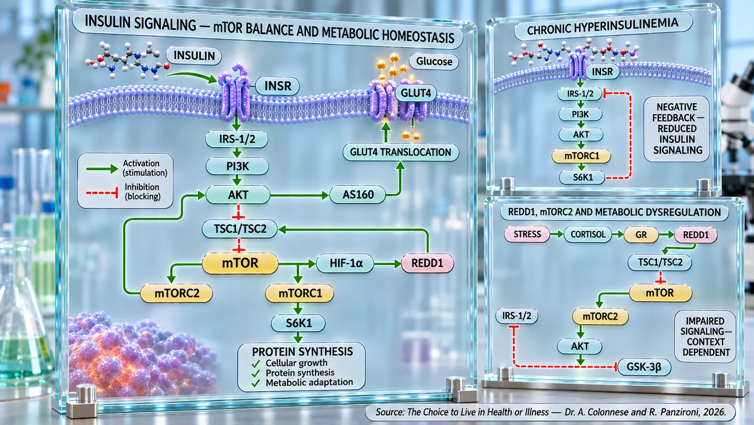
Molecular infographic of a cell membrane with green activation arrows and red inhibition arrows between the insulin receptor, GLUT4, and mTOR complexes.
Insulin signaling scheme: INSR activates IRS-1/2, PI3K, and AKT, promoting GLUT4 translocation and glucose entry; TSC1/TSC2, mTORC1/2, S6K1, HIF-1α, and REDD1 modulate protein synthesis and metabolic adaptation. The boxes link chronic hyperinsulinemia and stress to the reduction or alteration of the signal.

ABSTRACT

Insulin signaling coordinates energy availability, glucose utilization, and cellular adaptation through a molecular regulatory network. In the book "La scelta di Vivere in Salute o in Malattia" (The Choice to Live in Health or Disease), Dr. A. Colonnese and R. Panzironi delve into the PI3K/AKT/mTOR pathway, highlighting how its functional balance contributes to the maintenance of metabolic homeostasis.

The binding of insulin to its receptor activates IRS-1 and IRS-2, which transmit the signal to PI3K and AKT. The latter coordinates metabolic processes and modulates the TSC1/TSC2 complex, attenuating its inhibitory action on mTORC1. mTOR activity thus integrates nutritional, energetic, and hormonal signals, regulating protein synthesis, growth, and cellular adaptation. The REDD1 protein intervenes in the regulation of this system through TSC1/TSC2, while mTORC2 contributes to the activation of AKT.

The persistence of excessive signals can compromise the functionality of the cascade. In particular, the hyperactivation of mTORC1 promotes, via S6K1, a negative feedback on IRS-1/2 substrates, attenuating insulin transmission. Interactions with HIF-1α and REDD1 introduce further levels of regulation. A reduction in mTORC2 activity can also hinder the activation of AKT, modifying the control over TSC1/TSC2 and GSK-3β. These mechanisms indicate that both persistent activation and inappropriate inhibition of regulatory nodes can contribute to metabolic dysfunction.

The perspective described also highlights the value of temporal signal modulation: under physiological conditions, phases of activation and attenuation allow the cell to adapt its responses to the energy and nutritional needs of the evolving organism.

Insulin resistance therefore emerges as a possible consequence of interconnected alterations, rather than a single molecular defect. The relationship with hyperinsulinemia also appears to be bidirectional: alongside the compensatory response to reduced insulin sensitivity, chronic high hormonal exposure could promote further cellular desensitization. Understanding this dynamic allows us to consider the regulation of insulin signaling as a relevant goal for metabolic prevention, while requiring caution in translating molecular mechanisms into general clinical conclusions.

Insulin: at the origins of the signal

In the silent language of our body, insulin is the conductor. A single hormone, produced by a handful of cells in the pancreas, yet capable of coordinating an entire metabolic symphony. It is insulin that, at the end of every meal, interprets the presence of glucose as a signal and orchestrates the opening of cells so they can welcome it as a source of energy. But the role of insulin goes far beyond sugar management: it regulates protein synthesis, promotes the storage of nutrients in tissues, limits lipolysis (the process by which the body breaks down stored fats to use them as energy), and keeps the endogenous production of glucose (the body's ability to produce glucose internally, especially in the liver, without needing to ingest it through food) in check.

The first step for insulin to exert its effects within what is defined as the “insulin pathway”—the complex of all its biochemical reactions—is binding to the insulin receptor (IR), a transmembrane protein (a protein capable of crossing the cell membrane) that completely spans the lipid bilayer of the cell membrane. The insulin receptor functions like a two-part lock: an external part that recognizes and binds to insulin, and an internal part that transmits the signal inside the cell. When insulin attaches to the external part, the receptor activates, changing shape as if turning a key, and thus can begin to communicate with the interior of the cell" (Ullrich et al., 1985) [1].

This event, defined as “autophosphorylation,” is crucial as it allows for the initiation of signal transduction (the process by which a cell receives a signal from the outside and translates it into a response within itself). In this phase, the presence and proper functioning of the receptor are decisive. Any alteration in the number, structure, or functionality of the receptor can compromise the effectiveness of the signal.

Some clinical conditions can compromise the expression of the insulin receptor, as we will explore in the following paragraphs. It has been observed, for example, that the administration of prednisone, a drug similar to cortisol (a hormone produced by the adrenal glands, known as the “stress hormone,” which helps the body respond to difficult situations and also regulates metabolism), reduces receptor expression in muscle and liver, interfering with insulin's ability to activate its target (Pagano et al., 1983) [2]. Other studies indicate that situations of altered energy balance and chronic inflammation can contribute to reduced expression and activity of the insulin receptor in peripheral tissues (Huybrechts et al., 2014) [3].

Under physiological conditions, however, insulin interacts with its receptor efficiently, initiating an orderly and highly regulated intracellular response (a process controlled with great precision by the organism). It is this initial, specific, reversible, and tightly controlled binding that marks the beginning of the metabolic cascade, the foundation of the body's energy regulation.

IRS-1/2: the messengers of insulin signaling

In this section, let's take a closer look at how insulin binds to its receptor to trigger its activity within the cell. The substrates (molecules upon which enzymes act) of the insulin receptor are a family of proteins called IRS (Insulin Receptor Substrates), specifically the IRS-1 and IRS-2 isoforms, which play a crucial role in amplifying and directing the insulin response within the cell.

IRS proteins are adapter proteins that lack autonomous enzymatic activity (proteins that link other proteins together to facilitate signal transmission, but do not perform chemical reactions on their own), equipped with specific sites that allow them to interact with various intracellular molecules. Immediately after the activation of the insulin receptor, IRS proteins are recruited near the plasma membrane and phosphorylated (modified by the addition of a phosphate group, a step that activates or deactivates many proteins within the cell). This phosphorylation process transforms IRS proteins into actual docking platforms for other enzymes, thus paving the way for the activation of intracellular signaling pathways (Ola, 2022) [4].

The strategic importance of IRS-1 and IRS-2 in the activation of the insulin signal is underscored by the fact that alterations in their levels or activity compromise the entire cascade. Some studies have highlighted that IRS-1 expression can be reduced in response to treatments with glucocorticoids (hormones produced by the adrenal glands that regulate metabolism, stress response, and inflammation), leading to reduced phosphorylation following insulin stimulation (Pagano et al., 1983) [2]. The cellular distribution of IRS proteins can also be altered under conditions of metabolic stress, contributing to reduced insulin sensitivity (Huybrechts et al., 2014) [3].

IRS-1 and IRS-2 are not interchangeable: although they share many structural characteristics, they have distinct functions in different tissues. IRS-1 is predominant in skeletal muscle (a type of muscle connected to bones, which allows for voluntary body movements, such as walking or lifting objects) where it mediates glucose uptake, while IRS-2 has a more pronounced role in the liver and the central nervous system (the system composed of the brain and spinal cord, which controls all body functions and coordinates thoughts, movements, and sensations). The efficiency of the insulin signal therefore also depends on the correct balance between these two isoforms, their tissue-specific expression, and their functional integrity (Rabiee et al., 2018) [5].

Under physiological conditions, the phosphorylation of IRS proteins therefore allows for a linear and orderly passage of the insulin signal. However, aberrant phosphorylation—an event that can be induced by chronic inflammation or nutrient excess—can transform IRS proteins into signal inhibitors, blocking the proper functioning of the insulin pathway (Ola, 2022) [4].

The role of IRS proteins, therefore, is to act as a bridge between the receptor and intracellular pathways, ensuring faithful, selective, and controlled transmission. Their proper functioning is essential for metabolic health.

PI3K and AKT: the beginning of the pathway

The activation of insulin receptors and the subsequent phosphorylation of IRS-1 and IRS-2 substrates represent the first steps of the molecular cascade that converts the presence of insulin into biological action. However, it is only from this point that the signal begins to propagate deep into the cell, initiating a complex network of intracellular activations that determine the hormone's metabolic, anabolic, and regulatory effects. The two central protagonists in this process are phosphoinositide 3-kinase (PI3K) and protein kinase AKT, known for their fundamental role in regulating metabolism, cell growth, and survival.

If we imagine IRS proteins as magnets, PI3K is drawn toward the cell membrane. Once there, PI3K acts like a technician preparing a special platform: it modifies a molecule that becomes the starting point for AKT, the next protein in the chain. It is somewhat like a relay race, where PI3K passes the baton to AKT so that the insulin signal can continue its journey inside the cell (Jiang and Liu, 2009) [6].

Subsequently, the protein kinase AKT reaches the membrane, where it is phosphorylated for full functional activation. Once activated, AKT performs a multitude of functions: it stimulates glucose transport in tissues, promotes protein synthesis, inhibits apoptotic processes (related to apoptosis, i.e., the natural process by which cells self-destruct when they are damaged or no longer needed), and regulates lipid metabolism (Hamada et al., 2005) [7].

The robustness and efficiency of the PI3K-AKT signal are crucial for ensuring an effective insulin response. Alterations in any link of this chain can compromise signal transduction and, consequently, affect the cell's ability to adapt to metabolic demands.

It is through the activation of PI3K and AKT that the insulin signal acquires strength and directionality, transforming from a receptor impulse (IRS-1 and IRS-2) into a concrete biological response capable of orchestrating numerous vital cellular functions.

AKT - AS160 - Glut 4: the journey of glucose into the cell

The activation of PI3K and the protein kinase AKT represents one of the most important moments in the entire insulin pathway. It is, in fact, through these molecules that the signal, originating from the binding between insulin and its receptor, acquires the ability to produce concrete metabolic effects within the cell. However, although AKT constitutes one of the main hubs of the insulin cascade, its activation is not in itself sufficient to allow glucose to enter the cells. For this to happen, an additional step is required—less well-known but equally essential—which involves a protein called AS160 (Akt Substrate of 160 kDa). It is precisely through this molecular target that AKT translates the signal received from insulin into a physical and observable event: the translocation (the movement of a molecule or protein from one part of the cell to another) of the GLUT4 transporter toward the cell membrane (Sharma and Dey, 2021) [8].

To better understand this mechanism, it may be useful to imagine the cell as a large warehouse. Inside, there are numerous "doors" already built, but not yet installed on the outer walls. These doors are represented by GLUT4 (Glucose Transporter type 4) molecules, the main glucose transporter in insulin-sensitive tissues, such as skeletal muscle, adipose tissue, and the heart. In resting conditions, however, GLUT4 is not found on the cell surface; instead, it is stored within the cytoplasm (the inner part of the cell surrounding the nucleus) in specific intracellular compartments called Glucose Storage Vesicles (GSV), small vesicles that act as a depot and keep the transporter inactive until the insulin signal arrives. This organization allows the cell to regulate glucose entry with extreme precision, preventing it from occurring when not necessary; therefore, AKT's task is precisely to give the order for these vesicles to be mobilized. This occurs through the phosphorylation of AS160, a protein that acts as a regulator of intracellular traffic (Sharma and Dey, 2021) [8].

This step represents the true concluding moment of the metabolic response to insulin. All the events described in the previous paragraphs, from the binding of the hormone to the receptor up to the activation of IRS-1, IRS-2, PI3K, and AKT, converge toward a single goal: to increase the number of GLUT4 transporters exposed on the cell membrane and thus allow glucose to enter the cell rapidly. In other words, the effectiveness of the entire insulin pathway is measured by the ability to successfully complete this final stretch of the cascade (Sharma and Dey, 2021) [8].

A particularly interesting aspect is that insulin, in most cases, does not induce the synthesis of new GLUT4 molecules. Its effect consists rather in modifying their distribution within the cell. Studies conducted on cardiomyocytes derived from human pluripotent stem cells (heart muscle cells obtained in the laboratory from stem cells) have shown that insulin stimulation leads to a rapid increase in the phosphorylation of AKT and AS160, accompanied by a marked translocation of GLUT4 toward the plasma membrane, without significantly changing the total amount of the protein present in the cell. This means that the control exerted by insulin concerns primarily the localization of the transporter and not its production. This observation is of great importance, as it demonstrates that evaluating only the total amount of GLUT4 can be misleading: what truly determines glucose uptake is the portion of the transporter actually exposed on the cell membrane (Querio et al., 2024) [14].

It is therefore evident how the AKT-AS160-GLUT4 axis constitutes the direct link between insulin signal transduction and the final metabolic response. If any of its components loses efficiency, the entire cascade suffers. Alterations in AKT phosphorylation, functional defects in AS160, or anomalies in the mobilization of vesicles containing GLUT4 progressively compromise the cells' ability to absorb glucose, paving the way for the development of insulin resistance (Sharma and Dey, 2021) [8].

Observations obtained in experimental studies confirm that this mechanism does not represent merely a theoretical model, but constitutes the main determinant of insulin sensitivity in the body's various tissues. Regardless of the type of intervention used, in fact, the recovery of the response to insulin is consistently associated with the reactivation of the IRS-1–PI3K–AKT–AS160–GLUT4 axis, while its inhibition coincides with the onset of insulin resistance. It is precisely this extraordinary convergence of results, observed in different experimental models, that underscores the central role of this pathway in maintaining glycemic homeostasis.

One of the most convincing confirmations comes from studies conducted on neuregulin-1β (a protein involved in the development and metabolism of skeletal muscle). In muscle cells rendered insulin-resistant through exposure to palmitic acid, treatment with neuregulin-1β led to a marked increase in AKT phosphorylation, accompanied by the restoration of GLUT4 translocation toward the plasma membrane and a significant increase in glucose uptake. When AKT activity was pharmacologically blocked, these effects disappeared almost completely, demonstrating that the activation of this kinase represents an indispensable step for the insulin signal to reach AS160 and allow the correct trafficking of vesicles containing GLUT4. The same results were subsequently confirmed in murine models (mice or rats used in scientific research) of type 2 diabetes, in which the treatment improved glycemic control and increased the presence of GLUT4 on the membrane of muscle cells (Yu et al., 2023) [11].

A completely overlapping picture emerged in studies dedicated to luteolin (a natural substance of plant origin with antioxidant and anti-inflammatory biological activities). In adipocytes and animal models of obesity, this natural flavonoid promoted the reactivation of the entire insulin cascade, increasing the phosphorylation of IRS, PI3K, AKT, and AS160 and promoting the translocation of GLUT4 toward the cell membrane. Particularly significant is the fact that the pharmacological inhibition of PI3K completely nullified these effects, demonstrating that the improvement in glucose uptake depends on the integrity of the entire sequence of events that connects PI3K to AKT, AS160, and, finally, to GLUT4 (Park et al., 2026) [10].

Similar results were also obtained with trilobatin and stevioside (natural substances found in plants). Although these are different compounds, both showed the ability to counteract insulin resistance by restoring the functionality of the IRS-1–AKT–GLUT4 pathway in skeletal muscle. In experimental models, a recovery of IRS-1 and AKT phosphorylation, greater GLUT4 translocation, and a consequent increase in glucose absorption were observed. In the case of stevioside, these effects were also accompanied by a marked reduction in oxidative stress and the recovery of the expression of the insulin receptor, IRS-1, AKT, and GLUT4, suggesting that the correct functioning of this cascade depends not only on the integrity of the proteins involved but also on the redox balance of the intracellular environment (Liu et al., 2020) [12]; (Deenadayalan et al., 2021) [13].

The study conducted with montelukast (an anti-inflammatory drug), in combination with dapagliflozin (a drug that promotes the elimination of glucose through urine), also reinforces this interpretation. Although these are drugs with different mechanisms of action, the improvement in insulin sensitivity was again associated with the reactivation of the IRS-1/AKT/GLUT4 pathway. In this case, the recovery of insulin signaling was accompanied by a significant reduction in cellular stress, inflammation, and a recovery of glucose metabolism. Once again, the point of convergence of these processes is represented by the cell's ability to correctly mobilize GLUT4 toward the plasma membrane and restore efficient glucose uptake (Fleifel et al., 2022) [9].

Taken together, these findings show that the AKT-AS160-GLUT4 axis constitutes the main link between the activation of the insulin receptor and the final biological effect of the hormone. However, AKT's function is not limited to the regulation of GLUT4 traffic. Once activated, this kinase continues to transmit the signal to numerous other intracellular targets involved in protein synthesis, cell growth, and the control of energy metabolism. Among these, the complex formed by the TSC1 and TSC2 proteins assumes particular importance, as it represents one of the main control points through which AKT modulates the activity of mTOR, the cell's major anabolic regulator. It is precisely from this further level of regulation that the next step of the insulin pathway will begin.

TSC1/TSC2: the guardians of anabolism

Reaching the heart of insulin signaling, the activation of the AKT kinase represents a crucial junction: it not only propagates the molecular cascade but acts as a true bridge toward the final destination of the insulin pathway. In fact, AKT mediates the direct inhibition of the TSC1/TSC2 tuberous sclerosis complex (two proteins that work together to regulate cell growth and metabolism), finding its functional outlet in mTOR, the orchestrator of cell growth, protein synthesis, and metabolic regulation that we will address in the next paragraph.

This step marks the point where the signal, having started from the insulin receptor (IRS-1 and IRS-2), translates into a tangible biochemical action, transforming insulin into an intracellular operational order. Understanding how AKT deactivates the molecular brake represented by TSC1/TSC2 is essential for deciphering the final activation of mTOR: the true goal of the pathway.

TSC1 (hamartin) and TSC2 (tuberin) are two proteins that act together to restrain mTOR activity when environmental conditions are not favorable.

When the TSC1/TSC2 complex is active, mTOR cannot be activated. Conversely, in the presence of favorable signals such as insulin, amino acids, or energy availability, TSC2 is inhibited through phosphorylation by AKT, and the anabolic cascade proceeds (Inoki et al., 2003) [15].

In fact, the absence or malfunction of hamartin or tuberin (TSC1 and TSC2) leads to a deregulation of the quintessential cell growth complex “mTOR,” which results in uncontrolled development, with serious clinical consequences as demonstrated in pathologies related to tuberous sclerosis (a rare genetic disease that causes the formation of benign tumors in various organs, such as the brain, kidneys, heart, and skin, and can affect neurological and cognitive development) (Wong, 2009) [16].

From an evolutionary perspective, the TSC1/TSC2 complex has allowed cells to develop a capacity for discernment as well as a negative feedback system; in fact, not all situations in which insulin is present (or an excess of insulin) are necessarily favorable for growth. This complex acts as a protective metabolic barrier, ensuring that mTOR activation is granted only when all conditions—oxygen, nutrients, energy, and hormones—are adequate.

TSC1 and TSC2, therefore, are not simple growth inhibitors: they are the guarantors of anabolic balance (a condition in which the body builds more tissue than it destroys, promoting growth, repair, and the accumulation of muscle mass and energy reserves), capable of interpreting and integrating extracellular signals coming from multiple pathways.

mTOR: the master anabolic regulator

And here we arrive at the central hub of the signaling network that insulin activates within the cell: mTOR, an acronym for mechanistic Target of Rapamycin. It is a fundamental, highly conserved protein (a protein that has remained identical across many species throughout evolution, a sign of its essential function) that acts as a sensor and integrator of nutrient, energy, and hormonal stimuli. mTOR does not function alone: it exists in two functionally distinct complexes, mTORC1 and mTORC2, which specifically orchestrate different aspects of cellular physiology (Brugarolas et al., 2004) [17].

mTORC1 is the most studied complex: it is responsible for promoting cell growth and proliferation. It does this by activating protein synthesis, inhibiting autophagy (the process by which the cell "recycles" its own damaged or no longer useful components), and stimulating lipid biosynthesis (the process through which the body produces fats, such as triglycerides and cholesterol, from simpler substances). As we have seen previously, this complex responds directly to insulin signals through the IRS–PI3K–AKT pathway: the activation of AKT inhibits TSC1/TSC2 (which acts as a brake on mTORC1) and thus allows for the full activation of the mTOR complex (Reiling & Hafen, 2004) [18].

mTORC2, on the other hand, is less studied. However, it is essential for regulating the organization of the cytoskeleton (the internal network of filaments present in the cell that maintains its shape, enables movement, and organizes the transport of substances within it), for cell survival, and for the full activation of AKT, given that it is mTORC2 that phosphorylates AKT at one of the two sites necessary for its activation (Brugarolas et al, 2004) [17].

It has been observed that the hyperactivation of mTOR can lead to negative metabolic consequences, such as reduced insulin sensitivity and the promotion of pathological conditions like obesity and type 2 diabetes (Mao & Zhang, 2018) [19]. Maintaining balanced mTOR activation is therefore essential for metabolic health and cellular longevity.

Finally, it should be emphasized that mTOR is not just a target of insulin signaling: it is a convergence point for signals coming from nutrients (such as amino acids), from cellular energy status via AMPK (an enzyme present in all cells that acts as a sensor to regulate metabolism), and from oxidative stress (a condition in which too many free radicals accumulate—unstable molecules that can damage cells, proteins, and DNA, contributing to aging and various diseases), acting as a central regulator of cell fate.

mTORC1 - S6K1

The activation of mTORC1 represents one of the central moments in the entire insulin signaling cascade. However, the complex does not exert most of its biological effects directly. As already observed for AKT, mTORC1 also entrusts the execution of its metabolic decisions to specific effector proteins, which are responsible for transforming a molecular signal into a concrete cellular response. Among these, the most important is ribosomal protein S6 kinase 1 (S6K1), a kinase that constitutes the main downstream target of mTORC1 and coordinates numerous processes involved in cell growth, protein synthesis, metabolism, and proliferation (Ghosh & Kapur, 2017) [20].

To understand the role of this protein, it can be helpful to imagine mTORC1 as an orchestra conductor. The conductor sets the tempo and coordinates all the musicians, but does not produce the music directly. Similarly, mTORC1 represents the decision-making center of the anabolic response, while S6K1 acts as one of its main executors, tasked with transmitting the signal to numerous intracellular targets so that the cell can increase protein synthesis, support growth, and adapt its metabolism to the availability of nutrients and growth factors (Ghosh & Kapur, 2017) [20].

However, the activation of S6K1 does not occur randomly. Before it can be phosphorylated, the kinase must be recruited into proximity with the mTORC1 complex through a precise organization of the proteins that make up the signaling platform (Ahmed et al., 2019) [22].

Once its activation is complete, S6K1 becomes the primary interpreter of the signal coming from mTORC1. Through the phosphorylation of numerous intracellular targets, this kinase coordinates protein synthesis, cell growth, and metabolic adaptation, allowing the cell to translate nutrient availability into a coherent anabolic program. However, more recent research has shown that its functions are not limited to the cytoplasm. Once phosphorylated, S6K1 can also translocate into the nucleus, where it participates directly in the regulation of gene expression (the use of a gene by the cell to produce proteins), significantly expanding the biological reach of the signal transmitted by mTORC1 (Jeon et al., 2022) [21].

The physiological importance of this cascade emerges clearly in tissues characterized by continuous cell renewal. In the intestinal epithelium, for example, the selective activation of the mTORC1-S6K1 axis promotes the synchronized proliferation of intestinal stem cells. This finding suggests that the proliferative response depends primarily on the activation of biosynthetic programs regulated by S6K1 rather than a simple increase in energy production (Casanova-Maldonado et al., 2023) [23].

The complexity of S6K1's function also emerges when comparing results obtained in experimental models with those observed in humans. Although S6K1 phosphorylation is frequently used as an indicator of mTORC1 anabolic activation, available evidence suggests that it does not necessarily represent a direct quantitative index of protein synthesis. The final response depends, in fact, on the integration of numerous metabolic, nutritional, and mechanical signals that modulate the efficiency of protein translation and the adaptive response of tissues. This aspect underscores how the simple activation of S6K1 constitutes only one of the various regulatory levels that determine the final biological outcome (Borack et al., 2021) [26].

When S6K1 activation remains finely regulated, it contributes to the maintenance of cellular homeostasis (maintenance of the balance and proper functioning of the cell). Conversely, its persistent hyperactivation can transform a physiologically adaptive program into a pathological process. A significant example comes from the heart, where the loss of a specific enzyme that profoundly alters RNA regulation leads to persistent hyperactivation of the mTORC1-S6K1 cascade. This condition is associated with a marked reduction in autophagy, the accumulation of intracellular damage, and the onset of cardiomyocyte atrophy and apoptosis, accelerating the progression toward heart failure. Pharmacological inhibition of the pathway attenuates these alterations, confirming how excessive regulation of S6K1 can turn an anabolic signal into a major mechanism of cellular damage (Annamalai et al., 2025) [24].

A similar principle also emerges in the study of the blood-brain barrier (a natural filter that protects the brain from potentially harmful substances present in the blood). In conditions of acute liver failure, the marked reduction in the availability of arginine (an amino acid involved in protein production and numerous cellular processes) prevents the normal activation of the mTORC1-S6K1 pathway, leading to a cell cycle arrest of cerebral endothelial cells and a simultaneous activation of autophagy. Arginine supplementation restores S6K1 phosphorylation, promotes the resumption of cell proliferation, and contributes to the recovery of the integrity of the blood-brain barrier. These results identify S6K1 as one of the main sensors of nutrient availability and as a central element in the delicate balance between cell growth and autophagy (Yang et al., 2025) [25].

Taken together, these findings outline a much broader view of the role played by S6K1. Far from being a simple substrate of mTORC1, it represents the main translator of the anabolic response, integrating nutritional, metabolic, and transcriptional signals to coordinate growth, proliferation, protein synthesis, and cellular adaptation. Precisely this central position, however, also makes S6K1 one of the main culprits behind the unfavorable effects associated with chronic hyperactivation of the mTORC1/S6K1 pathway. When the anabolic signal persists beyond physiological limits, the same kinase that supports cell growth can, in fact, activate feedback mechanisms intended to limit insulin signaling. It is this self-regulatory circuit, exerted through the phosphorylation of IRS-1, that will constitute the direct link between chronic mTORC1 hyperactivation and the development of insulin resistance, the topic of the next section (Ghosh & Kapur, 2017) [20].

S6K1 - IRS-1 – Insulin resistance

The activation of the insulin pathway described in the previous sections is not a process intended to remain constantly switched on. Like all biological systems dedicated to maintaining homeostasis, this cascade also possesses sophisticated self-regulation mechanisms that limit its intensity once the necessary metabolic response has been achieved. Among these, the most important is the negative feedback circuit exerted by the S6K1 kinase on the insulin receptor substrate IRS-1. Under physiological conditions, this mechanism prevents excessive stimulation of the PI3K-AKT pathway, helping to maintain cellular balance. However, when the activation of mTORC1 and S6K1 becomes persistent, this same circuit loses its adaptive function and progressively transforms into one of the main molecular events responsible for the onset of insulin resistance (Um et al., 2006) [27].

This change depends primarily on the chronic availability of nutrients. In addition to lipids, a persistent excess of amino acids is also capable of keeping the mTORC1 pathway active: under these conditions, S6K1 continues to receive a constant anabolic stimulus and phosphorylates IRS-1, progressively reducing its ability to transmit the signal from the insulin receptor. The consequence is lower PI3K activation, reduced AKT phosphorylation, and a progressive impairment of glucose uptake in peripheral tissues. Studies conducted in humans confirm this model by showing that the simple infusion of amino acids is sufficient to increase S6K1 activity, reduce insulin sensitivity by about a quarter, and decrease muscle glucose utilization (Um et al., 2006) [27].

Chronic hyperactivation of the mTORC1 pathway is not, however, limited to functionally modifying IRS-1. The same study highlighted that continuous phosphorylation also promotes the progressive degradation of the protein, reducing its intracellular availability. In this way, the negative feedback takes on a dual nature: on one hand, it immediately limits the ability of IRS-1 to transmit the insulin signal, and on the other, it progressively decreases its quantity, making it increasingly difficult to recruit PI3K even in the presence of physiological concentrations of insulin. This mechanism provides a molecular explanation for the reduction in IRS-1 levels frequently observed in subjects with obesity and type 2 diabetes (Tzatsos & Kandror, 2006) [28].

Further evidence shows that the negative feedback exerted by S6K1 can also affect other regulatory sites of IRS-1. In cells lacking the TSC1-TSC2 complex, persistent hyperactivation of the mTOR-S6K1 pathway also leads to a reduction in the interaction between IRS-1 and the activated receptor, limiting PI3K recruitment and compromising the subsequent activation of AKT. At the same time, S6K1 also represses the transcription of the IRS-1 gene, reducing the total amount of available protein. The result is a dual negative control, both acute and chronic, that makes insulin signal transmission progressively inefficient. Treatment with rapamycin (an immunosuppressant drug that primarily inhibits the mTORC1 complex) or the silencing of S6K1 attenuates both alterations, allowing for a partial recovery of AKT activation and confirming the central role of the mTOR-S6K1 cascade in maintaining insulin resistance (Harrington et al., 2004) [29].

The importance of this feedback circuit emerges even more clearly when one of the main physiological regulators of the mTOR pathway is altered. The loss of function of the TSC1-TSC2 complex leads to an activation of the mTOR-S6K1 cascade sufficient to induce a marked reduction in the expression of IRS-1 and IRS-2. Consequently, signal transmission between the insulin receptor and PI3K is deeply compromised, while AKT activation in response to insulin is almost completely abolished. It is interesting to observe how signaling pathways that do not depend on IRS proteins remain substantially preserved, demonstrating that the defect selectively affects the link between the insulin receptor and PI3K. Once again, treatment with rapamycin restores IRS-1 and IRS-2 levels and recovers the ability of insulin to activate AKT, confirming that chronic hyperactivation of mTOR-S6K1 is the main driver of negative feedback (Shah et al., 2004) [30].

The clinical importance of this mechanism is further confirmed by studies conducted in humans. In subjects with obesity and type 2 diabetes, a persistent hyperactivation of the mTOR-S6K1 pathway has been observed, accompanied by an increase in the inhibitory phosphorylation of IRS-1 and reduced AKT activation in insulin-sensitive tissues. These alterations are closely associated with the loss of the normal metabolic response to insulin and confirm that the feedback circuit described in cellular and animal models is also present in human pathophysiology, representing one of the main links between nutritional excess, metabolic inflammation, and the development of insulin resistance (Denhez et al., 2020) [31].

Fortunately, the same evidence shows that this process can be at least partially countered. After nine weeks of spontaneous physical activity, the skeletal muscle of rats shows a significant reduction in the phosphorylation of mTOR, S6K1, and IRS-1, without changing the total amount of the main proteins in the insulin cascade. At the same time, muscle oxidative capacity increases, body mass decreases, and one of the main molecular mechanisms responsible for negative feedback on insulin signaling is reduced. These results suggest that the benefits of physical exercise depend not only on increased glucose consumption during muscle contraction but also on the rebalancing of the mTOR-S6K1 pathway and the progressive reduction of the inhibitory phosphorylation of IRS-1 (Glynn et al., 2008) [32].

Taken together, these findings outline a coherent pathophysiological model. Under normal conditions, the phosphorylation of IRS-1 mediated by S6K1 represents a sophisticated self-regulation mechanism, essential to avoid excessive stimulation of the insulin cascade. However, when nutrient availability remains persistently high or the mTORC1-S6K1 pathway is chronically hyperactivated, this system loses its homeostatic function. The progressive phosphorylation and depletion of IRS-1 and IRS-2 interrupt the link between the insulin receptor and PI3K, reduce AKT activation, and compromise GLUT4 translocation and all of insulin's main metabolic responses. It is thus understood that insulin resistance does not represent a sudden alteration of the insulin receptor, but the consequence of a progressive dysfunction of intracellular signal transmission. This observation completes the functional picture of the PI3K-AKT-mTOR cascade and naturally prepares for the next section, in which the role of further intracellular regulators involved in the control of insulin sensitivity will be explored in depth.

HIF-1α: the cellular oxygen sensor

The activation of mTOR, as we have seen previously, represents the heart of insulin signaling in terms of promoting growth and cellular biosynthesis (the set of processes by which a cell builds new molecules). However, the role of this complex is not limited to the control of protein synthesis and anabolism (the production of proteins from amino acids); on the contrary, mTOR also acts as a strategic node for the activation of a new phase of the cellular program: the regulation of gene expression, which is how a cell decides which genes to turn on and which to keep off, depending on the moment and the needs, as if it were a director turning on only the right lights on stage.

It is precisely thanks to its ability to modulate transcription factors (proteins that bind to DNA and regulate gene activity, deciding when and how much a gene should be "turned on") such as, for example, HIF-1α (hypoxia-inducible factor, a key protein that acts as a true cellular oxygen sensor), that mTOR extends its influence beyond immediate metabolism, guiding the cell toward wider-ranging adaptations. In this sense, HIF-1α does not represent a simple downstream target of insulin signaling, but the natural continuation of the biochemical trajectory activated by insulin. Understanding this step means delving into the heart of cellular function, where signals arriving from the environment or metabolism cause the cell to turn on only the genes it needs at that moment. It is like choosing the right recipe from a drawer: the cell reads only the instructions useful for producing the proteins necessary for the situation.

Under normoxic conditions (situations in which the oxygen supply to cells is normal, i.e., sufficient to ensure proper cellular function), HIF-1α is continuously synthesized but just as rapidly degraded, inevitably losing its transcriptional capacity. However, when oxygen is scarce, HIF-1α accumulates inside the cell and activates special genes that help the cell adapt to the oxygen deficiency. These genes serve, for example, to produce more energy without oxygen, create new blood vessels, increase red blood cells, and improve the cell's defenses to help it survive (Semenza, 2003) [33].

HIF-1α is influenced not only by oxygen but also by hormonal signals; insulin, for example, is able to promote the expression and stabilization of HIF-1α, even under normal oxygen conditions, through the activation of the PI3K/AKT/mTOR pathway. Studies conducted on human and rat smooth muscle cells have shown that insulin increases HIF-1α expression at the transcriptional level (transcription of genes involved in the cellular response for protein production). This suggests that HIF-1α can be considered a convergence point between oxygen metabolism and insulin signaling (Doronzo et al., 2006) [34].

Other studies have highlighted that PI3K and mTOR contribute to the regulation of HIF-1α, highlighting a multi-level interaction between metabolic signals and the hypoxic response. In this way, HIF-1α is configured not only as a hypoxic factor that therefore allows cellular function even in the absence of oxygen, but as an integrator of broader physiological signals, capable of shaping gene transcription based on energy and nutritional availability (Treins et al., 2002) [35].

The expression of HIF-1α can therefore be induced in a "non-hypoxic" way, that is, in the presence of oxygen, demonstrating that its activation represents a broader mechanism of physiological adaptation, in which even hormones like insulin play an active role. This positions the HIF-1α protein at the crossroads of metabolism, environment, and intracellular signaling.

REDD1: the genesis of a metabolic drift

With the activation of HIF-1α, insulin signaling reaches a new dimension where metabolic changes are translated into adaptive gene responses. However, among the transcriptional targets induced by HIF-1α, one in particular stands out for its importance and its role as a physiological brake: REDD1.

This protein, activated precisely in response to anabolic or hypoxic stimuli, represents a self-regulatory mechanism of the insulin pathway. Through its action, REDD1 activates a control system that inhibits mTOR activity, signaling to the cell that the action of insulin must be scaled back or modulated.

Several studies have shown that REDD1 acts by establishing a direct link between stress signals (stimuli indicating a condition of stress for the cell or organism, such as lack of nutrients, inflammation, toxins, or other factors that alter internal homeostasis) and the TSC1/TSC2 complex, as we saw in paragraph 4. Specifically, REDD1 promotes the activity of this complex, which subsequently acts as a brake on mTOR, preventing its activation in contexts of energy excess or oxygen deficiency (Brugarolas et al, 2004) [17].

It has been observed that REDD1 can also be induced by insulin itself, for the purpose of counter-regulating the insulin signal: in adipocytes, the hormone promotes its expression via HIF-1α, suggesting a negative feedback loop aimed at limiting the excessive activation of mTORC1 when anabolism is already stimulated (Regazzetti et al., 2010) [36]. This balancing system ensures that cell growth (through the action of insulin) occurs in a controlled manner, preventing metabolic damage.

Under conditions of hypoxia or cellular stress, REDD1 is rapidly activated and accumulates in the cytoplasm (the gelatinous substance inside the cell where organelles are located). Its stability is finely regulated by mTOR itself, through mechanisms that include protein modulation and the control of its degradation (Dennis et al., 2014) [37].

When active, REDD1 interacts with a specific protein, promoting the dephosphorylation (inactivation) of AKT and blocking the signal toward mTOR (DeYoung et al., 2008) [38].

It has also been documented that high and persistent levels of REDD1 are associated with conditions of insulin resistance and metabolic dysfunction, such as those observed in the skeletal muscles of subjects with type 2 diabetes (Williamson et al, 2015) [39]. In these contexts, the excessive inhibition of mTOR, induced by REDD1, can contribute to reduced insulin efficacy, demonstrating that even molecular brakes, if activated in a disordered manner, can have pathological effects.

And it is precisely here that the circle closes: the activation of REDD1, induced by HIF-1α and sustained by mTOR, does not represent just a momentary brake on the insulin pathway, but is configured as the key mechanism that determines its progressive dysfunction. In conclusion, therefore, REDD1, by stably inhibiting mTOR through the activation of the TSC1/TSC2 tuberous sclerosis complex, breaks the physiological flow of insulin signaling and thus the pathway, hindering downstream responses and establishing a functional block of insulin action that leads to the most feared clinical condition: insulin resistance.

mTOR – mTORC2 - AKT

At this point, a fundamental aspect of mTOR biology emerges that remained in the background for many years. Although research has focused primarily on mTORC1, we now know that the mTOR protein exerts its functions through two distinct but closely coordinated complexes: mTORC1, responsible primarily for controlling protein synthesis and cellular anabolism, and mTORC2, a complex that plays an equally essential role in regulating cell survival, cytoskeleton organization, metabolism, and, above all, the full functionality of AKT. Evidence accumulated in recent years has profoundly changed the traditional view of the insulin pathway, showing that many functions previously attributed generically to mTOR actually depend on the specific activity of mTORC2, which represents one of the main regulators of the kinase family, including AKT (Oh & Jacinto, 2011) [40].

For mTORC2 to perform this function, the complex must maintain a precise structural organization through a specific protein that, like a true assembly platform, binds mTOR and AKT simultaneously, positioning the substrate optimally for phosphorylation. It is interesting to note that the association between AKT and mTORC2 increases after insulin stimulation via the PI3K pathway, confirming that substrate availability constitutes an additional level of regulation of the insulin response (Yao et al., 2017) [41].

The activity of mTORC2, however, does not begin when AKT is already fully formed. Recent research has shown that the complex intervenes even during protein synthesis, and this mechanism demonstrates that mTORC2 directly links protein synthesis to protein quality control systems, ensuring that AKT acquires a stable conformation during its formation (Oh et al., 2010) [42].

This function is further confirmed by studies showing that in the absence of mTORC2 or its essential components, AKT loses constitutive phosphorylation, becomes structurally unstable, and is rapidly directed toward degradation. It follows that this complex does not only regulate the enzymatic activation of AKT, but also ensures its structural maturation (Facchinetti et al., 2008) [43].

The efficiency of this system also depends strictly on the energy state of the cell. It has been demonstrated that mTORC2 requires relatively high intracellular concentrations of ATP (the main immediate source of energy usable by the cell) to maintain its activity. The loss of this regulation simultaneously compromises the integrity of mTORC2 and the correct folding of AKT, demonstrating that energy availability is one of the main factors modulating the functionality of the complex. In other words, mTORC2 acts as a system capable of adapting the maturation of AKT to the metabolic state of the cell, avoiding the support of anabolic programs when energy resources are insufficient (Chen et al., 2013) [44].

Once protein maturation is complete, the phosphorylation of AKT mediated by mTORC2 becomes essential to allow for a correct metabolic response to insulin. In the liver, for example, the selective loss of Rictor (an essential protein that is part of the mTORC2 complex) prevents the phosphorylation of AKT, compromising the normal metabolic transition between fasting and the postprandial state. Under these conditions, the liver continues to produce glucose despite hyperinsulinemia, maintains high gluconeogenesis, and limits both glycogen synthesis (glucose storage in the form of glycogen) and lipogenesis (production of fats by the body). The experimental expression of a constitutively active form of AKT is able to almost completely restore these alterations, demonstrating how AKT represents the main metabolic effector (a component that modifies or regulates the metabolic activity of the cell) of mTORC2 in the regulation of glucose and lipid homeostasis (Hagiwara et al., 2012) [45].

From a pharmacological point of view, the relationship between mTORC2 and AKT also proves to be extremely important. For many years, it was believed that rapamycin inhibited only mTORC1. Subsequent studies have shown that prolonged treatment also prevents the formation of new mTORC2 complexes, progressively reducing the phosphorylation of AKT. This phenomenon contributes significantly to the antitumor effects of rapamycin, explaining at least in part the different sensitivity observed between different tissues and different neoplasms (Sarbassov et al., 2006) [46].

Paradoxically, however, the selective inhibition of mTORC1 can also lead to a compensatory reactivation of mTORC2. In models of diffuse large B-cell lymphoma (a cancer of the lymphatic system), it has been observed that treatment with rapamycin effectively reduces the phosphorylation of mTORC1 targets, but simultaneously induces an increase in AKT phosphorylation mediated by mTORC2. This adaptation represents an important mechanism of therapeutic resistance, as it keeps cell survival pathways active despite the blockade of anabolism. The simultaneous inhibition of both complexes or the direct destabilization of mTORC2 allows for the abolition of this compensatory mechanism, significantly enhancing the efficacy of the antitumor treatment (Gupta et al., 2009) [47].

AKT – GSK-3B

At this point in the cascade, one of the most important molecular targets of AKT clearly emerges: glycogen synthase kinase 3 beta (GSK-3β). This protein belongs to the kinase family and was initially identified as a regulator of glycogen synthesis. It was later understood that its role is much broader, extending to the control of cell survival, proliferation, differentiation, gene expression, protein synthesis, and cytoskeleton organization. Unlike many other kinases, GSK-3β has a peculiar characteristic: in healthy cells, it is constitutively active and performs a predominantly inhibitory function. Consequently, in most cases, the activation of a biological response does not require the activation of GSK-3β, but rather its inactivation. This is precisely one of the main tasks of AKT within the insulin pathway.

For many years, however, it was unclear which kinase was responsible for this inactivation. The question was clarified by elegant experiments in which a dominant-negative PI3K/AKT mutant was developed, capable of selectively preventing its activation. When AKT was blocked, insulin completely lost the ability to inactivate GSK-3β, demonstrating that AKT represents the essential link between PI3K activation and the inhibitory phosphorylation of this kinase. The authors also demonstrated that AKT and GSK-3β can physically interact to form a molecular complex, which makes the process extremely efficient (Van Weeren et al., 1998) [49].

This inactivation has an importance that goes well beyond glucose metabolism. Under physiological conditions, in fact, a constitutively active GSK-3β represents a stimulus that favors apoptosis. Experiments conducted through the overexpression of the active form of the protein have shown that the increase in its activity is sufficient, on its own, to induce programmed cell death in different cell lines. Conversely, the expression of a dominant-negative form effectively protects cells from apoptosis even when the PI3K pathway is pharmacologically inhibited. These results have profoundly changed the understanding of the insulin cascade, demonstrating that one of the main mechanisms through which AKT promotes cell survival consists precisely in keeping GSK-3β in an inactive state (Pap & Cooper, 1998) [48].

The biological importance of this axis also emerges clearly in skeletal muscle, where AKT simultaneously coordinates growth and the maintenance of muscle mass. Studies conducted in humans have shown that eight weeks of resistance training lead to an increase in AKT phosphorylation, accompanied by the simultaneous inactivation of GSK-3β and the activation of mTOR. During the subsequent de-training period, the opposite phenomenon is observed: AKT and GSK-3β phosphorylation decrease, favoring the loss of muscle mass. This results in the image of a perfectly coordinated circuit in which AKT acts as a switch capable of orienting muscle tissue toward anabolism or catabolism, simultaneously modulating GSK-3β and mTOR (Léger et al., 2006) [50].

The AKT-GSK-3β axis assumes an equally important role in the nervous system. In neurons, in fact, GSK-3β regulates numerous substrates involved in cytoskeleton stability, including the Tau protein (a protein that helps maintain the internal structure of neurons stable and functional). In human neuroblastoma cells (cancer cells of the nervous system), insulin stimulation significantly increases the phosphorylation of AKT and, consequently, the inhibitory phosphorylation of GSK-3β. This inactivation is accompanied by a marked reduction in Tau protein phosphorylation. Although these results were obtained in a cell model and do not represent a direct demonstration of neuroprotection, they highlight how the regulation of GSK-3β by AKT simultaneously influences Tau phosphorylation, redox homeostasis, and cell survival (Jorda et al., 2026) [51].

The importance of this relationship is also confirmed by numerous observations from research on neurodegenerative diseases. In Parkinson's disease, the reduction in PI3K/AKT pathway activity is associated with persistent hyperactivation of GSK-3β, favoring neuroinflammation, oxidative stress, dopaminergic neuron apoptosis, and the development of cerebral insulin resistance. Conversely, pharmacological activation of the PI3K/AKT pathway or selective inhibition of GSK-3β reduces these alterations in experimental models, so much so that numerous drugs already approved for other indications, such as metformin, pioglitazone, fenofibrate, and statins, are currently being studied for their ability to modulate this molecular cascade. The available evidence also suggests that the most promising therapeutic strategies are those capable of simultaneously activating AKT and inhibiting GSK-3β, restoring the physiological balance of the entire signaling pathway (AlRuwaili et al., 2025) [52].

In recent years, it has also emerged that the mTORC2-AKT-GSK-3β axis does not exclusively regulate metabolism and cell survival, but also controls the physical organization of the cell: this discovery further expands the physiological role of the insulin cascade, demonstrating that AKT and GSK3β also participate in the regulation of intracellular homeostasis, calcium transfer, energy metabolism, and communication between organelles, which are fundamental processes for the proper functioning of the cell (Kalarikkal et al., 2024) [53].

Finally, the biological relevance of this pathway also emerges in the oncological field. In glioblastoma, one of the most aggressive brain tumors, the persistent activation of AKT favors proliferation, cell survival, and the maintenance of cancer stem cells, while the modulation of GSK-3β simultaneously influences the cell cycle, metabolism, and apoptotic programs. Several experimental inhibitors directed against AKT or GSK-3β have been shown to reduce tumor growth, induce apoptosis, and limit the self-renewal of glioblastoma stem cells in preclinical models. Similarly, in platelets (blood components that help stop bleeding by forming clots), the AKT-GSK-3β axis also represents one of the main functional regulators: during platelet activation, AKT physiologically inactivates GSK-3β, favoring aggregation, granule secretion, and thrombus formation, while salidroside (one of the main bioactive compounds of Rhodiola rosea) interrupts this process by reducing the phosphorylation of both AKT and GSK-3β. In this way, GSK-3β remains functionally active, exerting its inhibitory effect on platelet function and reducing thrombus formation in experimental models (Majewska & Szeliga, 2017) [54]; (Wei et al., 2020) [55].

GSK-3β - IRS-1

At this point in the process, an additional level of regulation in insulin signaling emerges. While in the previous paragraphs we saw how AKT physiologically inactivates GSK-3β, it is equally important to understand that GSK-3β itself, when it remains active or is reactivated, is capable of exerting direct control over the main adapter of the insulin pathway: IRS-1. This creates a bidirectional regulatory circuit. On one hand, AKT limits GSK-3β activity to promote the metabolic response to insulin; on the other, when this inhibition wanes, GSK-3β modifies IRS-1, progressively reducing its functional efficiency and attenuating signal transduction. This mechanism represents one of the main links between the physiology of the insulin pathway and the development of insulin resistance.

The first experimental demonstrations of this phenomenon date back to the late 1990s, when IRS-1 was identified as one of the direct targets of GSK-3β. Until then, this kinase was known primarily as a regulator of glycogen synthase, while its involvement in the regulation of insulin signaling was still poorly defined. The experiments showed that GSK-3β is capable of directly phosphorylating IRS-1 and that this modification profoundly alters the protein's behavior. In fact, IRS-1 loses part of its ability to be phosphorylated by the insulin receptor and, even more interestingly, acquires the ability to hinder the receptor's own activity. In other words, a protein normally tasked with transmitting the insulin signal progressively transforms into an element that limits its propagation. The authors therefore proposed that excessive GSK-3β activity could represent one of the molecular mechanisms responsible for the reduced insulin sensitivity observed in obesity and type 2 diabetes (Eldar-Finkelman & Krebs, 1997) [56].

The interaction between GSK-3β and IRS-1, however, proved more complex than initially hypothesized. Although the phosphorylation of IRS-1 by the kinase reduces its functional activity, the constitutive activity of GSK-3β simultaneously contributes to maintaining intracellular levels of IRS-1 and IRS-2. Experiments conducted in specific cells have shown that the pharmacological inhibition of GSK-3β, or its insulin-induced inactivation, leads to a progressive reduction in the cellular quantities of both adapter proteins through a mechanism primarily dependent on their degradation. For IRS-2, there is also a reduction in gene expression. This apparent paradox suggests that GSK-3β performs a dual function: on one hand, it limits the functional efficiency of IRS-1, while on the other, it contributes to maintaining the quantitative balance of IRS proteins, preventing their excessive degradation (Nemoto et al., 2006) [57].

The pathophysiological importance of these mechanisms was subsequently confirmed in animal models of insulin resistance. In Zucker Diabetic Fatty rats, characterized by marked GSK-3β hyperactivity, selective inhibition of the kinase significantly improves the response to insulin. This improvement is accompanied by a recovery of IRS-1 phosphorylation, greater activation of the PI3K-AKT pathway, and an increase in GLUT4 translocation to the cell membrane, resulting in increased glucose transport in skeletal muscle. These effects are observed both in muscle preparations studied in vitro and after systemic administration of the inhibitors, indicating that GSK-3β hyperactivity is not just a marker of insulin resistance, but contributes directly to the post-receptor signaling defect (Henriksen et al., 2003) [58].

Further studies have shown that this recovery of insulin sensitivity also affects the more proximal events of the signaling cascade. Selective inhibition of GSK-3β increases the phosphorylation of both the insulin receptor and IRS-1, increases AKT phosphorylation, and simultaneously reduces the inhibitory phosphorylation of IRS-1, a modification closely associated with the loss of protein function (Henriksen & Teachey, 2007) [59].

Similar observations had already emerged in prediabetes models, in which pharmacological inhibition of GSK-3β improved insulin signaling without directly modifying receptor activation. The main effect consisted of restoring IRS-1 functionality, increasing its association with PI3K, and subsequently reactivating AKT, demonstrating that the primary target of GSK-3β is represented by the post-receptor events of the insulin cascade. This recovery was particularly evident in skeletal muscle, suggesting that different tissues may exhibit different susceptibility to the modulation of this kinase (Dokken et al., 2005) [60].

All these findings have progressively consolidated the role of GSK-3β as one of the main negative regulators of insulin sensitivity. Reviews published in subsequent years have shown how the overexpression and hyperactivity of the kinase are common features in animal models of obesity and in subjects with type 2 diabetes. Its inhibition simultaneously improves post-receptor signaling, promotes GLUT4 translocation, increases peripheral glucose utilization, and reduces hepatic glucose production, identifying GSK-3β as an important therapeutic target in the prevention and treatment of insulin resistance (Henriksen & Dokken, 2006) [61].

The most recent confirmation of the centrality of this mechanism comes from studies on kidney damage associated with hyperinsulinemia. In glomerular podocytes (renal cells that help filter the blood), chronic exposure to high concentrations of insulin leads to a progressive depletion of the insulin receptor, a reduction in the activity of the IRS1-PI3K-AKT pathway, and a consequent reactivation of GSK-3β. The hyperactive kinase directly phosphorylates IRS1, establishing a self-reinforcing circuit in which the loss of insulin sensitivity promotes further activation of GSK-3β and progressive deterioration of podocyte function. Pharmacological inhibition of the kinase using tideglusib (an experimental molecule that inhibits the GSK-3 enzyme) interrupts this pathological cycle, restores the functionality of the IRS1-PI3K-AKT pathway, and significantly reduces kidney damage. This study demonstrates how the GSK-3β/IRS1 circuit is not just a biochemical mechanism of the insulin cascade, but can constitute one of the main drivers of the progression of chronic insulin resistance and its organ complications (Chen et al., 2026) [62].

We have seen so far how the action of insulin, in a condition of hyperactivation, can generate an altered response of the pathway; however, there are other pathophysiological conditions that can negatively interact with the physiological activation of this biologically conserved mechanism, and in the next paragraphs, we will address how another hormone important for human physiology can negatively affect the insulin response of our cells.

Stress and cortisol: the hormonal response of insulin resistance

There is another factor, often underestimated, that can also compromise the proper activation and modulation of the insulin pathway: stress. It is therefore necessary to shift our focus toward other types of hormones, known as glucocorticoids, such as cortisol. In the complex landscape of metabolic regulation, glucocorticoids play a crucial role as stress-adaptive hormones. However, chronic exposure to them, whether endogenous (produced by the body itself) or pharmacological, can profoundly impair the efficiency of the PI3K-AKT-mTOR insulin cascade.

One of the most studied examples is the action of dexamethasone, a potent synthetic glucocorticoid (a drug used to reduce inflammation and immune reactions). Its administration leads to a marked reduction in insulin sensitivity by interfering with the pathway at multiple levels: it impairs the binding of insulin to its receptor by hindering the phosphorylation of IRS-1 and reduces the activation of the PI3K/AKT pathway, with clear effects on glucose uptake and utilization in muscle and adipose cells (Buren et al., 2002) [63].

In skeletal muscle tissue, dexamethasone alters anabolic homeostasis (the body's internal balance that promotes tissue building and repair processes, such as protein and muscle synthesis, while maintaining optimal conditions for growth and recovery) by stimulating atrophy (a reduction in the mass or functionality of a tissue or organ) and inhibiting insulin signaling. This also occurs through the negative modulation of mTOR and the induction of autophagy (a process by which the cell "recycles" its own damaged or no longer useful components, using them as an energy source or for renewal), which reduces protein synthesis and promotes muscle degradation (Britto et al., 2014) [64]. In parallel, in myoblasts (immature muscle cells that transform into mature muscle fibers during development or repair), dexamethasone activates specific genes, inducing catabolism (the breakdown of molecules to obtain energy), while suppressing the anabolic signal mediated by AKT (Permpoon et al., 2025) [65].

In adipose tissue, however, glucose uptake is inhibited due to reduced activation of the insulin transporter on the cell membrane (IRS-1 and IRS-2), compromising both the PI3K/AKT pathway and glucose oxidation (the process by which glucose is "burned" inside cells to produce energy, water, and carbon dioxide) (Olefsky, 1975) [66].

These effects translate into an increase in postprandial glycemia (blood glucose levels measured after a meal) and a state of systemic insulin resistance, which can be observed even in clinically healthy subjects after short exposures to prednisone, a drug similar to cortisol (Pagano et al., 1983) [2].

Furthermore, dexamethasone has been shown to act on both the TSC1/TSC2 complex and mTOR, indirectly modulating insulin action. Some studies indicate that this drug stimulates the synthesis of REDD1, a suppressive protein that acts by activating the TSC1/TSC2 complex, thereby inhibiting mTORC1 even under conditions where it would normally be active (Britto et al., 2018) [67].

When chronically elevated, cortisol can exert metabolic effects partly similar to those observed with pharmacological glucocorticoids like dexamethasone, promoting alterations in insulin sensitivity. In a 2015 study conducted on 766 Chinese workers, higher levels of work-related stress, particularly job demands and insecurity, were associated with both higher cortisol concentrations and greater insulin resistance. Cortisol also showed a positive correlation with glycemia, HOMA-IR (an index that estimates how resistant the body is to the action of insulin), and waist circumference in both sexes. Even after adjusting for potential confounding factors, cortisol remained an independent predictor of HOMA-IR, suggesting a possible contribution of chronic stress to the development of insulin resistance (Yan et al., 2016) [68].

Cortisol also exerts a long-term effect on intracellular signaling pathways. Specifically, it interferes with the activation of IRS-1 and the PI3K/AKT pathway, making glucose entry into target cells less efficient, as demonstrated in various animal and human models. This mechanism contributes to the development of peripheral insulin resistance (a condition in which muscle, liver, and adipose tissue cells respond poorly to insulin, making it difficult for glucose to enter cells and causing an increase in glycemia), which can precede disorders such as type 2 diabetes (Beaupere et al., 2021) [69].

Aberrant cortisol activation can also be caused by poor sleep quality or quantity; in fact, an observational study found a positive relationship between hair cortisol levels and poor sleep quality, a factor also linked to reduced insulin sensitivity in perimenopausal and menopausal women. These data suggest that not only acute cortisol activity but also its cumulative exposure over time can negatively influence metabolism (Mazgelytè et al., 2023) [76].

In a controlled study, subjects subjected to partial sleep deprivation (a condition that raises cortisol levels) showed clear insulin resistance in multiple metabolic pathways, confirming the modulating role of cortisol on insulin signaling (Donga et al., 2010) [71]. Even sleep fragmentation, in the absence of deprivation, has been shown to alter the insulin response in healthy subjects (Stamatakis & Punjabi, 2010) [72].

In conclusion, chronic stress and cortisol hyperactivity act as endocrine (substances that transmit hormonal signals in the body) disruptors that compromise the PI3K/AKT/mTOR pathway. These data reinforce the idea that stress is not just a psychological condition, but a true metabolic alteration that affects the efficacy of insulin, thus proving to be one of the causes that ultimately lead to insulin resistance. In the next paragraphs, we will address the physio-pathological conditions, also linked to an incorrect lifestyle, that can influence the dysregulation of this hormone.

Insomnia and cortisol: when sleep disrupts metabolic homeostasis

As we have just seen, stress raises circulating cortisol levels. However, if this activation becomes chronic, it does not end when the stimulus ceases; on the contrary, it infiltrates daily life, alters physiological rhythms, and affects one of the most vulnerable and underestimated biological functions: sleep. In fact, it is precisely during the night that its effects manifest most insidiously: cortisol remains elevated when it should be dropping, disturbing the depth and quality of sleep.

Sleep is therefore not just a moment of rest, but a fundamental physiological condition for maintaining metabolic homeostasis (the process by which the body keeps its metabolic functions stable, despite variations in the external or internal environment) and a proper insulin response. It is a central neuroendocrine regulator (an element that coordinates the activity of the nervous system and hormones) that intervenes in the modulation of insulin sensitivity, glucose metabolism, and hormonal secretion. Available research clearly demonstrates that a reduction in sleep duration, its fragmentation, or partial or total deprivation can significantly compromise insulin sensitivity, predisposing individuals to the onset of insulin resistance, even in healthy subjects. In fact, sleep deprivation results in a profound alteration of the hypothalamic-pituitary-adrenal axis (a complex neuroendocrine system that plays a crucial role in the body's stress response) with a significant increase in basal cortisol and its pulsatile secretion. This phenomenon has been widely documented by numerous studies that associate chronic insomnia or even single episodes of deprivation with a marked reduction in insulin sensitivity. One of the first observable signs is the increase in evening cortisol levels following sleep loss, which can activate counter-regulatory mechanisms capable of inhibiting glucose uptake in peripheral tissues (Leproult et al., 1997) [73].

Already after 24 consecutive hours of wakefulness, salivary cortisol levels are significantly higher compared to the normal sleep condition, with an alteration of the circadian rhythm (the biological cycle that regulates sleep and wake processes) and an increase in the evening and nighttime range (Balbo et al., 2010) [74]. In parallel, even a single night of partial sleep deprivation (e.g., 4 hours) is capable of generating systemic insulin resistance, altering the glycemic response to an OGTT (a test that involves an oral glucose load to analyze the insulin response) and reducing peripheral insulin efficiency. These effects are accompanied by an increase in morning cortisol, which further amplifies metabolic dysfunction (Donga et al., 2010) [71].

Acute sleep restriction can rapidly compromise insulin sensitivity even in young, healthy subjects. In a 2017 study, ten men were subjected to two nights with sleep duration reduced by 50%. Despite the absence of significant changes in glycemia, the insulin response was increased, while the Matsuda index (an indicator of insulin sensitivity) decreased by 18.6%. Simultaneously, PI3K activation in skeletal muscle showed a downward trend, although not statistically significant. These results suggest that even brief sleep restriction can early promote a condition of systemic insulin resistance (Sweeney et al., 2017) [75].

Another observational study showed that subjects with a sleep duration of less than six hours have significantly higher morning cortisol levels and a higher incidence of insulin resistance, regardless of BMI (a body mass index that relates a person's weight to their height), age, or dietary habits (Spiegel et al., 2005) [76]. Added to this are data from an epidemiological study (a scientific investigation that analyzes the distribution and causes of diseases) conducted on a large US population: in subjects subjected to sleep restriction, evidence of a progressive deterioration of insulin sensitivity emerges; in fact, after six nights with only four hours of sleep, the area under the HOMA-IR index curve was more than 50% higher than in the full rest condition, indicating greater insulin resistance. Parallel to this, glucose tolerance decreased by 40% and the disposition index, an indicator of the beta cells' ability to compensate for reduced insulin sensitivity, also decreased by 40%. In some participants, it reached values typical of populations at high metabolic risk, suggesting that sleep loss can rapidly induce alterations compatible with insulin resistance (Knutson & Van Cauter, 2008) [77].

Not only duration but also sleep quality plays a critical role: sleep fragmentation, even in the absence of quantitative reduction, is associated with a worsening of glucose tolerance (the body's ability to regulate blood sugar levels after glucose intake) and a reduction in pancreatic beta-cell functionality (cells responsible for insulin production). This suggests that for sleep to exert a protective effect on metabolism, it must be both adequate in duration and continuous and deep (Stamatakis and Punjabi, 2010) [72].

In perimenopausal and postmenopausal women, poorer sleep quality is associated with greater exposure to glucocorticoids. In a study conducted on 145 women between 50 and 64 years old, higher hair cortisol concentrations were significantly correlated with worse sleep quality scores. Total cortisol and glucocorticoids were also higher in participants with greater sleep latency, while lower cortisol concentrations were associated with better sleep efficiency. The results therefore suggest a close relationship between increased stress axis activity, prolonged exposure to glucocorticoids, and impaired sleep quality (Mazgelytė et al., 2023) [70].

Chronic sleep restriction can significantly compromise insulin sensitivity in women. In a randomized study, 38 participants reduced their sleep by approximately 1.34 hours per night for six weeks, reaching an average duration of about 6.2 hours. Compared to the adequate sleep condition, the restriction resulted in a significant increase in fasting insulin and HOMA-IR, highlighting the development of greater insulin resistance. The effect was particularly pronounced in postmenopausal women. These alterations were independent of changes in adiposity, indicating that chronic reduction in sleep duration can compromise insulin sensitivity even without significant changes in body composition (Zuraikat et al., 2024) [78].

Finally, a systematic review conducted on randomized controlled trials (experimental studies that evaluate the efficacy of an intervention by assigning participants randomly) confirmed that sleep manipulation, whether acute or chronic, compromises insulin action at the tissue level and promotes the accumulation of blood glucose (blood glucose levels). These alterations have important clinical implications, increasing the risk of developing metabolic syndrome, central obesity, and type 2 diabetes (Reutrakul and Van Cauter, 2018) [79].

In summary, insomnia and sleep deprivation, often underestimated, are effectively primary risk factors for the development of insulin resistance. In this scenario, as we saw in the previous paragraph, cortisol acts as a central pathophysiological hub, transforming even slight sleep alterations into a powerful accelerator of metabolic dysfunction, paving the way for insulin resistance, especially in predisposed individuals or those under chronic stress.

Coffee and caffeine: an unsuspected enemy

In the context of metabolic dysfunction induced by chronic stress, an often overlooked but potentially significant role is played by the habitual consumption of coffee. Although commonly consumed and socially accepted, caffeine—the primary active ingredient in the beverage—has the ability to significantly alter the insulin response. Scientific evidence suggests that its impact is not limited to an excitatory effect on the central nervous system (the nervous system that includes the brain and spinal cord), but directly involves glucose metabolism and insulin sensitivity.

In addition to interfering with insulin signaling at the peripheral level (target tissues), caffeine acts as a potent stimulator of the hypothalamic-pituitary-adrenal axis, known as the HPA axis (the neuroendocrine system that regulates the body's response to stress), inducing a hormonal response similar to that of acute stress (Lovallo et al., 2005) [80]. Controlled clinical studies have shown that the intake of caffeinated coffee compromises glycemic homeostasis (the process by which the body maintains constant blood sugar levels within a physiological range), both after high- and low-glycemic index meals, with an increase in postprandial glucose levels (2/3 hours after the meal) and a reduced insulin response. This effect is not mediated by the coffee itself, but is linked to caffeine, which exerts a temporary inhibition on the efficiency of insulin signaling (Moisey et al., 2008) [81].

On the other hand, several studies have documented a significant increase in cortisol secretion following caffeine ingestion, regardless of the perceived stress state; the mere consumption of a cup of coffee can raise blood cortisol within 30-60 minutes, with higher peaks in men (Lovallo et al., 2005) [80]. This activation of the HPA axis is also confirmed under controlled conditions: intravenous administration of caffeine during non-REM sleep (the phase of sleep in which the body progressively slows down its activities) caused an increase in levels of ACTH (the hormone that stimulates the production of glucocorticoids) and cortisol (Lin et al., 1997) [82].

From a mechanistic point of view, it has been observed that caffeine hinders the PI3K/AKT pathway, reducing the translocation of glucose transporters in target tissues and thus inhibiting the entry of glucose into cells. This effect is amplified by the simultaneous activation of cortisol, a hormone known for its ability to reduce insulin sensitivity through the dysfunction of the insulin receptor, IRS-1/2 substrates, and GLUT4 transporters (a protein that allows glucose to enter cells) (Akiba et al., 2004) [83].

Acute caffeine intake has been associated with a reduction in insulin sensitivity after just a single dose, an effect documented in randomized crossover studies (a type of clinical study in which each participant receives all study treatments, but in a random sequence) (Keijzers et al., 2002) [84]. Furthermore, the consumption of caffeinated coffee before a carbohydrate-rich meal induced a significantly higher insulin response compared to a placebo, without improving glucose tolerance (the body's ability to regulate blood sugar levels after glucose intake). Here, too, cortisol-mediated interference appears to be the predominant mechanism (Moisey et al., 2010) [85].

A meta-analysis (a statistical technique that quantitatively combines the results of different studies) found that caffeine can reduce insulin sensitivity by up to 15%, regardless of other metabolic factors, with a greater impact in subjects predisposed to glucose metabolism disorders (Shi et al., 2016) [86]. In support of this, another study showed that cellular glucose uptake is compromised even in non-obese and non-diabetic subjects, suggesting that the effect manifests early, well before the development of overt pathology (Lee et al., 2005) [87].

The situation worsens further under conditions of sleep deprivation: the combined intake of caffeine and lack of nighttime rest has shown a negative synergistic effect on glycemic homeostasis (the process by which the body maintains constant blood sugar levels within a physiological range). In a crossover study conducted on healthy subjects, insulin sensitivity was drastically reduced when the two factors were combined, compared to sleep deficit alone. It was also highlighted that in sleep-deprived subjects, caffeine caused a further increase in postprandial glycemia, insulin secretion, and HOMA-IR values (Rasaei et al., 2016) [88].

Even with chronic consumption, despite partial adaptations of the cortisol curve, the stimulating effect of caffeine on the HPA axis remains active during the central hours of the day. This activation is particularly harmful in contexts of chronic psychophysical stress or alteration of the circadian rhythm, where caffeine acts as a metabolic co-factor (Lovallo et al., 2005) [80].

In conclusion, all these data converge on a key point: caffeine, especially after a carbohydrate-rich meal and in moderately high quantities, has proven to be a potent modulator of the stress axis and, through persistent cortisol stimulation, can contribute significantly to the onset of insulin resistance. This interference, often underestimated, also manifests in young and healthy individuals, especially if they are exposed to stress or sleep deprivation.

Mechanisms of cortisol action on the pathway

The insulin pathway is structured to promote growth, synthesis, and regeneration, but this trajectory can be effectively interrupted by catabolic signals. Among these, one of the most potent is cortisol, the primary glucocorticoid hormone released in response to acute or chronic stress. When cortisol binds to its receptors, it activates a specific program aimed at conserving resources and halting anabolic processes. A crucial node in this response is the induction of the REDD1 gene.

REDD1 acts as a negative transducer that, once expressed, directly inhibits mTORC1, the central hub of cell growth. This effect has been clearly demonstrated in muscle models: treatment with dexamethasone, a potent synthetic glucocorticoid, induces marked expression of REDD1 in skeletal muscle and myoblasts, which leads to a clear reduction in mTOR activity, resulting in the suppression of protein synthesis (Wang et al., 2006) [89].

The mechanism through which REDD1 exerts this function involves multiple levels: on one hand, it promotes the activation of TSC2, which inhibits an enzyme essential for the activation of mTORC1. On the other hand, REDD1 also acts through the modulation of AKT, making the block on the anabolic signal even more robust; the importance of REDD1 as a functional switch is confirmed by experiments conducted on genetically modified animal models: in REDD1 knock-out mice (animals in which one or more specific genes have been deleted), the muscle atrophy typically induced by dexamethasone does not occur, and mTORC1 activity remains preserved. The absence of REDD1, therefore, is able to protect muscle from the catabolic effects of glucocorticoids (Britto et al., 2014) [64].

However, the action of REDD1 is not limited to muscle: in lymphocytes (immune system cells), cortisol also induces its expression, resulting in the inhibition of mTOR and the activation of autophagy, a process that can serve as a survival mechanism or, in certain contexts, as therapeutic resistance. In cell models lacking REDD1, treatment with dexamethasone was unable to either inhibit mTOR or activate autophagy, confirming that REDD1 is necessary to mediate these effects (Molitoris et al., 2011) [90].

Finally, REDD1 has also proven to be a sensitive biomarker of glucocorticoid receptor activation: in models of stress-induced hypercortisolemia, diabetes, and stroke, the increase in its expression was blocked, even though plasma cortisol levels remained unchanged. This demonstrates that REDD1 reflects receptor activity, and not simply hormonal concentration (Kumari et al., 2011) [91].

In summary, the cortisol–REDD1–mTOR axis represents a fundamental circuit through which stress, both physiological and pathological, is able to shut down the anabolic signal even in the presence of insulin. It is an evolutionarily conserved mechanism (a gene or mechanism that has remained similar throughout evolution) that ensures survival during times of scarcity, but which, if activated chronically, can contribute to the development of insulin resistance and progressive metabolic impairment. Recognizing its impact means including, in our understanding of the insulin pathway, not only energy abundance but also the organism's perception of stress.

Conclusions

Every insulin activation, therefore, represents much more than a simple response to food: it is the initiation of a complex decision-making network that informs the cell about the state of the organism. When insulin enters the bloodstream, it signals that energy is available and can be used safely. The binding to the insulin receptor is not an isolated event, but the trigger for an orderly and hierarchical cascade. IRS1 and IRS2 translate the initial signal and ensure its correct propagation; PI3K amplifies its direction; AKT carries out its concrete effects, coordinating glucose entry, cell survival, synthesis, and metabolic adaptation.

At this level, the pathway converges toward its central node: the TSC1/TSC2–mTOR system. TSC1 and TSC2 are not mere brakes, but true context sensors: only when AKT confirms that the environment is favorable, and there is no direct presence of REDD1 activity, is their action attenuated and mTOR can be activated. mTOR thus becomes the final integration point for all nutritional, energetic, and hormonal signals: it is the ultimate decision-maker that determines whether the cell can invest resources, increase protein synthesis, grow, and differentiate.

The activation of mTOR, however, can also become a pathophysiological condition if it becomes excessive in an incorrect metabolic context: in fact, the hyperactivation of mTORC1, through S6K1, implements a negative feedback control system upstream of the pathway by inhibiting IRS1/2; at the same time, this dysregulation of mTOR also induces the expression of HIF-1α which, through the activity of REDD1, increases the inhibitory action of TSC1/TSC2 toward mTOR.

However, even the complete inhibition of mTOR, as we have seen in the action of cortisol on REDD1, can itself cause an interruption of the insulin cascade signal: in fact, the inhibition of mTORC2, which acts as a promoter of AKT, can cause on one hand the failure to inactivate TSC1/TSC2 and at the same time the activation of GSK-3b, which is normally inhibited by the action of AKT, consequently causing an upstream block of IRS1/2; this demonstrates that the function of mTOR must be preserved, as both hyperactivation and inactivation can inevitably cause the blockage of the pathway, resulting in metabolic dysregulation culminating in insulin resistance.

In physiology, this system functions like an elastic circuit: insulin rises, performs its task, and then withdraws. mTOR turns on when needed and turns off when the work is finished. The result is an adaptable metabolism, capable of alternating between phases of use and phases of rest, growth, and conservation. Insulin resistance, therefore, does not arise from a single defect, but represents the outcome of a progressive imbalance of the entire insulin cascade. A pathway physiologically dedicated to coordinating metabolism, growth, and cellular adaptation can lose its ability to respond appropriately, reducing or exacerbating its activity. It is precisely this loss of balance that transforms a mechanism essential for homeostasis into fertile ground for metabolic dysfunction and, over time, for the development of numerous chronic-degenerative diseases.

At this point, it is evident that the analysis of the PI3K/AKT/mTOR insulin pathway and its downstream and upstream effectors is not just a theoretical construct, but a dynamic element of human physiology, observable, measurable, and, above all, modifiable even just with a change in lifestyle. But there is more: today we can state with increasing certainty that it is not only insulin resistance in certain pathophysiological contexts that determines compensatory hyperinsulinemia: it is hyperinsulinemia itself, chronic and persistent, that induces a progressive block of the pathway. This paradox reverses the perspective: it is not the cell that "gets sick" first, but the chronic exposure to high levels of insulin that makes it deaf to its signal. In this perspective, hyperinsulinemia is not just a risk biomarker, but an active pathogenetic engine: it acts directly on the crucial nodes of the intracellular signal, distorts them, and silences them. It is the excess insulin itself that turns off its own voice, and for this very reason, every truly effective therapeutic or preventive strategy must start from the recognition and reduction of the causes of hyperinsulinemia itself, well before evident glycemic imbalances appear.

Understanding the insulin pathway means understanding the pillar of modern human physiology: it means recognizing that insulin is not an enemy, but a very powerful signal that requires context, pauses, and modulation, and it is precisely in this balance that the true key to metabolic health resides.

References

[1] Human insulin receptor and its relationship to the tyrosine kinase family of oncogenes.

Ullrich et al., 1985

Nature. 1985;313(6005):756–761.

[2] An in vivo and in vitro study on the mechanism of prednisone-induced insulin resistance in healthy individuals.

Pagano et al., 1983

Institute of Internal Medicine, University of Turin, 10126 Torino-Corso Polonia 14, Italy; Lepetit Group spA, Milan, Italy Test clinico J Clin Investire.1983 novembre;72(5):1814-20.

[3] Mechanisms of stress, energy homeostasis and insulin resistance in European adolescents--the HELENA study

Huybrechts et al., 2014

Nutr Metab Cardiovasc Dis. 2014 Oct;24(10):1082-9.

[4] Reduced Tyrosine and Serine-632 Phosphorylation of Insulin Receptor Substrate-1 in the Gastrocnemius Muscle of Obese Zucker Rat

Ola, 2022

Curr Issues Mol Biol 2022 Nov 29;44(12):6015-6027.

[5] Distinct Signalling Properties of Insulin Receptor Substrate (IRS)-1 and IRS-2 in Mediating Insulin/IGF-1 Action

Rabiee et al., 2018

Cell Signal. 2018 Mar 14;47:1–15.

[6] PI3K/PTEN Signaling in Angiogenesis and Tumorigenesis

Jiang & Liu, 2009

Adv Cancer Res. 2009:102:19-65.

[7] The PTEN/PI3K pathway governs normal vascular development and tumor angiogenesis

Hamada et al., 2005

Genes Dev. 2005 Sep 1;19(17):2054–2065.

[8] AKT ISOFORMS-AS160-GLUT4: The defining axis of insulin resistance

Sharma e Dey, 2021

Rev Endocr Metab Disord. 2021 Dec;22(4):973-986.

[9] Preferential effect of montelukast on dapagliflozin: Modulation of IRS-1/AKT/GLUT4 signaling and endoplasmic reticulum stress response elements improves insulin sensitivity in soleus muscle of type 2 diabetic rat model

Fleifel et al., 2022

Life Sciences. 2022;307:120865.

[10] Luteolin extracted from Lonicera japonica Thunb. restores GLUT4-mediated glucose uptake through the PI3K/Akt signaling pathway in insulin resistance

Park et al., 2026

Biomedicine & Pharmacotherapy. 2026;201:119616.

[11] Neuregulin-1β increases glucose uptake and promotes GLUT4 translocation in palmitate-treated C2C12 myotubes by activating the PI3K/AKT signaling pathway

Yu et al., 2023

Frontiers in Pharmacology. 2023;13:1066279.

[12] Trilobatin ameliorates insulin resistance through the IRS-AKT-GLUT4 signaling pathway in C2C12 myotubes and ob/ob mice

Liu et al., 2020

Chinese Medicine. 2020;15:110.

[13] Stevioside attenuates insulin resistance in skeletal muscle by facilitating IR/IRS-1/Akt/GLUT4 signaling pathways: An in vivo and in silico approach

Deenadayalan et al., 2021

Molecules. 2021;26(24):7689.

[14] Insulin signaling pathway activation and GLUT4 membrane translocation in human induced pluripotent stem cell-derived cardiomyocytes

Querio et al., 2024

International Journal of Molecular Sciences. 2024;25(15):8197.

[15] Rheb GTPase is a direct target of TSC2 GAP activity and regulates mTOR signaling

Inoki et al., 2003

Genes Dev. 2003 Aug 1;17(15):1829-34.

[16] Mammalian Target of Rapamycin (mTOR) Inhibition as a Potential Antiepileptogenic Therapy: From Tuberous Sclerosis to Common Acquired Epilepsies

Wong, 2009

Epilepsia. 2009 Oct 8;51(1):27–36.

[17] Regulation of mTOR function in response to hypoxia by REDD1 and the TSC1/TSC2 tumor suppressor complex

Brugarolas et al., 2004

Genes Dev. 2004 Dec 1;18(23):2893-904.

[18] The hypoxia-induced paralogs Scylla and Charybdis inhibit growth by down-regulating S6K activity upstream of TSC in Drosophila

Reiling & Hafen, 2004

Genes Dev. 2004 Dec 1;18(23):2879-92.

[19] Role of mTOR in Glucose and Lipid Metabolism

Mao & Zhang, 2018

Int. J. Mol. Sci. 2018, 19, 2043.

[20] Role of mTORC1-S6K1 Signaling in Regulation of Hematopoietic Stem Cells and Acute Myeloid Leukemia

Ghosh & Kapur, 2017

Experimental Hematology. 2017;50:13–21.

[21] Nuclear S6K1 Regulates cAMP Response Element-Dependent Gene Transcription through Activation of the mTOR Signaling Pathway

Jeon et al., 2022

Biochemical and Biophysical Research Communications. 2022;594:101–108.

[22] Direct Visualization of S6K1 Recruitment and Phosphorylation in the mTORC1 Signaling Pathway in Living Cells

Ahmed et al., 2019

Scientific Reports. 2019;9:3408.

[23] Hyperbaric oxygen treatment increases intestinal stem cell proliferation through the mTORC1/S6K1 signaling pathway in Mus musculus

Casanova-Maldonado et al., 2023

Biol Res. 2023 Jul 13;56(1):41.

[24] Dysregulation of m6A RNA Methylation Impairs Adaptive Hypertrophic Response and Induces Maladaptation through Hyperactivation of mTORC1-S6K1 and Impaired Autophagy

Annamalai et al., 2025

Cell Communication and Signaling. 2025;23(1):522.

[25] Arginine Deficiency Induced by Acute Liver Failure Impairs the Blood-Brain Barrier by Inhibiting the mTORC1-S6K1/4EBP1 Pathway and Inducing Autophagy

Yang et al., 2025

Cell Death & Disease. 2025;16(1):842.

[26] Effect of the lysosomotropic agent chloroquine on mTORC1 activation and protein synthesis in human skeletal muscle

Borack et al., 2021

Nutr Metab (Lond). 2021 Jun 12;18(1):61.

[27] Nutrient Overload, Insulin Resistance, and Ribosomal Protein S6 Kinase 1, S6K1

Um et al., 2006

Cell Metabolism. 2006;3(6):393–402.

[28] Nutrients suppress phosphatidylinositol 3-kinase/Akt signaling via raptor-dependent mTOR-mediated insulin receptor substrate 1 phosphorylation

Tzatsos & Kandror, 2006

Molecular and Cellular Biology. 2006;26(1):63–76.

[29] The TSC1-2 tumor suppressor controls insulin-PI3K signaling via regulation of IRS proteins

Harrington et al., 2004

J Cell Biol. 2004 Jul 19;166(2):213-23.

[30] Inappropriate Activation of the TSC/Rheb/mTOR/S6K Cascade Induces IRS1/2 Depletion, Insulin Resistance, and Cell Survival Deficiencies

Shah et al., 2004

Current Biology. 2004;14(18):1650–1656.

[31] Saturated fatty acids induce insulin resistance in podocytes through inhibition of IRS1 via activation of both IKKβ and mTORC1

Denhez et al., 2020

Sci Rep. 2020 Dec 10;10(1):21628.

[32] Chronic Increase in Physical Activity Inhibits mTOR/S6K1 Signaling and Reduces IRS-1 Serine Phosphorylation in Rat Skeletal Muscle in the Fed State

Glynn et al., 2008

Applied Physiology, Nutrition, and Metabolism. 2008;33(1):93–101.

[33] Targeting HIF-1 for cancer therapy

Semenza, 2003

Nat Rev Cancer. 2003 Oct;3(10):721-32.

[34] Insulin activates hypoxia-inducible factor-1alpha in human and rat vascular smooth muscle cells via phosphatidylinositol-3 kinase and mitogen-activated protein kinase pathways: impairment in insulin resistance owing to defects in insulin signalling

Doronzo et al., 2006

Diabetologia. 2006 May;49(5):1049-63.

[35] Insulin stimulates hypoxia-inducible factor 1 through a phosphatidylinositol 3-kinase/target of rapamycin-dependent signaling pathway

Treins et al., 2002

J Biol Chem. 2002 Aug 2;277(31):27975-81.

[36] Insulin induces REDD1 expression through hypoxia-inducible factor 1 activation in adipocytes

Regazzetti et al., 2010

J Biol Chem. 2010 Feb 19;285(8):5157-64.

[37] REDD1 enhances protein phosphatase 2A-mediated dephosphorylation of Akt to repress mTORC1 signaling

Dennis et al., 2014

Sci Signal. 2014 Jul 22;7(335):ra68.

[38] Hypoxia regulates TSC1/2-mTOR signaling and tumor suppression through REDD1-mediated 14-3-3 shuttling

DeYoung et al., 2008

Genes Dev. 2008 Jan 15;22(2):239-51.

[39] Aberrant REDD1-mTORC1 responses to insulin in skeletal muscle from Type2 diabetics

Williamson et al., 2015

Am J Physiol Regul Integr Comp Physiol. 2015 Oct 15;309(8):R855-63. Epub 2015 Aug 12.

[40] mTOR complex 2 signaling and functions

Oh & Jacinto, 2011

Cell Cycle. 2011;10(14):2305–2316.

[41] Association of mSin1 with mTORC2, Ras, and Akt Reveals a Crucial Domain on mSin1 Involved in Akt Phosphorylation

Yao et al., 2017

Oncotarget. 2017;8(38):63392–63404.

[42] mTORC2 Associates with Ribosomes to Promote Cotranslational Phosphorylation and Stability of Nascent Akt Polypeptide

Oh et al., 2010

EMBO Journal. 2010;29(23):3939–3951.

[43] mTORC2 Controls Folding and Stability of Akt and Protein Kinase C

Facchinetti et al., 2008

EMBO Journal. 2008;27(14):1932–1943.

[44] Autoregulation of the Mechanistic Target of Rapamycin (mTOR) Complex 2 Integrity Is Controlled by an ATP-Dependent Mechanism

Chen et al., 2013

Journal of Biological Chemistry. 2013;288(38):27019–27030.

[45] Hepatic mTORC2 Activates Glycolysis and Lipogenesis Through Akt, Glucokinase, and SREBP1c

Hagiwara et al., 2012

Cell Metabolism. 2012;15(5):725–738.

[46] Prolonged Rapamycin Treatment Inhibits mTORC2 Assembly and Akt/PKB

Sarbassov et al., 2006

Molecular Cell. 2006;22(2):159–168.

[47] Inhibition of histone deacetylase overcomes rapamycin-mediated resistance in diffuse large B-cell lymphoma by inhibiting Akt signaling through mTORC2

Gupta et al., 2009

Blood. 2009;114(14):2926–2935.

[48] Role of Glycogen Synthase Kinase-3 in the Phosphatidylinositol 3-Kinase/Akt Cell Survival Pathway

Pap & Cooper, 1998

Journal of Biological Chemistry. 1998;273(32):19929–19932.

[49] Essential Role for Protein Kinase B (PKB) in Insulin-induced Glycogen Synthase Kinase-3 Inactivation. Characterization of Dominant-negative Mutant of PKB

Van Weeren et al., 1998

Journal of Biological Chemistry. 1998;273(21):13150–13156.

[50] Akt Signalling Through GSK-3β, mTOR and Foxo1 Is Involved in Human Skeletal Muscle Hypertrophy and Atrophy

Léger et al., 2006

The Journal of Physiology. 2006;576(Pt 3):923–933.

[51] Insulin Regulates AKT/GSK-3β Signaling, Tau Phosphorylation and Redox Homeostasis in SH-SY5Y Neuroblastoma Cells

Jorda et al., 2026

International Journal of Molecular Sciences. 2026;27(12):5565.

[52] Targeting of the PI3 K/AKT/GSK3β Pathway in Parkinson's Disease: A Therapeutic Blueprint

AlRuwaili et al., 2025

Molecular Neurobiology. 2025;62(11):15108–15131.

[53] Nup358 restricts ER-mitochondria connectivity by modulating mTORC2/Akt/GSK3β signalling

Kalarikkal et al., 2024

EMBO Reports. 2024;25(10):4226–4251.

[54] AKT/GSK3β Signaling in Glioblastoma

Majewska & Szeliga, 2017

Neurochemical Research. 2017;42(3):918–924.

[55] Salidroside Inhibits Platelet Function and Thrombus Formation Through AKT/GSK3β Signaling Pathway

Wei et al., 2020

Aging (Albany NY). 2020;12(9):8151–8166.

[56] Phosphorylation of IRS-1 by Glycogen Synthase Kinase 3 Impairs Insulin Action

Eldar-Finkelman & Krebs, 1997

Proceedings of the National Academy of Sciences of the United States of America. 1997;94(18):9660–9664.

[57] Constitutive activity of glycogen synthase kinase-3β: Positive regulation of steady-state levels of insulin receptor substrates-1 and -2 in adrenal chromaffin cells

Nemoto et al., 2006

Brain Res. 2006 Sep 19;1110(1):1-12.

[58] Modulation of Muscle Insulin Resistance by Selective Inhibition of Glycogen Synthase Kinase-3 in Zucker Diabetic Fatty Rats

Henriksen et al., 2003

American Journal of Physiology – Endocrinology and Metabolism. 2003;284(5):E892– E900.

[59] Short-term in vitro inhibition of glycogen synthase kinase 3 potentiates insulin signaling in type I skeletal muscle of Zucker Diabetic Fatty rats

Henriksen & Teachey, 2007

Metabolism, Volume 56, Issue 7, July 2007, Pages 931-938.

[60] Acute selective glycogen synthase kinase-3 inhibition enhances insulin signaling in prediabetic insulin-resistant rat skeletal muscle

Dokken et al., 2005

American Journal of Physiology – Endocrinology and Metabolism. 2005;288(6):E1188–E1194.

[61] Role of Glycogen Synthase Kinase-3 in Insulin Resistance and Type 2 Diabetes

Henriksen & Dokken, 2006

Current Drug Targets. 2006;7(11):1435–1441.

[62] Hyperinsulinemia drives glomerular podocyte injury and albuminuria via a self-perpetuating GSK3β-IRS1 insulin desensitization circuit

Chen et al., 2026

Metabolism. 2026 Jun:179:156571.

[63] Dexamethasone impairs insulin signalling and glucose transport by depletion of insulin receptor substrate-1, phosphatidylinositol 3-kinase and protein kinase B in primary cultured rat adipocytes

Buren et al., 2002

Eur J Endocrinol. 2002 Mar;146(3):419-29.

[64] REDD1 deletion prevents dexamethasone-induced skeletal muscle atrophy

Britto et al., 2014

Am J Physiol Endocrinol Metab. 2014 Dec 1;307(11):E983-93.

[65] Glucocorticoid-Mediated Skeletal Muscle Atrophy: Molecular Mechanisms and Potential Therapeutic Targets

Permpoon et al., 2025

Int. J. Mol. Sci. 2025, 26(15), 7616.

[66] Effect of dexamethasone on insulin binding, glucose transport, and glucose oxidation of isolated rat adipocytes

Olefsky, 1975

J Clin Invest. 1975 Dec;56(6):1499-1508.

[67] Glucocorticoid-dependent REDD1 expression reduces muscle metabolism to enable adaptation under energetic stress

Britto et al., 2018

BMC Biology (2018) 16:65.

[68] Investigation of the Relationship Between Chronic Stress and Insulin Resistance in a Chinese Population

Yan et al., 2016

J Epidemiol. 2016 Jul 5;26(7):355-60.

[69] Molecular Mechanisms of Glucocorticoid-Induced Insulin Resistance

Beaupere et al., 2021

Int. Journal of Molecular. Science. 2021, 22, 623.

[70] Association of hair glucocorticoid levels with sleep quality indicators: a pilot study in apparently healthy perimenopausal and menopausal women

Mazgelytė et al., 2023

Front Endocrinol (Lausanne). 2023 Jul 17:14:1186014.

[71] A single night of partial sleep deprivation induces insulin resistance in multiple metabolic pathways in healthy subjects

Donga et al., 2010

J Clin Endocrinol Metab. 2010 Jun;95(6):2963-8.

[72] Effects of Sleep Fragmentation on Glucose Metabolism in Normal Subjects

Stamatakis & Punjabi, 2010

Chest. 2010 Jan;137(1):95-101.

[73] Sleep loss results in an elevation of cortisol levels the next evening

Leproult et al., 1997

Sleep, Volume 20, Issue 10, October 1997, Pages 865–870.

[74] Impact of Sleep and Its Disturbances on Hypothalamo-Pituitary-Adrenal Axis Activity

Balbo et al., 2010

Int J Endocrinol. 2010:2010:759234.

[75] Skeletal muscle insulin signaling and whole-body glucose metabolism following acute sleep restriction in healthy males

Sweeney et al., 2017

Physiol Rep. 2017; Dec 12;5(23):e13498.

[76] Sleep loss: a novel risk factor for insulin resistance and Type 2 diabetes

Spiegel et al., 2008

J Appl Physiol 99: 2008 –2019, 2005.

[77] Associations between sleep loss and increased risk of obesity and diabetes

Knutson & Eve Van Cauter, 2008

Ann N Y Acad Sci. 2008; 1129: 287–304.

[78] Chronic Insufficient Sleep in Women Impairs Insulin Sensitivity Independent of Adiposity Changes: Results of a Randomized Trial

Zuraikat et al., 2024

Diabetes Care. 2024 Jan 1;47(1):117-125.

[79] Sleep influences on obesity, insulin resistance, and risk of type 2 diabetes

Reutrakul & Eve Van Cauter, 2018

Metabolism. 2018 Jul:84:56-66.

[80] Caffeine Stimulation of Cortisol Secretion Across the Waking Hours in Relation to Caffeine Intake Levels

Lovallo et al., 2005

Psychosom Med. 2005 Sep-Oct;67(5):734-9.

[81] Caffeinated coffee consumption impairs blood glucose homeostasis in response to high and low glycemic index meals in healthy men

Moisey et al., 2008

Am J Clin Nutr. 2008 May;87(5):1254-61.

[82] Effects of intravenous caffeine administered to healthy males during sleep

Lin et al., 1997

Depress Anxiety. 1997;5(1):21-8. <21::AID-DA4>3.0.CO;2-8.

[83] Inhibitory mechanism of caffeine on insulin-stimulated glucose uptake in adipose cells

Akiba et al., 2004

Biochem Pharmacol. 2004 Nov 15;68(10):1929-37.

[84] Caffeine Can Decrease Insulin Sensitivity in Humans

Keijzers et al., 2002

Diabetes Care 25:364–369, 2002.

[85] Consumption of caffeinated coffee and a high carbohydrate meal affects postprandial metabolism of a subsequent oral glucose tolerance test in young, healthy males

Moisey et al., 2010

Br J Nutr. 2010 Mar;103(6):833-41.

[86] Acute caffeine ingestion reduces insulin sensitivity in healthy subjects: a systematic review and meta-analysis

Shi et al., 2016

Nutr J. 2016 Dec 28;15(1):103.

[87] Caffeine Ingestion Is Associated With Reductions in Glucose Uptake Independent of Obesity and Type 2 Diabetes Before and After Exercise Training

Lee et al., 2005

Diabetes Care. 2005 Mar;28(3):566-72.

[88] Simultaneous coffee caffeine intake and sleep deprivation alter glucose homeostasis in Iranian men: a randomized crossover trial

Rasaei et al., 2016

Asia Pac J Clin Nutr. 2016 Dec;25(4):729-739.

[89] Dexamethasone represses signaling through the mammalian target of rapamycin in muscle cells by enhancing expression of REDD1

Wang et al., 2006

J Biol Chem. 2006 Dec 22;281(51):39128-34.

[90] Glucocorticoid elevation of dexamethasone-induced gene 2 (Dig2/RTP801/REDD1) protein mediates autophagy in lymphocytes

Molitoris et al., 2011

J Biol Chem. 2011 Aug 26;286(34):30181-9.

[91] REDD1 (regulated in development and DNA damage response 1) expression in skeletal muscle as a surrogate biomarker of the efficiency of glucocorticoid receptor blockade

Kumari et al., 2011

Biochem Biophys Res Commun. 2011 Sep 9;412(4):644-7.