
Scientific Book

Melatonin and the coordination of biological rhythms

Melatonin is a hormone produced primarily by the pineal gland, which is essential for regulating circadian rhythms and the sleep-wake cycle. Beyond its chronobiological function, research is investigating its antioxidant properties and potential interactions with inflammatory processes, energy metabolism, and mitochondrial function. In the book "The Choice to Live in Health or Disease," Dr. A. Colonnese and R. Panzironi delve into these mechanisms, distinguishing between established functions and applications that are still under investigation.

Cite this article
Colonnese, A. & Panzironi, R. (2026). Melatonin and the coordination of biological rhythms. The Choice to Live in Health or in Disease. https://lifesciencehub.info/en/la-scelta-di-vivere-in-salute-o-in-malattia/melatonin-and-the-coordination-of-biological-rhythms
The Choice to Live in Health or in Disease · ISBN 979-12-82692-03-8

DOI lsh.en.libro.28

  • Published 05/11/2026
  • Updated 05/11/2026
  • MELATONIN
  • MT1 RECEPTOR
  • CIRCADIAN RHYTHMS
  • OREXIN
  • LOCUS COERULEUS
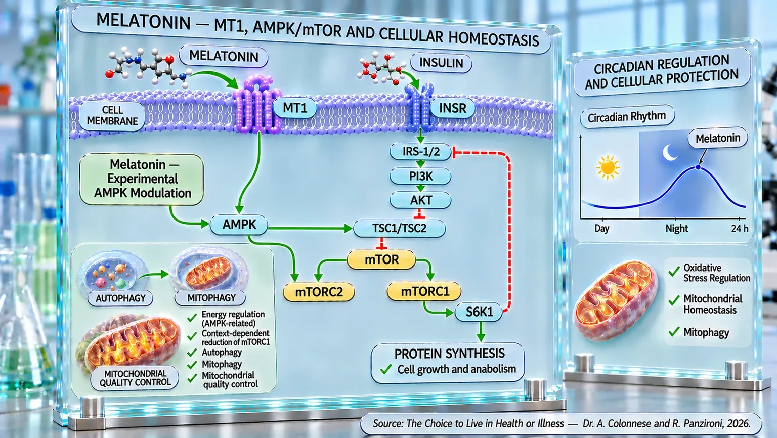
Molecular infographic featuring a cell membrane, MT1 and INSR receptors, the AMPK-mTOR cascade, and a circadian melatonin chart.
Diagram of the connections between melatonin, the MT1 receptor, AMPK, and the INSR–IRS-1/2–PI3K–AKT insulin pathway, including the regulation of TSC1/TSC2, mTORC1/2, S6K1, protein synthesis, autophagy, and mitophagy. The adjacent box indicates the nocturnal peak of melatonin and links the circadian rhythm to mitochondrial homeostasis, control of oxidative stress, and cellular protection.

ABSTRACT

Melatonin, synthesized primarily by the pineal gland and also by peripheral tissues, coordinates circadian rhythms and cellular protection processes. In the book "La scelta di Vivere in Salute o in Malattia" (The Choice to Live in Health or Disease), Dr. A. Colonnese and R. Panzironi explore its functions in sleep regulation, immunomodulation, and energy homeostasis. Its antioxidant and anti-inflammatory properties, along with its support for mitochondrial function, suggest a physiological role that extends beyond simply facilitating sleep onset.

Signaling through the MT1 receptor contributes to circadian synchronization and, according to experimental studies, to the organization of sleep stages. In the locus coeruleus, MT1 activation can reduce noradrenergic activity, which is normally high during wakefulness and strongly attenuated during REM sleep. Orexin, produced in the lateral hypothalamus, instead supports alertness and the release of noradrenaline. Further experimental observations suggest that melatonin may directly modulate orexinergic neurons, promoting non-REM sleep. The presence of MT1 in the pancreas, intestine, blood vessels, and immune cells also links these signals to peripheral metabolic and inflammatory functions.

Clinical studies report improvements in sleep quality and sleep latency, with variable results in adults with sleep disorders, patients with Parkinson's disease, children with neurodevelopmental disorders, and in hospital settings. In parallel, preclinical studies describe effects on mitochondrial protection and the regulation of autophagy, including mitophagy. In various models, melatonin promotes the activation of AMPK and limits mTORC1, modulating the recycling of cellular components; some tumor models also suggest an involvement of autophagy in neoplastic cell death.

Overall, melatonin integrates alertness circuits, circadian signals, and cellular maintenance mechanisms. However, the evidence regarding sleep does not automatically justify transferring experimental molecular findings to humans. Future research will need to clarify under which conditions the modulation of MT1, AMPK, and mTORC1 can translate into documentable clinical benefits, beyond the effects already observed on nighttime rest.

Its properties

Melatonin is a molecule that for many years was considered almost exclusively the sleep hormone. This definition, while correct, is now deeply reductive. Research over the last few decades has shown that melatonin is one of the most versatile molecules in the human body, involved in a vast network of biological processes ranging from the regulation of circadian rhythms to cellular protection, from the control of inflammation to the maintenance of energy balance, and even the safeguarding of the integrity of the nervous system and mitochondria (Kamfar et al., 2024) [1].

From a chemical point of view, melatonin is an indolamine, an amino acid derived from tryptophan through a series of reactions involving serotonin as an intermediate precursor. Although the pineal gland is the main organ responsible for its systemic secretion, we now know that its synthesis is not limited to the brain. Numerous tissues, including the gastrointestinal tract, retina, immune system, bone marrow, and other peripheral regions, are capable of producing melatonin locally, suggesting that this molecule performs functions much broader than the simple synchronization of the sleep-wake cycle (Jia et al., 2025) [2].

To understand the importance of melatonin, one can imagine the body as a large city that must constantly maintain order, communication, and adaptability. In this analogy, melatonin represents not only the clock that signals the arrival of night, but also a highly specialized maintenance crew that protects cellular infrastructure, coordinates emergency responses, and limits damage caused by daily wear and tear.

One of the most studied properties of melatonin concerns its antioxidant activity. During normal cellular metabolism, highly reactive molecules known as reactive oxygen species (ROS) and reactive nitrogen species (RNS) are continuously produced. In physiological amounts, these substances perform important signaling functions; however, when their production exceeds the body's defense capacity, they can damage proteins, lipids, cell membranes, and DNA. Melatonin possesses the extraordinary ability to counteract this phenomenon through multiple simultaneous mechanisms. It directly neutralizes numerous free radicals and, at the same time, stimulates the activity of the main endogenous antioxidant systems, including superoxide dismutase, catalase, and glutathione peroxidase (Kamfar et al., 2024) [1]; (Bocheva et al., 2024) [3]; (Jia et al., 2025) [2].

What makes melatonin particularly effective compared to other antioxidants is the fact that its activity does not end after the neutralization of a single reactive species. During the detoxification process, biologically active metabolites are generated, such as AFMK (a substance produced by the degradation of melatonin) and AMK, which in turn maintain antioxidant properties. This creates a true protective cascade capable of amplifying and prolonging the effects of the original molecule (Bocheva et al., 2024) [3].

Another fundamental characteristic is its high lipophilicity (the ability of a substance to dissolve in fats). Thanks to this property, melatonin easily crosses cell membranes, reaching compartments that are normally difficult to protect, such as the nucleus and, above all, the mitochondria. This ability is particularly important because mitochondria represent the main site of free radical production within the cell. The presence of melatonin near the very source of oxidative stress allows for rapid and effective intervention, limiting damage before it can spread (Kamfar et al., 2024) [1]; (Bao et al., 2026) [4]; (Reiter et al., 2024) [5].

In recent years, an even more fascinating view of melatonin physiology has emerged. For a long time, it was believed that the pineal gland was the main source of the molecule. Today, however, numerous pieces of evidence suggest that most of the melatonin produced in the body may derive from local synthesis within peripheral tissues and, likely, directly in the mitochondria. According to this model, pineal melatonin would act primarily as an endocrine signal dedicated to the temporal synchronization of the body, while intracellular melatonin would primarily perform metabolic, antioxidant, and cytoprotective functions (Reiter et al., 2024) [5].

The biological importance of this molecule appears even more evident when observing its evolutionary history. Phylogenetic analyses suggest that the ability to synthesize melatonin appeared long before the evolution of complex organisms, likely in primitive prokaryotes that populated the Earth billions of years ago. In an environment characterized by increasing oxygen concentrations and a consequent production of reactive species, melatonin would have represented a fundamental defense tool. This antioxidant function therefore seems to significantly predate the circadian role that is commonly associated with the hormone today (Reiter et al., 2024) [5].

Alongside its antioxidant activity, melatonin possesses significant anti-inflammatory properties. Inflammation is an essential mechanism for the survival of the organism, but when it becomes chronic, it can contribute to the development of numerous pathologies. Melatonin intervenes by modulating several central nodes of the inflammatory response. Of particular importance among these is the transcription factor NF-κB, considered one of the main regulators of the expression of proinflammatory genes. Through the inhibition of this signaling pathway, melatonin reduces the production of mediators such as TNF-α, IL-1β, and IL-6, helping to limit the amplification of the inflammatory response (Kamfar et al., 2024) [1]; (Jia et al., 2025) [2]; (Bocheva et al., 2024) [3].

Its actions, however, are not limited to systemic inflammation. Several studies have highlighted the ability of melatonin to modulate the activity of immune cells, influencing the production of cytokines and contributing to the maintenance of the balance between immune activation and tolerance. Specific receptors for melatonin have been identified in leukocytes, lymphocytes, bone marrow cells, and numerous other immune populations, confirming the role of the molecule as an important regulator of immune function (Kamfar et al., 2024) [1].

Particularly interesting is the so-called immune-pineal axis. In the presence of an inflammatory process, pineal melatonin production tends to decrease, while at the same time, local synthesis by macrophages and other immune cells present at the site of inflammation increases. This mechanism allows for the concentration of the molecule's production exactly where it is most needed, favoring the resolution of the inflammatory process and the protection of the involved tissues (Reiter et al., 2024) [5].

The cytoprotective properties of melatonin also extend to the maintenance of mitochondrial functionality. In addition to neutralizing free radicals, the molecule helps preserve mitochondrial membrane potential, improves ATP production, regulates mitochondrial fusion and fission processes, and participates in the control of mitophagy, the system through which the cell eliminates damaged mitochondria. Thanks to these actions, melatonin helps keep the cellular power plant efficient, reducing the risk of metabolic damage accumulation over time (Bao et al., 2026) [4]; (Bocheva et al., 2024) [3]; (Jia et al., 2025) [2].

The combination of these characteristics has led researchers to consider melatonin a pleiotropic molecule, meaning it is capable of simultaneously influencing numerous biological systems. Its actions involve the regulation of circadian rhythms, the control of oxidative stress, the modulation of inflammation, the maintenance of energy homeostasis, the protection of the nervous system, and the regulation of the immune response. In other words, melatonin does not act as a simple hormone specialized in a single function, but as a biological coordinator capable of linking seemingly distant processes, contributing to the maintenance of the balance of the entire organism (Minari and Pisani, 2025) [6].

Melatonin and sleep

Among all the functions attributed to melatonin, the one related to sleep is certainly the best known. However, defining melatonin simply as a hormone that promotes falling asleep would be reductive. Scientific evidence shows that this molecule plays a much more complex role, contributing to the regulation of the entire sleep architecture and the synchronization of the biological rhythms that govern the alternation between wakefulness and rest.

To understand its function, it is useful to imagine sleep as an orchestra composed of numerous musicians. Each instrument represents a different physiological system: the brain, metabolism, the endocrine system, the immune system, and the autonomic nervous system. Melatonin does not produce the music directly, but acts as the conductor who coordinates the exact moment each section must enter into action. When this coordination fails, sleep tends to become more fragmented, less efficient, and less restorative.

Clinical evidence gathered in recent years confirms this interpretation increasingly convincingly. A vast systematic review with meta-analysis that included twenty-three randomized controlled clinical trials demonstrated that melatonin supplementation significantly improves overall sleep quality in adults. Benefits have been observed in numerous clinical conditions, with effects particularly evident in subjects suffering from respiratory diseases, metabolic disorders, and primary sleep disorders. The analysis also showed a significant reduction in Pittsburgh Sleep Quality Index scores, indicating a clinically relevant improvement in the perceived quality of nocturnal rest. One of the most interesting aspects emerging from this meta-analysis concerns the fact that the improvement does not affect a single sleep parameter. Melatonin seems to act simultaneously on multiple dimensions of nocturnal rest, favoring faster sleep onset, better sleep continuity, and a reduction in nocturnal fragmentation. This suggests that its role is not limited to sleep induction, but involves the entire temporal organization of rest (Fatemeh et al., 2022) [7].

Particularly interesting confirmation comes from patients suffering from COMISA, a condition characterized by the coexistence of chronic insomnia and obstructive sleep apnea. This association is very frequent in clinical practice and represents one of the most complex forms of sleep disorder. The continuous micro-awakenings caused by apnea episodes profoundly alter the quality of rest, while insomnia in turn contributes to worsening sleep stability. In a randomized, placebo-controlled trial, the administration of 10 mg of melatonin for four weeks resulted in a marked improvement in sleep quality, a significant reduction in insomnia severity, and a decrease in daytime sleepiness. Patients also showed better adherence to CPAP therapy, an element particularly important for the control of sleep apnea (Motlaq et al., 2024) [8].

These results are particularly relevant because they demonstrate that melatonin does not act exclusively during the night. By improving sleep quality, it produces positive effects during the day as well, reducing fatigue, improving alertness, and promoting better functionality in daily activities. In other words, the benefit concerns not only sleeping better, but also living better during waking hours.

Another population in which melatonin has attracted great interest is that of patients suffering from Parkinson's disease. Sleep disorders represent one of the most frequent non-motor manifestations of the disease and can appear many years before the typical motor alterations. A recent meta-analysis that included randomized controlled trials highlighted that melatonin significantly improves sleep quality and reduces daytime sleepiness in subjects with Parkinson's. The best results were observed with dosages above 4 mg, with immediate-release formulations, and with relatively short-duration treatments. The data also suggest that the therapeutic response may depend on both the dose used and the pharmacokinetic characteristics of the formulation employed (Zrineh et al., 2025) [9].

The evidence in favor of melatonin does not only concern the adult population. In children with neurodevelopmental disorders, including autism, intellectual disabilities, and other neurological conditions, sleep onset problems represent one of the main causes of stress for the child and the entire family unit. The MENDS study, one of the largest randomized controlled trials conducted in the pediatric field, demonstrated that immediate-release melatonin significantly reduces the time required to fall asleep. On average, treated children fell asleep about forty-five minutes earlier than subjects who received a placebo. The increase in total sleep duration was more modest, suggesting that the main effect of melatonin is linked primarily to sleep onset rather than its prolongation (Appleton et al., 2012) [10].

This observation is particularly important because it helps to understand the true physiological nature of melatonin. Unlike traditional hypnotic drugs, which tend to force the body into a state of sedation, melatonin seems to facilitate the correct initiation of the biological processes that naturally lead to sleep. Once this process has begun, the maintenance and depth of rest depend on numerous other neurobiological systems.

Similar benefits have also been observed in the hospital setting. Hospitalization is indeed a particularly unfavorable condition for sleep due to noise, care procedures, pain, psychological stress, and alterations in normal circadian rhythms. A study conducted on hospitalized patients highlighted that evening administration of melatonin significantly improves sleep duration, reduces the time required to fall asleep, decreases the number of nocturnal awakenings, and increases the subjective quality of rest. These results take on particular relevance if one considers that sleep represents one of the main tools through which the body supports recovery and healing processes during illness (Ren et al., 2025) [11].

Considering the available evidence as a whole, an extremely coherent picture emerges. Melatonin should not be considered simply as a substance that induces sleep, but as a biological regulator capable of coordinating multiple aspects of nocturnal rest. Its effects are observed in very different populations—subjects with insomnia, patients with obstructive sleep apnea, people suffering from Parkinson's disease, children with neurodevelopmental disorders, and hospitalized patients—suggesting the existence of a common physiological mechanism. In all these contexts, melatonin seems to favor better synchronization of the processes that govern the transition from wakefulness to sleep, contributing to making rest more stable, more efficient, and more restorative.

MT1 receptors

Understanding the physiology of melatonin also means understanding the mechanisms through which this molecule communicates with cells. Like any hormone or signaling molecule, melatonin does not act alone. To exert its effects, it must be recognized by specific cellular structures capable of interpreting its message and translating it into a biological response. These structures are melatonin receptors, primarily identified as the MT1 and MT2 subtypes.

For many years, it was believed that both receptors performed substantially overlapping functions. More recent research, however, has revealed a much more complex reality. Although MT1 and MT2 are activated by the same molecule, they appear to participate in different and, in some cases, even opposite processes. This discovery has profoundly changed our understanding of the neurobiology of sleep and has opened new therapeutic perspectives for the treatment of sleep-wake rhythm disorders (Gobbi and Comai, 2019) [12]; (Gobbi and Comai, 2019) [13]; (Comai and Gobbi, 2024) [15].

The MT1 receptor belongs to the family of G protein-coupled receptors (proteins that transmit signals within cells) and is expressed in numerous brain areas involved in the regulation of sleep and circadian rhythms. Its presence has been documented in the suprachiasmatic nucleus (a small area of the brain located in the hypothalamus), the body's main biological clock, but also in the lateral hypothalamus, the pons, the amygdala, and the hippocampus. This anatomical distribution suggests that MT1 is not simply involved in receiving the melatonergic signal, but participates directly in the neurobiological organization of sleep (Comai and Gobbi, 2024) [15].

To understand the role of this receptor, one can imagine melatonin as a communication sent every evening by the central biological clock. The MT1 receptor represents one of the main recipients of this message. When activated, a series of cellular and neuronal modifications are triggered that progressively prepare the brain for the transition from wakefulness to sleep.

The first indications of the specific role of MT1 emerged from studies conducted on genetically modified animal models. Through the selective elimination of the gene responsible for producing the receptor, researchers were able to observe the consequences of its absence. The results showed that mice lacking the MT1 receptor exhibit a significant reduction in REM sleep, the phase of sleep associated with intense dream activity, memory consolidation, and emotional regulation (Comai et al., 2013) [14].

This observation became even more significant when researchers analyzed brain electrical activity during sleep. In animals lacking MT1, a reduction in theta oscillations (rhythmic electrical waves produced by brain neurons) was documented during REM sleep. Theta waves represent one of the main neurophysiological signatures of this sleep phase and participate in the processes of memory consolidation and the reorganization of information acquired during the day. Their reduction suggests that MT1 influences not only the quantity of REM sleep but also the functional quality of this phase (Comai et al., 2013) [14].

Evidence collected subsequently has progressively strengthened the hypothesis that the MT1 receptor plays a fundamental role in the stabilization of REM sleep. Several literature reviews have highlighted how the activation of MT1 is associated with the consolidation of REM phases and the modulation of the neuronal circuits that regulate their onset and maintenance throughout the night (Gobbi and Comai, 2019) [12]; (Gobbi and Comai, 2019) [13]; (Comai and Gobbi, 2024) [15].

Another aspect of great interest concerns the relationship between MT1 and circadian regulation. Numerous pieces of evidence suggest that this receptor participates in the mechanisms through which the suprachiasmatic nucleus synchronizes the body's biological functions with the light-dark environmental cycle. In other words, MT1 could represent one of the main connection points between temporal information coming from the environment and the internal organization of sleep (Gobbi and Comai, 2019) [13]; (Comai and Gobbi, 2024) [15].

Observations from knockout models further reinforce this interpretation. Animals lacking MT1 show not only alterations in REM sleep but also greater sleep fragmentation, a reduction in the continuity of rest, and less stability in circadian organization. These results suggest that the receptor contributes to maintaining a stable and physiologically efficient sleep structure (Comai and Gobbi, 2024) [15].

From a therapeutic point of view, these discoveries have opened particularly interesting perspectives. Most currently available preparations activate both MT1 and MT2 simultaneously. However, if the two receptors perform different functions, it becomes plausible to imagine drugs capable of acting selectively on only one of the two targets. In this perspective, specific MT1 agonists could be useful in conditions characterized by REM sleep alterations, sleep fragmentation, and difficulty in initiating the physiological processes that lead to nighttime rest (Gobbi and Comai, 2019) [12]; (Gobbi and Comai, 2019) [13]; (Comai and Gobbi, 2024) [15].

Overall, the available evidence outlines the MT1 receptor as much more than a simple mediator of melatonin's effects. It appears as a sophisticated neurobiological regulator that connects the temporal signal of the night to the organization of sleep phases, the stability of alertness, and the control of the neuronal circuits involved in REM sleep. This modern view allows us to understand how melatonin does not act simply by inducing sleep, but by orchestrating a complex network of biological signals that prepare the brain and the body for nighttime rest.

The Locus Coeruleus and norepinephrine

To fully understand how melatonin influences sleep, it is necessary to delve into one of the most important neurobiological systems involved in the regulation of alertness: the locus coeruleus–noradrenaline system. If melatonin can be imagined as the signal announcing the arrival of night, the locus coeruleus represents one of the main brain centers that decide how alert, attentive, and ready to react to environmental stimuli the brain should remain.

The locus coeruleus is a small nucleus located in the brainstem that, despite its small size, exerts an extraordinarily broad influence on the entire central nervous system. Its neurons produce noradrenaline, a neurotransmitter fundamental for attention, alertness, memory, stress response, and the maintenance of the waking state. In recent decades, it has emerged that the locus coeruleus–noradrenaline system constitutes one of the main regulators of the sleep-wake cycle, participating not only in the maintenance of alertness but also in the organization of the different stages of sleep (Van Egroo et al., 2022) [17].

Electrophysiological recordings have shown that the activity of locus coeruleus neurons varies continuously throughout the day. During active wakefulness, the firing rate is high, allowing the brain to maintain high levels of attention and reactivity. As sleep approaches, noradrenergic activity decreases progressively, reaching even lower levels during non-REM sleep. The situation changes radically during REM sleep, when the neurons of the locus coeruleus become almost completely silent and extracellular levels of noradrenaline plummet to the lowest values observable in the entire sleep-wake cycle (Van Egroo et al., 2022) [17].

For many years, it was believed that the locus coeruleus was simply "turned off" during sleep. However, modern noradrenaline monitoring technologies have profoundly changed this view. Today we know that during non-REM sleep, the noradrenergic system continues to function through extremely precise rhythmic oscillations that contribute to the organization of sleep architecture. Noradrenaline levels rise and fall cyclically every thirty to fifty seconds, synchronizing with fundamental phenomena such as sleep spindles (brief electrical waves produced by the brain during light sleep), slow cortical oscillations, and heart rate variations. This behavior suggests that the locus coeruleus is not simply a wakefulness center, but a sophisticated regulator of sleep quality itself (Osorio-Forero et al., 2022) [20].

One of the most interesting aspects concerns the role of noradrenaline in memory consolidation. During non-REM sleep, the controlled oscillations of the noradrenergic system seem to favor synaptic plasticity and the transfer of information from the hippocampus to the cerebral cortex. Subsequently, during REM sleep, the almost complete disappearance of noradrenaline creates a completely different neurochemical environment that favors the reorganization of neuronal connections and the processing of emotional information accumulated during the day. This drastic reduction of noradrenaline during REM sleep does not represent a simple side effect of sleep, but a biological condition necessary for the brain to properly enter this phase. The most recent observations have in fact shown that the drop in noradrenaline levels begins several seconds before the start of a REM episode. In other words, the silencing of the locus coeruleus seems to represent one of the events that prepare the brain for entry into REM sleep (Osorio-Forero et al., 2022) [20].

In this context, the direct link with melatonin and the MT1 receptor described in the previous paragraph emerges. A study published in 2024 demonstrated that the selective activation of MT1 receptors present on the noradrenergic neurons of the locus coeruleus reduces their electrical activity in a dose-dependent manner. This inhibition translates into a significant increase in REM sleep without altering non-REM sleep. When researchers pharmacologically blocked the MT1 receptor or genetically silenced its expression in the neurons of the locus coeruleus, the effect disappeared completely. This result provided one of the strongest pieces of evidence that melatonin can directly modulate the noradrenergic system through the MT1 receptor, facilitating the onset of REM sleep (López-Canul et al., 2024) [19].

This discovery allows for a better understanding of the role of melatonin in sleep physiology. Rather than acting as a simple sleep inducer, melatonin seems to intervene in specific neuronal circuits that control the transition between different states of alertness. Through the activation of MT1 receptors present in the locus coeruleus, it contributes to progressively reducing noradrenergic activity, creating the neurochemical conditions necessary for entry into REM sleep (López-Canul et al., 2024) [19].

The importance of the locus coeruleus emerges with particular evidence when sleep is chronically altered. An experimental study that simulated a condition very similar to that experienced by many people in modern life—a few days of insufficient sleep followed by short recovery periods—documented surprising consequences. After several weeks of intermittent reduced sleep, the animals showed a significant loss of noradrenergic neurons in the locus coeruleus, accompanied by persistent alterations of the sleep-wake cycle, reduction of neuronal projections, and biological markers compatible with accelerated cellular aging processes (Zhu et al., 2016) [18].

The vulnerability of the locus coeruleus to sleep loss is also confirmed by studies that have analyzed selective REM sleep deprivation. In these conditions, an increase in oxidative stress, apoptotic activation, and degeneration of noradrenergic neurons is observed. However, the administration of melatonin after sleep deprivation was found to be able to significantly counteract these phenomena. Researchers observed an increase in reduced glutathione levels, a decrease in caspase-3 activation, and a preservation of the noradrenergic neuronal population of the locus coeruleus. In other words, melatonin did not just promote sleep, but showed a true neuroprotective action toward one of the most important systems for the regulation of alertness (Jameie et al., 2019) [16].

This observation takes on particular relevance when considering that the locus coeruleus is now recognized as one of the first brain structures involved in neurodegenerative processes associated with aging and Alzheimer's disease. Numerous pieces of evidence suggest that the accumulation of pathological alterations in this region may begin many years before the appearance of cognitive symptoms. In parallel, chronic sleep fragmentation accelerates the loss of noradrenergic neurons and favors the accumulation of pathological proteins such as tau and β-amyloid, establishing a vicious cycle between sleep deterioration and neurodegeneration (Van Egroo et al., 2022) [17].

Overall, the available evidence describes the locus coeruleus–noradrenaline system as one of the main orchestrators of sleep physiology. Its activity regulates transitions between wakefulness and sleep, contributes to memory consolidation, coordinates important autonomic functions, and participates in neuroprotective processes that keep the brain efficient throughout life. Melatonin, through its interaction with the MT1 receptors present in the noradrenergic neurons of the locus coeruleus, seems to fit directly into this regulatory network, contributing not only to the organization of REM sleep but also to the long-term protection of one of the most important neuronal systems for brain health.

The lateral hypothalamus, orexin, and the Locus Coeruleus

In the previous paragraph, we saw how the locus coeruleus represents one of the main brain centers responsible for the production of norepinephrine and the regulation of alertness. However, to fully understand how this system functions, it is necessary to go upstream and identify what controls the activity of the locus coeruleus itself. Research over the last few decades has shown that one of its most important regulators is a small group of neurons located in the lateral hypothalamus: the neurons that produce orexin, also known as hypocretin.

The discovery of orexin has profoundly changed our understanding of sleep physiology. Initially, this neuropeptide was primarily associated with the control of appetite and energy balance. Over the years, however, it has emerged that its main function is to coordinate the organism's state of alertness with its current biological needs. Orexinergic neurons act as a sort of integration center that collects information from metabolism, emotions, the reward system, and the external environment, transforming it into signals capable of modulating the level of brain activation (Sakurai, 2007) [24].

This function appears particularly logical from an evolutionary perspective. An organism that is in a state of energy deficiency, that perceives danger, or that must actively search for food resources cannot afford to reduce its level of alertness. In these circumstances, the orexinergic system increases its activity, favoring a state of sustained attention and promoting survival-oriented behaviors. Conversely, during sleep, and particularly during REM sleep, the activity of orexin neurons decreases drastically (Sakurai, 2007) [24].

Despite the relatively small number of orexinergic neurons present in the lateral hypothalamus, their projections reach numerous brain regions involved in the regulation of alertness. Among all these structures, the locus coeruleus is likely the most important target. Neuroanatomical analyses have shown that the locus coeruleus receives one of the densest orexinergic innervations in the entire brain. Fibers originating from the lateral hypothalamus form numerous direct excitatory synapses on the noradrenergic neurons of the locus coeruleus, creating an extremely robust anatomical connection between the two systems (Horvath et al., 1999) [21].

Electron microscope observations have highlighted that practically all noradrenergic neurons of the locus coeruleus receive synaptic contacts from terminals containing orexin. Even more interesting is the fact that these synapses exhibit characteristics typical of excitatory connections, suggesting that the orexinergic system is designed to directly increase the activity of locus coeruleus neurons; subsequent electrophysiological recordings have confirmed this hypothesis. When orexin is applied directly to locus coeruleus neurons, their firing rate increases rapidly and consistently. This effect is the result of both cell membrane depolarization and a reduction in the mechanisms that normally limit neuronal excitability. In other words, orexin makes noradrenergic neurons more active and more prone to maintaining high levels of alertness (Horvath et al., 1999) [21].

These results were further confirmed by subsequent studies that analyzed the role of orexin A, one of the two main biologically active forms of the peptide. Microinjections of orexin A directly into the locus coeruleus result in a marked increase in wakefulness, accompanied by a significant reduction in REM sleep and deep sleep. In some experiments, the increase in the state of wakefulness reached values exceeding 70%, demonstrating the extraordinary power of this neuronal activation system; it is particularly interesting that these effects are mediated primarily by the orexin receptor type 1 (OX1R). This suggests that communication between the lateral hypothalamus and the locus coeruleus occurs through a highly specialized mechanism, optimized to rapidly promote the state of wakefulness when physiological conditions require it (Hagan et al., 1999) [22].

The role of the orexin–locus coeruleus circuit emerges with particular clarity when analyzing the transitions between wakefulness and REM sleep. As seen in the previous paragraph, the locus coeruleus is practically silent during REM sleep. Orexinergic activation, on the other hand, produces the opposite effect: it increases the activity of noradrenergic neurons and makes entering this phase of sleep more difficult. A study specifically dedicated to this phenomenon demonstrated that local administration of orexin A in the locus coeruleus suppresses REM sleep in a dose-dependent manner, while simultaneously increasing the duration of wakefulness. This effect is accompanied by marked neuronal activation documented both electrophysiologically and through the increased expression of a specific protein (Bourgin et al., 2000) [23].

These observations have helped clarify one of the mechanisms through which the brain stabilizes behavioral states. When the orexinergic system is active, the locus coeruleus continues to produce norepinephrine, keeping the level of arousal high and preventing the intrusion of REM phenomena during wakefulness. When orexinergic activity decreases, as occurs physiologically during the night, the locus coeruleus can progressively diminish its activity, favoring the transition to the different phases of sleep (Bourgin et al., 2000) [23].

The importance of this circuit becomes even more evident when observing what happens in narcolepsy (a neurological sleep disorder that causes severe daytime sleepiness). This pathology is characterized by the selective loss of orexinergic neurons in the lateral hypothalamus. In the absence of the stabilizing action of orexin, the brain loses the ability to maintain a clear separation between sleep and wakefulness. Consequently, daytime sleepiness, sleep fragmentation, and REM phenomena that inappropriately break through during wakefulness occur, as happens in episodes of cataplexy (a sudden and temporary loss of muscle strength) (Sakurai, 2007) [24].

A particularly elegant study allowed for the identification of the main circuits responsible for these phenomena. By selectively restoring orexinergic signaling in different neuronal populations of narcoleptic mouse models, researchers observed that the recovery of orexin transmission in the noradrenergic neurons of the locus coeruleus significantly improves the consolidation of wakefulness. The effect was directly proportional to the number of locus coeruleus neurons in which sensitivity to orexin had been restored. This result demonstrated that the locus coeruleus represents one of the main effectors through which the orexinergic system maintains a continuous and stable state of alertness (Hasegawa et al., 2014) [25].

Taken together, these findings describe the lateral hypothalamus as a sort of command center capable of continuously modulating the organism's level of alertness. Through the release of orexin, it activates the locus coeruleus, increases the production of norepinephrine, and supports the state of wakefulness when the organism must remain attentive to the surrounding environment. This system represents the physiological counterweight to the mechanisms that promote sleep described in the previous paragraphs. On one hand, melatonin and the MT1 receptor contribute to reducing the activity of the locus coeruleus and facilitating REM sleep; on the other, orexin exerts an opposite action, stimulating noradrenergic neurons and promoting alertness. The delicate balance between these opposing forces allows the brain to alternate between states of sleep and wakefulness in an orderly and stable manner.

Melatonin, the lateral hypothalamus, and orexin

In the previous paragraphs, we saw how the locus coeruleus and the orexinergic system represent some of the main promoters of alertness. However, a fundamental question remains: how does melatonin, the biological signal of the night, manage to counteract these powerful brain activation systems and promote sleep?

For many years, the answer remained incomplete. It was known that melatonin promoted sleep and that orexin supported wakefulness, but the direct link between these two neurobiological systems had not yet been clarified. More recent research, however, has identified an important anatomical and functional meeting point within the perifornical lateral hypothalamus, a region that houses most of the orexinergic neurons responsible for maintaining the state of alertness (Sharma et al., 2018) [26].

To understand the importance of this discovery, it is useful to remember that orexin neurons function as a sort of accelerator of consciousness. When active, they stimulate numerous arousal systems, including the locus coeruleus, the serotonergic raphe nuclei, the cholinergic systems of the brainstem, and other structures involved in alertness. Through these diffuse connections, they contribute to maintaining the brain in a state of activation that is stable and consistent with the demands of the surrounding environment (Hung and Yamanaka, 2023) [27].

Modern immunofluorescence techniques have shown that these neurons express MT1 receptors for melatonin. This observation suggests that the melatonergic system can exert direct control over the main wake-promoting neurons through a highly selective mechanism. Experimental verification of this hypothesis has produced particularly interesting results. When melatonin is administered directly into the perifornical lateral hypothalamus, a significant increase in non-REM sleep is observed, accompanied by a reduction in wakefulness. In parallel, the activity of orexinergic neurons decreases drastically, as demonstrated by the marked reduction in the expression of the neuronal marker c-Fos (a protein used as a marker of neuronal activation). In other words, melatonin seems to literally turn down the volume of one of the main brain systems responsible for maintaining alertness (Sharma et al., 2018) [26].

Particularly significant is the fact that the opposite effect is observed when melatonergic signaling is blocked. Pharmacological inhibition of melatonin receptors in the lateral hypothalamus indeed leads to an increase in wakefulness and a reduction in sleep propensity. This result suggests that the control exerted by melatonin over orexin neurons does not represent an artificial phenomenon observable only in the laboratory, but constitutes a physiological mechanism normally active during the night (Sharma et al., 2018) [26].

This discovery has helped to redefine how the action of melatonin is interpreted. For a long time, it was imagined that the hormone acted primarily as a circadian signal capable of informing the brain of the arrival of night. Today, it appears increasingly evident that melatonin also plays an active role in the modulation of the neuronal circuits that govern states of consciousness. In this perspective, sleep would not result solely from the presence of a favorable temporal signal, but also from the progressive deactivation of the systems that support wakefulness.

The importance of this mechanism becomes even more evident by observing the physiological behavior of orexinergic neurons during the sleep-wake cycle. Electrophysiological recordings and modern imaging techniques have shown that these neurons reach maximum activity during active wakefulness, exploration of the environment, exposure to new stimuli, and motivated behaviors. Conversely, their activity decreases progressively during quiet wakefulness and becomes minimal during non-REM sleep. This trend suggests that the inhibition of orexin represents one of the fundamental steps that allow the brain to transition from alertness to sleep (Hung and Yamanaka, 2023) [27].

Genetic evidence further reinforces this interpretation. Animal models lacking orexin or its receptors show a marked instability of states of consciousness, with fragmentation of wakefulness, episodes of early REM sleep, and manifestations similar to human narcolepsy. These observations demonstrate that the orexinergic system represents one of the main mechanisms through which the brain maintains the continuity of the state of wakefulness (Hung and Yamanaka, 2023) [27].

From this perspective, a particularly elegant picture of sleep physiology emerges. During the day, the activity of orexinergic neurons helps to support alertness and activate systems such as the locus coeruleus and noradrenergic transmission. With the arrival of the night, the increase in melatonin secretion allows this activity to be progressively reduced through the MT1 receptors expressed on orexin neurons. The decrease in orexinergic activity therefore translates into less stimulation of arousal systems, facilitating the entry into sleep and its stabilization (Sharma et al., 2018) [26].

The clinical importance of this relationship has also been confirmed by recent pharmacological studies. The observation that melatonin receptor agonists and orexin receptor antagonists produce similar benefits in various pathological conditions suggests that both therapeutic approaches may converge on the same neurobiological circuits. In a study conducted on elderly patients hospitalized for stroke, the early administration of a melatonin receptor agonist or orexin receptor antagonists was associated with a significant reduction in the incidence of delirious symptoms. The authors hypothesized that this benefit derives at least in part from the improvement in sleep quality and the normalization of the sleep-wake rhythm, confirming the importance of the balance between the melatonergic system and the orexinergic system in the stability of cognitive functions (Miyoshi et al., 2024) [28].

These observations open up particularly interesting therapeutic perspectives. If melatonin promotes sleep by reducing the activity of orexin neurons and if orexinergic receptor antagonists produce similar clinical effects through a different mechanism, it becomes plausible to consider these two systems as complementary parts of the same biological network. In such a model, melatonin would represent the physiological signal that communicates the arrival of the night, while orexin would constitute the main system that must be progressively silenced so that sleep can emerge in a stable and effective way.

Overall, the available evidence indicates that the relationship between melatonin and orexin represents one of the main meeting points between the circadian system and the neuronal circuits of alertness. Through the activation of MT1 receptors present in the lateral hypothalamus, melatonin appears to directly inhibit one of the most powerful promoters of wakefulness present in the brain. This mechanism allows the biological signal of the night to be linked to the reduction of neuronal arousal, creating the necessary conditions for sleep to develop in a physiological and coordinated manner.

MT1 receptors in the human body

Up to this point, we have followed the path of melatonin within the brain circuits that regulate sleep, observing how it interacts with the locus coeruleus, norepinephrine, and the orexinergic system of the lateral hypothalamus. At this stage, a common misconception might arise: imagining that MT1 receptors are confined almost exclusively to the brain and that their primary function is to regulate sleep. In reality, research in recent years has revealed a much more fascinating and complex reality. MT1 receptors are distributed across numerous organs and tissues of the body and participate in the regulation of biological processes involving metabolism, the cardiovascular system, immunity, endocrine function, and even cellular protection against oxidative stress (Jockers et al., 2016) [29]; (Slominski et al., 2012) [31].

From a structural standpoint, the MT1 receptor belongs to the large family of G protein-coupled receptors. When melatonin binds to this receptor, a cascade of intracellular signals is activated, modifying the cell's behavior. Among the most important effects are the reduction of intracellular levels of cyclic AMP (a molecule that transmits signals within cells, regulating important functions), the modulation of protein kinases (enzymes that regulate the activity of other proteins by adding phosphate groups), the control of ion fluxes (the movement of electrically charged particles (ions) across the cell membrane), and the regulation of gene expression. In some tissues, the receptor can also activate the phospholipase C pathway (an enzyme that transforms certain cell membrane fats into chemical signals) and protein kinase C, further expanding the variety of possible biological responses (Jockers et al., 2016) [29]; (Tripathy and Bhattamisra, 2025) [33].

One of the peripheral systems richest in melatonergic signaling is the gastrointestinal tract. For many years, it was believed that the pineal gland was the body's main source of melatonin. Today, we know that the gut produces extremely high quantities of melatonin, often exceeding those present in the bloodstream. Studies conducted on human tissues have identified the MT1 receptor throughout the entire gastrointestinal tract, including the intestinal epithelium, the myenteric plexus, the submucosal plexus (a network of neurons located between the muscle layers of the intestine), and the vascular structures of the intestinal wall (Söderquist et al., 2015) [30].

This distribution suggests that melatonin participates directly in the regulation of intestinal motility, digestive secretion, barrier function, and communication between the enteric nervous system and the digestive tract. In other words, the gastrointestinal tract does not represent merely a target organ for melatonin, but a true peripheral melatonergic system endowed with significant functional autonomy (Söderquist et al., 2015) [30]; (Slominski et al., 2012) [31].

Particularly interesting is the presence of MT1 receptors in the endocrine pancreas as well. Cells of the pancreatic islets, in fact, express complete components of melatonergic signaling. This observation has attracted the attention of researchers because it directly links melatonin to the control of glucose metabolism. Through its receptors, melatonin contributes to the modulation of insulin secretion and the circadian synchronization of energy metabolism. Alterations in melatonergic signaling have been associated with insulin resistance, impaired β-cell function, and an increased risk of type 2 diabetes (Söderquist et al., 2015) [30]; (Tripathy and Bhattamisra, 2025) [33].

The cardiovascular system also possesses significant expression of MT1 receptors. These receptors have been identified in the wall of blood vessels, both in smooth muscle cells and in the endothelium. Research indicates that MT1 activation is generally associated with vasoconstrictor phenomena mediated by the reduction of cyclic AMP and the modulation of calcium-dependent potassium channels. This mechanism contributes to the fine-tuning of vascular tone and blood pressure, integrating circadian signals with the hemodynamic needs of the body (da Rocha-Carvalho et al., 2025) [32]; (Slominski et al., 2012) [31].

Interest in these effects has increased upon observing that MT1 receptor expression tends to decrease with aging and in various pathological cardiovascular conditions. Some researchers have hypothesized that the progressive loss of sensitivity to melatonin may contribute to age-related vascular alterations, promoting endothelial dysfunction, increased arterial stiffness, and reduced capacity to adapt to circadian rhythms (da Rocha-Carvalho et al., 2025) [32].

Another particularly relevant area concerns the immune system. Numerous immune cells express MT1 receptors and respond directly to the presence of melatonin. Through these mechanisms, melatonin can influence lymphocyte proliferation, cytokine production, and the regulation of inflammatory responses. In particular, the MT1 receptor appears to be involved in the modulation of adaptive immunity, contributing to the production of interleukin-2 (a protein of the immune system that stimulates the growth and multiplication of T lymphocytes) and the control of T lymphocyte activity (Jockers et al., 2016) [29]; (Slominski et al., 2012) [31].

The peripheral distribution of MT1 also extends to the kidney, bone tissue, skin, reproductive organs, and the placenta. In the kidney, it contributes to the regulation of hydro-electrolytic homeostasis and protection against inflammatory processes. In bone tissue, it promotes osteoblast activity and the maintenance of skeletal mass. In the skin, it participates in protection against oxidative stress and the regulation of cell proliferation. During pregnancy, the presence of melatonergic receptors in the placenta and uterus contributes to fetal protection and the regulation of gestational processes (Slominski et al., 2012) [31].

An aspect that is sparking growing interest concerns the role of MT1 receptors in chronic diseases. Alterations in receptor expression have been observed in Alzheimer's disease, Parkinson's disease, Huntington's disease, and some neurodevelopmental disorders. In many of these conditions, alterations in sleep, circadian rhythms, and metabolic function are observed simultaneously, suggesting that dysfunction of melatonergic signaling may contribute to disease progression (Jockers et al., 2016) [29].

Overall, the body of available evidence profoundly changes the traditional image of melatonin as a simple sleep hormone. The MT1 receptor emerges, in fact, as a component of a vast biological network distributed throughout the body. Through its presence in the brain, gut, pancreas, blood vessels, immune system, and numerous other tissues, it helps coordinate functions ranging from circadian regulation to energy metabolism, and from immunity to cellular protection. This systemic view allows us to understand how the nocturnal signal of melatonin is not destined exclusively for the brain, but represents a global biological message capable of synchronizing the entire organism with the rhythm of the day.

Melatonin and autophagy

To fully understand many of the functions attributed to melatonin, it is necessary to introduce a biological process fundamental to cellular survival: autophagy. The term derives from Greek and literally means "self-eating," but this definition can be misleading. Autophagy is not a self-destructive mechanism, but rather an extraordinary internal maintenance system through which the cell identifies, sequesters, and degrades damaged components, misfolded proteins, and non-functional organelles, subsequently reusing their constituents to produce new cellular structures (Coto-Montes et al., 2012) [34]; (Boga et al., 2019) [35].

This process could be compared to what happens in a well-organized city. Every day, waste is collected, damaged materials are removed, and useful raw materials are recovered for new construction. If this collection and recycling system were to stop, the streets would progressively fill with debris, compromising the functioning of the entire city. Similarly, when autophagy is impaired, cells accumulate damaged structures that promote inflammation, metabolic dysfunction, aging, and disease (Coto-Montes et al., 2012) [34].

In recent years, it has emerged that melatonin is one of the most important physiological regulators of this system. Initially, it was believed that its role was limited to the direct neutralization of free radicals and the regulation of the sleep-wake cycle. More recent evidence has shown that melatonin intervenes at a much deeper level, coordinating numerous cellular mechanisms involved in intracellular quality control (Boga et al., 2019) [35]; (Lei et al., 2024) [38].

One of the elements that closely links melatonin and autophagy is oxidative stress. During normal cellular metabolism, reactive oxygen species—commonly called ROS—are continuously generated. In physiological amounts, these molecules perform important signaling functions. However, when their production exceeds the cell's defensive capacity, damage to proteins, lipids, DNA, and mitochondria accumulates. Under these conditions, autophagy is activated as an emergency mechanism to eliminate compromised structures and restore cellular homeostasis (Coto-Montes et al., 2012) [34]; (Lei et al., 2024) [38].

Melatonin actively participates in this process through numerous molecular pathways. Among the most important are the activation of AMPK, one of the main cellular energy sensors, and the inhibition of mTOR, a protein that normally suppresses autophagy when energy resources are abundant. Through this regulation, melatonin promotes the activation of ULK1 (a protein that initiates autophagy) and the start of autophagosome formation, the structures that engulf the material to be degraded (Qiu et al., 2026) [40]; (Zhu et al., 2025) [39].

Among the various forms of autophagy, one of the most important is mitophagy, a selective process dedicated exclusively to the removal of damaged mitochondria. Mitochondria constitute the main energy powerhouses of the cell, but they are also one of the largest intracellular sources of free radicals. When they become dysfunctional, they can transform from energy producers into generators of cellular damage. For this reason, the organism has developed sophisticated mitochondrial quality control systems that allow for the selective elimination of compromised organelles and their replacement with new, functional mitochondria (Coto-Montes et al., 2012) [34]; (Lei et al., 2024) [38].

The most recent evidence suggests that melatonin plays a particularly important role in mitochondrial quality control. In addition to directly protecting mitochondria from oxidative damage, it regulates mitochondrial fusion and fission, promotes the biogenesis of new organelles, and stimulates mitophagy through specific systems. This coordinated set of mechanisms allows for the preservation of cellular energy efficiency and limits the accumulation of dysfunctional structures (Lei et al., 2024) [38]; (Zhu et al., 2025) [39].

A particularly fascinating aspect that has emerged from research is that melatonin does not act as a simple switch that always activates autophagy. On the contrary, it acts as an adaptive regulator: when the autophagic flux is insufficient and the cell accumulates damaged structures, melatonin tends to enhance it; when, however, autophagy becomes excessive and risks turning into a self-destructive mechanism, melatonin can limit it. In other words, its goal is not to increase or reduce autophagy indiscriminately, but to maintain the optimal balance necessary for cellular survival (Boga et al., 2019) [35]; (Zhu et al., 2025) [39]; (Lei et al., 2024) [38].

This bidirectional nature appears particularly evident in the nervous system. Neurodegenerative diseases such as Alzheimer's and Parkinson's are characterized by the progressive accumulation of aggregated proteins and damaged mitochondria. In these contexts, melatonin promotes autophagy and mitophagy, facilitating the removal of pathological structures, improving mitochondrial function, and reducing neuroinflammation. In other neurological scenarios, however, excessive activation of autophagy can contribute to neuronal death; in such conditions, melatonin appears to exert a moderating effect, helping to restore cellular balance (Zhu et al., 2025) [39]; (Boga et al., 2019) [35].

The same regulatory capacity has been observed in the liver. In a mouse model of steatohepatitis associated with obesity and chronic intermittent hypoxia—a condition frequently observed in subjects suffering from obstructive sleep apnea syndrome—melatonin reduced liver damage by restoring autophagic activity. The recovery of autophagic flux was associated with a reduction in inflammation, an improvement in liver structure, and a slower progression of metabolic disease (Ren et al., 2019) [36].

Bone tissue also seems to benefit from this regulation. In models of diabetic osteoporosis, chronic hyperglycemia compromises the regenerative capacity of mesenchymal stem cells (immature cells capable of transforming into different types of cells, such as those of bone, cartilage, and adipose tissue) and promotes a persistent inflammatory state. Melatonin was found to be capable of activating the NRF2-autophagy pathway, reducing oxidative stress, and promoting the polarization of macrophages toward the anti-inflammatory M2 phenotype. This change in the cellular microenvironment favored osteogenic differentiation and improved bone formation (Zhang et al., 2026) [41].

Particularly interesting is the role of autophagy in oncology as well. Tumor cells frequently use autophagic mechanisms to survive in hostile environments characterized by nutrient deficiency, hypoxia, and metabolic stress. In some contexts, melatonin appears to exploit this very system to counteract neoplastic growth. In neuroblastoma cells, for example, it induces a form of cytotoxic autophagy through the activation of the Pak2/AMPK/mTOR axis and the modulation of endoplasmic reticulum stress, contributing to the suppression of tumor growth (Qiu et al., 2026) [40].

Similar results have been observed in colorectal carcinoma. In this model, melatonin increases the production of reactive oxygen species, activates the endoplasmic reticulum stress response, and induces the expression of specific autophagic proteins. Modifications of the AMPK and PI3K/AKT/mTOR pathways have also emerged. The final outcome is an activation of autophagy associated with the death of tumor cells, suggesting a possible role for melatonin as an adjuvant in oncological therapeutic strategies (Chok et al., 2021) [37].

Overall, the currently available evidence shows that melatonin represents much more than a simple sleep regulator. Through the control of autophagy and mitophagy, it intervenes in some of the most important mechanisms for maintaining cellular homeostasis. The ability to modulate AMPK, mTOR, and numerous other signaling pathways allows melatonin to adapt the cellular response to the specific needs of each tissue and each pathological condition. In this perspective, autophagy appears as one of the main tools through which melatonin protects the organism from aging, metabolic dysfunction, neurodegeneration, and chronic cellular damage.

AMPK and autophagy

In the previous paragraph, we saw how melatonin can influence autophagy through numerous intracellular signaling pathways, including AMPK and mTOR. To fully understand this mechanism, it is necessary to focus on one of the most important metabolic sensors present in the organism: AMP-activated protein kinase, better known as AMPK.

Every cell in the human body is constantly faced with a fundamental challenge: balancing energy consumption and production. When available energy is abundant, the cell can invest resources in growth, protein synthesis, and proliferation. When energy reserves decrease, however, it becomes necessary to adopt conservation and recovery strategies. In this context, AMPK acts as a true metabolic control center, capable of continuously monitoring the ratio between AMP and ATP (two molecules linked to cellular energy availability) and adapting cellular behavior to the energy conditions of the moment (Smiles et al., 2024) [43]; (Mandic et al., 2025) [45].

AMPK could be compared to a sophisticated emergency management system installed in a large city. When energy resources are sufficient, activities proceed normally. If, however, there is an energy shortage, the system intervenes immediately by reducing non-essential consumption and activating mechanisms designed to restore balance. This principle represents the biological foundation of AMPK's action (Mandic et al., 2025) [45].

To perform this function, AMPK works in close collaboration with another important metabolic regulator: mTORC1. If AMPK represents the sensor of energy scarcity, mTORC1 can be considered the promoter of cell growth. When nutrients and energy are abundant, mTORC1 promotes protein synthesis, proliferation, and anabolism. When energy decreases, however, AMPK intervenes by suppressing mTORC1 activity and orienting metabolism toward resource conservation and recovery processes (Smiles et al., 2024) [43]; (Al-Kuraishy et al., 2025) [46].

This antagonistic relationship represents one of the most important metabolic checkpoints in the entire organism. Through the phosphorylation of proteins such as TSC2, AMPK is able to reduce mTORC1 activity, temporarily interrupting high-energy-consuming cellular programs and favoring the activation of catabolic pathways oriented toward survival (Smiles et al., 2024) [43]; (Mandic et al., 2025) [45].

Among the main targets of this regulation is ULK1, a kinase considered the starting point of the autophagy program. ULK1 represents the convergence node through which AMPK and mTOR communicate with the autophagy system. In the presence of abundant nutrients, mTORC1 phosphorylates ULK1 at inhibitory sites, preventing the initiation of autophagy. When energy resources decrease, however, AMPK modifies ULK1 through different phosphorylation events that allow for the activation of the autophagy program and the beginning of autophagosome formation (Smiles et al., 2024) [43].

For many years, this relationship was interpreted according to a relatively simple model: energy deficiency, AMPK activation, mTOR inhibition, ULK1 activation, and consequent induction of autophagy. This paradigm dominated the scientific literature for over two decades and has been confirmed by numerous experimental studies conducted on cells, animal models, and human tissues (Kim, 2024) [44]; (Mandic et al., 2025) [45].

According to this classic view, autophagy represents a tool through which the cell recovers nutrients and energy substrates during periods of metabolic stress. By eliminating damaged organelles, protein aggregates, and other non-functional components, the cell can recycle precious materials and use them to support its vital functions (Mandic et al., 2025) [45].

From these observations, a new interpretative model has emerged. According to this perspective, AMPK would not simply be an activator of autophagy, but a dynamic regulator capable of deciding when and to what extent the process should be activated. During an initial phase of severe energy depletion, AMPK could preserve the autophagy machinery by keeping proteins like ULK1 in a functionally ready but temporarily inactive state. Once more favorable energy conditions are restored, autophagy could be initiated in a more efficient and sustainable way (Kim, 2024) [44].

This concept has been defined by some authors as the maintenance of "autophagic competence." In other words, AMPK would not only control the switching on or off of autophagy, but would ensure that the entire system remains ready to respond when metabolic conditions allow it (Kim, 2024) [44]; (Mandic et al., 2025) [45].

The AMPK/mTOR/autophagy axis also plays a central role in skeletal muscle. During physical exercise, the increase in energy consumption leads to the activation of AMPK, the suppression of mTORC1, and the coordinated stimulation of autophagy and lysosomal biogenesis (the process by which cells produce new lysosomes, small organelles that degrade and recycle waste substances). This process promotes the removal of damaged proteins and dysfunctional mitochondria, improving cellular quality, insulin sensitivity, and metabolic flexibility. In sarcopenic obesity, these adaptations contribute simultaneously to the preservation of muscle mass and the reduction of adipose accumulation (Zhang et al., 2026) [42].

The importance of AMPK also emerges in cardiovascular diseases. In atherosclerosis, chronic activation of mTOR and the impairment of autophagy promote endothelial dysfunction, vascular inflammation, and the progression of atherosclerotic plaque. Conversely, AMPK activation improves lipid metabolism, reduces the production of inflammatory cytokines, and promotes the restoration of physiological autophagy, contributing to the protection of the vascular wall (Al-Kuraishy et al., 2025) [46].

Overall, current evidence shows that AMPK represents much more than a simple energy sensor. It constitutes one of the main conductors of cellular homeostasis, capable of coordinating growth, survival, metabolism, and autophagy through an extremely sophisticated regulatory network. The modern view no longer describes AMPK as a simple activator of autophagy, but as a contextual regulator that continuously adapts the cellular response to the energy conditions of the moment. Through its dialogue with mTOR, AMPK contributes to maintaining that dynamic balance that allows cells to survive, adapt, and function correctly even in the presence of significant metabolic stress.

The MT1 → AMPK ↑ → mTORC1 ↓ pathway

In the previous paragraphs, we saw how melatonin is much more than just a hormone involved in sleep regulation. Scientific evidence shows that it participates in the control of energy metabolism, autophagy, mitochondrial function, the response to oxidative stress, and cell survival. At the heart of many of these functions emerges a common signaling pathway, consisting of the MT1–AMPK–mTORC1 axis, which represents one of the main mechanisms through which melatonin translates the circadian rhythm signal into concrete biological adaptations.

When melatonin binds to its receptors, particularly MT1, a cascade of intracellular events is initiated that leads to the activation of AMPK, the main cellular energy sensor. AMPK activation signals to the cell the need to optimize the use of available resources, reducing high-energy-consuming anabolic processes and favoring mechanisms of adaptation and cellular maintenance. One of the main targets of this regulation is mTORC1, a protein complex that promotes growth, protein synthesis, and nutrient accumulation when metabolic conditions are favorable (Chen et al., 2020) [47]; (Zhang et al., 2019) [50].

In simple terms, one could imagine AMPK and mTORC1 as the two ends of a metabolic scale. When mTORC1 prevails, the cell invests energy in growth and the construction of new structures. When AMPK is activated, however, the organism prioritizes resource conservation, the recycling of damaged components, and cellular protection. Melatonin appears to possess the ability to shift this balance toward metabolic programs more oriented toward cellular resilience, especially during nighttime hours (Wang et al., 2019) [51]; (Chen et al., 2020) [47].

One of the clearest examples of this mechanism comes from studies on vascular calcification. In this pathological condition, the smooth muscle cells of blood vessels tend to progressively take on characteristics similar to those of bone cells, promoting the deposition of calcium in the arterial wall. Chen and colleagues demonstrated that melatonin activates AMPK, inhibits mTORC1, and increases the activity of ULK1, promoting autophagy and simultaneously reducing calcium deposition and the apoptotic activity of vascular cells. When researchers blocked AMPK or artificially reactivated mTOR, most of the protective effects of melatonin disappeared, confirming the central role of the AMPK–mTORC1 axis (Chen et al., 2020) [47].

The same molecular architecture has also been observed in the nervous system. In neuroblastoma cells, a particularly aggressive form of tumor, melatonin was found to be able to increase the expression of the Pak2 protein, which in turn activates AMPK and suppresses mTOR signaling. In this context, autophagy does not perform a protective function for the tumor cell, but contributes to its elimination, suggesting that melatonin can exploit the AMPK–mTORC1 axis to limit neoplastic growth (Qiu et al., 2026) [40].

The evidence gathered in the reproductive system shows remarkable consistency with these results. In the corpus luteum of sheep, the phase characterized by the highest progesterone production coincides with the highest expression of MT1 and MT2 receptors and the most intense autophagic flux, suggesting that melatonin may support steroidogenesis (the process by which the organism transforms cholesterol into steroid hormones) precisely through the modulation of autophagy mediated by the AMPK–mTOR axis. In this scenario, autophagy does not appear as a degradation mechanism for its own sake, but as a tool with which the luteal cell optimizes its metabolism to support hormone synthesis (Duan et al., 2024) [48].

A similar picture emerges in the senescent ovary. With advancing age, the accumulation of reactive oxygen species, the deterioration of mitochondrial function, and the alteration of homeostasis progressively compromise oocyte quality. Studies conducted on murine models have shown that chronic administration of melatonin increases the number of retrievable oocytes, improves embryonic quality, and increases fertility. These effects are strictly dependent on the activation of the MT1 receptor and the AMPK pathway, as demonstrated by the marked reduction in fertility observed in animals lacking the MT1 receptor. The activation of this axis is also associated with a reduction in oxidative stress, greater antioxidant activity, and better preservation of mitochondrial function (Zhang et al., 2019) [50].

The male reproductive system also seems to benefit from the same biological regulation. During sperm cryopreservation, freezing and thawing cause a massive production of reactive oxygen species that compromises motility, cellular integrity, and fertilizing capacity. Zhu and colleagues observed that melatonin significantly increases AMPK phosphorylation, reduces intracellular ROS levels, limits lipid peroxidation, and improves sperm survival after thawing. The fact that these effects are attenuated by blocking melatonin receptors suggests once again the existence of a functional connection between MT1, AMPK, and cellular protection mechanisms (Zhu et al., 2019) [49].

One of the most interesting aspects that emerged from the research concerns lipid metabolism. In bovine mammary epithelial cells, melatonin induces an increase in MT1 receptor expression and a marked reduction in the phosphorylation of mTOR and its main effectors. This change is accompanied by a decrease in the synthesis of triglycerides, the formation of intracellular lipid droplets, and the accumulation of fat in milk. When mTOR is experimentally reactivated, the effects of melatonin are nullified, demonstrating that the suppression of mTOR represents the crucial step of this mechanism (Wang et al., 2019) [51].

Considering these results as a whole, a surprisingly consistent biological model emerges. In very different tissues and conditions—blood vessels, brain, ovary, corpus luteum, sperm, and mammary gland—melatonin tends to converge on the same regulatory nodes. The activation of MT1 receptors favors the activation of AMPK; AMPK reduces the activity of mTORC1; the decrease in mTORC1 signaling allows for the activation of adaptive programs such as autophagy, mitochondrial protection, control of oxidative stress, and metabolic regulation.

This observation suggests that melatonin does not act simply as a signal of darkness, but as a systemic coordinator of cellular energy economy. During the night, when the organism reduces many of the activities associated with the external environment, melatonin seems to favor processes of maintenance, repair, recycling, and metabolic optimization. Through the MT1–AMPK–mTORC1 axis, it helps maintain the balance between growth and conservation, between synthesis and recycling, between energy consumption and cell survival.

In light of the currently available evidence, the MT1–AMPK–mTORC1 pathway can be considered one of the main biological mechanisms through which melatonin exerts its systemic effects. The extraordinary conservation of this axis in different tissues suggests that it represents a central element of melatonergic physiology and a possible therapeutic target for numerous conditions characterized by metabolic dysfunction, altered autophagy, oxidative stress, and cellular aging.

Conclusions

The journey undertaken in this chapter has allowed us to understand how melatonin is much more than just the sleep hormone. Produced primarily by the pineal gland, but also by numerous peripheral tissues, this molecule participates in the regulation of circadian rhythms and cellular protection. Research describes its antioxidant, anti-inflammatory, and immunomodulatory properties. Melatonin also contributes to preserving mitochondrial functionality, supporting energy production and limiting the damage caused by oxidative stress. These activities show how a molecule associated with rest can instead influence numerous fundamental health processes.

We then explored its role in sleep regulation. Clinical studies show benefits for sleep quality and the time required to fall asleep, although effects vary according to individual conditions. Favorable results have been observed in adults with sleep disorders, patients with Parkinson's disease, children with neurodevelopmental disorders, and in certain hospital settings. Therefore, melatonin does not simply act by causing drowsiness, but contributes to synchronizing the biological mechanisms that regulate the transition from wakefulness to rest.

To understand this process, we examined the MT1 receptor, through which melatonin transmits part of its signals to nerve cells. Experimental research indicates that MT1 contributes to the organization of REM sleep, the stability of its phases, and the regulation of circadian rhythms. A particularly important role emerges in the locus coeruleus, a brain region that produces noradrenaline, which is essential for maintaining attention and alertness. During wakefulness, this system is active, while in REM sleep its activity decreases drastically. The activation of MT1 can promote this reduction, facilitating the entry into REM sleep.

But the locus coeruleus does not act in isolation. Its activity is also supported by orexin, a molecule produced by neurons in the lateral hypothalamus. When orexin increases, it stimulates the release of noradrenaline and promotes the maintenance of wakefulness. This connection allows us to understand the relationship between two systems with opposing functions. On one hand, melatonin contributes to reducing the activity of alertness circuits; on the other, orexin keeps them active. Experimental studies also suggest that melatonin may act directly on orexinergic neurons through MT1, promoting non-REM sleep.

The role of MT1, however, is not limited to the brain. Research documents its presence in the gut, pancreas, blood vessels, immune cells, and numerous other tissues. This distribution links melatonin to the regulation of glucose metabolism, insulin secretion, vascular tone, and inflammatory responses. The circadian signal can therefore influence functions that involve the entire organism.

From this systemic view, an additional mechanism emerges through which melatonin contributes to cellular protection: autophagy. This process allows cells to eliminate and recycle damaged components, keeping their functioning efficient. Melatonin can regulate its activity, also promoting the removal of compromised mitochondria through mitophagy. Experimental studies have highlighted protective effects in various tissues, while some tumor models show how autophagy can contribute to the death of neoplastic cells.

But through which mechanisms does melatonin manage to control this cellular recycling system? The answer primarily involves AMPK and mTORC1, two proteins that regulate energy balance and autophagy. AMPK promotes energy conservation and resource recovery, while mTORC1 supports cellular growth and synthesis. In various experimental models, melatonin promotes the activation of AMPK and the reduction of mTORC1 activity. This balance allows for the modulation of autophagy according to cellular needs, contributing to tissue protection and the maintenance of mitochondrial functionality.

Thus, the central principle of the chapter emerges: melatonin connects sleep regulation to the mechanisms that maintain energy and cellular balance. Its potential therapeutic applications remain a subject of research, but its physiological role appears much broader than the simple induction of sleep.

References

[1] Comprehensive review of melatonin as a promising nutritional and nutraceutical supplement

Kamfar et al., 2024

Heliyon. 2024;10(2):e24266.

[2] Melatonin: beyond circadian regulation— exploring its diverse physiological roles and therapeutic potential

Jia et al., 2025

Sleep Medicine Reviews. 2025;82:102123.

[3] Melatonin, Brain Aging and Neuroprotection: New Evidence on the Role of a Potent Anti-Aging Modulator

Bocheva et al., 2024

International Journal of Molecular Sciences. 2024;25(10):5122.

[4] Melatonin and Mitochondrial Neuroprotection: New Therapeutic Perspectives in Neurodegenerative Diseases

Bao et al., 2026

Biology (Basel). 2026;15(2):189.

[5] Dual Sources of Melatonin and Evidence Supporting Distinct Primary Functions

Reiter et al., 2024

Frontiers in Endocrinology. 2024;15:1414463.

[6] Melatonin Supplementation: New Perspectives on Health and Disease

Minari e Pisani, 2025

Sleep and Breathing. 2025;29(2):169.

[7] The Effects of Melatonin Supplementation on Sleep Quality: A Systematic Review and Meta-Analysis of Randomized Controlled Trials

Fatemeh et al., 2022

Journal of Neurology. 2022;269(1):205-216.

[8] Effect of Melatonin on Insomnia and Daytime Sleepiness in Patients with Obstructive Sleep Apnea and Insomnia (COMISA): A Randomized Double-Blind Placebo-Controlled Trial

Motlaq et al., 2024

Journal of Pharmaceutical Health Care and Sciences. 2024;10(1):25.

[9] The Effect of Melatonin on Sleep Quality and Daytime Sleepiness in Parkinson's Disease: A Systematic Review and Meta-Analysis of Placebo-Controlled Randomized Trials

Zrineh et al., 2025

Sleep Medicine. 2025;131:106540.

[10] Use of Melatonin in Children with Neurodevelopmental Disorders and Impaired Sleep (MENDS): A Randomised, Double-Blind, Placebo-Controlled Parallel Study

Appleton et al., 2012

Health Technology Assessment. 2012;16(40):i-239.

[11] Improving Sleep Quality and Quantity in Hospitalized Patients with Melatonin: A Quality Improvement Project at HCA Oak Hill Hospital

Ren et al., 2025

Cureus. 2025;17(12):e100510.

[12] Sleep Well. Untangling the Role of Melatonin MT1 and MT2 Receptors in Sleep

Gobbi e Comai, 2019

Journal of Pineal Research. 2019;66(3):e12544.

[13] Differential Function of Melatonin MT1 and MT2 Receptors in REM and NREM Sleep

Gobbi e Comai, 2019

Frontiers in Endocrinology. 2019;10:87.

[14] Characterization of the Sleep-Wake Cycle in MT1/MT2 Double Knockout Mice and Comparison with MT1 and MT2 Receptor Knockout Mice

Comai et al., 2013

Behavioural Brain Research. 2013;243:231-238.

[15] Melatonin, Melatonin Receptors and Sleep: Beyond Traditional Views

Comai e Gobbi, 2024

Journal of Pineal Research. 2024;76(7):e13011.

[16] Exogenous Melatonin Neuroprotective Effect on Noradrenergic Neurons of the Locus Coeruleus Following REM Sleep Deprivation in Adult Male Rats

Jameie et al., 2019

Journal of Chemical Neuroanatomy. 2019;100:101656.

[17] Importance of the Locus Coeruleus-Norepinephrine System in Sleep-Wake Regulation: Implications for Aging and Alzheimer's Disease

Van Egroo et al., 2022

Sleep Medicine Reviews. 2022;62:101592.

[18] Chronic Sleep Restriction Causes Enduring Sleep-Wake Disturbances and Degeneration of Locus Coeruleus and Orexinergic Neurons

Zhu et al., 2016

Sleep. 2016;39(8):1601-1611.

[19] Selective Enhancement of REM Sleep in Male Rats by Melatonin MT1 Receptor Activation in Locus Coeruleus Noradrenergic Neurons

López-Canul et al., 2024

Journal of Neuroscience. 2024;44(29):e0914232024.

[20] When the Locus Coeruleus Wakes Up During Sleep: Recent Insights and Emerging Perspectives

Osorio-Forero et al., 2022

International Journal of Molecular Sciences. 2022;23(9):5028.

[21] Hypocretin (orexin) activation and synaptic innervation of the locus coeruleus noradrenergic system

Horvath et al., 1999

Journal of Comparative Neurology. 1999;415(2):145-159. <145::AID-CNE1>3.0.CO;2-2.

[22] Orexin A Activates Locus Coeruleus Cell Firing and Increases Arousal in the Rat

Hagan et al., 1999

Proceedings of the National Academy of Sciences of the United States of America. 1999;96(19):10911-10916.

[23] Hypocretin-1 Modulates Rapid Eye Movement Sleep Through Activation of the Locus Coeruleus Neurons

Bourgin et al., 2000

Journal of Neuroscience. 2000;20(20):7760-7765.

[24] The Neural Circuit of Orexin (Hypocretin): Maintaining Sleep and Wakefulness

Sakurai, 2007

Nature Reviews Neuroscience. 2007; 8(3):171-181.

[25] Orexin Neurons Suppress Narcolepsy via 2 Distinct Effector Pathways

Hasegawa et al., 2014

Journal of Clinical Investigation. 2014;124(2):604-616.

[26] Melatonin Inhibits Orexin Neurons in the Perifornical Lateral Hypothalamus and Promotes Non-Rapid Eye Movement Sleep in Mice

Sharma et al., 2018

Journal of Pineal Research. 2018;65(2):e12498.

[27] The Role of Orexin Neuronal Activity in Sleep-Wake Regulation

Hung e Yamanaka, 2023

Peptides. 2023;165:171007.

[28] Effectiveness of a Melatonin Receptor Agonist and Orexin Receptor Antagonists in Preventing Delirium Symptoms in Elderly Patients With Stroke: A Retrospective Study

Miyoshi et al., 2024

Journal of Pharmaceutical Health Care and Sciences. 2024;10(1):74.

[29] Update on Melatonin Receptors: IUPHAR Review 20

Jockers et al., 2016

British Journal of Pharmacology. 2016;173(18):2702-2725.

[30] Human Gastroenteropancreatic Expression of Melatonin and its MT1 and MT2 Receptors

Söderquist et al., 2015

PLoS One. 2015;10(3):e0120195.

[31] Melatonin Membrane Receptors in Peripheral Tissues: Distribution and Functions

Slominski et al., 2012

Molecular and Cellular Endocrinology. 2012;351(2):152-166.

[32] Melatonin Membrane Receptors in the Vascular System: Function, Modulation and Clinical Relevance

da Rocha-Carvalho et al., 2025

Journal of Pineal Research. 2025;77(6):e70090.

[33] Cellular Signaling of Melatonin and its Role in Metabolic Disorders

Tripathy e Bhattamisra, 2025

Molecular Biology Reports. 2025;52(1):193.

[34] Role of Melatonin in the Regulation of Autophagy and Mitophagy: A Review

Coto-Montes et al., 2012

Molecular and Cellular Endocrinology. 2012;361(1-2):12-23.

[35] Potential Therapeutic Actions of Melatonin in the Regulation of Autophagy: A Review

Boga et al., 2019

Journal of Pineal Research. 2019;66(1):e12534.

[36] Melatonin Prevents Chronic Intermittent Hypoxia-Induced Injury by Inducing Sirtuin 1-Mediated Autophagy in Steatotic Liver of Mice

Ren et al., 2019

Sleep and Breathing. 2019;23(3):825-836.

[37] Melatonin Induces Autophagy via Reactive Oxygen Species-Mediated Endoplasmic Reticulum Stress Pathway in Colorectal Cancer Cells

Chok et al., 2021

Molecules. 2021;26(16):5038.

[38] Potential Influence of Melatonin on Mitochondrial Quality Control: A Review

Lei et al., 2024

Frontiers in Pharmacology. 2024;14:1332567.

[39] Modulation of Autophagy by Melatonin and Its Receptors: Implications in Brain Disorders

Zhu et al., 2025

Acta Pharmacologica Sinica. 2025;46(3):525-538.

[40] Melatonin Induces Autophagy in Neuroblastoma by Alleviating Pak2-Mediated Endoplasmic Reticulum Stress

Qiu et al., 2026

Molecular Medicine Reports. 2026;33(2):74.

[41] Melatonin Improves Osteogenic Differentiation in a High-Glucose Environment by Activating NRF2 to Promote Autophagy Through Regulation of Macrophage–BMSC Crosstalk

Zhang et al., 2026

Inflammation Research. 2026;75(1):112.

[42] Exercise as a Metabolic Regulator: Targeting AMPK/mTOR-Autophagy Crosstalk in Sarcopenic Obesity

Zhang et al., 2026

Aging and Disease. 2026;17(4):1904–1915.

[43] New Developments in the Interplay Between AMPK and mTORC1

Smiles et al., 2024

Essays in Biochemistry. 2024;68(3):321-336.

[44] Contrasting Views on the Role of AMPK in Autophagy

Kim, 2024

BioEssays. 2024;46(3):e2300211.

[45] AMPK and Autophagy: The Dual Nature of an Energy Sensor in the Regulation of Cellular Survival

Mandic et al., 2025

American Journal of Physiology - Cell Physiology. 2025;329(5):C1380-C1397.

[46] Targeting the AMPK/mTOR Signaling Pathway in Atherosclerosis Management: The Ultimate Leverage

Al-Kuraishy et al., 2025

International Journal of Biological Macromolecules. 2025;309(Pt 2):142933.

[47] Melatonin Attenuates Vascular Calcification by Activating Autophagy via an AMPK/mTOR/ULK1 Signaling Pathway

Chen et al., 2020

Experimental Cell Research. 2020;389(1):111883.

[48] Melatonin Promotes Progesterone Secretion in Sheep Luteal Cells by Regulating Autophagy Through the AMPK/mTOR Pathway

Duan et al., 2024

Theriogenology. 2024;214:342-351.

[49] Melatonin Protects Rabbit Sperm During Cryopreservation by Reducing Oxidative Stress Through AMPK Activation

Zhu et al., 2019

Cryobiology. 2019;88:1-8.

[50] Melatonin Regulates Ovarian Activity and Delays Fertility Decline Through the MT1/AMPK Signaling Pathway

Zhang et al., 2019

Journal of Pineal Research. 2019;66(3):e12550.

[51] Melatonin Suppresses Milk Fat Synthesis Through MT1-Mediated Inhibition of mTOR Signaling in Bovine Mammary Epithelial Cells

Wang et al., 2019

Journal of Pineal Research. 2019;67(3):e12593.