Scientific Book

Tumors and the organization of the metabolic microenvironment

The development of tumors involves genetic alterations, uncontrolled cell proliferation, and modifications to the metabolic programs that regulate growth and survival. The PI3K/AKT/mTOR and HIF-1α pathways, as well as angiogenesis processes, represent important areas of oncological research, also due to their interactions with the tumor environment and the immune system. In the book "The Choice to Live in Health or Disease," Dr. A. Colonnese and R. Panzironi explore these molecular mechanisms and their relevance in tumor biology.

Cite this article
Colonnese, A. & Panzironi, R. (2026). Tumors and the organization of the metabolic microenvironment. The Choice to Live in Health or in Disease. https://lifesciencehub.info/en/la-scelta-di-vivere-in-salute-o-in-malattia/tumors-and-the-organization-of-the-metabolic-microenvironment
The Choice to Live in Health or in Disease · ISBN 979-12-82692-03-8

DOI lsh.en.libro.16

  • Published 05/11/2026
  • Updated 05/11/2026
  • TUMOR METABOLISM
  • PI3K/AKT/MTOR
  • HIF-1Α
  • WARBURG EFFECT
  • LACTATE
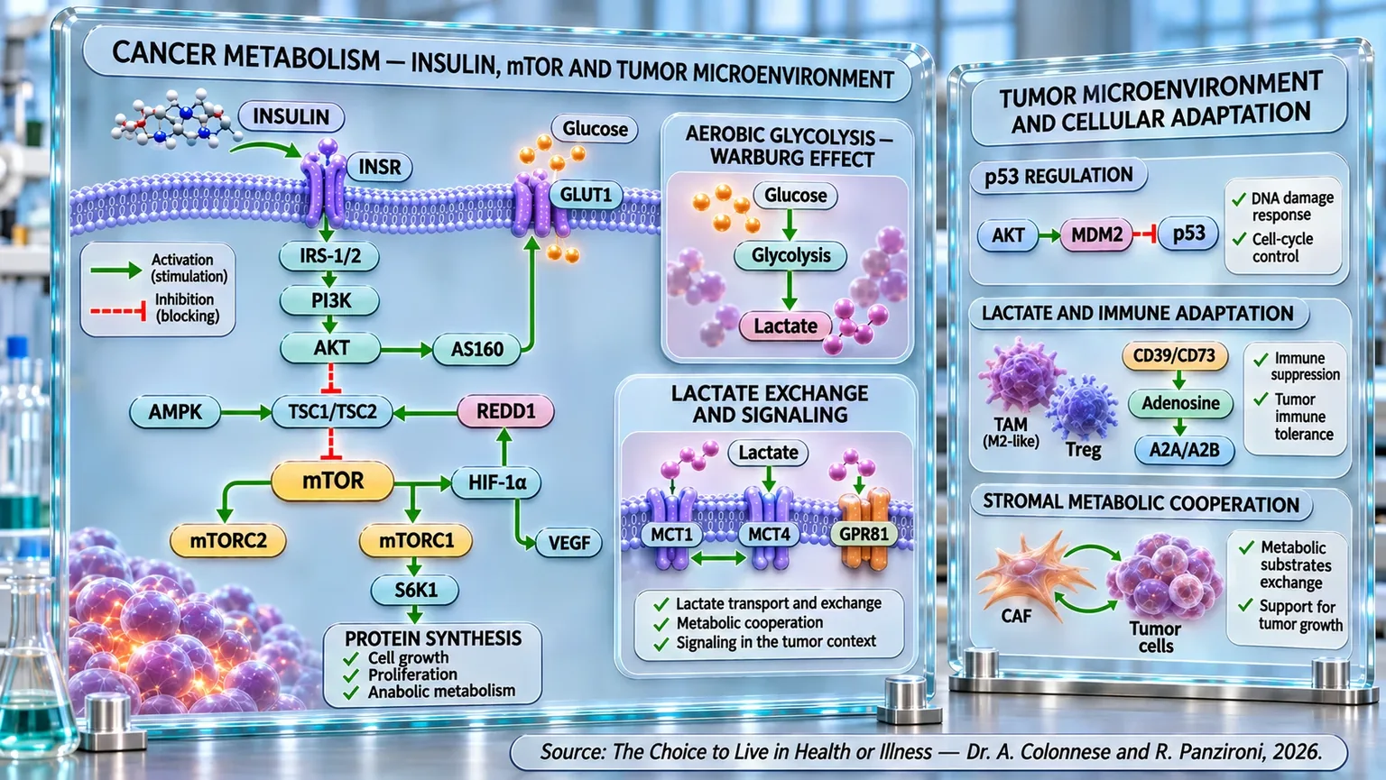
Two scientific panels connect insulin signaling, aerobic glycolysis, lactate, p53 regulation, immune adaptation, and cooperation between stroma and tumor.
Diagram of tumor metabolism: insulin activates IRS-1/2–PI3K–AKT and mTOR, while GLUT1 fuels aerobic glycolysis and lactate production. HIF-1α, VEGF, MCT1/MCT4 transporters, p53, adenosine, immune cells, and cancer-associated fibroblasts illustrate adaptation and cooperation in the neoplastic microenvironment.

ABSTRACT

Tumor progression results from the interaction between genetic alterations, metabolism, proliferative signals, and the microenvironment. In the book "La scelta di Vivere in Salute o in Malattia" (The Choice to Live in Health or Disease), Dr. A. Colonnese and R. Panzironi explore these processes in depth, examining the potential contribution of insulin signaling to neoplastic growth. Epidemiological studies associate hyperinsulinemia, insulin resistance, and the insulinemic potential of the diet with specific oncological outcomes, without necessarily demonstrating causality.

Activation of the insulin receptor can involve PI3K/AKT/mTOR, regulating protein synthesis, proliferation, and survival. In numerous tumors, glycolysis increases even in the presence of oxygen, according to the Warburg effect. mTOR can support HIF-1α, which under hypoxic conditions promotes metabolic adaptations and the expression of GLUT1. This reprogramming provides intermediates necessary for growth and links glucose consumption to metabolic cooperation between tumor regions with different oxygen levels. The lactate produced is exchanged via MCT1 and MCT4, can fuel different cells, and modulate GPR81, intracellular signals, and protein lactylation.

These circuits interact with cellular control systems. AKT can promote MDM2, reducing p53 activity, while p53 counteracts mTOR through AMPK and TSC1/TSC2. In the microenvironment, cancer-associated fibroblasts contribute to metabolic exchanges through the reverse Warburg effect. Lactate and hypoxia can orient macrophages toward immunoregulatory phenotypes. HIF-1α promotes adenosinergic processes mediated by CD39 and CD73; adenosine, through A2A and A2B receptors, can attenuate antitumor immunity. Regulatory T cells can adapt to local conditions and contribute to immune tolerance, making the relationship between metabolism and antitumor defense more complex. Regulatory T cells participate in this context, while VEGF associated with hypoxia supports angiogenesis.

Overall, the evidence describes the tumor as a dynamic ecosystem in which energy availability, proliferation, stromal cooperation, and immune evasion influence each other. The integration of these mechanisms offers targets for further research, distinguishing experimental observations from epidemiological associations and clinically validated therapeutic applications.

Insulin and cancer

Cancer is not a single disease, but an extremely heterogeneous group of conditions characterized by the progressive loss of the normal systems that regulate cell growth, survival, and behavior. To understand the scale of the problem, one only needs to look at the numbers: in 2022, nearly 20 million new cancer cases and approximately 9.7 million deaths were estimated worldwide. The cumulative risk of developing a neoplasm before the age of 75 was estimated at around 20% of the global population, with a difference between the sexes: approximately 21.8% in men and 18.5% in women. In the same year, about 10.3 million new cancers were diagnosed in men and 9.7 million in women. Age is also one of the main determinants of risk: cancer becomes progressively more frequent with advancing years, as a consequence of the accumulation of genetic, epigenetic, and metabolic alterations over time. Taken together, these data describe a pathology that affects a considerable portion of the population and whose onset depends on the interaction of multiple biological and environmental factors (Bray et al., 2024) [1].

Among the metabolic factors that have attracted increasing attention in recent decades is insulin. We are accustomed to considering it primarily as the hormone that acts after a meal to keep glycemia under control. This representation is correct, but incomplete. Insulin is also a biological signal capable of communicating to the cell that energy and nutrients are available in the environment and that, when conditions allow, growth, synthesis, and proliferation can be supported. It is precisely this second function that becomes particularly interesting when exposure to insulin is no longer just transient, but remains chronically elevated.

One of the most important observations comes from a study conducted on 9,778 adults without diabetes and without a previous oncological diagnosis. Hyperinsulinemia, defined by a fasting insulin level of 10 μU/mL or higher, was present in 30.6% of non-obese participants. In the total population, hyperinsulinemic subjects had approximately double the risk of cancer mortality compared to subjects without hyperinsulinemia; most importantly, the association remained significant even when considering only non-obese individuals, in whom the risk was increased by 89%. The data takes on further importance because the association was observable even in the presence of normal fasting glycemia (Tsujimoto et al., 2017) [2].

This means that glycemia and insulin should not necessarily be considered as two overlapping photographs of the same phenomenon. The body can manage to maintain apparently normal glycemia for a long time by increasing the amount of insulin required to achieve that result. It is a bit like keeping a car's speed constant by pressing harder and harder on the accelerator: by observing only the speed, the change might go unnoticed; by observing how much the pedal is being pressed, however, it would become evident. Another large cohort, consisting of 12,238 Japanese individuals followed for a median of ten years, has reinforced this interpretation. The group with the highest fasting insulin values had a 55% higher risk of cancer mortality compared to the group with the lowest values, and the association persisted even after adjustment for fasting glycemia and HbA1c. Each 1 μU/mL increase in fasting insulin was associated with a 4% increase in the risk of death from cancer (Kira et al., 2020) [3].

Evidence regarding the colon-rectum shows that this association may already be present in the stages preceding the actual carcinoma. A meta-analysis of 27 studies highlighted that high concentrations of insulin, C-peptide, and HOMA-IR were associated with increases in the risk of colorectal adenoma of 33%, 44%, and 33%, respectively. The adenoma represents one of the main precancerous lesions of the colon-rectum and, importantly, most of the associations remained significant even after adjustment for adiposity (Yoon et al., 2015) [4]. A subsequent meta-analysis including 35 studies, 25,566 patients, and over 5.7 million participants found an association between the risk of colorectal carcinoma and all the main indicators of glucose metabolism alteration. Elevated fasting insulin was associated with a 42% increase in risk, while HOMA-IR showed the strongest association, with a 47% increase (Xu et al., 2016) [5].

The epidemiological data gains greater strength when combined with experimental evidence. In a mouse model of human prostate carcinoma, a diet rich in refined carbohydrates resulted in higher concentrations of insulin and IGF-1 and significantly greater tumor growth: after nine weeks, the average tumor volume reached 1,695 mm3 compared to 980 mm3 in the group fed a low carb diet. Furthermore, the serum of the most hyperinsulinemic animals stimulated the proliferation of tumor cells more intensely in vitro (Venkateswaran et al., 2007) [6].

Even more interesting is the experiment conducted on the pancreas, because it allowed for the separation, at least in part, of the effect of insulin from that of glycemia. In mice genetically predisposed to the development of pancreatic lesions associated with the Kras oncogenic mutation (a genetic alteration that can continuously stimulate cell growth), a moderate reduction in endogenous insulin production, achieved without significantly modifying glycemia in the analyzed females, approximately halved the pancreatic area occupied by lesions and tumor: 12.7% versus 25.4%. Collagen deposition in the stroma also decreased from 41.3% to 20.1%. The extent of the lesions correlated with fasting insulin, but not with glycemia (Zhang et al., 2019) [7]. These results do not prove that insulin is sufficient, on its own, to generate a tumor; rather, they indicate that, in a tissue already predisposed by oncogenic alterations, a hyperinsulinemic environment can contribute to creating conditions more favorable to tumorigenesis.

The same relationship emerges when insulin exposure is studied indirectly through diet and lifestyle. In 78,356 postmenopausal women followed for over twenty years, the quartile with the highest hyperinsulinemic potential of the diet had a 68% higher risk of liver carcinoma compared to the lowest quartile (Zhang et al., 2023) [8]. In endometrial carcinoma, the picture is more complex: in 60,441 women, indices based exclusively on diet did not show a significant independent association after adjustments, while overall lifestyle patterns characterized by higher hyperinsulinemic and insulin-resistant potential were associated with risks 2.58 and 2.89 times higher, respectively (Habeshian et al., 2025) [9]. This result invites us not to reduce the phenomenon to a single nutrient: body weight, physical activity, diet, and metabolic status interact with each other.

Insulin exposure could also be relevant after diagnosis. In 1,718 patients with stage I-III colorectal carcinoma, a high insulinemic potential of the diet after diagnosis was associated with a 66% higher risk of colorectal carcinoma-specific mortality. Patients who maintained a strongly hyperinsulinemic diet over time also showed a less favorable prognosis compared to those who maintained a low insulinemic potential dietary pattern (Tabung et al., 2020) [10].

Data on exogenous insulin also deserve attention, although they require particular caution in interpretation. In a cohort of 127,031 diabetic patients, a positive relationship was observed between insulin dose and the incidence of neoplasms. After adjustment for dose, insulin glargine (a long-acting synthetic insulin) also showed a dose-dependent association with oncological risk compared to human insulin, while aspart and lispro (rapid-acting human insulin analogs) did not show significant differences. As this is an observational study, with a relatively short follow-up and important potential confounding factors, these results do not allow us to establish that insulin therapy or glargine causes cancer (Hemkens et al., 2009) [11].

Taken together, this evidence outlines a coherent picture that must, however, be interpreted with precision. Insulin cannot be defined simply as a cause of cancer: carcinogenesis requires much more complex genetic, epigenetic, and microenvironmental alterations. The data suggest instead that chronic hyperinsulinemia can represent, in certain biological contexts, an environment favorable to neoplastic initiation, growth, or progression. Insulin can, in fact, provide the cell with signals that promote proliferation and survival and can increase the bioavailability of IGF-1, further amplifying mitogenic stimuli (Tsujimoto et al., 2017) [2]; (Kira et al., 2020) [3]. The decisive point, therefore, is not to imagine insulin as a switch capable of turning on a tumor by itself, but as one of the metabolic signals that can modify the terrain in which a transformed cell is born, survives, and expands. To understand how this signal can be translated within the tumor cell, we must now follow its molecular path and observe what happens when PI3K, AKT, and mTOR are involved.

PI3K/AKT/mTOR and cancer

As has emerged so far, insulin can help create a biological environment favorable to neoplastic growth; the next step, now, is to understand what happens when a growth signal reaches the inside of the cell. This is where one of the most important communication networks in cell biology comes into play: the PI3K/AKT/mTOR pathway. Under physiological conditions, as seen in depth in the dedicated chapter 4, this cascade functions as a sophisticated control system that allows the cell to adapt growth, proliferation, metabolism, and protein synthesis to the availability of nutrients and signals from the environment. The problem arises when this system stops responding correctly to its normal regulatory mechanisms and remains persistently active. In tumor cells, in fact, genetic or functional alterations can transform a physiological pathway essential for cellular life into a circuit capable of sustaining uncontrolled growth, survival, motility, and disease progression (Wu et al., 2022) [12].

We can imagine PI3K, AKT, and mTOR as three stations placed along the same transmission line. When PI3K is activated, the signal is progressively transferred toward AKT and then toward a series of intracellular effectors, among which mTOR occupies a fundamental position. In a normal cell, this line does not remain constantly on: it is activated when needed and braked when the stimulus wanes. In a tumor, however, the hyperactivity of PI3K, the loss of function of negative regulators like PTEN, or activating alterations of AKT can contribute to keeping the circuit persistently operational. The result is that the cell continues to receive signals favorable to growth and survival even when, physiologically, it should slow down or stop (Jiang et al., 2024) [13].

mTOR represents one of the main convergence points of this system and operates through two functionally distinct complexes, mTORC1 and mTORC2. mTORC1 controls processes strictly linked to growth and protein synthesis; mTORC2, on the other hand, participates in the full activation of AKT, thus contributing to the maintenance of the signal. The relationship should therefore not be imagined simply as a row of dominoes falling one after the other. It is more like a network in which some elements located downstream can contribute to keeping those that precede them active. This organization helps to understand why the deregulation of the axis can become particularly effective in supporting the tumor cell (Ye et al., 2020) [14].

The clinical relevance of this hyperactivation emerges clearly in nasopharyngeal carcinoma. By analyzing 119 patients and comparing them with 30 healthy subjects, some researchers found greater expression of PI3K, AKT, and mTOR in tumor tissues. But the most interesting data concerned the behavior of the disease: higher levels of the three proteins were associated with more advanced tumor stages, greater lymph node involvement, recurrence, and distant metastases. Furthermore, PI3K correlated positively with AKT and AKT with mTOR, showing in human tissue the same functional continuity predicted by the molecular cascade. Patients characterized by greater expression of these components also presented lower survival and, in multivariate analysis, the overexpression of PI3K, AKT, and mTOR emerged as an independent prognostic factor (Yu et al., 2019) [15].

A similar picture was observed in oral squamous cell carcinoma. In tumor tissues, positivity for PI3K, AKT, and mTOR reached 58.8%, 62.5%, and 51.3% respectively, while in adjacent normal tissues it was only 10%, 15%, and 10%. The activation of the cascade was associated with advanced stage and lymph node metastases; furthermore, PI3K and AKT were associated with lower tumor differentiation. Particularly significant was the position of AKT: AKT positivity remained independently associated with worse survival, with a hazard ratio (an indicator that compares the risk of an event between two groups over time) of 2.156 (Gu, 2026) [16].

These observations help to understand an essential concept: the activation of PI3K/AKT/mTOR does not seem to be limited to making a tumor mass grow more rapidly. It can also contribute to the acquisition of characteristics that allow cells to become more aggressive and to disseminate. In lung carcinoma, for example, this cascade participates in the regulation of the epithelial-mesenchymal transition, or EMT (transformation of epithelial cells into more mobile cells, capable of moving and invading other tissues). During this process, the cell progressively loses some characteristics typical of epithelial tissue, including part of its adhesive properties, and acquires a more mobile and invasive phenotype. It is as if a cell normally bound to its place in the tissue progressively acquired the tools necessary to detach, move, and colonize other environments. Numerous molecular regulators converge on the PI3K/AKT/mTOR pathway, influencing this very behavior; its persistent activation has been linked not only to proliferation but also to invasion, metastasis, angiogenesis, and therapeutic resistance (Moghbeli, 2024) [17].

A particularly intuitive experimental example comes from thyroid carcinoma. Midkine, a growth protein indicated by the acronym MDK, is overexpressed in tumor cells and its expression increases with the progression of the disease. When researchers experimentally reduced MDK, the ability of tumor cells to migrate and invade decreased along with the levels of PI3K, AKT, and mTOR. But the most interesting step happened subsequently: by pharmacologically reactivating the cascade via IGF-1, migration and invasiveness were restored. In short, turning off the signal reduced the aggressive behavior of the cell, while turning it back on allowed the cell to recover it (Yuan et al., 2024) [18].

A similar principle emerges in triple-negative breast carcinoma. In these cells, microRNA (tiny single-stranded RNA molecules that do not produce proteins) is reduced, while the proto-oncogene MYB (a normal gene that helps cells grow and divide in a healthy way) is more highly expressed. Experimentally increasing the microRNA resulted in a reduction of proliferation, migration, and invasion; conversely, its silencing favored these phenomena. MYB proved to be a direct target of this microRNA and its overexpression was able to reactivate PI3K/AKT/mTOR and restore cellular proliferation. The experiment therefore shows how different molecular signals can converge on the same cascade and use it as a sort of common node through which to sustain malignant behavior (Tang et al., 2025) [19].

But a cell that proliferates does not only need growth signals. It must also procure energy and, above all, material with which to build new cells. For this reason, the PI3K/AKT pathway takes on an even broader significance: it connects the proliferative signal to the reorganization of metabolism. The evidence collected by some researchers shows how AKT can increase glucose entry into the cell through the regulation of GLUT1 and favor the activity of glycolytic enzymes. The tumor cell, therefore, does not use PI3K/AKT only as a command that tells it to grow: it uses the same circuit to reorganize its metabolic resources in function of growth (Fontana et al., 2024) [20].

Making the picture even more complex is the fact that this pathway can also be influenced by the cells surrounding the tumor. In osteosarcoma, cancer-associated fibroblasts were found to be capable of modifying the metabolism of neoplastic cells by increasing the expression of phosphoserine aminotransferase 1, PSAT1, an enzyme involved in serine biosynthesis. The increase in PSAT1 was accompanied by the activation of the PI3K/mTOR/S6K1 axis and greater migration and epithelial-mesenchymal transition. The silencing of PSAT1 reduced these phenomena, while the inhibition of mTOR via rapamycin limited both cellular migration and the formation of metastases in animal models (Feng et al., 2025) [21]. This demonstrates that PI3K/mTOR can also represent one of the points through which the surrounding microenvironment communicates with the neoplastic cell and modifies its behavior.

The centrality of this cascade becomes even more evident by observing what happens when it is experimentally blocked. In renal carcinoma, the dual inhibitor GNE-477 reduced the growth, viability, proliferation, migration, and invasion of tumor cells, simultaneously inducing apoptosis. The compound blocked the phosphorylation of various components of the cascade, including AKT, simultaneously interfering with mTORC1 and mTORC2. Above all, when researchers artificially restored a constitutively active AKT, the antitumor effect of the compound was substantially canceled. Conversely, genetically eliminating AKT produced effects similar to those produced by the drug (Ye et al., 2020) [14].

However, the very network structure of the cascade explains why blocking it pharmacologically is more difficult than it might seem. The inhibition of a single point can remove some physiological feedback and allow the cell to reactivate AKT or use alternative circuits. For this reason, inhibitors capable of acting simultaneously on PI3K and mTOR have been developed: the goal is to close multiple escape routes at the same time and obtain a more complete suppression of the signal. The available data show that these compounds can reduce proliferation and favor apoptosis, but pharmacological resistance, toxicity, and molecular differences between tumors continue to limit their clinical efficacy (Wu et al., 2022) [12]. Even in head and neck carcinoma, in fact, PI3K, AKT, and mTOR inhibitors have produced heterogeneous results, and the blocking of a single node can be followed by the compensatory reactivation of the cascade (Jiang et al., 2024) [13].

Taken together, this evidence allows us to see PI3K/AKT/mTOR not as a simple sequence of proteins, but as a true biological infrastructure of the tumor cell. Its activation can link extracellular signals, growth, survival, protein synthesis, metabolism, migration, invasion, and metastatic capacity. Studies conducted in profoundly different tumors, from nasopharyngeal to oral carcinoma, from lung to breast, from thyroid to kidney and osteosarcoma, therefore converge on the same principle: when this network remains persistently active, the cell has a system capable of coordinating many of the functions necessary for neoplastic progression.

But among these functions, there is one that deserves to be followed separately. A cell that grows continuously must find a way to sustain exceptional energy and biosynthetic demands, even when the environment surrounding it becomes poor in oxygen and nutrients. It is precisely at this point that mTOR meets one of the most important regulators of cellular adaptation, HIF-1α, and that the metabolism of the tumor cell begins to take on characteristics profoundly different from those we would expect in a normal cell. This will be the necessary step to understand, in the next paragraph, the relationship between mTOR, HIF-1α, and the Warburg effect.

mTOR, HIF-1α and Warburg

In light of what has been observed, the activation of the PI3K/AKT/mTOR pathway is capable of providing the tumor cell with continuous growth and survival signals. But a rapidly proliferating cell does not only need the order to grow: it must also obtain energy and, above all, a large quantity of material with which to build new membranes, proteins, and nucleic acids. It is precisely in this step that mTOR takes on even greater significance, because it links the proliferative signal to the reorganization of cellular metabolism. One of the most characteristic phenomena of this transformation is the so-called Warburg effect, that is, the tendency of many tumor cells to strongly increase the use of glycolysis (the process by which cells break down glucose to obtain energy) and the production of lactate (a substance produced by cells during glucose metabolism) even when oxygen is available. This does not necessarily mean that the mitochondria have stopped working: in numerous tumors, aerobic glycolysis and oxidative phosphorylation can coexist. AKT can in fact simultaneously increase glucose uptake, glycolytic flux, and, in some contexts, even mitochondrial energy production, so much so that it has been proposed as a possible “Warburg kinase” (Robey & Hay, 2009) [22].

To understand the advantage of this, one must abandon the idea that the cell chooses its metabolism solely on the basis of the maximum amount of ATP obtainable from a glucose molecule. A tumor cell must duplicate rapidly and therefore also requires metabolic intermediates that can be used to synthesize nucleotides (small molecules that constitute the fundamental building blocks of DNA and RNA), lipids, and other macromolecules. Highly active glycolysis allows part of the glucose carbon to be diverted toward these biosynthetic pathways. It is as if a factory, instead of using all its raw material exclusively to produce energy, retained a portion along the production chain to build new machinery and new structures. The neoplastic cell can thus transform its metabolism from a simple energy system into a true infrastructure for proliferation (Robey & Hay, 2009) [22].

One of the most important molecular links between the proliferative signal and this metabolic transformation passes through mTOR and HIF-1α. Normally, HIF-1α is known primarily as a component of the cellular response to reduced oxygen. However, in cells characterized by oncogenic activation of mTOR, the program normally associated with hypoxia can be sustained even when oxygen is available. In this context, mTOR promotes the increase of HIF-1α, which in turn contributes to the expression of proteins involved in glucose utilization and glycolysis. The cell can therefore behave metabolically as if it had to adapt to an oxygen deficiency even while in normoxic conditions: a sort of “pseudohypoxia” generated by intracellular signaling (Sun et al., 2011) [23].

Some researchers have shown this phenomenon using cells in which the loss of TSC1, TSC2, or PTEN (a protein that functions as a natural brake on the PI3K/AKT/mTOR pathway) resulted in strong activation of mTOR. These cells developed intense aerobic glycolysis even in the presence of oxygen; when mTOR was inhibited with rapamycin, the glycolytic phenotype decreased significantly. In parallel, there was an increase in the expression of pyruvate kinase M2, PKM2, an isoform associated with proliferating and tumor cells. When PKM2 was experimentally suppressed, both aerobic glycolysis and tumor growth decreased. The Warburg effect thus appeared not as an isolated metabolic phenomenon, but as a functional consequence of the hyperactivation of upstream oncogenic signaling (Sun et al., 2011) [23].

Further demonstration comes from pancreatic carcinoma. In these cells, ARHGAP4 (a human gene that produces an enzymatic protein) is reduced, while PKM2 is more highly expressed. When ARHGAP4 was experimentally restored, mTOR phosphorylation, HIF-1α and PKM2 levels decreased, along with glucose uptake and lactate production. Conversely, silencing ARHGAP4 increased all these parameters. Above all, the metabolic effects produced by the loss of ARHGAP4 were countered by both the inhibition of mTOR with rapamycin and the inhibition of HIF-1α. The experiment thus outlines a functional sequence in which mTOR activation sustains HIF-1α, HIF-1α contributes to the glycolytic program, and PKM2 represents one of the effectors through which this program is concretely realized (Shen et al., 2019) [24].

The same principle emerges in colorectal carcinoma. The long non-coding RNA CRNDE (an RNA molecule that does not produce proteins but regulates gene activity) was found to be capable of sustaining the Warburg effect through the AKT/mTORC1 pathway. Its silencing reduced glucose uptake, ATP and lactate production, and GLUT1 expression, accompanied by lower proliferation, greater apoptosis, and increased sensitivity to cisplatin (a life-saving platinum-based chemotherapy drug). Conversely, the overexpression of CRNDE intensified glycolytic metabolism, but these effects were abolished by AKT and mTOR inhibitors (Yang et al., 2022) [25]. The important consequence is that the Warburg effect does not appear simply as the result of tumor growth: in these models, it actively participates in maintaining cellular proliferation and survival.

In gemcitabine-resistant pancreatic carcinoma (an antitumor drug that blocks DNA replication and limits tumor cell proliferation), mTOR overexpression indeed produced an increase in glucose consumption, lactate production, and ATP, along with an increase in GLUT1 and PKM2. Inhibition of mTOR using everolimus (a drug that inhibits mTORC1, helping to reduce cell growth) reversed this metabolic program and simultaneously reduced cell growth and migration, promoting apoptosis; in animal models, it also decreased the growth of gemcitabine-resistant tumors (Cui et al., 2021) [26]. Also in non-small cell lung carcinoma, the experimental reduction of PI3K/AKT/mTOR activity through treatment with 1,25-dihydroxy-vitamin D3 was accompanied by a decrease in GLUT1 and PKM2 and the suppression of the glycolytic phenotype, observed both in vitro and in experimental in vivo models (Yiyan et al., 2022) [27].

The link with HIF-1 has also been observed in the cells of a type of triple-negative breast carcinoma. An experimental extract of Holothuria scabra reduced the activity of the AKT/mTOR/HIF-1 axis, accompanied by a decrease in lactate production and an increase in apoptosis. Although this was exclusively a cellular model and not clinical therapeutic evidence, the experiment provides further confirmation of the functional link between AKT/mTOR signaling, HIF-1, and the maintenance of tumor glycolysis (Yurasakpong et al., 2021) [28].

Other experiments show that it is also possible to modify metabolism by intervening upstream in the circuit. In specific tumor cells, berberine (a plant substance that can influence energy metabolism) reduced AKT/mTOR activity, GLUT1 availability, glucose entry, and ATP production; by experimentally reactivating AKT, most of these effects were attenuated, reinforcing the hypothesis of a functional relationship between the signaling cascade and glucose utilization (Guo et al., 2021) [29]. In docetaxel-resistant prostate carcinoma (a chemotherapy drug), however, the reduction of PI3K, AKT, mTOR, and HIF-1α was accompanied by a decrease in lactate, ATP, and PKM2 and an increase in pyruvate dehydrogenase, suggesting a partial reversal of the glycolytic programming associated with drug resistance (Xu et al., 2023) [30].

Further mechanistic confirmation, although coming from non-tumor cells, shows how closely proliferative signaling and metabolism can be linked. In pulmonary artery smooth muscle cells, PDGF (a platelet-derived growth factor) simultaneously increases proliferation, activation of the PI3K/AKT/mTOR pathway, HIF-1α, and aerobic glycolysis; blocking the cascade reduces both the Warburg effect and proliferation. This model shows that a growth signal can be transformed directly into a metabolic program intended to support cell multiplication (Xiao et al., 2017) [31].

Taken together, these findings allow us to interpret the Warburg effect as part of a coordinated metabolic strategy. The activation of AKT and mTOR does not just tell the cell to grow: it helps prepare the metabolism so that such growth is materially possible. HIF-1α represents one of the main points of connection between these two worlds, because it translates the signal coming from mTOR into a program capable of increasing glucose utilization and glycolysis even when oxygen is available. Fully understanding this step, however, requires looking at HIF-1α more closely: not just as a metabolic regulator, but as one of the main systems through which the tumor cell modifies its behavior and adapts to a progressively more hostile environment.

HIF-1α in cancer

In this context, HIF-1α represents one of the main meeting points between the proliferative signals of the tumor cell and its metabolic reprogramming. To fully understand its significance, however, it is necessary to observe what happens inside a solid tumor as the neoplastic mass grows. A cell population that is rapidly increasing in size requires ever-increasing amounts of oxygen and nutrients, but the vascular network that should supply it does not necessarily develop at the same speed. Furthermore, tumor vessels are often structurally and functionally abnormal. This leads to the formation of regions where the oxygen supply becomes insufficient relative to the cells' needs. It is important, however, to clarify what it really means to talk about hypoxia. The 20-21% oxygen present in the atmosphere, frequently used as a “normoxia” condition in cell experiments, does not correspond to the normal oxygenation of our tissues. In healthy tissues, physiological concentrations, more accurately defined as physioxia, generally range between approximately 3% and 7.4%, with an average around 5%. In solid tumors, values can drop further, to approximately between 0.3% and 4.2%, and in most neoplasms, median values of less than 2% have been observed, with particularly low levels in some prostate and pancreatic cancers (McKeown, 2014) [32].

This oxygen deficiency is not just a passive consequence of tumor growth. As the disease progresses, it becomes a true selective pressure: cells unable to adapt to the more difficult conditions are penalized, while those that possess or acquire systems capable of tolerating hypoxia gain an advantage. A greater presence of hypoxic cells has indeed been associated with a worse prognosis, and hypoxic regions can favor cell populations endowed with particularly resistant and aggressive characteristics (McKeown, 2014) [32]. In this scenario, HIF-1α can be imagined as an emergency switch that allows the cell to understand that the environment has changed and to rapidly modify its behavior.

HIF-1, an acronym for Hypoxia-Inducible Factor 1, is a transcription factor consisting of two components, HIF-1α and HIF-1β. The decisive component in the response to oxygen is HIF-1α. When oxygen is sufficiently available, specific enzymes called PHDs modify HIF-1α. This modification allows it to be directed toward the system through which it is rapidly degraded. In other words, in the presence of oxygen, the cell produces HIF-1α but, at the same time, has a system that prevents its accumulation. When oxygen decreases, the activity of PHDs is limited; HIF-1α escapes normal degradation, stabilizes, accumulates, and reaches the nucleus. Here, it associates with HIF-1β to form functional HIF-1, capable of recognizing specific DNA sequences defined as Hypoxia Response Elements, or HREs, and modifying the expression of numerous genes (Basheeruddin and Qausain, 2024) [33].

This mechanism originated as a physiological adaptation system. The problem in a tumor is that it is exploited to survive in an environment that might otherwise hinder the growth of the neoplastic mass. Once stabilized, HIF-1α coordinates a response much broader than simple energy conservation: it intervenes in metabolism, cell survival, angiogenesis (the formation of new blood vessels), invasiveness, and the cell's ability to interact with the microenvironment. Furthermore, its activation does not depend exclusively on oxygen deficiency. Oncogenic signals and pathways such as PI3K/AKT/mTOR can increase its expression or activity even when oxygen is not drastically reduced (Qannita et al., 2024) [34]. This connection is particularly important because it directly links what was described in the previous paragraphs to the hypoxic response: in a tumor, HIF-1α can be sustained both by the physical characteristics of the microenvironment and by molecular alterations within the cell.

One of the most profound consequences of its activation concerns the ability to reorganize the entire cellular metabolism. HIF-1 does not only modify the fate of glucose, but also intervenes in lipid and amino acid metabolism and mitochondrial function. Under hypoxic conditions, it limits the entry of pyruvate into oxidative metabolism through the induction of PDK1 (an enzyme that contributes to activating AKT), modifies the functioning of the respiratory chain, reduces oxygen consumption, and helps control the production of reactive oxygen species. It can also promote, through two proteins, mitophagy (a biological process in which the cell selectively eliminates its old, damaged, or no longer useful mitochondria) and reduce mitochondrial biogenesis. The consequence is therefore not simply the “switching off” of the mitochondrion, but its reorganization aimed at better utilizing the little oxygen available and preventing an excessive imbalance of reactive oxygen species from compromising cell survival (Infantino et al., 2021) [35].

Lipid and amino acid metabolism are also adapted to the new needs. Cells can increase the accumulation of lipid droplets, modify the synthesis and utilization of fatty acids, and rely more on glutamine and serine to support biosynthesis and the control of redox balance (Infantino et al., 2021) [35]. HIF-1α must therefore be interpreted not as a simple oxygen sensor, but as a coordinator capable of redesigning the metabolic priorities of the tumor cell.

This adaptability extends well beyond metabolism. HIF-1α promotes processes involved in proliferation and survival, contributes to the epithelial-mesenchymal transition, and can increase the expression of molecules that facilitate the remodeling of the extracellular matrix, migration, and invasion. In this way, hypoxia can transform from an obstacle to growth into a stimulus capable of selecting more adaptable and invasive phenotypes (Ghosh et al., 2022) [36].

The hypoxic (oxygen-poor) environment modifies the relationships between tumor cells, fibroblasts, and immune cells. These changes favor tumor survival and growth, reducing the effectiveness of the immune response. Hypoxia also alters the metabolism of tumor cells, increases oxidative stress, and hinders apoptosis (programmed cell death). A central role is played by HIF-1α, which allows cells to adapt to oxygen deficiency and supports various processes linked to tumor progression. Understanding these mechanisms can favor the development of new therapies, including glycolysis and HIF inhibitors, anti-angiogenic drugs, and hypoxia-activated treatments (Chen et al., 2022) [37].

A particularly important aspect concerns cancer stem cells, small cell populations capable of self-renewal and considered relevant in the persistence and recurrence of the disease. HIF-1α contributes to the maintenance of stemness and, under hypoxic conditions, has been associated with the expansion of populations characterized by specific markers and the increase of factors linked to self-renewal. This phenomenon has been described in various neoplastic contexts, including glioblastoma, breast carcinoma, hepatocellular carcinoma, lung, colorectal, and bladder carcinoma, as well as some hematological neoplasms (Zhao et al., 2023) [38]. It is as if the less oxygenated areas of the tumor could become true protective niches in which cells particularly capable of surviving, regenerating the tumor population, and resisting environmental pressures are favored.

The role of HIF-1α, in fact, does not seem limited to solid tumors. Its high activity has also been observed in leukemias, lymphomas, and multiple myeloma. In diffuse large B-cell lymphoma, high levels of HIF-1α have been associated with a worse prognosis in patients treated with immunochemotherapy; in multiple myeloma, HIF-1α has been linked to the production of pro-angiogenic signals, proliferation, invasion, and drug resistance (Zhao et al., 2023) [38]. The phenomenon cannot therefore be considered exclusively the product of poorly vascularized regions of a solid mass: HIF-1α is inserted into broader oncogenic networks that can sustain its activity in different neoplastic contexts.

In breast carcinoma, this network appears particularly evident. The persistent activation of HIFs is associated with metabolic reprogramming, invasive capacity, the maintenance of cancer stem cells, and chemoresistance. In particular, HIF-1α can increase the expression of transporters responsible for drug efflux, contributing to the reduction of their intracellular concentration (Luo et al., 2022) [39]. This helps to understand why hypoxia does not represent only a metabolic problem, but can also concretely influence the therapeutic response.

The relationship between HIF-1α and treatment resistance is further strengthened by its ability to simultaneously coordinate metabolism, extracellular matrix remodeling, invasion, and microenvironment modification. Furthermore, the reactive oxygen species produced at the mitochondrial level can contribute to the stabilization of HIF-1α, generating reinforcement circuits through which the adaptive response tends to be maintained (Magar et al., 2024) [40]. This results in an extremely dynamic system in which hypoxia, metabolism, and intracellular signaling feed each other.

HIF-1α thus assumes the role of a true coordination center for tumor adaptation. The decrease in oxygen, which might initially seem like a limit to growth, is translated into a biological program that allows the cell to change its metabolism, preserve its survival, modify the microenvironment, acquire invasive characteristics, and increase its ability to resist therapies. It is therefore not surprising that its overexpression is frequently associated with more aggressive phenotypes and unfavorable prognosis. But HIF-1α does not directly carry out all these transformations: it does so by activating specific target genes. In the next sections, it will therefore be necessary to follow separately some of the main tools through which this program is concretely realized, starting from one of the most important for the energy supply of the tumor cell: GLUT1.

HIF-1α, GLUT1 and cancer

Based on what has been described, HIF-1α allows the tumor cell to adapt to an environment where oxygen becomes progressively harder to find. But adapting to hypoxia does not just mean managing to survive with less oxygen: it also means finding an alternative way to ensure the cell has a sufficient flow of nutrients to support proliferation, biosynthesis, and survival. In this process, one of the most important targets of HIF-1α is GLUT1, the glucose transporter. If we imagine the cell membrane as the border of a city, GLUT1 can be compared to one of the main gates through which glucose manages to enter. Increasing the number and activity of these gates means allowing the tumor cell to intercept a greater quantity of glucose from the surrounding environment and thus fuel the glycolytic metabolism upon which many neoplasms particularly rely. This relationship between HIF-1α and GLUT1 therefore represents one of the points through which the response to hypoxia is transformed into a true functional modification of tumor metabolism.

A first, particularly intuitive demonstration of this relationship comes from studies that have linked HIF-1α and GLUT1 to the uptake of the 18F-FDG radiopharmaceutical used in PET scans. The principle is also important for clinically understanding what happens in a tumor: 18F-FDG is a glucose analog, and its increased uptake can therefore reflect, at least in part, the increased capacity of neoplastic cells to acquire glucose. In an experimental rabbit lung tumor model, researchers observed that tumor uptake of the radiopharmaceutical was positively correlated with both GLUT1 and HIF-1α. Furthermore, GLUT1 expression was directly correlated with that of HIF-1α, while tracer uptake also increased in relation to the size of the tumor mass. These results allow us to visualize, almost from the outside, what is happening inside the cell: as the tumor grows and develops hypoxic regions, HIF-1α increases, GLUT1 increases, and the capacity to intercept glucose grows in parallel (Xu et al., 2013) [41].

The strength of the HIF-1α/GLUT1 axis emerges even more clearly when observed during the progressive transformation of normal tissue into neoplastic tissue. In cervical carcinoma, 158 samples were analyzed, including 28 cases of normal cervical epithelium, 32 cervical intraepithelial neoplasias, and 98 invasive cervical carcinomas. The expression of HIF-1α and GLUT1 increased progressively along this sequence, moving from normal tissue to the precancerous lesion and finally to invasive carcinoma. HIF-1α was associated with the degree of pathological differentiation, clinical stage, extent of lesions, and patient age, while GLUT1 showed a significant association with lymph node metastases. Above all, a strong positive correlation emerged between HIF-1α and GLUT1 (Cheng et al., 2013) [42].

When specific cervical carcinoma cells were experimentally placed in hypoxic conditions, the phenomenon became even more evident. Proliferation and invasiveness increased, apoptosis decreased, and glycolytic activity grew significantly. Simultaneously, both the gene and protein expression of GLUT1 and hexokinase II (the enzyme that initiates glucose utilization within cells) increased. HIF-1α, on the other hand, showed a different behavior: its mRNA did not increase significantly, while the amount of protein increased markedly (Cheng et al., 2013) [42].

This detail connects directly to what was described in the previous paragraph: during hypoxia, HIF-1α can increase not because the cell necessarily needs to produce much more mRNA, but because the protein is stabilized and protected from normal degradation mechanisms. Once accumulated, it can act on the cell's transcriptional program and increase the expression of genes necessary for glycolysis, including GLUT1. In this way, the decrease in oxygen is translated into an increase in the capacity to acquire glucose.

The functional importance of GLUT1 emerges with particular clarity in hepatocellular carcinoma. In this context, analysis of tumor tissues showed that 68.2% of samples exhibited an increase in GLUT1 mRNA compared to the corresponding non-neoplastic liver tissue. Protein expression detectable by immunohistochemistry was present in 13.2% of tumors and was absent in non-tumor liver tissues. In positive samples, GLUT1 was associated with a higher proliferative index, more advanced tumor stages, and lower histological differentiation (Amann et al., 2009) [43]. The transporter, therefore, did not seem to simply accompany neoplastic transformation, but rather characterized cells endowed with more aggressive biological behavior.

The most important proof, however, came from experiments in which GLUT1 was directly silenced. Reducing its expression via siRNA (short double-stranded RNA molecules) significantly decreased both the proliferation and migratory capacity of hepatocellular carcinoma cells. At the same time, glucose uptake and lactate secretion were reduced (Amann et al., 2009) [43]. This step is fundamental: GLUT1 does not appear only as an indicator of a metabolically active cell, but as a functional element that contributes to the maintenance of the tumor phenotype. If the gate through which glucose enters is narrowed, the cell loses part of its capacity to support both its own metabolism and certain behaviors associated with neoplastic progression.

The functional dependence between HIF-1α and GLUT1 was further tested in laryngeal carcinoma using genetic engineering. Under hypoxic conditions, tumor cells developed greater radioresistance, increased 18F-FDG uptake, and simultaneously showed activation of the PI3K/AKT/mTOR pathway and greater expression of HIF-1α and GLUT1. When HIF-1α or GLUT1 were eliminated, cell viability and glucose uptake decreased; when they were eliminated simultaneously, the effects were even more marked (Bao et al., 2022) [44].

This study also reveals something particularly important for understanding the complexity of tumor signaling: the relationship does not seem to be simply a unidirectional line in which PI3K/AKT/mTOR activates HIF-1α, HIF-1α increases GLUT1, and the process ends there. PI3K/AKT/mTOR activation increased HIF-1α and GLUT1, but genetic inactivation of HIF-1α or GLUT1 in turn reduced the activity of the PI3K/AKT/mTOR cascade. The authors therefore proposed the existence of a reciprocal regulation circuit, without being able to definitively establish which component occupied the position hierarchically further upstream (Bao et al., 2022) [44]. The tumor cell thus appears less like a linear chain of switches and more like a network of mutually reinforcing circuits: the proliferative signal supports metabolism, and metabolism, in turn, contributes to keeping survival signals active.

The importance of the HIF-1α/GLUT1 axis is further supported by experiments in which its interruption was associated with a reduction in tumor growth. In hypopharyngeal carcinoma, treatment of tumor cells with pseudoginsenoside DQ (a species of medicinal plant similar to true ginseng) reduced both HIF-1α and GLUT1 at the mRNA and protein levels, simultaneously decreasing glucose uptake. Microscopy analysis also showed something that simple quantification of expression could not have highlighted: not only did the total amount of GLUT1 decrease, but the molecules present on the plasma membrane were also reduced and their organization into clusters was altered (Wang et al., 2021) [45]. The availability of a transporter, in fact, does not depend only on how much is produced, but also on how much manages to reach and organize itself on the membrane, which is exactly the point where it must perform its function.

Further confirmation comes from breast carcinoma. In different experimental models, honokiol (a natural substance found in the bark of the magnolia plant) reduced the amount of HIF-1α and simultaneously the expression of GLUT1. The metabolic consequence was a decrease in glucose uptake, lactate production, and ATP generation, accompanied by a reduction in extracellular acidification and an increase in oxygen consumption. From a biological point of view, these modifications were associated with lower proliferation, greater apoptosis, and reduced tumor growth in vivo (Yi et al., 2022) [46].

Here too, HIF-1α was regulated primarily at the level of protein stability. Honokiol did not significantly modify its mRNA, but promoted its degradation through a mechanism involving specific ligases. By reducing HIF-1α, its downstream glycolytic targets, including GLUT1, were therefore decreased (Yi et al., 2022) [46]. This experiment reinforces a principle that already emerged in cervical carcinoma: to profoundly modify tumor metabolism, it is not always necessary to change the transcription of HIF-1α; it may be sufficient to modify its protein stability and, through this, the entire metabolic program it controls.

A conceptually analogous mechanism was observed in colorectal carcinoma through the study of sodium butyrate. In cells of this tumor line, treatment increased SIRT4 (a protein found in mitochondria), promoting the autophagic degradation of HIF-1α. The decrease in HIF-1α was accompanied by a reduction in GLUT1, lower glucose uptake, lower lactate production, and lower ATP generation. In parallel, the survival and clonogenic capacity (the ability of a cell to reproduce and generate other cells) of the tumor cells decreased, while in a xenograft model, butyric acid inhibited tumor growth (Zhang et al., 2024) [47].

The data is particularly interesting because it links the availability of HIF-1α to the amount of GLUT1 and, through GLUT1, to the very possibility of the cell effectively fueling its glycolytic metabolism. When SIRT4 promoted the autophagic elimination of HIF-1α, the entire downstream metabolic flux decreased: less GLUT1 meant less available glucose, and this was associated with lower levels of lactate and ATP (Zhang et al., 2024) [47].

Further, very recent evidence expands the biological significance of the HIF-1α/GLUT1 axis even more. In highly metastatic osteosarcoma, a link was identified between lysosomal zinc metabolism (zinc contained in lysosomes) and glucose metabolism through the HIF-1α/GLUT1 axis. In experimental models, the activation of TRPML1 (a protein that forms an ion channel located on the lysosomal membrane) modified the intracellular flux of the zinc ion and signaling mediated by reactive oxygen species, leading to the inhibition of HIF-1α, the reduction of GLUT1, and a consequent decrease in glucose uptake (Wang et al., 2026) [48].

In this study, the impairment of glucose metabolism produced consequences that went beyond the simple reduction of energy availability. The lower utilization of glucose compromised the regeneration of antioxidant systems, reducing the cell's ability to control oxidative stress. The experimental platform used also induced immunogenic programmed cell death and, in in vivo models, reduced both osteosarcoma growth and lung metastases; the combination with an oncological immunotherapy drug further increased the antitumor response (Wang et al., 2026) [48]. It is still a preclinical result, but it suggests how deeply the control of glucose entry can be intertwined not only with energy production but also with redox homeostasis and the tumor immune microenvironment.

Putting these pieces of evidence together, a coherent picture emerges. HIF-1α allows the tumor cell to perceive and cope with reduced oxygen availability; GLUT1 represents one of the tools through which this response is concretely transformed into a metabolic advantage. When HIF-1α stabilizes, GLUT1 expression can increase and the cell membrane becomes more efficient at intercepting glucose from the environment. The more glucose that enters the cell, the greater the amount of substrate available to support glycolytic metabolism. Studies on cervical, hepatocellular, laryngeal, hypopharyngeal, breast, and colorectal carcinoma, together with experimental models of specific rabbit tumors and osteosarcoma, converge in showing that interfering with HIF-1α or GLUT1 can reduce glucose uptake and be accompanied by a decrease in proliferation, survival, or tumor growth, albeit with important differences between the various experimental models.

GLUT1, therefore, should not be considered just a passive gate. In the context of tumor metabolic reprogramming, it becomes one of the points through which the cell adjusts its glucose supply to its own needs. HIF-1α helps increase this capacity especially when oxygen becomes scarce, allowing the cell to continue fueling a metabolism that is heavily dependent on glycolysis. But once it has entered through GLUT1, the glucose must be metabolized, and this process produces consequences that do not remain confined within the cell. One of the most important is the increasing production of lactate, an element that will progressively assume a central role not only in the metabolism of the neoplastic cell but also in the construction of the tumor microenvironment.

HIF-1α, lactate increase, and MCT4

HIF-1α plays a fundamental role in the adaptation of tumor cells to oxygen deficiency. When oxygen levels decrease, this factor promotes cell survival through two important mechanisms. On one hand, it stimulates the production of VEGF, a protein that promotes the formation of new blood vessels. On the other, it modifies energy metabolism, increasing the reliance on glycolysis (a process that allows energy to be derived from glucose). This metabolic shift leads the tumor cell to use more glucose and to convert a greater portion of pyruvate into lactate. In this way, even when oxygen is scarce, the cell can continue to produce energy and sustain its activities.

However, the increased production of lactate creates a new requirement. To avoid excessive accumulation, the cell must have systems capable of transporting it across the membrane. This is where monocarboxylate transporters (MCTs) come into play—proteins that facilitate the passage of lactate. Among these, MCT4 assumes particular importance in tumor cells characterized by high glycolytic activity.

The general link between hypoxia, increased glycolysis, and lactate production can also be observed outside of tumors. In human adipocytes exposed to 1% oxygen, researchers observed that lactate release increased 2.3-fold after just 24 hours. After 48 hours, MCT1 mRNA increased 8.5-fold and MCT4 mRNA by as much as 14.3-fold. Returning to normoxia also reversed the observed changes, indicating that this was a dynamic response to oxygen availability (Pérez de Heredia et al., 2010) [49]. Although these are not tumor cells, this experiment is useful for understanding the physiological principle: when oxygen is scarce, increased lactate production and the remodeling of its transport systems can proceed together.

In tumors, this adaptation takes on an even more complex significance. In breast carcinoma, other researchers have shown that the constitutive activation of HIF-1α in fibroblasts (cells that produce the structural support for tissues) induces a shift toward aerobic glycolysis, with greater production and secretion of L-lactate, an increase in MCT4, and a simultaneous reduction in mitochondrial activity. When these fibroblasts were co-injected with specific breast carcinoma cells, they significantly promoted tumor growth (Chiavarina et al., 2012) [50]. This finding introduces a fundamental concept: the lactate produced in response to HIF-1α should not be considered simply a "waste product" of glycolysis. Its production and transport can become part of the metabolic organization of the tumor and the microenvironment that surrounds it.

The distribution of MCT4 in human neoplastic tissue makes this concept even more intuitive. By analyzing advanced head and neck carcinomas, researchers observed that GLUT1 and MCT4 exhibited a very similar spatial pattern. As the distance from blood vessels increased—and therefore in regions where oxygen tends to become progressively less available—the expression of both increased. At a distance of approximately 150–200 μm from the vessels, the positive fraction was two to four times higher. Furthermore, GLUT1 and MCT4 showed high colocalization (Rademakers et al., 2011) [51]. It is almost possible to imagine a metabolic map of the tumor: as one moves away from the vessels, GLUT1 increases, supporting the entry of glucose necessary for glycolysis, and simultaneously MCT4 increases, consistent with the need for glycolytic cells to manage and export the produced lactate.

A similar response has been observed directly in brain and breast tumor cells. Researchers compared specific glioblastoma cells and breast carcinoma cells and found that hypoxia increased MCT1 and MCT4 in both lines. MCT2, however, showed a response dependent on the cell type; they interpreted the increase in MCT4 as consistent with its role in the efflux of lactate produced by highly glycolytic cells (Cheng et al., 2012) [52]. This step is essential: if glycolysis can be imagined as an accelerated production line, MCT4 represents one of the main pathways through which the cell prevents the final product of that line from accumulating uncontrollably.

However, available data show that not all tumors rely primarily on MCT4 for this function. In glioblastoma, researchers observed that hypoxia primarily increased the expression of MCT1 on the plasma membrane. This increase was accompanied by greater glucose consumption, lactate production, and an increase in glycolytic markers such as GLUT1 and HKII (an enzyme that initiates the use of glucose by cells). Reducing MCT1 decreased lactate production and compromised proliferation and invasiveness, while in human tissues, MCT1 was associated with hypoxic regions and specific markers (Miranda-Gonçalves et al., 2016) [53]. The significance of this observation is important: MCT4 represents a central element of hypoxic glycolytic adaptation, but lactate management cannot be described as a system entrusted to a single transporter.

The same principle emerges in bladder carcinoma. High expression of MCT1 was associated with lymph node metastases, distant metastases, and lower overall survival. When MCT1 was experimentally reduced, proliferation, migration, and invasion decreased, along with the lactate present in the extracellular medium and the expression of HK2, GLUT1, and LDHB (a component of an enzyme that allows cells to transform lactate into pyruvate). Above all, hypoxia simultaneously increased HIF-1α and MCT1, while silencing HIF-1α resulted in a reduction of MCT1 (Zhang et al., 2018) [54]. These results therefore link the hypoxic response, glycolytic reprogramming, and lactate metabolism to biological characteristics associated with tumor aggressiveness.

In colon cancer, other researchers provided further evidence of how closely HIF-1α can integrate with this metabolic reorganization. The depletion of TRAP1 (a protein located in the mitochondria) compromised mitochondrial function, increased ROS, and pushed cells toward a higher glycolytic flux, with an increase in glucose consumption and lactate secretion. Simultaneously, HIF-1α showed greater nuclear localization and higher transcriptional activity of hypoxia-response elements, while GLUT1 and MCT1 were upregulated (Tsai et al., 2025) [55]. In other words, the system does not act only by increasing glycolysis: it can simultaneously limit the entry of pyruvate into oxidative metabolism, favoring a metabolic configuration in which the production and management of lactate acquire increasing importance.

An even more direct relationship between HIF-1α and a lactate transporter emerges from the study of other researchers on hexavalent chromium. In lung tumor cells, the metal increased HIF-1α, MCT1, and MCT4 and favored the use of lactate. Experimental induction of HIF-1α increased MCT1, while inhibition of HIF-1α or MCT1 reduced lactate use and cell growth. The most mechanistically relevant aspect is that HIF-1α was found to be capable of binding directly to the promoter region of MCT1, promoting its transcription (Li et al., 2025) [56]. This study therefore shows how HIF-1α can intervene directly in the apparatus necessary to use lactate, even if in this specific model the functional protagonist is MCT1 and not MCT4.

Finally, a non-tumor model offers further confirmation of the functional relationship between HIF-1α and lactate metabolism. In bovine mammary epithelial cells, stimulation with LPS (a laboratory technique used in immunology) increased glycolysis, intracellular lactate, HIF-1α, and MCT1. Induction of HIF-1α further increased lactate accumulation, while its inhibition produced the opposite effect and modified MCT1 in parallel (Cheng et al., 2025) [57].

Overall, an adaptive system emerges in which the reduction of oxygen and the activation of HIF-1α can favor the shift of metabolism toward glycolysis, consequently increasing lactate production. At that point, the cell must necessarily manage this metabolite through MCT transporters. MCT4 appears particularly associated with hypoxic and glycolytic tumor regions and the function of lactate export, while MCT1 shows greater versatility and can assume a significant role in the efflux or use of lactate depending on the metabolic context. The tumor thus builds a sort of monocarboxylate management network that allows it to adapt to the profound differences in oxygenation present within it. But the lactate that is produced and exported does not disappear. It accumulates in the microenvironment, can be used by other cell populations, and can profoundly modify their behavior. It is precisely this transformation of lactate from a product always considered a waste of glycolysis into a biologically active component of the tumor microenvironment that we will address in the next paragraph.

Lactate and cancer

As has emerged so far, the activation of HIF-1α promotes a metabolism more oriented toward glycolysis, increasing lactate production and necessitating the strengthening of the systems responsible for its transport. At this point, however, it is necessary to definitively abandon the idea that lactate represents only the final product of particularly intense glycolysis. For a long time, it was described almost as a waste material that the cell simply had to remove. Evidence accumulated over the last few decades has profoundly changed this interpretation. Lactate can be used as fuel, transferred from one cell to another, participate in cell signaling, and influence the behavior of the cells that make up the tumor microenvironment. More recently, the possibility has even emerged that it can directly link metabolism to the regulation of gene expression through lactylation. In a tumor, therefore, what initially appears as the end point of glycolysis can become the beginning of numerous other biological processes.

One of the most radical interpretations of this phenomenon was proposed by researchers San-Millán and Brooks. The authors of the study suggest shifting the focus from the high glucose consumption characteristic of the Warburg effect to the resulting lactate production, introducing the concept of “lactogenesis.” According to this hypothesis, persistent lactate production would not simply represent a consequence of tumor metabolic reprogramming, but could constitute one of its functional goals. Oncogenes, loss of tumor suppressor gene activity, increased GLUT1, activation of glycolysis and LDHA (a gene that encodes a subunit of the enzyme L-lactate dehydrogenase), inhibition of pyruvate dehydrogenase (an enzyme complex that allows pyruvate to be used for energy production through the Krebs cycle), and an increase in MCT transporters would all converge toward a system capable of maintaining high lactate production and distribution. The accumulated lactate could then participate in feedback circuits capable of further supporting glycolysis, angiogenesis, migration, and tumor adaptation (San-Millán and Brooks, 2017) [58].

What emerges most solidly is that in tumors, lactate can reach very high concentrations and its presence is associated with unfavorable biological characteristics. Some researchers report tumor concentrations up to forty times higher than in normal tissues and a relationship between high lactate levels, lymph node or distant metastases, and reduced survival. Lactate can also contribute to the maintenance of the glycolytic phenotype, migration, and angiogenesis, and can profoundly modify the tumor microenvironment (Zhang et al., 2023) [59]. This means that lactate production does not end the moment the molecule is expelled from the cell that generated it. From that moment on, the lactate enters a shared space and can become a resource or a signal for other cells.

A review by other researchers describes this very conceptual transformation, defining lactate as a true oncometabolite. Intense tumor glycolysis can cause significant acidification of the extracellular microenvironment, while MCT1 and MCT4 allow for a continuous exchange of lactate between different cell populations. Increased expression of these systems has been observed in numerous neoplasms and is frequently associated with greater invasiveness, metastasis, and unfavorable prognosis (de la Cruz-López et al., 2019) [60]. In other words, the tumor should not be imagined as a collection of metabolically identical cells, but as an ecosystem in which different populations can produce, export, import, and use metabolites in different ways.

One of the most elegant experimental demonstrations of this cooperation was provided by other researchers. In solid tumors, oxygen distribution is not uniform: some cells are located in hypoxic regions and depend more on glycolysis, while others are located near blood vessels and have more oxygen available. The authors demonstrated that the lactate produced by hypoxic cells can be taken up through MCT1 by oxygenated tumor cells and converted into pyruvate to fuel oxidative metabolism. In this way, the better-oxygenated cells can use the lactate and save glucose, leaving it available to the hypoxic cells that need it more. When MCT1 was pharmacologically inhibited or silenced, the oxygenated cells were forced to return to glucose; this increased competition for this resource, and the hypoxic cells died from energy deprivation. In mouse models and human xenografts, the disruption of this system also slowed tumor growth and increased sensitivity to radiotherapy (Sonveaux et al., 2008) [61].

Lactate can therefore be imagined as a sort of “energy currency” that circulates between different compartments of the tumor. Cells that produce it in abundance can release it, while other cells can recover and use it. Some researchers have described this organization as a true metabolic symbiosis. Hypoxic and glycolytic cells tend to be characterized by MCT4 and release lactate; those that are more oxygenated can instead express MCT1, import it, convert it into pyruvate through lactate dehydrogenase, and use it to fuel the Krebs cycle and oxidative phosphorylation. The availability of oxygen, nutrients, oncogenic alterations, and the microenvironment therefore determine the appearance of metabolically specialized populations within the same tumor (Nakajima and Van Houten, 2013) [62].

A particularly intuitive example of this plasticity comes from glioblastoma (a malignant brain tumor). Some researchers subjected specific cells to glucose deprivation and observed that the addition of lactic acid allowed them to maintain higher intracellular ATP levels and reduced cell death. At the same time, MCT1 and MCT4 increased, HIF-1α decreased, and proteins associated with oxidative phosphorylation increased. In the presence of lactate, therefore, cells subjected to glucose scarcity were able to modify their energy program and orient themselves more toward mitochondrial respiration (Duan et al., 2018) [63]. This is a fundamental conceptual shift: a tumor cell that produces lactate does not necessarily have to be the same one that subsequently uses it. Within the tumor mass, there can be actual production and consumption circuits.

But lactate does not act only as fuel. Its accumulation also modifies the physical-chemical conditions of the surrounding environment. Other researchers report tumor microenvironment pH values between approximately 6.3 and 6.9 in relation to lactate accumulation, resulting in the activation of metalloproteinases, cathepsins, and hyaluronidases (enzymes that break down tissue components) that can facilitate the degradation of the extracellular matrix. In parallel, hyaluronic acid can increase, creating conditions favorable to motility and invasion (Zhang et al., 2023) [59]. A 2019 review similarly describes how the acidic environment contributes to tumor progression, angiogenesis, therapeutic resistance, and the selection of more aggressive phenotypes (de la Cruz-López et al., 2019) [60].

This ability of lactate to modify the behavior of surrounding cells extends to the immune system. Some researchers describe lactate as a regulatory molecule capable of acting through MCT transporters. In the tumor microenvironment, its accumulation can reduce the cytotoxic function of NK cells (the ability to recognize and destroy infected or tumor cells) and CD8+ T lymphocytes and promote characteristics associated with M2 macrophage polarization. Lactate can also participate in angiogenesis through its entry into endothelial cells mediated by MCT1, with stabilization of HIF-1α and an increase in VEGF (Liu et al., 2024) [64]. These aspects will be explored in the following paragraphs; what is of interest here is to understand that a molecule produced by tumor metabolism can become a means through which the tumor influences cells that do not genetically belong to the neoplasm.

In recent years, this interpretation has further expanded with the discovery of lactylation, a post-translational modification through which groups derived from lactate metabolism can be attached to lysine residues of histone and non-histone proteins (proteins that perform various cellular functions, including regulating and protecting DNA). Other researchers describe lactylation as a possible bridge between metabolism, epigenetics (the set of mechanisms that can activate or reduce gene activity without changing the DNA), and cell signaling. Through modifications of chromatin and non-histone proteins, this process can influence gene transcription, DNA repair, the cell cycle, and therapeutic response. The analyzed evidence links lactate and lactylation to proliferation, invasion, metastasis, recurrence, and resistance to treatments (Fang et al., 2026) [65]. The concept is fascinating because it implies that a metabolic variation can leave a functional trace on the apparatus that controls which genes are used by the cell.

The same relationship emerges from the review by other researchers. The lactylation of histone and non-histone proteins has been associated with the regulation of genes involved in proliferation, migration, autophagy, epithelial-mesenchymal transition, and cell survival. In several tumors, high levels of lactylation are associated with more aggressive biological characteristics; furthermore, specific modifications have been linked to resistance to bevacizumab (a drug that hinders the formation of new blood vessels), DNA repair, chemoresistance, and radioresistance (Li et al., 2026) [66]. Even in this case, however, most therapeutic strategies directed against these mechanisms remain in the experimental phase and require clinical confirmation.

Thus, a radical transformation of the way we must observe lactate is taking shape. It is not simply the substance that remains at the end of glycolysis. It can be produced by one cell population and consumed by another; it can allow oxygenated cells to save glucose for hypoxic ones; it can contribute to the acidification of the microenvironment; it can influence angiogenesis, invasion, and immune cells; and, through lactylation, it can even link metabolic status to the regulation of gene expression. The most recent reviews bring these aspects together, describing lactate and lactylation as integrated components of the tumor's metabolic and epigenetic plasticity (Fang et al., 2026) [65].

The tumor therefore appears less and less as a mass of cells that indiscriminately consume glucose and more and more as an ecosystem capable of distributing its resources. Some cells produce lactate, others use it; some live predominantly on glycolysis, others maintain significant oxidative activity. Lactate thus becomes one of the languages through which these populations communicate metabolically. And it is precisely this ability to transform a metabolic product into a signal and an energy resource that opens the next step: understanding through which receptors and transporters lactate can be perceived and used by the tumor cell and how this communication can reach AKT and mTOR again.

Lactate, GPR81, MCT1, AKT and mTOR

In light of these observations, lactate is not merely the end product of glycolysis; it can become a fuel, a signal, and a tool through which different cell populations communicate within a tumor. For this to happen, however, the cell must be able to "perceive" the presence of lactate or transport it across its membrane. Two systems are therefore of particular importance: GPR81, a lactate-sensitive membrane receptor, and monocarboxylate transporters, including MCT1. Through these two entry points—one primarily for signaling and the other for metabolism—lactate can reach intracellular networks fundamental to growth and survival, including AKT and mTOR.

The importance of GPR81 in tumors emerged clearly from the work of researcher Roland in 2014. The receptor was found to be expressed in cell lines belonging to numerous types of neoplasia and, above all, was strongly present in human pancreatic carcinoma: 148 of the 158 samples analyzed, or about 94%, showed high expression of GPR81. Silencing the receptor had relatively modest effects when glucose was abundant, but it became rapidly lethal when glucose availability decreased and lactate served as an alternative energy substrate. Furthermore, lactate increased MCT1 only in cells that retained GPR81. Conversely, the loss of the receptor reduced lactate uptake and mitochondrial activity and, in in vivo models, resulted in a marked reduction in tumor growth and metastasis (Roland et al., 2014) [67].

GPR81 can therefore be imagined as a sensor placed on the cell surface: it detects that lactate is available in the microenvironment and helps activate the adaptations necessary to utilize it. This means that signaling and metabolism can proceed together. This concept appears with particular clarity in non-small cell lung carcinoma resistant to EGFR inhibitors. In the presence of erlotinib (a targeted molecular anticancer drug), the addition of lactate increased the viability of specific cells and reduced their apoptosis. Simultaneously, GPR81 and AKT increased. When GPR81 was silenced, AKT activation decreased and the cells regained sensitivity to erlotinib (a drug that blocks EGFR, reducing growth-promoting signals). At the same time, lactate increased MCT1, supporting cellular metabolic capacity; in clinical samples, GPR81 and MCT1 were more highly expressed in resistant tumors compared to sensitive ones (Ma et al., 2022) [68].

Lactate therefore seems to be able to act through two complementary arms: through GPR81, it can transmit a survival-promoting signal via AKT, while through MCT1, it can enter the cell and participate directly in metabolism. This distinction is important because it avoids viewing GPR81 and MCT1 as two components necessarily placed on the same line. In some contexts, lactate may indeed act primarily through the receptor; in others, what matters most is its entry through the transporters.

A demonstration of the first scenario comes from osteoblasts (specialized cells of bone tissue): in specific cells, lactate enhanced parathyroid hormone-induced differentiation through GPR81 and a cascade involving AKT. Inhibition of MCT1 did not abolish this effect, while interference with GPR81 or AKT significantly reduced it (Wu et al., 2018) [69]. Although not an oncological model, the study provides an important mechanistic demonstration: lactate can be recognized extracellularly and transformed into an intracellular signal capable of reaching AKT without its transport via MCT1 necessarily representing the determining step.

The opposite was observed in adult hippocampal progenitor cells. In this model, lactate increased proliferation, but the effect depended on entry through MCT1 and MCT2 transporters and not on the presence of GPR81. Parallel to this, AKT phosphorylation increased transiently. The deletion of GPR81 did not prevent the proliferative response, whereas blocking MCTs abolished it (Pötzsch et al., 2021) [70]. This is also a non-tumor model, but it demonstrates how lactate can use different pathways to reach the same signaling nodes.

The connection can extend further to mTOR. In pancreatic carcinoma treated with radiotherapy, lactate produced by surviving tumor cells increased persistently and modified the behavior of myeloid-derived suppressor cells (MDSCs). In these cells, lactate activated a sequence involving GPR81, mTOR, and HIF-1α. Inhibition of PI3K or mTOR reduced the lactate-induced accumulation of HIF-1α; furthermore, interference with HIF-1α in turn modified AKT, suggesting the existence of reinforcement circuits between these pathways. By blocking tumor lactate production or eliminating HIF-1α in MDSCs, the antitumor activity of T cells was restored and disease control after radiotherapy was improved (Yang et al., 2020) [71].

Further evidence of lactate's ability to activate AKT comes from endothelial cells. Some researchers have shown that lactate enters through MCT1 and promotes the activation of specific receptor tyrosine kinases, which converge on the PI3K/AKT pathway. This activation was necessary for the formation of tubular structures and new blood vessels. Blocking the receptors or their respective ligands substantially reduced the angiogenic response (Ruan and Kazlauskas, 2013) [72]. In this case, too, lactate acts as a metabolic messenger capable of reaching one of the main growth pathways already encountered in the previous paragraphs.

The relationship with mTOR finds further confirmation in physiological systems. In rat skeletal muscle, acute oral administration of lactate increased the expression of AKT and mTOR and resulted in a significant increase in mTOR phosphorylation after just thirty minutes, even though AKT phosphorylation did not vary significantly (Kyun et al., 2020) [73]. In a subsequent experiment lasting five weeks in mice, lactate administration increased AKT and mTOR mRNA, indicating a modification of signals associated with protein synthesis and degradation (Kyun et al., 2024) [74]. These studies do not naturally prove that the same mechanism operates automatically in tumors, but they confirm that lactate can influence the AKT/mTOR network even in non-neoplastic tissues.

MCT1 can also directly link lactate to the regulation of mTOR through different mechanisms. In human periodontal ligament stem cells, high concentrations of lactate interfered with autophagy and osteogenic differentiation. Blocking MCT1 partially recovered autophagic markers, while rapamycin restored both autophagy and several osteogenic markers, indicating the functional involvement of an MCT1-mTOR axis (Luo et al., 2022) [75]. The data once again underline that lactate entry can modify mTOR in a context-dependent manner and that the biological consequence is not necessarily always proliferative.

Taken together, these findings show that lactate can re-enter the PI3K/AKT/mTOR network through multiple doors. It can be recognized by GPR81, it can cross the membrane through MCT1, and it can indirectly modify the activity of receptor tyrosine kinases or other metabolic circuits. The result depends on the cell type and the context: in some models, GPR81-AKT prevails, in others MCT1 takes the lead role, while in the tumor immune microenvironment, a GPR81/mTOR/HIF-1α axis emerges directly. Lactate, therefore, should not be imagined as a simple linear signal that invariably leads to AKT and mTOR, but as a metabolite capable of entering different molecular networks and using these central nodes to modify survival, metabolism, and response to the microenvironment. It is precisely the activation of AKT that now opens a new step in our sequence: the relationship between AKT, MDM2, and p53 (a protein that protects cells from DNA damage).

AKT, MDM2 and p53

As previously highlighted, lactate can reach the PI3K/AKT network and, in certain contexts, contribute to its activation. At this point, it becomes necessary to follow the signal even further downstream, because AKT does not only control metabolism and cell growth: it can also interfere with one of the most important protection systems against neoplastic transformation, the one governed by p53. The connection passes through MDM2, a protein that functions as the main negative regulator of p53. To understand this mechanism, we can imagine p53 as an inspector who continuously checks the genomic integrity of the cell. If it detects sufficiently significant damage, it can slow down the cell cycle, promote repair, or prevent that cell from continuing to proliferate. MDM2, on the other hand, represents one of the systems through which the activity of this inspector is physiologically limited. When AKT enhances MDM2, the balance can shift further toward the reduction of p53.

One of the fundamental molecular demonstrations of this relationship was provided by some researchers in 2002: the activation of AKT reduced the amount of the p53 protein without decreasing the relative mRNA, indicating that the phenomenon did not derive from a lower production of the genetic messenger, but from a greater degradation of the already synthesized protein. The intermediate step was MDM2: AKT promoted its phosphorylation and increased the ability of MDM2 to ubiquitinate p53, that is, to apply a molecular signal that favors its degradation (Ogawara et al., 2002) [76].

This mechanism clarifies why the AKT–MDM2 axis can become particularly relevant in tumors. AKT provides survival and proliferation signals; p53, conversely, can impose an arrest when the cell accumulates dangerous alterations. Enhancing MDM2 therefore means reducing one of the main brakes that could counteract the survival of a damaged cell. In colorectal cancer, for example, involucrasin A (a natural flavonoid) reduced the phosphorylation of AKT and MDM2, while increasing p53. This was associated with a cell cycle arrest and an increase in apoptosis. When a constitutively active form of AKT was introduced or p53 was eliminated, the antiproliferative and pro-apoptotic effects of the compound were significantly reduced (Wei et al., 2023) [77].

A similar relationship has been observed in glioma (the primary tumor of the central nervous system). FOXN3 (a human gene that regulates the cell cycle) is reduced in tumor tissues, and low levels of the protein are associated with lower overall survival. Its overexpression reduces proliferation, colony formation, migration, and invasion, while simultaneously increasing apoptosis. From a molecular point of view, the increase in FOXN3 reduces AKT and MDM2 and increases p53; silencing it, on the other hand, produces the opposite profile. In xenografts, FOXN3 overexpression also slows tumor growth (Wang et al., 2021) [78]. Once again, when the pressure exerted by AKT and MDM2 decreases, p53 returns to accumulate and the tumor behavior is attenuated.

An experiment conducted in a non-tumor context helps to understand how powerful this circuit can be in controlling cell survival. In doxorubicin-induced cardiotoxicity (a chemotherapy drug), licochalcone A (a natural flavonoid extracted from licorice roots) increases the phosphorylation of PI3K, AKT, and MDM2 and reduces p53, protecting cells from ferroptosis (a form of programmed cell death). The inhibition of PI3K/AKT or the overexpression of p53 eliminates much of this protective effect (Chen et al., 2024) [79]. The significance for our journey is not therapeutic, but mechanistic: the same AKT–MDM2–p53 axis can be used physiologically to favor the survival of a cell subjected to stress. In a tumor, however, this capacity can become advantageous for cells that would otherwise be better to eliminate.

In esophageal cancer, genistein (a natural substance of plant origin with a structure similar to human estrogens) offers confirmation in the opposite direction. Treatment reduces EGFR and the phosphorylation of AKT and MDM2, while simultaneously increasing p53. This modification is accompanied by greater apoptosis and cell cycle arrest. In xenograft models, a dose-dependent reduction in tumor growth is also observed (Gao et al., 2020) [80]. The axis therefore appears as a point where proliferative signals coming from membrane receptors can be translated into a reduction of the control exerted by p53.

Signals coming from the extracellular matrix can also use this mechanism. In stem cells derived from adipose tissue, fibronectin and vitronectin increase AKT and MDM2 and reduce p53, attenuating cellular senescence. When MDM2 is inhibited, p53 increases and the share of senescent cells increases simultaneously (Tragoonlugkana et al., 2024) [81]. In this case, too, it is not an oncological model, but the result clearly shows that the AKT/MDM2/p53 axis constitutes a general system through which extracellular signals can influence the cell's decision to continue to proliferate or to arrest.

The oncological importance of MDM2 phosphorylation has also been demonstrated directly in vivo. Some researchers studied a specific residue of MDM2, a target of AKT, observing that preventing its phosphorylation does not substantially change the p53 response to ionizing radiation, but profoundly changes the response to oxidative stress. The mutation reduces the nuclear stability of MDM2, increases p53 activity, and promotes senescence. In murine models of liver cancer, lung cancer, and skin carcinogenesis, the inability to phosphorylate this residue is accompanied by a significant reduction in tumorigenesis (Chibaya et al., 2021) [82]. The study therefore demonstrates that the AKT–MDM2 axis is not limited to modifying the amount of p53 in a test tube, but can concretely influence the ability of a tissue subjected to oncogenic stress to develop a neoplasm.

At this point, we can also reconnect the mechanism to the lactate metabolism described in the previous paragraphs. In hepatocellular carcinoma, lung adenocarcinoma, and ovarian cancer cells, lactate increases the expression of ESM1 (a specific gene that regulates the formation of new blood vessels). ESM1 subsequently activates AKT and MDM2, determining the functional inhibition of p53 and attenuating the response to DNA damage caused by cisplatin (an antitumor drug that damages cell DNA) (Tan et al., 2026) [83]. A particularly interesting connection thus emerges: lactate → ESM1 → AKT → MDM2 → p53, through which a metabolic change can directly reach a central system of the DNA damage response.

The same architecture also contributes to chemoresistance. In oral squamous cell carcinoma, MINK1 (a specific gene that regulates cell movements, cell shape, and the response to biological stress) promotes the activation of AKT and the phosphorylation of MDM2, contributing to the degradation of p53. The knockout of MINK1 reduces AKT and MDM2 and increases phosphorylated forms of p53 associated with its stabilization. The same effect was obtained experimentally through pharmacological inhibition of MINK1 (Mohanty et al., 2022) [84].

In bladder cancer, the mechanism appears even more direct. TPI1, known mainly as a glycolytic enzyme, acquires an oncogenic function independent of its traditional metabolic activity: it forms a complex with AKT and MDM2, facilitating their encounter. In this way, it increases the phosphorylation of MDM2 and promotes ubiquitination (a process by which the cell marks certain proteins to regulate or eliminate them) and degradation of p53. The inhibition of AKT counteracts this effect and preserves p53. High levels of TPI1 are also associated with a worse prognosis, while its silencing reduces proliferation, migration, invasion, and tumor growth (Wang et al., 2025) [85].

All these pieces of evidence therefore show how AKT, MDM2, and p53 constitute a fundamental node in which metabolic, receptor, and oncogenic signals can converge. AKT can enhance MDM2; MDM2 can ubiquitinate p53 and favor its degradation; the reduction of p53 can finally allow the cell to avoid some of the main programs that would normally limit the proliferation of a damaged cell. To fully understand why the functional loss of p53 represents such an important advantage for the tumor, however, it is necessary to observe separately everything that this protein is capable of doing. It is therefore from p53 and its multiple tumor-suppressive actions that our journey must continue.

p53 and genomic integrity

Based on what has been described, the activation of AKT reinforces the action of MDM2, thereby promoting the reduction of p53. To understand why this step can represent such a significant advantage for a tumor cell, we must now observe what happens when p53 is fully functional. Its definition as the “guardian of the genome” does not stem simply from its ability to arrest the cell cycle or induce the death of a severely damaged cell. p53 participates in a much broader surveillance system, which includes damage recognition, chromatin organization (a complex of DNA and proteins that organizes genetic material), DNA repair, protection of chromosomal ends (the terminal parts of chromosomes), and even the maintenance of mitochondrial DNA. In other words, p53 does not intervene only when a problem is already evident: it continuously contributes to limiting the accumulation of errors that, if left to propagate, could become the substrate for neoplastic transformation.

As early as the late 1990s, it was proposed that this protection had at least two levels. Under basal conditions, even before significant genotoxic damage occurs, p53 may contribute to DNA surveillance through its own biochemical activities. When more significant damage occurs, however, the protein modifies its functional state, and sequence-specific DNA binding becomes more important, which is necessary to activate transcriptional stress-response programs. The two activities appear to be reciprocally regulated: the activation of sequence-specific binding is associated with a reduction in DNA strand degradation activity, suggesting that p53 can change its operational mode depending on cellular conditions (Janus et al., 1999) [86].

It is as if the cell possessed a security system capable of functioning both during normal daily maintenance and during an emergency. Under ordinary conditions, p53 would help prevent small errors from accumulating; when the damage exceeds a certain threshold, the response becomes more structured and involves a much larger number of genes and cellular processes. This capability depends largely on the extremely sophisticated relationship between p53 and DNA.

Indeed, p53 does not only recognize a specific sequence of bases. As a transcription factor, it can bind to response elements consisting of specific consensus sequences (stretches of DNA, RNA, or proteins that function as recognition signals for specific molecules), but its activity is also influenced by the shape locally assumed by the DNA. In addition to the classic double helix, DNA can fold into different three-dimensional structures. p53 is capable of recognizing many of these conformations, and in some cases, the presence of a particular structure can significantly increase the protein's affinity for that genomic region (Brázda and Fojta, 2019) [87].

This means that p53 does not read DNA only as a text composed of letters, but seems to be capable, at least in part, of perceiving its shape as well. A region that is folded, deformed, or organized in a particular configuration can therefore become an important element for recognition. Some researchers have highlighted how this relationship between sequence and topology extends the possible functions of p53 to repair, replication, recombination, and the organization of chromatin itself (Brázda and Coufal, 2017) [88].

But recognizing damage is not enough. Nuclear DNA is not found free within the cell: it is wrapped around histones (proteins that organize DNA) and organized into chromatin. To repair a lesion, the proteins responsible for maintenance must be able to physically reach the damaged tract. A recent study showed that p53 also participates directly in this step: following the formation of double-strand breaks, the protein is rapidly recruited to the damaged sites and the surrounding chromatin, promoting its relaxation. When p53 is silenced or poorly expressed, the repair of breaks is less efficient, with a particularly evident contribution to the non-homologous end joining pathway (a system by which the cell repairs broken DNA by directly joining the damaged ends) (Chen et al., 2025) [89].

The comparison with a maintenance crew is particularly useful. Knowing where a breakdown is located is not enough if the area is closed and inaccessible. p53 seems to contribute not only to identifying the problem but also to opening the space necessary for repair systems to intervene. This function appears to be very early: the recruitment of p53 to damaged regions can occur rapidly and precede its better-known transcriptional consequences, such as cell cycle arrest or apoptosis (Chen et al., 2025) [89].

The protective effect of p53 on chromatin also emerges in senescent cells. A 2025 study showed that p53 activation reduces the formation of cytoplasmic chromatin fragments (CCF, fragments of genetic material that exit the nucleus and can trigger inflammatory responses in the cell) that can originate from damaged nuclear DNA. Increased p53 activity promotes better DNA repair and preserves genomic integrity, while simultaneously reducing the expulsion of chromatin into the cytoplasm. Since these fragments can fuel inflammatory signals, the function of p53 thus ends up linking genome stability and inflammation control (Miller et al., 2025) [90].

The protection of chromosome ends is particularly delicate. Telomeres can be imagined as the caps that protect chromosomal ends, preventing them from being interpreted as DNA breaks. In human and murine subtelomeres, binding sites for p53 derived from retrotransposon-like elements (DNA fragments that can copy themselves and insert into other parts of the genetic heritage) have been identified. In response to damage, these sites can locally modify chromatin and stimulate the production of an RNA associated with telomere regulation. The loss of p53 or the deletion of these sites increases telomeric instability (Lieberman, 2016) [91].

The concept of the “guardian of the genome” must also be extended beyond the nucleus. Mitochondria possess their own DNA, mtDNA, which is particularly vulnerable to oxidative stress and characterized by a higher mutation rate than nuclear DNA. Here, too, p53 seems to participate in surveillance. It can regulate genes necessary for the maintenance of mtDNA and can interact directly with proteins involved in mitochondrial replication and repair. Of particular importance is the repair of the main systems through which the mitochondrion corrects oxidative damage (Park et al., 2016) [92].

p53 therefore appears as a system capable of coordinating the stability of two different genetic heritages: the nuclear and the mitochondrial. This is particularly significant because mitochondrial damage does not only concern energy production: it can modify oxidative stress and, consequently, further increase the pressure exerted on the nuclear genome.

Observing these functions as a whole, it becomes easier to understand why genomic stability depends so deeply on p53. The protein can temporarily arrest proliferation so that DNA can be repaired, directly promote certain repair processes, and, if necessary, prevent a genetically compromised cell from continuing to propagate. When this surveillance fails, cells carrying chromosomal alterations can instead survive and multiply. Evidence from human tumors, animal models, and “Li-Fraumeni syndrome” (a rare hereditary genetic disease that increases the risk of developing various types of cancer) indeed links the impairment of the p53 pathway to aneuploidy (a genetic anomaly in which cells have an altered number of chromosomes compared to the normal set for the species), chromosomal instability, and telomeric alterations (Eischen, 2016) [93].

Finally, it is important to understand that p53 does not react to all stresses in the same way. The response depends on their intensity and duration. Under moderate conditions of energy deficiency, hypoxia, or oxidative stress, p53 can initially contribute to adaptation, slowing growth and favoring more efficient resource management. However, when the stress becomes severe or persistent and cellular integrity cannot be adequately preserved, the response can shift toward the elimination of the damaged cell (Humpton and Vousden, 2016) [94].

p53 is therefore not simply a switch that decides whether a cell should live or die. It is a dynamic surveillance system that first attempts to protect the genome, make DNA accessible for repair, preserve telomeres and mitochondrial DNA, and limit the propagation of errors. Only when the damage exceeds the possibility of recovery do more drastic responses come into play. Precisely for this reason, the loss of p53 has much deeper consequences than the simple decrease in apoptosis: it means depriving the cell of one of the main systems capable of preventing genomic instability from being transmitted to subsequent cell generations. It is from this loss of control that we must therefore continue in order to understand the relationship between the loss of p53 and tumorigenesis.

p53 loss and tumorigenesis

In the previous subsection, we observed how p53 contributes to preserving genomic integrity through multi-level surveillance of DNA, chromatin, telomeres, and even the mitochondrial genome. At this point, it becomes intuitive to understand why its loss can represent such a favorable step for neoplastic transformation. When p53 is inactivated, the cell does not just lose a brake on proliferation: it loses a system capable of preventing potentially dangerous genetic alterations from being maintained, propagated, and selected. The consequence is not necessarily an immediate tumor transformation, but the creation of an evolutionary terrain in which genomic instability can increase and certain cell populations can progressively acquire proliferative and survival advantages.

A particularly elegant example comes from human gastric organoids (small models of the human stomach grown in the laboratory to study its functioning). Researchers inactivated both copies of TP53 (the human gene that produces the p53 protein) and followed the cultures for about two years, observing pre-malignant evolution in real time. The loss of TP53 progressively led to aneuploidy, copy number alterations, and structural variants similar to those observed in gastric carcinoma with chromosomal instability. But the most interesting aspect was that this evolution was not completely random. Some alterations appeared in a preferential order, and initially rare subclones with greater fitness (groups of cells that were initially rare but had a greater capacity to survive and proliferate) repeatedly tended to become dominant. In parallel, the cells acquired transcriptional programs increasingly similar to those observed in malignant cells (Karlsson et al., 2023) [95].

This result modifies an intuitive but incomplete representation of genomic instability. The loss of p53 does not simply seem to transform the genome into a chaotic system in which every alteration has the same probability of being maintained. It is more like opening an evolutionary space in which many variants are generated, but selection favors those capable of conferring an advantage. Some cells acquire characteristics that allow them to grow better than others and thus end up progressively occupying a larger share of the population. Tumorigenesis therefore appears as the result of the interaction between genomic instability and clonal selection.

An even more orderly dynamic has been observed in pancreatic ductal carcinoma. Researchers longitudinally followed the consequences of p53 loss in a mouse model and identified a reproducible sequence of genomic events. The first step was the loss of heterozygosity (a genetic condition in which an individual possesses two different versions of the same gene) of TP53; this was followed by an accumulation of chromosomal deletions; subsequently, whole-genome doubling occurred, and only in the most advanced stages did chromosomal gains and amplifications appear (Baslan et al., 2022) [96].

In this case, too, the loss of p53 does not produce disorder devoid of logic. Alterations are acquired in a succession that can progressively confer new properties to the cell. Deletions predominate in the early stages, while polyploidy appears later and can offer a sort of compensation for the loss of genetic material. After genome doubling, the cell has a greater amount of DNA on which further oncogenic gains and amplifications can accumulate. The loss of p53 thus creates the permissiveness necessary for the genome to be progressively remodeled by tumor selection.

However, it is fundamental to emphasize that the complete genetic loss of TP53 does not necessarily have to be the first event. In colorectal carcinoma, some researchers have proposed a model in which p53 can be functionally reduced as early as the initial stages through its protein degradation. In this model, therefore, the protein can be functionally eliminated even before the TP53 gene is lost or mutated. Restoring p53 levels, achieved experimentally by increasing p53, prevented tumor onset and prolonged animal survival (Herranz-Montoya et al., 2025) [97].

This study introduces an important concept: what matters biologically is not only the genetic state of TP53, but also the amount of functional p53 actually available in the cell. If the protein is degraded too rapidly, the tumor-suppressive barrier can be reduced even in the absence of an initial gene mutation. According to the proposed model, the early functional loss of p53 allows for abnormal cell cycle progression, increases hyperproliferation, and promotes replicative stress and DNA damage, creating the conditions for the acquisition of further alterations associated with tumor progression (Herranz-Montoya et al., 2025) [97].

The consequences of p53 loss do not only concern chromosomal instability. In inflammation-associated colorectal carcinoma, TP53 deletion also modifies the relationship between tissue damage, stemness, and tumorigenesis. In an AOM/DSS mouse model, animals completely lacking TP53 developed more severe colitis, showed significantly reduced tumor latency, and reached a 100% neoplastic incidence. The tumors also presented more aggressive characteristics and a selective increase in cells associated with tumor stemness properties (Cunha et al., 2024) [98].

This means that the loss of p53 can modify not only the genome of a cell but also the functional state of the cell populations that make up the tissue. The increase in these specific cells suggests that, in the absence of p53, populations more capable of self-renewal and sustaining tumor growth may be favored. In an environment simultaneously exposed to inflammation and damage, this combination can create conditions particularly favorable to neoplastic progression.

A conceptually similar phenomenon has been observed in neural stem cells. In the adult brain, many of these cells remain physiologically quiescent, meaning they are metabolically active but outside the proliferative cycle. p53 helps maintain this condition by promoting fatty acid oxidation. When p53 is eliminated, the cells exit quiescence prematurely and begin to proliferate (Amodeo et al., 2023) [99].

The significance is important because an oncogenic mutation can remain biologically silent if the cell carrying it does not proliferate. The loss of p53, on the other hand, can allow that same cell to enter the cycle and transform a genetic predisposition into a biologically active phenomenon. In the glioblastoma mouse model analyzed by the authors, modulation of the metabolic axis was able to restore the quiescence of p53-deficient cells and delay tumor onset (Amodeo et al., 2023) [99].

The relationship between p53 loss and changes in cellular identity emerges even more clearly in reprogramming experiments. Researchers have observed that reduced p53 activity progressively increases the efficiency with which somatic cells (cells of the organism, excluding reproductive ones, which constitute tissues and organs) can be reprogrammed toward a pluripotent state (a cellular state capable of differentiating into numerous specialized cell types). A mutated form of p53 increased reprogramming even more than the complete absence of the protein. However, this greater efficiency had a biological cost: cells reprogrammed in the presence of mutated p53 more easily lost the ability to differentiate correctly and generated malignant tumors (Sarig et al., 2010) [100].

p53 therefore also seems to act as a barrier to excessive cellular plasticity. Eliminating this control makes it easier to change identity, but it can allow genomically altered cells to go through the reprogramming process without adequate DNA quality checks. It is like accelerating a reconstruction while eliminating safety checks: the process becomes faster, but the probability that errors are incorporated into the final structure increases.

However, one must carefully distinguish between two different phenomena: the loss of normal p53 function and the potential acquisition of new properties by mutated proteins. For years, it has been proposed that many p53 mutants acquire true oncogenic gain-of-function activities (when a genetic mutation causes a protein to acquire new abnormal properties or hyperactivity). Some researchers recently tested this hypothesis by eliminating various TP53 mutants in numerous tumor lines, organoids, and animal models. The removal of the mutated protein generally did not modify proliferation, survival, metastasis, or response to chemotherapy. Even the analysis of 391 tumor lines containing a total of 158 different TP53 mutations did not show a general dependence on the mutated proteins. Conversely, the restoration of the wild-type form, i.e., the original and most common form of TP53, resulted in the arrest of tumor proliferation (Wang et al., 2024) [101].

This result does not mean that no p53 mutant can acquire new properties, but it suggests that, for many tumors, the fundamental advantage derives primarily from having lost the normal tumor-suppressive function of the protein. This is an important distinction: the tumor cell might not necessarily need a "more powerful" mutated p53 in an oncogenic sense; it might be sufficient to no longer have normal p53 available.

One of the most intuitive demonstrations of the biological weight of this loss comes from mouse models carrying nonsense mutations (DNA alterations that prematurely interrupt the production of a protein). Researchers studied a specific mutation: in homozygous mice, 95% developed macroscopic tumors, and the neoplasms could appear as early as about 2 months; no animal survived beyond 8 months. Even in heterozygotes, 73% developed tumors, and in 71% of the analyzed neoplasms, the residual wild-type copy was lost. Multicentric or metastatic disease was also frequent (Strandgren et al., 2025) [102].

Taken together, these findings show that the loss of p53 does not simply represent the absence of a single molecular brake. It modifies the way the cell manages its own genome, its identity, and its proliferative state. It allows for the accumulation of chromosomal alterations, favors the selection of more competitive subclones, facilitates the exit from quiescence, can increase plasticity and stemness, and makes it easier for cells that would normally be eliminated to survive. In some contexts, moreover, this functional loss occurs long before the definitive mutation of the TP53 gene, through excessive degradation of the protein.

The tumorigenesis that follows p53 loss is therefore not simply the result of the random accumulation of errors. It is an evolutionary process in which instability generates variability and selection progressively preserves the configurations most favorable to growth. Once this important surveillance system is eliminated, other proliferative and metabolic pathways can act with fewer restrictions. Among these, mTOR and HIF-1α take on particular importance, because p53 normally interacts with both of these systems and contributes to limiting some of their consequences. Understanding what happens when this dialogue is interrupted therefore represents the next step.

p53, inhibition, mTOR and HIF-1α

In this context, we have therefore seen how p53 protects the integrity of the genome and how its loss progressively promotes chromosomal instability, clonal selection, and tumorigenesis. However, the tumor-suppressive function of p53 is not limited to DNA control. The protein also intervenes in one of the central systems that regulate growth, metabolism, and the cell's response to nutritional status: the mTOR pathway. This connection is particularly important because it allows us to understand how p53 can translate a stress condition into a metabolic decision. When the cell is damaged or resources are insufficient, continuing to grow and synthesize new structures would be biologically risky. p53 therefore contributes to curbing the anabolic and proliferative programs that depend on mTOR, coordinating the stress response with growth control.

One of the clearest demonstrations of this relationship was provided by a 2005 study: the activation of p53 following DNA damage leads to a marked inhibition of mTOR activity. This effect does not simply depend on cell cycle arrest, because it persists even in the absence of p21 (a protein that functions as a brake on cell division). The mechanism instead requires the activation of AMPK and the integrity of the TSC1/TSC2 complex: when AMPK or TSC1/TSC2 are inactivated, p53 loses the ability to suppress mTOR (Feng et al., 2005) [103].

The physiological significance of this sequence is intuitive. mTOR functions as a powerful promoter of anabolism: when nutrients and growth factors are available, it promotes protein synthesis, growth, and proliferation. AMPK, conversely, signals a condition of energy insufficiency. Through AMPK and TSC1/TSC2, p53 can therefore transform a stress signal into a reduction of biosynthetic programs. It is as if, faced with structural damage, the cell temporarily halts the expansion of the factory to focus resources on maintenance.

This inhibition can occur on different timescales. In the early stages of the response, p53 rapidly activates AMPK and the TSC1/TSC2 complex; subsequently, it can increase the expression of PTEN (a protein that limits the activity of the PI3K/AKT/mTOR pathway) and TSC2, reinforcing the blockade of mTOR over 12–24 hours (Feng et al., 2005) [103]. In parallel, glucose deprivation induces both the inhibition of mTOR and a transient phosphorylation of p53, showing that communication does not proceed in only one direction: p53 controls mTOR, but systems that sense energy status can in turn communicate with p53.

The influence of p53 on the proliferative axis is actually even broader. Some researchers have described a coordinated network through which p53 limits IGF-1/AKT/mTOR signaling; PTEN limits AKT activity, while TSC2, AMPK, and REDD1 converge on the inhibition of mTORC1 (Feng and Levine, 2010) [104]. In this way, p53 does not act on a single switch, but builds a true network of brakes that progressively reduces the cell's ability to keep growth signals active during stress.

The same work shows that this regulation is closely intertwined with energy metabolism. The loss of p53 is in fact associated with a reduction in oxygen consumption and mitochondrial respiration and a greater reliance on aerobic glycolysis. Conversely, p53 supports respiration, limits glycolysis, and represses components involved in glucose entry and utilization, including GLUT1 and GLUT4 (Feng and Levine, 2010) [104]. This results in a picture where p53 simultaneously limits the proliferative signal and the metabolic program that should support it.

This response, however, is not identical in every cell and every tissue. The analysis by some researchers on PTEN, TSC2, and AMPK has shown a marked dependence on the type of stress and the cellular context. Some stimuli induce PTEN, TSC2, and AMPK simultaneously; others produce different combinations. Tissue response also varies: PTEN and TSC2 are regulated by p53 primarily in tissues highly dependent on insulin-sensitive energy metabolism, such as skeletal muscle, heart, white adipose tissue, and liver (Feng et al., 2007) [105].

This specificity is important because it prevents us from describing p53 as a rigid switch. The protein functions rather as a signal integrator: it modifies its response based on the type of damage, the energy status, and the tissue involved. The final result is nonetheless consistent: limiting growth when proliferation could further compromise cellular integrity.

Another level of control concerns the endosomal compartment (a system of small vesicles present inside cells). p53 induces PTEN, TSC2, and AMPK, reducing IGF-1/AKT/mTOR; in parallel, it increases the production of exosomes (tiny vesicles released by cells), autophagy, and the internalization of membrane receptors (Feng, 2010) [106]. It is a further way to reduce proliferative pressure: not only is the signal curbed inside the cell, but the ability to receive it from the outside is also reduced.

At this point, the connection with HIF-1α becomes particularly important. As we have seen in the previous paragraphs, HIF-1α supports adaptation to hypoxia, VEGF expression, and glycolytic reprogramming. p53 can directly counteract this system. Some researchers have demonstrated that p53 interacts with HIF-1α and promotes its ubiquitination through MDM2, directing it toward proteasomal degradation (a system by which the cell eliminates and recycles proteins that are no longer needed). When p53 is eliminated, HIF-1α accumulates more during hypoxia, VEGF transcription increases, and neoangiogenesis and tumor xenograft development grow (Ravi et al., 2000) [107].

In breast carcinoma, the balance between these networks can be shifted in the opposite direction. MARK2 (an enzyme that controls microtubule stability and cell polarity) is increased in tumor tissues and associated with poor prognosis. From a functional point of view, MARK2 stimulates the transcriptional activity of HIF-1α through mTOR and, simultaneously, represses that of p53. Its inhibition reduces aerobic glycolysis and cell growth and increases cycle arrest and apoptosis. In the data, MARK2 also shows positive correlations with mTOR, S6K1, GLUT1, HIF-1α and negative correlations with p53 (Ponnusamy and Natarajan, 2022) [108].

This model represents almost the mirror image of the physiological p53 response. On one hand, p53 tends to limit mTOR and HIF-1α; on the other, MARK2 promotes mTOR/HIF-1α and attenuates p53. The result is a molecular environment more favorable to aerobic glycolysis, growth, and tumor survival.

Overall, these findings show that p53 occupies a central position in countering a tumor program that combines growth and metabolic adaptation. Through AMPK, TSC1/TSC2, and PTEN, p53 reduces the PI3K/AKT/mTOR signaling pathway; in parallel, it limits glycolysis and supports mitochondrial respiration. On another front, it can contain HIF-1α both by promoting its degradation and by reducing its synthesis. When p53 is inactivated or repressed, these brakes are progressively lost: mTOR can remain more active, HIF-1α can accumulate, and the cell can more easily orient itself toward a glycolytic metabolism and a phenotype adapted to hypoxia.

The loss of p53, therefore, does not just mean giving up genome protection. It also means removing a system that normally limits growth, biosynthesis, and metabolic adaptation when cellular conditions are unfavorable. And it is precisely this metabolic dimension that leads us to the final subsection dedicated to the actions of p53: the relationship between lactate, lactylation, and p53.

Lactylation and p53

In the previous sections, we saw how p53 represents much more than a simple cell cycle controller. Its activity helps preserve genome integrity, limits proliferation when the cell is in unfavorable conditions, and counteracts some of the main metabolic programs that support tumor growth, including those dependent on mTOR and HIF-1α. However, there is an additional level of regulation that allows us to reconnect p53 to one of the metabolic protagonists encountered several times in this chapter: lactate. Long considered primarily a terminal product of glycolysis, lactate can in fact behave as a true regulatory molecule. One of the mechanisms through which it exerts this function is lactylation, a post-translational modification in which groups derived from lactate are attached to specific lysine residues of proteins. In this way, a metabolic variation can be translated directly into a functional variation of cellular proteins.

This relationship assumes particular importance in tumors, where high glycolytic activity can lead to a considerable production of lactate. The biological significance is profound: tumor metabolism does not only modify the amount of energy and metabolites available, but can intervene directly on the systems that should normally counteract neoplastic transformation. p53 constitutes one of the most significant examples of this communication between metabolism and tumor control.

A fundamental step in understanding the phenomenon came from the identification of AARS1 as an intracellular lactate sensor and as an enzyme capable of promoting lactylation. AARS1 is traditionally known as alanyl-tRNA synthetase, and thus as a component of the machinery necessary for protein translation. However, some researchers have shown that it possesses a second function: it can recognize lactate, bind it, and catalyze the formation of a lactate-AMP intermediate, to then transfer the lactyl group to specific lysine residues of target proteins. In other words, AARS1 can transform the intracellular availability of lactate into a chemical modification of the proteome (the set of all proteins produced by a cell) (Zong et al., 2024) [109].

Among the numerous targets identified, p53 itself appears. In particular, AARS1 promotes the lactylation of specific residues located in the domain through which p53 interacts with DNA. The position of the modification is fundamental to understanding its consequences. To effectively exert its tumor-suppressive activity, p53 must be able to recognize specific DNA sequences and regulate the transcription of its target genes. Lactylation hinders the clustering of p53 molecules, reduces their ability to bind DNA, and consequently alters its transcriptional activity (Zong et al., 2024) [109].

We can imagine p53 as a key capable of recognizing specific locks present on DNA. In this case, the TP53 gene may be perfectly intact and the key may be present, but a modification of its structure makes its correct use more difficult. This introduces a particularly important concept: the tumor-suppressive function of p53 can be compromised even without TP53 necessarily being mutated. A metabolic environment rich in lactate can intervene directly on the already produced protein and modify its properties.

The oncological relevance of this phenomenon is also supported by the observation that high levels of AARS1 and p53 lactylation are associated with an unfavorable prognosis in cancer patients with wild-type p53. Furthermore, experimentally interfering with the binding between lactate and AARS1 using β-alanine reduces global lactylation, decreases p53 lactylation, and attenuates tumorigenesis in animal models (Zong et al., 2024) [109].

An even more direct example comes from bone metastases of lung carcinoma. Some researchers have identified the SLC2A3 gene of the GLUT3 glucose transporter as an important metabolic regulator of bone metastasis. SLC2A3 is more highly expressed in metastatic bone cells and tissues, and its increased activity increases glucose entry, supports glycolysis, and leads to an increase in lactate production (Ding et al., 2026) [110].

At this point, the sequence becomes particularly significant in light of what we have built in the previous paragraphs. The increase in glucose uptake mediated by SLC2A3 intensifies glycolysis; increased glycolysis increases the availability of lactate; lactate promotes the lactylation of p53. This modification antagonizes the acetylation of the same residue and reduces the transcriptional and tumor-suppressive activity of p53 (Ding et al., 2026) [110]. This creates a direct link between glucose availability, glycolytic metabolism, and the functional loss of one of the main tumor suppression systems.

The same relationship can be relevant in the response to therapies. In advanced prostate cancer, other researchers have studied the mechanisms through which tumor cells acquire resistance to enzalutamide (a hormonal drug). Prolonged exposure to the drug leads to the activation of NF-κB and STAT3 (two proteins that regulate gene activity), which promote increased expression of SLC4A4. This response is accompanied by metabolic alterations associated with glycolysis, an increase in lactate production, and greater p53 lactylation (Chen et al., 2024) [111].

When SLC4A4 is experimentally reduced, extracellular bicarbonate accumulation increases, intracellular acidosis appears, and lactate production derived from glycolysis decreases. In parallel, p53 lactylation decreases and sensitivity to enzalutamide is restored, with results observed both in vitro and in vivo (Chen et al., 2024) [111]. The metabolic modification of p53 can therefore play a role not only in tumor progression but also in the adaptive processes through which a neoplastic population manages to progressively survive pharmacological pressure.

The relationship between lactate and p53 becomes even more complex when considering that p53 does not necessarily have to be the direct target of lactylation. In pancreatic adenocarcinoma, some researchers have identified a prognostic signature associated with lactylation composed of five genes. Among these, PRKCG is downregulated in the tumor, and its lower expression is associated with more advanced disease and unfavorable prognosis. When PRKCG is experimentally overexpressed, proliferation, migration, and invasion of tumor cells decrease, apoptosis increases, and xenograft growth is reduced (Zhu et al., 2026) [112].

Lactate, however, can lactylate PRKCG without substantially changing its quantity. What changes is its function: the lactylated protein loses part of its ability to support the activation of phosphorylated p53. When the predicted lactylation sites are experimentally modified, this activity is at least partially recovered (Zhu et al., 2026) [112]. Lactate metabolism can therefore reach p53 through different paths. In some cases, it directly modifies the protein; in others, it modifies one of its regulators and alters p53 indirectly. The biological result can, however, converge towards a reduction in tumor-suppressive action.

The studies we have seen are more relevant precisely because of the Warburg effect and the increased glycolysis that do not simply produce more lactate to be eliminated. That lactate can become raw material for post-translational modifications capable of intervening directly on the cell's genomic control systems. In the case of p53, the connection is particularly significant because it creates a possible reinforcement circuit: tumor metabolism increases lactate, and lactate can contribute to weakening p53; the functional loss of p53, as we have seen, in turn reduces some of the brakes placed on glycolytic and proliferative programs. In this way, metabolism and tumor control can influence each other.

The neoplastic cell can therefore manage to neutralize p53 through different levels. It can lose or mutate TP53, it can promote the degradation of the protein, it can alter the circuits that regulate its activity, or, as these more recent findings show, it can use a consequence of its own glycolytic metabolism to chemically modify it with lactylation. This is a conceptually important step because it makes lactate not only the result of the tumor's metabolic reprogramming but one of the molecules through which that reprogramming can be transformed into biological information.

And it is precisely from here that the discussion can expand beyond the single tumor cell. The lactate produced by the tumor can accumulate in the tumor microenvironment, reach stromal and immune cells, and contribute to modifying their behavior and function. The next step will therefore be to understand how this metabolic communication can contribute to the construction of an environment that supports the tumor, introducing the TME and the reverse Warburg effect.

TME and the reverse Warburg effect

So far, we have mainly observed what happens inside the tumor cell: the activation of proliferative pathways, the intervention of HIF-1α, the increase in glycolysis, the production of lactate, and the progressive loss of the control systems exerted by p53. But a tumor never grows in isolation. Neoplastic cells are immersed in a complex environment with which they communicate continuously and which, over time, is profoundly transformed by the very presence of the neoplasm. This set of elements is called the tumor microenvironment, or TME, and includes stromal and immune cells, fibroblasts, the extracellular matrix, blood and lymphatic vessels, as well as physical and metabolic conditions such as hypoxia, acidity, interstitial pressure, and nutrient availability. The TME must therefore be imagined not as the passive background in which the tumor grows, but as a true dynamic ecosystem in which the different components influence each other (Wang et al., 2025) [115].

Tumor cells actively participate in the construction of this ecosystem. Through cytokines, chemokines, and numerous other mediators, they can modify the behavior of surrounding stromal cells (cells that support and protect tissues and organs) and immune cells, progressively inducing them to adopt characteristics favorable to proliferation, angiogenesis, invasion, and metastasis. Fibroblasts (cells that produce collagen and other substances necessary to support tissues), the extracellular matrix, and the vascular compartment also participate in maintaining these conditions, creating a network of signals in which clearly distinguishing what belongs to the tumor from what belongs to the surrounding tissue becomes increasingly difficult (Hinshaw & Shevde, 2019) [116].

This cooperation also concerns metabolism. The representation that all tumor cells use glucose in the same way through very intense aerobic glycolysis is now insufficient to describe the heterogeneity observed within neoplasms. In the same tumor, predominantly glycolytic populations and populations with a more oxidative metabolism can coexist. This diversity allows for the development of true metabolic cooperation, in which the products generated by one compartment can become energy substrates for another (Wilde et al., 2017) [113].

It is within this perspective that the concept of the reverse Warburg effect arises. In the classic Warburg effect, it is the tumor cell that strongly increases glycolysis and produces lactate even in the presence of oxygen. In the reverse model, however, part of this glycolytic behavior is assumed by stromal cells, particularly cancer-associated fibroblasts, or CAFs. These fibroblasts can acquire a predominantly glycolytic metabolism and produce lactate, pyruvate, and other high-energy metabolites that are then used by tumor cells, in which oxidative mitochondrial metabolism can remain very active (Fu et al., 2017) [118].

A sort of division of labor is thus created. The stromal compartment becomes more catabolic: it consumes substrates and produces metabolites. The neoplastic compartment, on the other hand, can use those metabolites to support energy production, biosynthesis, and growth. In this organization, what one cell eliminates can become the fuel for another.

One of the experimental mechanisms proposed to explain how this transformation of the stroma can begin involves oxidative stress. Some researchers, using a co-culture of specific tumor cells and fibroblasts, have observed that neoplastic cells release hydrogen peroxide and increase oxidative stress in the surrounding fibroblasts. In fibroblasts, mitochondrial activity decreases and glucose uptake increases, while in tumor cells the opposite phenomenon occurs: mitochondrial activity increases and glucose uptake and GLUT1 expression decrease (Martinez-Outschoorn et al., 2011) [117].

Oxidative stress can therefore push fibroblasts toward autophagy, mitophagy, and progressive impairment of mitochondrial function, favoring the shift to glycolysis. This process has also been associated with the activation of HIF-1α and NF-κB. Thus, glycolytic enzymes and systems dedicated to the production and transport of metabolites increase, creating a circuit through which the stromal compartment becomes progressively more suitable for producing substrates usable by the tumor (Fu et al., 2017) [118].

Lactate naturally occupies a central position in this system. Glycolytic CAFs can export it primarily through MCT4, while tumor cells can acquire it via MCT1 and subsequently use it in oxidative metabolism. This metabolic shuttle allows the two compartments to specialize: one produces and exports, the other imports and uses (Wilde et al., 2017) [113].

The importance of this model does not derive only from cellular experiments. In prostate cancer, some researchers analyzed samples from 480 patients and observed a marked metabolic difference between the stroma and the neoplastic compartment. CAFs strongly expressed MCT4, while in tumor cells, intense membranous expression of MCT1 was evident. Furthermore, proteins associated with fatty acid oxidation were predominantly localized in tumor cells, consistent with a more oxidative metabolism (Pértega-Gomes et al., 2014) [119].

A particularly interesting aspect was the early onset of stromal modifications: MCT4 showed alterations already during the first phases of malignant transformation. Furthermore, the combination of high stromal expression of MCT4 and strong tumor expression of MCT1 was associated with the pT3 stage (a tumor that has exceeded the boundaries of the area in which it formed) and other unfavorable prognostic characteristics (Pértega-Gomes et al., 2014) [119]. The stroma, therefore, can be metabolically reprogrammed very early and become an integral part of the evolution of the neoplasm.

Clinical-pathological confirmation comes from a subsequent analysis of 535 patients with prostate cancer. Some researchers observed that neither MCT1 nor MCT4, considered in isolation, were significantly associated with biochemical recurrence-free survival. The data changed, however, when they were analyzed together. The profile characterized by high MCT1 expression in tumor cells and high stromal MCT4 expression was associated with a significantly worse prognosis and maintained its value even after adjustment for other prognostic factors, with a hazard ratio (a value that indicates how much the risk of an event occurring increases or decreases) of 1.99 (Andersen et al., 2015) [114]. The significance is important: what matters is not necessarily the presence of a single transporter, but the combination of the two compartments that makes metabolic exchange possible.

An even more marked result was observed in triple-negative breast cancer. Some researchers studied over 180 patients, finding that high stromal expression of MCT4 was associated with a ten-year overall survival of less than 18%, while in patients without stromal MCT4 expression, survival reached approximately 97%. Conversely, MCT4 expressed by epithelial tumor cells did not show the same prognostic value (Witkiewicz et al., 2012) [120].

This data makes it particularly evident how reductive it can be to observe exclusively the neoplastic cell. In that specific cohort, a metabolic characteristic of the stroma provided much more relevant prognostic information than the same marker expressed directly by the tumor.

The TME, however, does not only concern metabolism and fibroblasts. Its physical, metabolic, and immune compartments are deeply interconnected. Hypoxia and acidity favor tumor adaptation and resistance to treatments; HIF-1α increases glycolytic and angiogenic programs; the accumulation of lactate contributes to the acidification of the medium and modifies the behavior of immune cells. In parallel, CAFs, the extracellular matrix, and altered vascularization participate in invasion, migration, and the difficulty with which drugs and immune cells manage to penetrate the tumor mass (Wang et al., 2025) [115].

However, it is fundamental not to replace one overly simple model with another that is equally rigid. The reverse Warburg effect does not imply that all tumor cells use stromal lactate nor that the classic Warburg effect disappears. The two configurations can coexist in the same tumor. Some neoplastic populations can remain strongly glycolytic, while others can use oxidative phosphorylation more and exploit metabolites produced by the stromal compartment (Fu et al., 2017) [118].

The tumor thus appears as a metabolically organized and plastic ecosystem, in which resources are not simply consumed, but redistributed. Lactate can be produced by one cell, exported into the extracellular space, and subsequently recovered by another population. Tumor-associated fibroblasts assume, in this system, a particularly important function: they are no longer just structural cells of the stroma, but can become true metabolic partners of the neoplasm. It is therefore precisely on the relationship between lactate and CAFs that the next step must now focus.

Lactate and CAF

As we have seen, the tumor microenvironment (TME) can organize itself as a true metabolic ecosystem, in which neoplastic cells and the stromal compartment continuously exchange nutrients, metabolites, and signals. Within this network, cancer-associated fibroblasts (CAFs) play a particularly important role. They are not simply structural cells located around the tumor mass: they can be progressively reprogrammed to acquire properties capable of supporting proliferation, invasion, fibrosis, immunosuppression, and therapeutic resistance. Lactate represents one of the main tools through which the tumor cell can influence this transformation. The relationship, however, is more complex than a simple transfer of fuel from one cell to another: lactate can be used metabolically by CAFs, can modify their redox state, can reprogram their epigenome (the set of chemical signals that regulate gene activity), and can orient their secretory profile toward phenotypes capable of altering the immune compartment as well.

A particularly clear demonstration comes from pancreatic ductal adenocarcinoma. In this tumor, neoplastic cells, which are highly glycolytic, compete effectively for glucose and divert a significant portion of it away from the surrounding stromal cells. CAFs, therefore forced to live in conditions of relative glucose restriction, can adapt by using the very lactate produced by the tumor cells. Through MCT1, they import it, incorporate it into oxidative metabolism, and use it to fuel the Krebs cycle, thus supporting their own proliferation even when glucose is scarce (Kitamura et al., 2023) [121].

This behavior profoundly changes the significance of lactate in the microenvironment. The same tumor cell that monopolizes glucose simultaneously produces a metabolite that allows the surrounding fibroblasts to survive. But the effect is not only energetic. Exposure to lactate increases the production of immunosuppressive mediators in CAFs, including IL-6, and fibroblasts stimulated in this way reduce the activity of cytotoxic immune cells. Furthermore, in PDAC models (the most common and aggressive type of pancreatic cancer), high tumor LDHA (an enzyme that helps cells produce energy by converting pyruvate to lactate) is associated with a greater presence of CAFs and lower infiltration of CD8+ T cells (immune system cells that recognize and destroy tumor cells), while experimental inhibition of LDHA reduces both tumor growth and the fibroblastic component and increases the infiltration and cytotoxic activity of CD8+ cells (Kitamura et al., 2023) [121].

However, lactate can act even earlier, contributing to the actual formation of the CAF phenotype. In breast carcinoma, it directly increases the transcription of LDHA and pushes tumor cells toward greater production and secretion of lactate. The latter not only increases the proliferation, migration, and invasiveness of neoplastic cells but also induces their conversion in fibroblasts toward a CAF-associated phenotype. When LDHA is reduced, tumor growth and the metabolic activity of xenografts decrease in parallel (Martínez-Ordoñez et al., 2021) [122].

The general principle that emerges is therefore one of bidirectional communication. Tumor cells can produce lactate and use it to “educate” the fibroblast; once transformed, the CAF can in turn produce metabolites, cytokines, and signals capable of supporting the neoplasm. The most recent evidence describes lactate as a central node in this dialogue, with effects on stromal remodeling, epithelial-mesenchymal transition, CAF heterogeneity, and therapeutic resistance. In addition to this metabolic and signaling function, there is also lactylation, through which lactate can modify proteins and chromatin and directly link metabolism to gene regulation (Tan et al., 2025) [123].

A particularly significant example of this link between metabolism and epigenetics has been observed in pancreatic carcinoma: a study shows that lactate produced by tumor cells can promote tumor growth and spread. Lactate modifies the behavior of neighboring cells, transforming them into cells capable of supporting tumor development. This process involves TET enzymes, which modify the activity of certain genes, also favoring the increase of a receptor involved in tumor invasion; lactate can therefore modify the molecular memory of the stromal cell. It does not just induce a temporary signal, but can participate in an epigenetic reorganization that helps stabilize new functional characteristics (Bhagat et al., 2019) [124].

Gastric carcinoma offers a complementary demonstration. Tumor cells produce lactate both in normoxia and hypoxia, but production increases when oxygen decreases. Lactate is transported into bone marrow-derived mesenchymal stem cells and promotes their acquisition of a pro-tumor phenotype similar to iCAFs. Blocking transport or reducing lactate production attenuates this transformation. Cells activated in this way show greater migration and invasion of tumor cells (Tao et al., 2023) [125].

Even in hepatocellular carcinoma, the link is strictly dependent on glycolytic reprogramming: a study shows that lactate produced by liver tumor cells promotes the formation of an environment that facilitates cancer spread. In particular, lactate contributes to transforming neighboring cells into cancer-associated fibroblasts (CAFs), capable of supporting their growth and spread. Researchers have identified a specific protein as an important regulator of this process. Blocking this protein reduced lactate production, CAF activation, and metastasis formation in experimental models (Mai et al., 2025) [126].

Overall, this evidence shows that lactate can support CAFs on at least three levels. It can become a metabolic fuel, it can function as a signal capable of modifying their redox and transcriptional state, and it can contribute to relatively stable epigenetic reprogramming. However, the fibroblast transformed in this way does not remain confined to its own compartment: it produces IL-6, IL-8, and other mediators that modify tumor cells and immune populations. Lactate thus becomes the starting point of a multi-step communication: tumor cell → CAF → immune cells. Among the latter, tumor-associated macrophages play a particularly relevant role, because some of the signals produced by CAFs promote their recruitment and polarization toward M2 phenotypes (immune system cells that help reduce inflammation). It is therefore from the relationship between lactate, TAMs, and M2 polarization that our journey must continue.

Lactate, TAMs, and M2 macrophages

In light of these observations, the lactate produced by the tumor cell profoundly modifies the behavior of cancer-associated fibroblasts, transforming a product of glycolytic metabolism into a true signal capable of remodeling the stroma (the supporting tissue surrounding the cells of an organ or tumor). The same principle also extends to the immune compartment. Among the cells most influenced by the metabolic microenvironment are tumor-associated macrophages, or TAMs. These cells possess remarkable plasticity and can assume different functional states in response to signals they receive from the surrounding tissue. The distinction between M1 and M2 macrophages represents a useful simplification to describe two extremes of this spectrum: the former are generally associated with inflammatory and antitumor functions, while the latter exhibit more immunoregulatory, pro-angiogenic, and reparative characteristics, and, in the neoplastic context, are frequently favorable to tumor progression. Lactate is one of the metabolic signals capable of shifting this balance toward M2 characteristics.

One of the fundamental demonstrations of this phenomenon was provided by a 2014 scientific study: in mouse models of Lewis lung carcinoma and B16 melanoma, lactic acid produced by tumor cells induced the expression of markers associated with the M2 phenotype in macrophages. Tumor lines that produced more lactate induced a greater macrophage response, while reducing tumor lactate production by silencing PKM2 reduced tumor growth. The elimination of a specific gene in macrophages also significantly limited the development of the neoplasm (Colegio et al., 2014) [127].

The significance is important: tumor metabolism can directly modify the functional identity of an immune cell. The neoplastic cell produces lactate to support its own metabolism, but this metabolite diffuses into the microenvironment and reaches the macrophages, where it can activate tumor-promoting transcriptional programs. In gastric carcinoma, this relationship has been further clarified. High glycolytic activity was associated with a greater presence of M2 macrophages. Reducing LDHA in tumor cells decreased their ability to induce this polarization. Even more importantly, blocking monocarboxylate transporters (MCTs) almost completely abolished the M2 response, as did the silencing of HIF-1α in macrophages (Zhang and Li, 2020) [128].

A particularly linear sequence thus emerges: the tumor increases glycolysis, produces lactate, the lactate enters the macrophage through MCTs and, via HIF-1α, modifies the expression of genes that characterize the M2 phenotype. It is therefore not only the acidification of the microenvironment that is relevant: it is the metabolite itself, once transported into the cell, that actively participates in the reprogramming of the macrophage.

In esophageal carcinoma, however, a second pathway emerges. Exposure to lactate increases the phosphorylation of AKT. The intensity of the response increases as the lactate concentration rises, while reducing the production of this metabolite using oxalate attenuates both M2 polarization and AKT activation. These reprogrammed macrophages, in turn, increase the proliferation and migration of tumor cells; when AKT signaling is attenuated, these protumoral effects decrease (Zhang et al., 2023) [129].

This introduces a principle we will encounter repeatedly: the communication between tumor and TAMs is bidirectional. The tumor cell modifies the macrophage through its metabolism, but the modified macrophage returns signals to the neoplasm capable of increasing its aggressiveness.

Pituitary adenoma provides a particularly clear example of this circuit. In invasive tumors, lactate production is higher and is associated with higher infiltration of M2 TAMs. Lactate induces polarization through mTORC2; subsequently, M2 macrophages produce CCL17: this chemokine acts on tumor cells and activates mTORC1, increasing invasive behavior. High expression of CCL17 is also associated with larger tumor size, invasiveness, and greater susceptibility to postoperative recurrence (Zhang et al., 2021) [130].

In melanoma, an additional metabolic level has been identified. Tumor-conditioned medium induces an increase in key markers associated with the M2 phenotype in macrophages; hypoxia further amplifies the phenomenon. Lactate is one of the main mediators, and blocking its transport attenuates M2 polarization. In this model, however, the process depends primarily on oxidative metabolism: inhibition of the Krebs cycle reduces key markers, while blocking glycolysis does not produce equivalent effects. Macrophages polarized in this way increase the proliferation, clonogenicity (the ability of a single cell to multiply and form a colony of new cells), migration, and invasion of melanoma cells, and reduce their apoptosis (Wang et al., 2025) [131].

Lactate can also modify the macrophage through epigenetics. In melanoma, some researchers have shown that interfering with the MCT1 complex using crizotinib (an antitumor drug) reduces both tumor secretion and macrophage uptake of lactate. This reduction is associated with less M2 polarization and a better response to immune checkpoint blockade (control mechanisms that regulate the activity of the immune system). In macrophages, lactate increases another specific chemokine, and this phenomenon is accompanied by greater lactylation in the gene promoter. Reducing lactate transport decreases both the genetic modification and the chemokine (Zhou et al., 2025) [132].

This step is particularly significant because it adds a new level to the sequence: lactate does not necessarily have to be limited to activating a kinase or stabilizing a transcription factor. It can contribute to modifying the chromatin and, therefore, the genetic program with which the macrophage responds to the tumor.

The most recent reviews confirm this integration between metabolism and epigenetics. In macrophages, lactate is predominantly associated with the promotion of M2 phenotypes, and the lactylation of other proteins can support immunosuppressive transcriptional programs, favoring invasion, metastasis, chemoresistance, and reduced phagocytosis (Dong et al., 2025) [133].

Intratumoral Tregs represent an example of cells particularly adapted to this environment. Unlike effector T lymphocytes, which suffer more from glucose scarcity and the accumulation of metabolites, Tregs can maintain significant immunosuppressive activity thanks to their greater dependence on oxidative phosphorylation and their flexibility in using different energy substrates (Rao et al., 2021) [134]. This study does not directly demonstrate M2 polarization mediated by lactate, but it helps to understand why a metabolically hostile TME can simultaneously disadvantage effector cells and select for immunoregulatory populations.

Finally, an experimental demonstration in the opposite direction further reinforces the role of lactate on TAMs. In small-cell lung carcinoma, ACT001 (an experimental drug of natural origin) reduces glycolytic activity and leads to a decrease in lactate production. This metabolic change corresponds to a reduction in M2 macrophage infiltration and an increase in M1 characteristics, along with reduced tumor growth, invasion, and metastasis (Ding et al., 2025) [135].

Overall, therefore, lactate represents one of the main points of communication between tumor metabolism and the macrophage compartment. Through MCT, HIF-1α, AKT, mTOR, mitochondrial metabolism, and lactylation, it can orient TAMs toward M2-like functional states. These macrophages then produce other mediators capable of supporting angiogenesis, invasion, growth, and immune evasion. Lactate does not only modify what the tumor cell does: it also modifies those surrounding it, progressively creating an immune ecosystem that is more tolerant toward the neoplasm.

And it is precisely this ability to transform the immune microenvironment that leads us to the next step: the activation of HIF-1α and CD73, through which tumor metabolism will begin to link directly to the production of adenosine (a substance produced naturally by our body, which helps regulate sleep, brain activity, and blood circulation).

HIF-1α, CD39, CD73 and adenosine

In the journey followed so far, we have seen how hypoxia is not merely a consequence of disordered tumor growth, but a condition capable of profoundly modifying the behavior of neoplastic cells and the microenvironment surrounding them. When oxygen becomes insufficient, the stabilization of HIF-1α allows the cell to adapt through extensive transcriptional reprogramming. Part of this adaptation concerns energy metabolism, another concerns angiogenesis, and yet another modifies the relationship between the tumor and the immune system. It is precisely in this latter context that a new protagonist appears: adenosine. In tumor tissues, in fact, this molecule can reach higher extracellular concentrations compared to normal tissues, and its production is strictly linked to the activity of two ectoenzymes, CD39 and CD73 (proteins present on the outer surface of cells to transform or degrade substances present in the surrounding environment) (Arab et al., 2019) [136].

To understand the mechanism, we can imagine a small assembly line placed on the surface of the cells. When ATP and ADP (molecules used by cells to provide energy for their activities) accumulate in the extracellular space following stress, ischemia, or cell damage, CD39 converts them into AMP; subsequently, CD73 transforms AMP (a molecule present in cells that helps signal a reduction in available energy) into adenosine. Under physiological conditions, this system helps contain excessive inflammation. In a tumor, however, the same pathway can be exploited within a chronically altered environment. Hypoxia promotes this process through HIF-1α, which increases the expression of CD73, thereby helping to create favorable conditions for the persistent production of adenosine (Arab et al., 2019) [136].

An important experimental confirmation of this connection comes from hepatocellular carcinoma. Some researchers have studied a specific microRNA (microRNA-223) (small RNA molecules that regulate gene activity) that is particularly expressed in healthy cells and reduced in HCC tumor tissues. Its absence aggravated tumorigenesis and was accompanied by greater angiogenesis, hypoxia, and immunosuppression. The fundamental step was represented precisely by HIF-1α: this microRNA acted directly on this factor, and in models lacking it, CD39, CD73, and adenosine were increased. Conversely, increasing microRNA-223 in tumor cells subjected to hypoxia decreased the ectoenzymes and the concentration of adenosine (Fu et al., 2023) [137].

These results allow us to read the phenomenon as a physiological sequence: hypoxia favors HIF-1α; HIF-1α supports the CD39/CD73 pathway; the activity of these ectonucleotidases (enzymes present on the cell surface that transform molecules such as ATP and ADP) increases the microenvironment's ability to produce adenosine. In the same model, in fact, the inhibition of CD73 or the adenosine A2A receptor reduced both certain immunosuppressive signals and tumor size, reinforcing the functional relevance of the entire circuit (Fu et al., 2023) [137].

HIF-1α can also coordinate CD73 with other tumor adaptation mechanisms. In triple-negative breast cancer, exposure to different chemotherapeutic agents—carboplatin, doxorubicin, gemcitabine, and paclitaxel—results in a coordinated increase of CD47 and CD73 in surviving cells. When HIF-1α activity is inhibited genetically or pharmacologically, this enrichment is prevented; conversely, hypoxia is sufficient to promote a similar phenotype even in the absence of chemotherapy (Samanta et al., 2018) [138].

This data suggests that CD73 should not be considered an isolated element. It can be part of a broader program through which HIF-1α simultaneously modifies different characteristics of the tumor cell. A complementary example is represented by CD47. In hypoxic breast cancer, HIF-1α directly increases its transcription; reducing HIF-1α or CD47 instead increases the phagocytosis (a defense process through which certain immune system cells recognize and destroy tumor cells) of tumor cells by macrophages. CD47 is also more highly expressed in tumor stem cells, and its reduction significantly decreases their population (Zhang et al., 2015) [139]. Even though CD47 belongs to a different pathway than the adenosinergic one, this observation shows how extensive the adaptive program governed by hypoxia can be.

However, there is an even more interesting aspect: HIF-1α can contribute to the increase of CD39 and CD73 even when oxygen is not necessarily the initial signal. In myeloid-derived suppressor cells of patients with non-small cell lung cancer, TGF-β (a protein that regulates cell activity, contributing to the control of inflammation) present in the tumor microenvironment induces the phosphorylation of mTOR; downstream of this response, HIF-1α increases, which is necessary for the induction of CD39 and CD73 even under normoxic conditions (Li et al., 2017) [140].

A connection is thus created with mechanisms encountered much earlier in our journey: TGF-β → mTOR → HIF-1α → CD39/CD73. CD39+CD73+ myeloid cells are particularly immunosuppressive, and their frequency is associated with lymph node involvement, metastasis, tumor stage, and a less favorable response to chemotherapy (Li et al., 2017) [140]. HIF-1α therefore emerges not only as a sensor of hypoxia but as a convergence point through which different microenvironment signals can fuel the enzymatic machinery responsible for adenosine production.

One of the most intuitive proofs of the relationship between hypoxia, HIF-1α, and adenosine comes from experiments in which the pathway is modified in the opposite direction. Some researchers used a breathing mixture containing 60% oxygen in mouse models. By reducing tumor hypoxia, HIF-1α and its dependent proteins decreased; simultaneously, the ectoenzymes involved in adenosine production and the extracellular adenosine concentration in the microenvironment decreased (Hatfield et al., 2014) [141].

It is almost like intervening on the switch placed at the beginning of the cascade: by increasing oxygen availability, the signal that maintains HIF-1α is reduced, and by decreasing HIF-1α, a part of the network that supports adenosine accumulation is weakened. In parallel, the recognition of tumor cells by cytotoxic T lymphocytes improved (Hatfield et al., 2014) [141].

The principle was subsequently developed by considering the entire hypoxia–adenosine axis. The evidence collected by Steingold and Hatfield describes hypoxia, HIF-1α, CD39/CD73, adenosine, and the A2A receptor as functionally linked components of an immunosuppressive barrier. CD39 and CD73 control the extracellular generation of adenosine, while the correction of hypoxia can reduce HIF-1α, adenosine, and several elements of the same pathway (Steingold and Hatfield, 2020) [142].

In the same year, other researchers proposed interpreting hypoxia and adenosinergic signaling as different ends of the same circuit: hypoxia and HIF-1α occupy the upstream part, while adenosine and its receptors represent the downstream part. Experimental reversal of hypoxia through respiratory hyperoxia weakens various stages of the axis and makes the microenvironment more permissive to the immune response (Halpin-Veszeleiova and Hatfield, 2020) [143].

More recent experimental approaches have sought to overcome one of the problems of solid tumors: delivering oxygen into an irregular and disorganized vasculature. A stable mixture of synthetic chemical compounds formed exclusively by carbon and fluorine atoms, associated with respiratory hyperoxia, reduced microenvironment hypoxia and weakened the HIF-1α–adenosine axis. In parallel, intratumoral infiltration and the activity of T and NK cells increased (Halpin-Veszeleiova et al., 2025) [144].

Further confirmation comes from a nanoreactor designed to intervene simultaneously on oxygen production and consumption. By reducing hypoxia, this system caused a significant decrease in HIF-1α, CD39, and CD73; in the analyzed cells, CD39 and CD73 decreased by 50.7% and 31.5%, respectively. Extracellular ATP metabolism slowed down, and adenosine formation was reduced (Gu et al., 2024) [145].

Overall, a coherent relationship emerges: the hypoxic tumor microenvironment stabilizes HIF-1α and favors the activation of a network that includes CD39 and especially CD73, progressively shifting the metabolism of extracellular nucleotides from ATP toward adenosine. Even signals independent of hypoxia, through mTOR, can converge on HIF-1α and fuel the same system. The opposite experiment completes the picture: when hypoxia is attenuated, HIF-1α decreases, CD39/CD73 are reduced, and adenosine accumulation is limited.

At this point, however, the most important question remains: why does adenosine represent an advantage for the tumor? Having understood how it is produced does not yet fully tell us what happens to immune cells when they are immersed in a microenvironment rich in this metabolite. This is precisely the step we will address next, by analyzing CD73, adenosine, and immune evasion.

CD73, adenosine, and immune evasion

To fully understand how a tumor manages to evade immune system surveillance, it is necessary to momentarily abandon the idea that immune evasion depends solely on the inability of defense cells to recognize the neoplasm. In many tumors, immune cells actually reach the tissue, recognize danger signals, and, at least initially, possess the necessary tools to react. The problem is that the tumor microenvironment can progressively transform into a place where these cells are restrained, metabolically weakened, and functionally silenced. In this process, extracellular adenosine represents one of the most important mediators. Under conditions of stress, cellular damage, and hypoxia, ATP is released into the extracellular space; here, CD39 promotes its degradation into AMP, while CD73 completes the transition by transforming AMP into adenosine. A sort of change in the biological meaning of the signal thus occurs: an extracellular molecule potentially capable of supporting inflammatory and immunostimulatory signals is progressively converted into a metabolite endowed with potent immunosuppressive properties (Kaplinsky et al., 2024) [146].

Adenosine, therefore, does not simply represent a terminal product of nucleotide metabolism, but can become a true tool through which the tumor modifies the behavior of the cells surrounding it. In the tumor microenvironment, it can reach high concentrations and act through specific receptors expressed on immune cells, particularly A2A and A2B. The result does not concern a single cell population: CD4+ and CD8+ T lymphocytes, natural killer cells, dendritic cells, regulatory T cells, tumor-associated macrophages, and myeloid-derived suppressor cells are all involved. Adenosine thus ends up functioning as an environmental signal capable of communicating to the immune defenses that this territory must be tolerated rather than attacked (Xing et al., 2023) [147].

This effect becomes particularly evident when observing T lymphocytes, one of the main weapons of antitumor immunity. The T cell may be present in the tumor and may even recognize what it should attack, but the signals necessary to transform that recognition into an effective response are weakened. In fact, in the presence of adenosine, effector differentiation, proliferation, adhesion, migration, and cytotoxicity decrease, and the production of fundamental mediators is reduced. Parallel to this, the same environment favors the differentiation and activity of regulatory T cells, further shifting the balance from an antitumor response toward tolerance (Vigano et al., 2019) [148].

A particularly clear example of this mechanism comes from gastric carcinoma. In human tumor samples, greater infiltration of FoxP3+ regulatory T cells and a greater presence of CD8+ T cells have been observed, but the abundance of Tregs was associated with the stage of the disease and the presence of metastases. Functional experiments showed an even more important step: Tregs isolated from patients were capable of using CD39 and CD73 to degrade extracellular ATP and generate adenosine. When these cells were placed together with CD8+ T lymphocytes, they promoted their apoptosis, limited their proliferation, and reduced their activity. By blocking adenosine receptors, a significant portion of this suppression was attenuated (Shi et al., 2019) [149].

This finding is fundamental because it allows for the experimental tracking of almost the entire sequence: extracellular ATP, CD39 and CD73 activity, adenosine formation, increase in Tregs, and progressive loss of CD8+ T cell efficiency. In other words, immune evasion can be at least partially constructed metabolically. The tumor cell does not necessarily have to become invisible: it can make those who should eliminate it less efficient.

Furthermore, regulatory T cells occupy a particularly interesting position in this circuit because they can simultaneously be producers and beneficiaries of the adenosinergic environment. The co-expression of CD39 and CD73 allows them to contribute to the local production of adenosine, while stimulation of the A2A receptor can further favor their suppressive function and their recruitment into the tumor. This creates a potentially self-reinforcing mechanism: while effector cells are restrained, those tasked with immune regulation are supported (Xing et al., 2023) [147].

The phenomenon, however, is not limited to Tregs. A particularly significant step emerges from studies on ovarian carcinoma, in which it was demonstrated that the neoplastic cells themselves can possess an efficient apparatus for producing adenosine. CD39 was detected in 29 of the 36 tumor samples analyzed and CD73 in 31 of the 34 carcinomas; in tumor cell cultures, both ectonucleotidases were functionally active. Even more interestingly, ovarian carcinoma cells showed a capacity to consume extracellular ATP and generate adenosine superior to that of activated Tregs (Häusler et al., 2011) [150].

This profoundly changes the perspective. It is not only the immunoregolatory cells recruited by the tumor that build the immunosuppressive environment: the neoplastic cell itself can participate directly in its creation. It is as if the tumor were capable of building a metabolic shield around itself. And that this shield has a functional significance is suggested by the fact that the silencing or inhibition of CD39 and CD73 increased the proliferation of CD4+ T cells (a type of white blood cell that coordinates immune defenses), the activation of cytotoxic T cells, and the ability of NK cells to eliminate tumor cells. Similar results were obtained by blocking the A2A gene that produces the adenosine receptor, thus linking tumor production of adenosine to the effective suppression of immune cells (Häusler et al., 2011) [150].

Immune impairment, in fact, does not end with T lymphocytes. Adenosine can also reduce the maturation, migration, and cytotoxic activity of natural killer cells and compromise the ability of dendritic cells (immune system cells that recognize and capture foreign substances, such as viruses and bacteria) to support an effective immune response. Simultaneously, it can favor more tolerogenic dendritic phenotypes and support immunosuppressive populations such as Tregs, myeloid-derived suppressor cells, and tumor-associated macrophages (Xing et al., 2023) [147]. The overall result is therefore deeper than the simple inhibition of a single cell: the entire immune ecosystem is progressively reorganized.

This network also appears particularly evident in non-small cell lung carcinoma. CD73 is expressed not only by neoplastic cells but by different immune and stromal populations. The locally generated adenosine can suppress CD4+ and CD8+ T cells and interfere with dendritic cells; in parallel, macrophages, fibroblasts, and endothelial cells participate in the adenosinergic network, contributing respectively to immunosuppression, the stromal component, and angiogenic processes (Saigí et al., 2023) [151]. In this way, CD73 should not be imagined as a switch present exclusively on the surface of the tumor cell, but as one of the nodes of a metabolic network distributed among multiple components of the microenvironment.

The same principle emerges in pancreatic ductal adenocarcinoma, where the CD39-CD73-adenosine axis simultaneously involves immune cells, cancer-associated fibroblasts, and neoplastic cells. A2A and A2B contribute to the impairment of CD8+ T cells and NK cells and to the support of immunoregolatory populations; tumor-associated fibroblasts can express CD39 and CD73 and contribute directly to the local production of adenosine (Liu et al., 2025) [152]. The adenosinergic pathway thus takes on the characteristics of a multicellular circuit in which the tumor, stromal, and immune compartments cooperate, albeit in different ways, in the construction of an environment less favorable to immune surveillance.

It is particularly interesting to observe that this mechanism could become relevant as early as the initial phases of neoplastic transformation. In a genetically modified mouse model characterized by the spontaneous development of pancreatic intraepithelial lesions, the inhibition of CD73 reduced pancreatitis, early neoplasms, and advanced neoplasms. Simultaneously, the immune microenvironment changed: M1 macrophages increased, while M2 macrophages decreased, and the infiltration of CD4+ T lymphocytes, CD8+ T lymphocytes, and mature B cells (immune system cells that have completed their development and are ready to recognize specific foreign substances) increased (Strickland et al., 2024) [153].

The concept of a “metabolic checkpoint” becomes even more evident when observing hematological neoplasms. In this context, CD39 and CD73 participate in the transformation of extracellular ATP into adenosine, and the consequent activation of adenosinergic signaling interferes with important factors involved in T cell activation. CD8+ T cells that express CD39 can exhibit characteristics of immunological exhaustion, with lower production of specific proteins that allow immune system cells to communicate with each other and higher expression of co-inhibitory molecules (Jiang et al., 2023) [154].

However, the balance between extracellular ATP and adenosine can support both pro-inflammatory and immunostimulatory signals, while its degradation through CD39 and CD73 shifts the environment toward the accumulation of adenosine and immunosuppression. Nevertheless, extracellular ATP does not possess exclusively antitumor properties and, through different receptors, can in certain contexts also participate in proliferation, invasion, and metastasis (Kaplinsky et al., 2024) [146]. Tumor physiology, once again, does not function through simple on or off switches, but through dynamic balances.

Overall, what emerges is a progressive reversal of the metabolic meaning of the extracellular environment. Damage and stress can release ATP; CD39 and CD73 progressively transform it until adenosine is produced; adenosine meets its receptors on immune cells and modifies their behavior. On one hand, the proliferation, cytokine production, and cytotoxic capacity of effector cells decrease; on the other, populations and cellular programs with immunoregolatory characteristics are supported. Adenosine thus becomes a connecting point between the tumor's altered metabolism and immune evasion. The growing relevance of this axis is also evidenced by a bibliometric analysis of 1,721 publications, in which immunotherapy, immunosuppression, T cells, Tregs, immune checkpoints, and immune infiltration emerge among the main nuclei of contemporary research on CD39-CD73 (Huang et al., 2024) [155].

The most interesting proof of the biological relevance of this pathway derives from the experimental attempt to interrupt it. In preclinical models, the blockade of CD73 can reduce tumor growth and metastasis and increase the intratumoral accumulation of CD8+ T cells, while the antagonism of A2A and A2B receptors can favor a greater presence of effector immune cells (Vigano et al., 2019) [148].

A 2024 review also highlights how the enzymes CD39 and CD73 favor the ability of tumors to escape immune defenses. These enzymes transform ATP into adenosine, and its accumulation therefore contributes to creating an environment favorable to tumor growth. CD39 could also help predict the response to immunotherapy. Blocking CD39 and CD73 represents a possible therapeutic strategy, currently being studied through experimental drugs, including in combination with other immunotherapies (Kaplinsky et al., 2024) [146].

Tumor immune evasion therefore does not appear as the result of a single signal, but as the consequence of a network in which metabolism, hypoxia, neoplastic cells, stroma, and the immune system communicate continuously. Within this network, CD73 occupies a strategic position because it participates in the transformation of AMP into adenosine and therefore in the construction of an environment capable of restraining the cells that should eliminate the tumor. Adenosine, in turn, is not limited to turning off a single response: it can simultaneously weaken multiple arms of antitumor immunity and strengthen those tasked with regulation (Xing et al., 2023) [147].

A fundamental principle is thus outlined: to survive, a neoplasm does not necessarily have to completely escape the gaze of the immune system. It can also modify the environment in which that immune system is forced to operate. Through CD39, CD73, and the accumulation of adenosine, a metabolic signal can be transformed into an immunological signal; and what was initially a territory marked by cellular damage and ATP release can progressively become an environment in which the antitumor response is contained. It is precisely in this transformation that metabolism and immune evasion end up becoming two sides of the same process.

Adenosine and M2 macrophage polarization

Based on what has been described, we have seen how adenosine can contribute to immune evasion by directly weakening T cells, NK cells, and other components of the antitumor response. However, there is a second, equally important effect: adenosine does not merely shut down the cells that should be attacking the tumor, but can actively modify the behavior of the macrophages present in the microenvironment. These cells possess remarkable plasticity and can assume different functional states in relation to the metabolic, inflammatory, and tissue signals to which they are exposed. The traditional distinction between M1 and M2 macrophages remains useful for describing the extremes of this spectrum, but it should not be interpreted as a rigid classification. Adenosine represents precisely one of the signals capable of orienting macrophages toward anti-inflammatory, pro-angiogenic, and immunoregulatory characteristics that, in the tumor context, can become favorable to the progression of the neoplasm.

This step is fundamental because it prevents us from considering “M2” as a single cellular identity. The macrophage can achieve protumoral functions through different molecular programs. In the case of adenosine, the increase in IL-10 (molecules produced mainly by immune system cells, with an anti-inflammatory function) and VEGF is particularly significant: the former contributes to reducing inflammatory tone and immune activity, while the latter promotes angiogenesis and tissue adaptation. Adenosine can therefore transform a cell born to react to damage into a cell that participates in the resolution and repair phase; in a tumor, however, a program physiologically useful for healing can be exploited to protect and vascularize the neoplastic tissue.

The A2A receptor represents one of the main mediators of this response. Mechanistic evidence comes from a model of chronic cerebral hypoperfusion, in which the activation of A2AR under conditions of hypoxia and low glucose availability favored an M2 phenotype in macrophages, increasing IL-10 and reducing TNF-α and IL-1β (two molecules produced mainly by immune cells, which act as chemical messengers that activate and regulate inflammation). When the nuclear receptor was silenced, the increase in IL-10 and M2 polarization were attenuated (Mou et al., 2022) [156]. Although this is not a tumor model, this study shows that the A2A signal can directly reach a transcriptional program associated with anti-inflammatory characteristics through the receptor.

In a tumor, this relationship becomes particularly evident when observing the spatial distribution of adenosine. In pancreatic ductal adenocarcinoma, some researchers have shown that extracellular adenosine is not uniformly distributed in the neoplastic mass. The highest concentrations are found primarily in hypoxic regions, exactly where myeloid cells accumulate. M2 macrophages show high levels of the human gene that produces the A2A adenosine receptor, which are particularly marked in tumors resistant to immuno-oncology therapies. When the production of adenosine via CD73 and its signaling via Adora2a (genes that produce A2A adenosine receptors) are blocked simultaneously, M2 macrophages and Tregs decrease, and tumor growth and metastasis are reduced (Graziano et al., 2023) [157].

These results allow us to connect many of the mechanisms described in the previous paragraphs into a single sequence. Tumor growth produces poorly oxygenated regions; hypoxia increases HIF-1α and promotes the CD39/CD73 system; this system increases adenosine; adenosine encounters macrophages rich in A2A receptors and helps support their M2 characteristics. We no longer have a simple succession of independent events, but a network in which hypoxia, purinergic metabolism (a communication system between cells that uses molecules such as ATP and adenosine), and myeloid polarization reinforce each other.

An equally clear example comes from chronic lymphocytic leukemia. In the lymph nodes of patients, a marked activation of HIF-1α was observed, associated with increased adenosine production. Under hypoxic conditions, monocytes (immune system cells present in the blood) are directed toward macrophages with M2 characteristics, increasing the production of IL-6. At the same time, these macrophages acquire greater expression of CD73 and the ability to generate adenosine, while A2A increases in the various cell populations of the niche. A2A antagonism attenuates the hypoxia-induced macrophage phenotype and recovers part of the compromised immune functions (Serra et al., 2016) [158].

Here, an even more interesting element appears: the macrophage is not simply the target of adenosine, but can also become a source of it. Under hypoxic conditions, it acquires CD73, increases the generation of adenosine, and simultaneously expresses more of the receptor through which it responds to that same molecule. This creates the possibility of an autocrine and paracrine circuit (in the autocrine circuit, the cell communicates with itself; in the paracrine one, it communicates with surrounding cells) in which the microenvironment induces an immunosuppressive phenotype that, once established, contributes to maintaining the same environment.

Ovarian carcinoma offers an even more direct demonstration of this self-amplifying mechanism. Tumor cells that express CD39 and CD73 produce adenosine and promote the migration of monocytes toward the neoplasm. Once recruited, these monocytes can differentiate into TAM-like macrophages with M2 characteristics, showing higher IL-10 compared to IL-12. The resulting macrophages in turn express CD39 and CD73 and produce biologically active adenosine at concentrations comparable to those generated by tumor cells (Montalbán del Barrio et al., 2016) [159].

The circuit thus becomes: tumor adenosine → myeloid recruitment → TAM/M2 differentiation → further adenosine production. The reprogrammed macrophages are also capable of suppressing the proliferation of CD4+ T cells. The tumor thus builds a system in which the myeloid compartment becomes both a consequence and an amplifier of adenosinergic metabolism.

Particularly useful evidence also comes from the reverse process. In a mouse model of pancreatic intraepithelial neoplasia, the inhibition of CD73 significantly reduces the population of M2 macrophages and increases that of M1. This immune remodeling is accompanied by a decrease in fibrosis and a reduction in the progression of PanINs (pancreatic intraepithelial neoplasias) (Strickland et al., 2024) [153]. The result takes on a specific meaning in this context: by reducing the ability to produce adenosine via CD73, the macrophage balance of the tissue changes concretely.

Furthermore, the production of adenosine does not depend solely on tumor cells and macrophages. Regulatory T cells can also contribute significantly. Some researchers have shown that the stabilization of HIF-1α strongly increases CD73 in human Tregs. When CD73 is co-expressed with CD39, these cells acquire the entire enzymatic apparatus necessary to convert extracellular ATP into adenosine. The metabolically reprogrammed Tregs thus show a particularly high suppressive capacity (Jarvis et al., 2021) [160].

This data adds another layer to the network. In the hypoxic tumor microenvironment, HIF-1α can simultaneously promote CD73 in immunoregulatory cells and the production of adenosine; adenosine can then act on macrophages and support M2 states, while Tregs and TAMs cooperate in further reducing antitumor immunity. The review on Treg metabolism reinforces this very connection between hypoxia, HIF-1α, lactate, and adenosine, describing regulatory cells as metabolically adaptable populations capable of maintaining their immunosuppressive activity even in a TME poor in oxygen and nutrients (Mortezaee, 2026) [161].

This leads to a vision very different from the one with which we began. The M2 macrophage is not simply “present” in the tumor: it can be progressively built by the microenvironment. Hypoxia modifies HIF-1α; HIF-1α promotes CD73 and adenosine; adenosine acts on the A2A and A2B receptors of the myeloid compartment; macrophages reduce inflammatory programs and increase IL-10, VEGF, and other characteristics associated with immunoregulatory and protumoral phenotypes. Once reprogrammed, these same macrophages can produce further adenosine and amplify the circuit. Several times in this chapter, we have introduced the vascular endothelial growth factor VEGF, which, as we will see in the next paragraph, also represents an important effector downstream of the PI3K/AKT/mTOR/HIF-1α cascade.

HIF-1α, VEGF and cancer

As a tumor mass grows, its demand for oxygen and nutrients increases rapidly. However, the pre-existing vascularization is not always able to support this expansion, and hypoxic areas form within the tumor. As we have seen, hypoxia leads to the stabilization of HIF-1α, which modifies the cell's transcriptional program, allowing it to adapt to unfavorable environmental conditions. One of the most significant consequences of this response is the increased expression of factors belonging to the VEGF family, particularly VEGFA, one of the main regulators of tumor angiogenesis (the process by which a tumor stimulates the formation of new blood vessels). Intuitively, a neoplastic cell experiencing oxygen deficiency does not simply modify its own metabolism: through HIF-1α, it can send a genuine "distress signal" to the surrounding vascular compartment, stimulating the formation of new blood vessels. This process may initially seem advantageous because it attempts to restore oxygen supply, but in the neoplastic context, it ends up providing the tumor mass with new resources to support growth, survival, and spread.

A particularly clear example of this connection comes from hepatocellular carcinoma. Researchers have identified a mechanism in which the loss of a specific enzyme leads to the accumulation of the metabolite succinylacetone (a highly toxic chemical byproduct): the latter promotes the stabilization of HIF-1α and increases the expression of VEGFA. In parallel, the proliferation, migration, and angiogenic capacity of endothelial cells increase. In clinical samples of hepatocellular carcinoma, the authors also observed an inverse relationship between the enzyme and HIF-1α (Luo et al., 2023) [162].

However, the relationship between HIF-1α and VEGF does not depend exclusively on hypoxia or metabolites. The proliferative pathways described in the previous paragraphs can also converge on this axis. In osteosarcoma, the overexpression of a gene activates the PI3K/AKT/mTOR cascade, subsequently increasing HIF-1α and VEGF. This phenomenon is accompanied by the maintenance of characteristics similar to those of tumor stem cells, greater ability to form spheroids (small groups of cells cultured in the laboratory that organize into three-dimensional structures), migration, invasion, chemoresistance, and lung metastases. When PI3K or HIF-1α are inhibited, the oncogenic effects of the gene overexpression are substantially nullified (Wang et al., 2026) [163]. This data reinforces the idea that VEGF is not merely a passive response to oxygen deficiency, but can represent the end of a signaling network that starts from upstream oncogenic pathways.

Experimental confirmation of this same principle comes from hepatocellular carcinoma. A derivative of epigallocatechin gallate (a natural principle found in green tea) reduced tumor proliferation, microvessel density, and xenograft growth. These effects were associated with a decrease in the PI3K/AKT/mTOR pathway and, in parallel, a reduction in HIF-1α and VEGF at both the transcriptional and protein levels (Liao et al., 2020) [164]. The importance of this experiment lies not so much in the compound used, but in the observation that interfering with upstream proliferative pathways is accompanied by a reduction in the HIF-1α/VEGF axis and decreased tumor vascular formation.

A similar phenomenon has been observed in breast cancer. Echinacoside (an active ingredient of plant origin) reduced the expression of PI3K, AKT, HIF-1α, and VEGFA in MCF-7 cells (human tumor cells derived from breast carcinoma) and in animal models, accompanied by lower proliferation, increased apoptosis, and reduced tumor growth (Liang et al., 2024) [165]. In this case, too, the cascade appears continuous: the reduction of upstream growth signals is reflected in the hypoxic response and, ultimately, in the availability of the main angiogenic stimulus.

The HIF-1α/VEGF axis also assumes particular importance in metastasis. In cervical cancer, a specific gene is more highly expressed in patients with lymph node metastases. Its overexpression increases the migration and invasion of tumor cells, induces the epithelial-mesenchymal transition, and simultaneously increases HIF-1α and VEGFA at the mRNA and protein levels. Conversely, silencing the involved gene reduces migratory and invasive capacity (Peng et al., 2024) [166]. These results suggest that the activation of the HIF-1α/VEGFA axis may be part of a broader program through which the tumor cell simultaneously acquires invasive capacity and vascular support.

In melanoma, the same relationship was observed by studying the effect of luteolin (a flavonoid, a natural substance belonging to the polyphenol family). At concentrations that were not significantly cytotoxic, the compound reduced migration, invasion, adhesion, and the formation of tubular structures by endothelial cells. At the same time, AKT, HIF-1α, and VEGF-A levels decreased (Li et al., 2019) [167]. In this model, too, angiogenesis and invasiveness do not appear to be independent processes, but parts of the same coordinated molecular response.

Hepatocellular carcinoma offers further confirmation. Experimental treatment with arsenic sulfide reduced HIF-1α and VEGF, decreased VEGF secretion, and compromised the ability of tumor cells to induce the formation of vascular structures. Simultaneously, a reduction in migration and invasion and a modification of EMT markers were observed (Lu et al., 2023) [168]. The data therefore support the existence of a functional relationship between HIF-1α, VEGF, angiogenesis, and metastatic behavior.

Finally, in glioblastoma, the relationship between HIF-1α and VEGFA is directly involved in the dialogue between tumor cells and the microenvironment. A specific gene increases proliferation, migration, invasion, and angiogenic capacity through the HIF-1α/VEGFA axis. Glioblastoma cells also release exosomes capable of modifying surrounding astrocytes, inducing them to express higher levels of HIF-1α and VEGFA, and contributing to the transmission of resistance to chemotherapy. Reducing the drug decreases the activation of the HIF-1α/VEGFA axis in both tumor cells and the microenvironment (Shen et al., 2025) [169]. This study therefore shows that VEGF should not be considered only as the product of a single hypoxic cell: it can become part of a communication network that involves stromal cells and contributes to therapeutic resistance.

Overall, this evidence shows how the HIF-1α/VEGF axis represents one of the main tools through which the tumor cell translates oxygen deficiency and oncogenic signals into a response capable of actively modifying its environment. By stabilizing HIF-1α, the cell increases the expression of VEGF factors, stimulating angiogenesis. In this way, the tumor does not simply adapt to an oxygen-poor environment but attempts to remodel it to its own advantage. This response also intertwines with EMT, invasion, stemness, and therapeutic resistance, showing once again how metabolism, signaling, and the microenvironment are inseparable aspects of neoplastic progression.

Conclusions

The scientific research analyzed in this chapter shows how cancer is the result of closely linked biological processes. Genetic alterations are certainly a component, but they do not explain the complexity of the disease on their own. Metabolism, growth signals, and cellular interactions also contribute to tumor progression. To understand these relationships, our journey began with insulin, an essential hormone for the regulation of glycemia. Beyond controlling metabolism, insulin transmits signals that influence cell growth, proliferation, and survival. Several epidemiological studies associate hyperinsulinemia with an increase in cancer mortality, even in non-obese individuals. Other research links insulin resistance and the insulinemic potential of the diet to the risk of specific neoplasms.

To understand this relationship, one must follow the insulin signal inside the cell. Receptor activation can involve PI3K and AKT, subsequently reaching mTOR. This complex regulates protein synthesis, cell growth, and numerous metabolic activities. When such signals become persistent, they can contribute to uncontrolled proliferation and resistance to treatments. However, a rapidly proliferating cell also requires energy and materials to build new structures. It is precisely this need that introduces the next step: the transformation of tumor metabolism.

Many neoplastic cells increase glucose consumption and rely heavily on glycolysis, even in the presence of oxygen. This behavior, known as the Warburg effect, supports the production of intermediates necessary for growth. In this context, mTOR can contribute to the regulation of HIF-1α, a protein involved in metabolic adaptation. When oxygen decreases, HIF-1α stabilizes and promotes the expression of genes useful for cell survival. Among these is GLUT1, a transporter that facilitates glucose entry. The increased availability of substrates thus supports glycolysis, paving the way for the production of another fundamental molecule: lactate.

For a long time, lactate was considered primarily an end product of energy metabolism. The research examined shows, however, that it can perform much broader functions. Through transporters such as MCT1 and MCT4, lactate is exchanged between different cells and tumor regions. Some cells produce it abundantly, while others can use it as fuel. This cooperation favors adaptation to different oxygenation conditions. Furthermore, lactate can influence specific receptors, including GPR81, and modify various intracellular pathways, among which are PI3K/AKT/mTOR. Lactylation adds a further level of regulation, linking metabolism to protein activity and gene expression.

These metabolic changes intertwine with the mechanisms that normally protect the cell from tumor transformations. Among these, p53, a protein involved in DNA repair and the control of proliferation, takes on particular importance. AKT activation can promote the activity of MDM2, contributing to the reduction of p53. When this protection decreases, damaged cells may acquire greater chances of survival. In parallel, p53 can counteract mTOR activity through mechanisms involving AMPK and TSC1/TSC2. An equilibrium is thus outlined between signals that support growth and systems that seek to limit it.

But the tumor is not formed only by neoplastic cells. A microenvironment composed of fibroblasts, immune cells, blood vessels, and metabolites develops around them. Cancer-associated fibroblasts, called CAFs, can contribute to the metabolic support of malignant cells. In the reverse Warburg effect, some stromal cells produce metabolites that can be used by tumor cells. Lactate can also modify the behavior of CAFs, strengthening the interactions between metabolism and the surrounding tissue. This cooperation demonstrates how tumor progression also depends on the resources provided by neighboring cells.

The connection becomes even more evident by observing the immune system. Lactate and hypoxia can modify the activity of tumor-associated macrophages, favoring immunoregulatory and protumor characteristics. These phenotypes are often described as M2-like, although they represent heterogeneous biological conditions. Simultaneously, HIF-1α can promote mechanisms that increase extracellular adenosine through enzymes such as CD39 and CD73. Adenosine interacts with specific receptors, including A2A and A2B, contributing to the suppression of antitumor immune responses. Regulatory T cells can also adapt to this environment, further supporting immune tolerance.

Finally, hypoxia can favor the production of VEGF, a molecule that stimulates the formation of new blood vessels. Angiogenesis thus contributes to the supply of oxygen and nutrients to the tumor tissue, favoring its progression.

Overall, a network emerges in which insulin, mTOR, HIF-1α, glycolysis, increased lactate, adenosinergic metabolism, p53 inhibition, fibroblasts, and immunity interact continuously. Each change can influence subsequent ones, creating circuits capable of mutually sustaining themselves. The tumor therefore appears as a dynamic ecosystem in which metabolism, cell growth, and immune response become inseparable. Understanding these connections offers new perspectives for research and treatment, while keeping experimental evidence distinct from already proven clinical applications.

References

[1] Global cancer statistics 2022: GLOBOCAN estimates of incidence and mortality worldwide for 36 cancers in 185 countries

Bray et al., 2024

CA: A Cancer Journal for Clinicians, 2024, 74(3):229–263.

[2] Association between hyperinsulinemia and increased risk of cancer death in nonobese and obese people: A population-based observational study

Tsujimoto et al., 2017

International Journal of Cancer, 2017, 141(1):102–111.

[3] Increased cancer mortality among Japanese individuals with hyperinsulinemia

Kira et al., 2020

Metabolism Open, 2020, 7:100048.

[4] Hyperinsulinemia, insulin resistance and colorectal adenomas: A meta-analysis

Yoon et al., 2015

Metabolism, 2015, 64(10):1324–1333.

[5] Association between markers of glucose metabolism and risk of colorectal cancer

Xu et al., 2016

BMJ Open, 2016, 6(6):e011430.

[6] Association of Diet-Induced Hyperinsulinemia With Accelerated Growth of Prostate Cancer (LNCaP) Xenografts

Venkateswaran et al., 2007

Journal of the National Cancer Institute, 2007, 99(23):1793–1800.

[7] Endogenous Hyperinsulinemia Contributes to Pancreatic Cancer Development

Zhang et al., 2019

Cell Metabolism, 2019, 30(3):403–404.

[8] Association of insulinemic and inflammatory dietary potential with risk of liver cancer and chronic liver disease mortality in postmenopausal women: a prospective cohort study

Zhang et al., 2023

The American Journal of Clinical Nutrition, 2023, 118(3):530–537.

[9] Inflammatory and insulinemic dietary and lifestyle patterns and incidence of endometrial cancer: the multiethnic cohort

Habeshian et al., 2025

The American Journal of Clinical Nutrition, 2025, 121(6):1236–1245.

[10] Post-diagnosis dietary insulinemic potential and survival outcomes among colorectal cancer patients

Tabung et al., 2020

BMC Cancer, 2020, 20(1):817.

[11] Risk of malignancies in patients with diabetes treated with human insulin or insulin analogues: a cohort study

Hemkens et al., 2009

Diabetologia, 2009, 52(9):1732–1744.

[12] Recent Advances in Dual PI3K/mTOR Inhibitors for Tumour Treatment

Wu et al., 2022

Frontiers in Pharmacology, 2022, 13:875372.

[13] The Role of the PI3K/Akt/mTOR Axis in Head and Neck Squamous Cell Carcinoma

Jiang et al., 2024

Biomedicines, 2024, 12(7):1610.

[14] PI3K-Akt-mTOR inhibition by GNE-477 inhibits renal cell carcinoma cell growth in vitro and in vivo

Ye et al., 2020

Aging (Albany NY), 2020, 12(10):9489–9499.

[15] PI3K-PKB-mTOR hyperactivation in relation to nasopharyngeal carcinoma progression and prognosis

Yu et al., 2019

Journal of Cellular Biochemistry, 2019, 120(6):10186–10194.

[16] Molecular heterogeneity and individualized therapeutic targets of oral squamous cell carcinoma: A study of PI3K/AKT/mTOR pathway and PD-L1 expression

Gu, 2026

Journal of Stomatology, Oral and Maxillofacial Surgery, 2026, 127(6):102856.

[17] PI3K/AKT pathway as a critical regulator of epithelial-mesenchymal transition in lung cancer cells

Moghbeli, 2024

Cancer Cell International, 2024, 24:165.

[18] Midkine promotes thyroid cancer cell migration and invasion by activating the phosphatidylinositol 3 kinase/protein kinase B/mammalian target of rapamycin pathway

Yuan et al., 2024

CytoJournal, 2024, 21:41.

[19] MicroRNA-195-5p Targets MYB to Regulate Proliferation and Malignant Metastasis in Triple-Negative Breast Cancer via PI3K/AKT/mTOR Signaling

Tang et al., 2025

The Breast Journal, 2025, 2025:7303173.

[20] The PI3K/Akt Pathway and Glucose Metabolism: A Dangerous Liaison in Cancer

Fontana et al., 2024

International Journal of Biological Sciences, 2024, 20(8):3113–3125.

[21] Cancer-associated Fibroblast-like Cells Promote Osteosarcoma Metastasis by Upregulation of Phosphoserine Aminotransferase 1 and Activation of the mTOR/S6K Pathway

Feng et al., 2025

International Journal of Biological Sciences, 2025, 21(9):4153–4171.

[22] Is Akt the “Warburg kinase”?—Akt-energy metabolism interactions and oncogenesis

Robey et al., 2009

Seminars in Cancer Biology, 2009, 19(1):25–31.

[23] Mammalian target of rapamycin up-regulation of pyruvate kinase isoenzyme type M2 is critical for aerobic glycolysis and tumor growth

Sun et al., 2011

Proceedings of the National Academy of Sciences of the United States of America, 2011, 108(10):4129–4134.

[24] ARHGAP4 mediates the Warburg effect in pancreatic cancer through the mTOR and HIF-1α signaling pathways

Shen et al., 2019

OncoTargets and Therapy, 2019, 12:5003–5012.

[25] CRNDE silencing promotes apoptosis and enhances cisplatin sensitivity of colorectal carcinoma cells by inhibiting the Akt/mTORC1-mediated Warburg effect

Yang et al., 2022

Oncology Letters, 2022, 23(2):70.

[26] Everolimus regulates the activity of gemcitabine-resistant pancreatic cancer cells by targeting the Warburg effect via PI3K/AKT/mTOR signaling

Cui et al., 2021

Molecular Medicine, 2021, 27(1):38.

[27] Vitamin D Affects the Warburg Effect and Stemness Maintenance of Non-Small-Cell Lung Cancer Cells by Regulating the PI3K/AKT/mTOR Signaling Pathway

Yiyan et al., 2022

Current Cancer Drug Targets, 2022, 22(1):86–95.

[28] Holothuria scabra Extract Induces Cell Apoptosis and Suppresses Warburg Effect by Down-Regulating Akt/mTOR/HIF-1 Axis in MDA-MB-231 Breast Cancer Cells

Yurasakpong et al., 2021

Nutrition and Cancer, 2021, 73(10):1964–1975.

[29] Berberine exerts its antineoplastic effects by reversing the Warburg effect via downregulation of the Akt/mTOR/GLUT1 signaling pathway

Guo et al., 2021

Oncology Reports, 2021, 46(6):253.

[30] Icariin-Curcumol promotes docetaxel sensitivity in prostate cancer through modulation of the PI3K-Akt signaling pathway and the Warburg effect

Xu et al., 2023

Cancer Cell International, 2023, 23(1):190.

[31] PDGF Promotes the Warburg Effect in Pulmonary Arterial Smooth Muscle Cells via Activation of the PI3K/AKT/mTOR/HIF-1α Signaling Pathway

Xiao et al., 2017

Cellular Physiology and Biochemistry, 2017, 42(4):1603–1613.

[32] The definition of hypoxia in tumours and its implications for treatment response

McKeown, 2014

British Journal of Radiology, 2014, 87(1035):20130676.

[33] Hypoxia-Inducible Factor 1-Alpha (HIF-1α) and Cancer: Mechanisms of Tumor Hypoxia and Targeted Therapeutic Strategies

Basheeruddin e Qausain, 2024

Cureus, 2024, 16(10):e70700.

[34] Targeting Hypoxia-Inducible Factor-1 (HIF-1) in Cancer: Emerging Therapeutic Strategies and Pathway Regulation

Qannita et al., 2024

Pharmaceuticals, 2024, 17(2):195.

[35] Cancer Cell Metabolism in Hypoxia: Role of HIF-1 as Key Regulator and Therapeutic Target

Infantino et al., 2021

International Journal of Molecular Sciences, 2021, 22(11):5703.

[36] Targeting HIF-1α by Natural and Synthetic Compounds: A Promising Approach for Developing Anticancer Therapeutics

Ghosh et al., 2022

Molecules, 2022, 27(16):5192.

[37] Role of hypoxia in the tumor microenvironment and targeted therapy

Chen et al., 2022

Frontiers in Oncology, 2022, 12:961637.

[38] HIF-1α signaling: Essential roles in tumorigenesis and implications in targeted therapies

Zhao et al., 2024

Genes & Diseases, 2024, 11(1):234–251.

[39] Targeting hypoxia-inducible factors for breast cancer therapy: A narrative review

Luo et al., 2022

Frontiers in Pharmacology, 2022, 13:1064661.

[40] A Molecular Perspective on HIF-1α and Angiogenic Stimulatory Networks and Their Role in Solid Tumors: An Update

Magar et al., 2024

International Journal of Molecular Sciences, 2024, 25(6):3313.

[41] Correlation between 18F-FDG uptake and the expression of glucose transporter-1 and hypoxia-inducible factor-1α in transplanted VX2 tumors

Xu et al., 2013

Nuclear Medicine Communications, 2013, 34(10):953–958.

[42] How does hypoxia inducible factor-1α participate in enhancing the glycolysis activity in cervical cancer?

Cheng et al., 2013

Annals of Diagnostic Pathology, 2013, 17(3):305–311.

[43] GLUT1 Expression Is Increased in Hepatocellular Carcinoma and Promotes Tumorigenesis

Amann et al., 2009

The American Journal of Pathology, 2009, 174(4):1544–1552.

[44] Effect of Glut-1 and HIF-1α double knockout by CRISPR/CAS9 on radiosensitivity in laryngeal carcinoma via the PI3K/Akt/mTOR pathway

Bao et al., 2022

Journal of Cellular and Molecular Medicine, 2022, 26(10):2881–2894.

[45] Pseudoginsengenin DQ exerts antitumour activity against hypopharyngeal cancer cells by targeting the HIF-1α-GLUT1 pathway

Wang et al., 2021

Cancer Cell International, 2021, 21(1):382.

[46] Honokiol Inhibits HIF-1α-Mediated Glycolysis to Halt Breast Cancer Growth

Yi et al., 2022

Frontiers in Pharmacology, 2022, 13:796763.

[47] Sodium butyrate blocks the growth of colorectal cancer by inhibiting the aerobic glycolysis mediated by SIRT4/HIF-1α

Zhang et al., 2024

Chemical-Biological Interactions, 2024, 403:111227.

[48] A High-Loading Zn Single-Atom Nanozyme Targets the Zn/HIF-1α/GLUT1 Axis to Disrupt Glucose Metabolic Reprogramming and Remodel the Tumor Immune Microenvironment

Wang et al., 2026

Advanced Science, 2026, online ahead of print:e76780.

[49] Hypoxia stimulates lactate release and modulates monocarboxylate transporter (MCT1, MCT2, and MCT4) expression in human adipocytes

Pérez de Heredia et al., 2010

Pflügers Archiv, 2010, 459(3):509–518.

[50] Metabolic reprogramming and two-compartment tumor metabolism

Chiavarina et al., 2012

Pestell RG, Sotgia F, Lisanti MP. Cell Cycle, 2012, 11(17):3280–3289.

[51] Metabolic markers in relation to hypoxia; staining patterns and colocalization of pimonidazole, HIF-1α, CAIX, LDH-5, GLUT-1, MCT1 and MCT4

Rademakers et al., 2011

BMC Cancer, 2011, 11:167.

[52] Alterations of monocarboxylate transporter densities during hypoxia in brain and breast tumour cells

Cheng et al., 2012

Cellular Oncology, 2012, 35(3):217–227.

[53] Hypoxia-mediated upregulation of MCT1 expression supports the glycolytic phenotype of glioblastomas

Miranda-Gonçalves et al., 2016

Oncotarget, 2016, 7(29):46335–46353.

[54] MCT1 regulates aggressive and metabolic phenotypes in bladder cancer

Zhang et al., 2018

Journal of Cancer, 2018, 9(14):2492– 2501.

[55] Restricting metabolic plasticity enhances stress adaptation through the modulation of PDH and HIF-1Α in TRAP1-depleted colon cancer

Tsai et al., 2025

Cancer Letters, 2025, 632:217977.

[56] Cr (VI) induces lactate utilization through HIF-1α/MCT1 dependent on p53 protein level

Li et al., 2025

Food and Chemical Toxicology, 2025, 202:115505.

[57] Regulation of lactate accumulation in bovine mammary epithelial cells by LPS-induced HIF-1α/MCT1 pathway

Cheng et al., 2025

Microbial Pathogenesis, 2025, 199:107261.

[58] Reexamining cancer metabolism: lactate production for carcinogenesis could be the purpose and explanation of the Warburg Effect

San-Millán e Brooks, 2017

Carcinogenesis, 2017, 38(2):119–133.

[59] Function and mechanism of lactate and lactylation in tumor metabolism and microenvironment

Zhang et al., 2023

Genes & Diseases, 2023, 10(5):2029–2037.

[60] Lactate in the Regulation of Tumor Microenvironment and Therapeutic Approaches

de la Cruz-López et al., 2019

Frontiers in Oncology, 2019, 9:1143.

[61] Targeting lactate-fueled respiration selectively kills hypoxic tumor cells in mice

Sonveaux et al., 2008

Journal of Clinical Investigation, 2008, 118(12):3930–3942.

[62] Metabolic symbiosis in cancer: refocusing the Warburg lens

Nakajima e Van Houten, 2013

Molecular Carcinogenesis, 2013, 52(5):329–337.

[63] Lactic acid induces lactate transport and glycolysis/OXPHOS interconversion in glioblastoma

Duan et al., 2018

Biochemical and Biophysical Research Communications, 2018, 503(2):888–894.

[64] Lactate: a rising star in tumors and inflammation

Liu et al., 2024

Frontiers in Immunology, 2024, 15:1496390.

[65] Lactate metabolism and lactylation in cancer: from pathogenesis to therapeutic advances

Fang et al., 2026

Signal Transduction and Targeted Therapy, 2026, 11(1):190.

[66] Role of lactate and lactylation in tumor immunosuppression and drug resistance

Li et al., 2026

Frontiers in Immunology, 2026, 17:1836534.

[67] Cell surface lactate receptor GPR81 is crucial for cancer cell survival

Roland et al., 2014

Cancer Research, 2014, 74(18):5301– 5310.

[68] Dual roles of lactate in EGFR-TKI-resistant lung cancer by targeting GPR81 and MCT1

Ma et al., 2022

Journal of Oncology, 2022, 2022:3425841.

[69] Lactate enhanced the effect of parathyroid hormone on osteoblast differentiation via GPR81-PKC-Akt signaling

Wu et al., 2018

Biochemical and Biophysical Research Communications, 2018, 503(2):737–743.

[70] L-lactate exerts a pro-proliferative effect on adult hippocampal precursor cells in vitro

Pötzsch et al., 2021

iScience, 2021, 24(2):102126.

[71] Lactate-modulated immunosuppression of myeloid-derived suppressor cells contributes to the radioresistance of pancreatic cancer

Yang et al., 2020

Cancer Immunology Research, 2020, 8(11):1440–1451.

[72] Lactate activates Axl, Tie2, and VEGF receptor 2 tyrosine kinases to activate phosphoinositide 3-kinase/Akt and promote angiogenesis

Ruan e Kazlauskas, 2013

Journal of Biological Chemistry, 2013, 288(29):21161–21172.

[73] The effects of exogenous lactate administration on the IGF1/Akt/mTOR pathway in rat skeletal muscle

Kyun et al., 2020

International Journal of Environmental Research and Public Health, 2020, 17(21):7805.

[74] Lactate administration induces skeletal muscle synthesis by affecting Akt/mTOR and MuRF1 in untrained mice, but not in trained mice

Kyun et al., 2024

Physiological Reports, 2024, 12(4):e15952.

[75] Lactate inhibits osteogenic differentiation of human periodontal ligament stem cells through autophagy via the MCT1-mTOR signaling pathway

Luo et al., 2022

Bone, 2022, 162:116444.

[76] Akt enhances Mdm2-mediated ubiquitination and degradation of p53

Ogawara et al., 2002

Journal of Biological Chemistry, 2002, 277(24):21843–21850.

[77] Antitumor effect of involucrasin A on colorectal cancer cells by modulating the Akt/MDM2/p53 pathway

Wei et al., 2023

Oncology Letters, 2023, 25(6):218.

[78] FOXN3 inhibits cell proliferation and invasion by modulating the AKT/MDM2/p53 axis in human glioma

Wang et al., 2021

Aging (Albany NY), 2021, 13(17):21587–21598.

[79] Licochalcone A alleviates ferroptosis in doxorubicin-induced cardiotoxicity through the PI3K/AKT/MDM2/p53 pathway

Chen et al., 2024

Naunyn-Schmiedeberg's Archives of Pharmacology, 2024, 397(6):4247–4262.

[80] Inhibition of esophageal-carcinoma cell proliferation by genistein via suppression of JAK1/2-STAT3 and AKT/MDM2/p53 signaling pathways

Gao et al., 2020

Aging (Albany NY), 2020, 12(7):6240–6259.

[81] Fibronectin and vitronectin attenuate adipose-derived stem cell senescence during long-term culture through the AKT/MDM2/P53 pathway

Tragoonlugkana et al., 2024

Scientific Reports, 2024, 14(1):14242.

[82] Mdm2 phosphorylation by Akt regulates the p53 response to oxidative stress to promote cell proliferation and tumorigenesis

Chibaya et al., 2021

Proceedings of the National Academy of Sciences of the United States of America, 2021, 118(4):e2003193118.

[83] Lactic acid stimulates ESM1 expression to attenuate DNA damage and CD8+ T-cell infiltration in cancer

Tan et al., 2026

Oncology Research, 2026, 34(4):28.

[84] CRISPR-based kinome screening revealed MINK1 as a druggable factor to reprogram 5-FU resistance in oral squamous cell carcinoma through the AKT/MDM2/p53 axis

Mohanty et al., 2022

Oncogene, 2022, 41(45):4929–4940.

[85] TPI1 promotes p53 ubiquitination in bladder cancer by recruiting AKT to enhance MDM2 phosphorylation

Wang et al., 2025

Pharmacological Research, 2025, 215:107695.

[86] The dual role model for p53 in maintaining genomic integrity

Janus et al., 1999

Cellular and Molecular Life Sciences, 1999, 55(1):12–27.

[87] The Rich World of p53 DNA Binding Targets: The Role of DNA Structure

Brázda e Fojta, 2019

International Journal of Molecular Sciences, 2019, 20(22):5605.

[88] Recognition of Local DNA Structures by p53 Protein

Brázda e Coufal, 2017

International Journal of Molecular Sciences, 2017, 18(2):375.

[89] p53-dependent chromatin relaxation is required for DNA double-strand break repair

Chen et al., 2025

Acta Biochimica et Biophysica Sinica, 2025, 57(5):701–711.

[90] p53 enhances DNA repair and suppresses cytoplasmic chromatin fragmentation and inflammation in senescent cells

Miller et al., 2025

Nature Communications, 2025, 16:2229.

[91] Retrotransposon-derived p53 binding sites enhance telomere maintenance and genome protection

Lieberman, 2016

BioEssays, 2016, 38(10):943–949.

[92] p53 as guardian of the mitochondrial genome

Park et al., 2016

FEBS Letters, 2016, 590(7):924–934.

[93] Genome stability requires p53

Eischen, 2016

Cold Spring Harbor Perspectives in Medicine, 2016, 6(6):a026096.

[94] Regulation of Cellular Metabolism and Hypoxia by p53

Humpton e Vousden, 2016

Cold Spring Harbor Perspectives in Medicine, 2016, 6(7):a026146.

[95] Deterministic evolution and stringent selection during preneoplasia

Karlsson et al., 2023

Nature, 2023, 618(7964):383–393.

[96] Ordered and deterministic cancer genome evolution after p53 loss

Baslan et al., 2022

Nature, 2022, 608(7924):795–802.

[97] p53 protein degradation redefines initiation mechanisms and drives transition mutations in colorectal cancer

Herranz-Montoya et al., 2025

Nature Communications, 2025, 16:3934.

[98] Trp53 deletion promotes colitis exacerbation, facilitates Lgr5+ cancer stem cell expansion and fuels tumorigenesis in AOM/DSS-induced colorectal cancer

Cunha et al., 2024

International Journal of Molecular Sciences, 2024, 25(20):10953.

[99] Diet suppresses glioblastoma initiation in mice by maintaining quiescence of mutation-bearing neural stem cells

Amodeo et al., 2023

Developmental Cell, 2023, 58(10):836–846.e6.

[100] Mutant p53 facilitates somatic cell reprogramming and augments the malignant potential of reprogrammed cells

Sarig et al., 2010

Journal of Experimental Medicine, 2010, 207(10):2127–2140.

[101] Loss-of-function but not gain-of-function properties of mutant TP53 are critical for the proliferation, survival, and metastasis of a broad range of cancer cells

Wang et al., 2024

Cancer Discovery, 2024, 14(2):362–379.

[102] Mice carrying p53 nonsense mutations frequently develop multicentric or metastatic tumors

Strandgren et al., 2025

Cell Death & Disease, 2025, 17(1):85.

[103] The coordinate regulation of the p53 and mTOR pathways in cells

Feng et al., 2005

Proceedings of the National Academy of Sciences. 2005;102(23):8204–8209.

[104] The regulation of energy metabolism and the IGF-1/mTOR pathways by the p53 protein

Feng e Levine, 2010

Trends in Cell Biology, 2010, 20(7):427–434.

[105] The regulation of AMPK beta1, TSC2, and PTEN expression by p53: stress, cell and tissue specificity, and the role of these gene products in modulating the IGF-1-AKT-mTOR pathways

Feng et al., 2007

Cancer Research, 2007, 67(7):3043–3053.

[106] p53 regulation of the IGF-1/AKT/mTOR pathways and the endosomal compartment

Feng, 2010

Cold Spring Harbor Perspectives in Biology, 2010, 2(2):a001057.

[107] Regulation of tumor angiogenesis by p53-induced degradation of hypoxia-inducible factor 1α

Ravi et al., 2000

Genes & Development, 2000, 14(1):34–44.

[108] MARK2 enhances aerobic glycolysis-mediated cell growth in breast cancer through regulating mTOR/HIF-1α and p53 signaling pathways

Ponnusamy e Natarajan, 2022

Journal of Cellular Biochemistry, 2022, 123:759–771.

[109] Alanyl-tRNA synthetase, AARS1, is a lactate sensor and lactyltransferase that lactylates p53 and contributes to tumorigenesis

Zong et al., 2024

Cell, 2024, 187(10):2375–2392.e33.

[110] SLC2A3-mediated lactate metabolism promotes lung cancer bone metastasis by modulating P53 lactylation and immune evasion

Ding et al., 2026

Advanced Science, 2026, 13(22):e16622.

[111] SLC4A4 is a novel determinant of enzalutamide resistance in prostate cancer

Chen et al., 2024

Cancer Letters, 2024, 597:217070.

[112] Integrative analysis of lactylation patterns reveals prognostic biomarkers and therapeutic targets in pancreatic cancer

Zhu et al., 2026

Cancer Letters, 2026, 651:218536.

[113] Metabolic coupling and the Reverse Warburg Effect in cancer: implications for novel biomarker and anticancer agent development

Wilde et al., 2017

Seminars in Oncology, 2017, 44(3):198–203.

[114] Organized metabolic crime in prostate cancer: the coexpression of MCT1 in tumor and MCT4 in stroma is an independent prognosticator for biochemical failure

Andersen et al., 2015

Urologic Oncology, 2015, 33(8):338. e9–338.e17.

[115] The microenvironment of solid tumors and corresponding targeting strategies: a brief review

Wang et al., 2025

Frontiers in Immunology, 2025, 16:1563858.

[116] The tumor microenvironment innately modulates cancer progression

Hinshaw e Shevde, 2019

Cancer Research, 2019, 79(18):4557–4566.

[117] Cancer cells metabolically “fertilize” the tumor microenvironment with hydrogen peroxide, driving the Warburg effect

Martinez-Outschoorn et al., 2011

Cell Cycle, 2011, 10(15):2504–2520.

[118] The reverse Warburg effect is likely to be an Achilles' heel of cancer that can be exploited for cancer therapy

Fu et al., 2017

Oncotarget, 2017, 8(34):57813–57825.

[119] A lactate shuttle system between tumour and stromal cells is associated with poor prognosis in prostate cancer

Pértega-Gomes et al., 2014

BMC Cancer, 2014, 14:352.

[120] Using the “reverse Warburg effect” to identify high-risk breast cancer patients: stromal MCT4 predicts poor clinical outcome in triple-negative breast cancers

Witkiewicz et al., 2012

Cell Cycle, 2012, 11(6):1108–1117.

[121] Cancer-associated fibroblasts reuse tumor-derived lactate to maintain a fibrotic and immunosuppressive microenvironment in pancreatic cancer

Kitamura et al., 2023

JCI Insight, 2023, 8(20):e163022.

[122] The transcription factor POU1F1 induces metabolic reprogramming and breast cancer progression through LDHA regulation

Martínez-Ordoñez et al., 2021

Oncogene, 2021, 40(15):2725–2740.

[123] Lactate-mediated crosstalk between cancer cells and cancer-associated fibroblasts: mechanisms and therapeutic opportunities

Tan et al., 2025

International Journal of Molecular Sciences, 2025, 26(12):5583.

[124] Lactate-mediated epigenetic reprogramming regulates formation of human pancreatic cancer-associated fibroblasts

Bhagat et al., 2019

eLife, 2019, 8:e50663.

[125] Lactate-induced activation of mesenchymal stem cells promotes migration and proliferation of gastric cancer cells

Tao et al., 2023

Experimental Cell Research, 2023, 424(1):113492.

[126] Increased ITGB4 expression promotes hepatocellular carcinoma metastasis by activating hypoxia-mediated glycolysis and cancer-associated fibroblasts

Mai et al., 2025

European Journal of Pharmacology, 2025, 986:177102.

[127] Functional polarization of tumour-associated macrophages by tumour-derived lactic acid

Colegio et al., 2014

Nature, 2014, 513(7519):559–563.

[128] Lactic acid promotes macrophage polarization through MCT-HIF1α signaling in gastric cancer

Zhang e Li, 2020

Experimental Cell Research, 2020, 388(2):111846.

[129] Lactate secreted by esophageal cancer cells induces M2 macrophage polarization through the AKT/ERK pathway

Zhang et al., 2023

Thoracic Cancer, 2023, 14(22):2139– 2148.

[130] Lactate-induced M2 polarization of tumor-associated macrophages promotes the invasion of pituitary adenoma by secreting CCL17

Zhang et al., 2021

Theranostics, 2021, 11(8):3839–3852.

[131] A375 melanoma-derived lactate controls A375 melanoma phenotypes by inducing M2 macrophage polarization through the TCA cycle and TGF-β signaling

Wang et al., 2025

PeerJ, 2025, 13:e18887.

[132] Crizotinib: a novel strategy to reverse immunosuppression in melanoma by targeting lactate transport

Zhou et al., 2025

MedComm, 2025, 6(8):e70286.

[133] Lactate at the crossroads of tumor metabolism and immune evasion: a new frontier in cancer therapy

Dong et al., 2025

Journal of Translational Medicine, 2025, 23(1):1239.

[134] Metabolic profiles of regulatory T cells in the tumor microenvironment

Rao et al., 2021

Cancer Immunology, Immunotherapy, 2021, 70(9):2417–2427.

[135] ACT001 suppresses malignant progression of small cell lung cancer by inhibiting lactate production and promoting antitumor immunity

Ding et al., 2025

Thoracic Cancer, 2025, 16(5):e70028.

[136] Adenosine Blockade in Tumor Microenvironment and Improvement of Cancer Immunotherapy

Arab e Hadjati, 2019

Immune Network, 2019, 19(4):e23.

[137] MicroRNA-223 attenuates hepatocarcinogenesis by blocking hypoxia-driven angiogenesis and immunosuppression

Fu et al., 2023

Gut, 2023, 72(10):1942–1958.

[138] Chemotherapy induces enrichment of CD47+/CD73+/PDL1+ immune evasive triple-negative breast cancer cells

Samanta et al., 2018

Proceedings of the National Academy of Sciences of the United States of America, 2018, 115(6):E1239–E1248.

[139] HIF-1 regulates CD47 expression in breast cancer cells to promote evasion of phagocytosis and maintenance of cancer stem cells

Zhang et al., 2015

Proceedings of the National Academy of Sciences of the United States of America, 2015, 112(45):E6215–E6223.

[140] Upregulation of CD39/CD73 on myeloid-derived suppressor cells via TGF-β-mTOR-HIF-1 signaling in patients with non-small cell lung cancer

Li et al., 2017

OncoImmunology, 2017, 6(6):e1320011.

[141] Systemic oxygenation weakens the hypoxia and hypoxia inducible factor 1α-dependent and extracellular adenosine-mediated tumor protection

Hatfield et al., 2014

Journal of Molecular Medicine, 2014, 92(12):1283–1292.

[142] Targeting hypoxia-A2A adenosinergic immunosuppression in antitumor T cells during cancer immunotherapy

Steingold e Hatfield, 2020

Frontiers in Immunology, 2020, 11:570041.

[143] Oxygenation and A2AR blockade to eliminate hypoxia/HIF-1α-adenosinergic immunosuppressive axis and improve cancer immunotherapy

Halpin-Veszeleiova e Hatfield, 2020

Current Opinion in Pharmacology, 2020, 53:84–90.

[144] Oxygen-carrying nanoemulsions and respiratory hyperoxia eliminate tumor hypoxia-induced immunosuppression

Halpin-Veszeleiova et al., 2025

JCI Insight, 2025, 10(6):e174675.

[145] Suppression of the hypoxia-adenosinergic axis by a tailored nanoreactor for enhanced photothermal immunotherapy

Gu et al., 2024

Small Science, 2024, 4(4):2300242.

[146] Regulatory role of CD39 and CD73 in antitumor immunity

Kaplinsky et al., 2024

Future Oncology, 2024, 20(19):1367-1380.

[147] The Immunomodulatory Role of Adenosine in the Tumor Microenvironment

Xing et al., 2023

International Journal of Molecular Sciences, 2023, 24(19):14928.

[148] Targeting Adenosine in Cancer Immunotherapy to Enhance T-Cell Function

Vigano et al., 2019

Frontiers in Immunology, 2019, 10:925.

[149] Regulatory T Cell-Derived Adenosine Induces CD8+ T Cell Exhaustion in Gastric Cancer through A2aR Pathway

Shi et al., 2019

BioMed Research International, 2019, 2019:4093214.

[150] Ectonucleotidases CD39 and CD73 on OvCA cells are potent adenosine-generating enzymes responsible for adenosine receptor 2A-dependent suppression of T cell function and NK cell cytotoxicity

Häusler et al., 2011

Cancer Immunology, Immunotherapy, 2011, 60(10):1405-1418.

[151] Unveiling the Complex Mechanisms of CD73/Adenosine Signaling: The Lung Immune and Stromal Tumor Microenvironment in Lung Cancer

Saigí et al., 2023

Cancers, 2023, 15(23):5706.

[152] The CD39-CD73-adenosine axis: A key regulator of immune evasion and therapeutic target in pancreatic ductal adenocarcinoma

Liu et al., 2025

Biochimica et Biophysica Acta (BBA) -Reviews on Cancer, 2025, 1880(5):189443.

[153] Preventive Treatment with a Small-Molecule CD73 Inhibitor Improves Immune Surveillance in K-Ras Mutant Pancreatic Intraepithelial Neoplasia

Strickland et al., 2024

Cancer Prevention Research, 2024, 17(10):457-470.

[154] Ectonucleotidases CD39 and CD73 on T cells: The new pillar of hematological malignancy

Jiang et al., 2023

Frontiers in Immunology, 2023, 14:1110325.

[155] Current research perspectives and trends of CD39-CD73-eAdo/A2aR in tumor microenvironment: a bibliometric analysis

Huang et al., 2024

Frontiers in Immunology, 2024, 15:1427380.

[156] Adenosine A2A receptor in bone marrow-derived cells mediates macrophage M2 polarization through PPARγ-P65 pathway in chronic hypoperfusion situations

Mou et al., 2022

Frontiers in Aging Neuroscience, 2022, 13:792733.

[157] Defining the spatial distribution of extracellular adenosine revealed a myeloid-dependent immunosuppressive microenvironment in pancreatic ductal adenocarcinoma

Graziano et al., 2023

Journal for ImmunoTherapy of Cancer, 2023, 11(8):e006457.

[158] Adenosine signaling mediates hypoxic responses in the chronic lymphocytic leukemia microenvironment

Serra et al., 2016

Blood Advances, 2016, 1(1):47–61.

[159] Adenosine-generating ovarian cancer cells attract myeloid cells which differentiate into adenosine-generating tumor associated macrophages – a self-amplifying, CD39- and CD73-dependent mechanism for tumor immune escape

Montalbán del Barrio et al., 2016

Journal for ImmunoTherapy of Cancer, 2016, 4:49.

[160] Therapeutically expanded human regulatory T-cells are super-suppressive due to HIF-1Α induced expression of CD73

Jarvis et al., 2021

Communications Biology, 2021, 4(1):1186.

[161] Regulatory T cell metabolism in cancer

Mortezaee, 2026

Immunology, 2026, 177(4):686–700.

[162] The signaling metabolite succinylacetone activates HIF-1α and promotes angiogenesis in GSTZ1-deficient hepatocellular carcinoma

Luo et al., 2023

JCI Insight, 2023, 8(23):e164968.

[163] ANXA3 Activates HIF-1α/VEGF Signaling via the PI3K/AKT/mTOR Pathway to Promote the Stem Cell-Like Phenotype of Osteosarcoma

Wang et al., 2026

Stem Cell Reviews and Reports, 2026, 22(6):2919–2937.

[164] Epigallocatechin gallate derivative Y6 inhibits human hepatocellular carcinoma by inhibiting angiogenesis in MAPK/ERK1/2 and PI3K/AKT/HIF-1α/VEGF dependent pathways

Liao et al., 2020

Journal of Ethnopharmacology, 2020, 259:112852.

[165] Echinacoside regulates the PI3K/AKT/HIF-1α/VEGF cross-talk signaling axis in breast cancer proliferation and apoptosis

Liang et al., 2024

Analytical Biochemistry, 2024, 684:115360.

[166] JAM3 promotes cervical cancer metastasis by activating the HIF-1α/VEGFA pathway

Peng et al., 2024

BMC Women’s Health, 2024, 24(1):293.

[167] Epithelial-mesenchymal transition and angiogenesis mediated by the HIF-1α/VEGF signaling pathway are critically involved in the anti-metastatic effect of luteolin in melanoma cells

Li et al., 2019

Phytotherapy Research, 2019, 33(3):798–807.

[168] Arsenic sulfide inhibits hepatocellular carcinoma metastasis by suppressing the HIF-1α/VEGF signaling pathway

Lu et al., 2023

Frontiers in Bioscience-Landmark, 2023, 28(7):152.

[169] KIF4A promotes malignant progression of glioblastoma and transmits temozolomide resistance in the tumor microenvironment through the HIF-1Α/VEGFA axis

Shen et al., 2025

CNS Neuroscience & Therapeutics, 2025, 31(10):e70550.