
Scientific Book

From nerve signal to hormonal response

The neuroendocrine response to stress involves complex communication between the brain, the pituitary gland, and the adrenal glands. The hypothalamic-pituitary-adrenal axis, through CRH, ACTH, and cortisol, coordinates adaptations affecting metabolism, the immune system, and cardiovascular function. Orexin circuits also participate in regulating arousal and the response to stimuli. In the book "The Choice to Live in Health or Disease," Dr. A. Colonnese and R. Panzironi explore these mechanisms and the consequences of their persistent alteration in depth.

Cite this article
Colonnese, A. & Panzironi, R. (2026). From nerve signal to hormonal response. The Choice to Live in Health or in Disease. https://lifesciencehub.info/en/la-scelta-di-vivere-in-salute-o-in-malattia/from-nerve-signal-to-hormonal-response
The Choice to Live in Health or in Disease · ISBN 979-12-82692-03-8

DOI lsh.en.libro.23

  • Published 05/11/2026
  • Updated 05/11/2026
  • NEUROENDOCRINE STRESS
  • GLUTAMATE
  • MTORC1/MTORC2
  • OREXIN
  • NOREPINEPHRINE
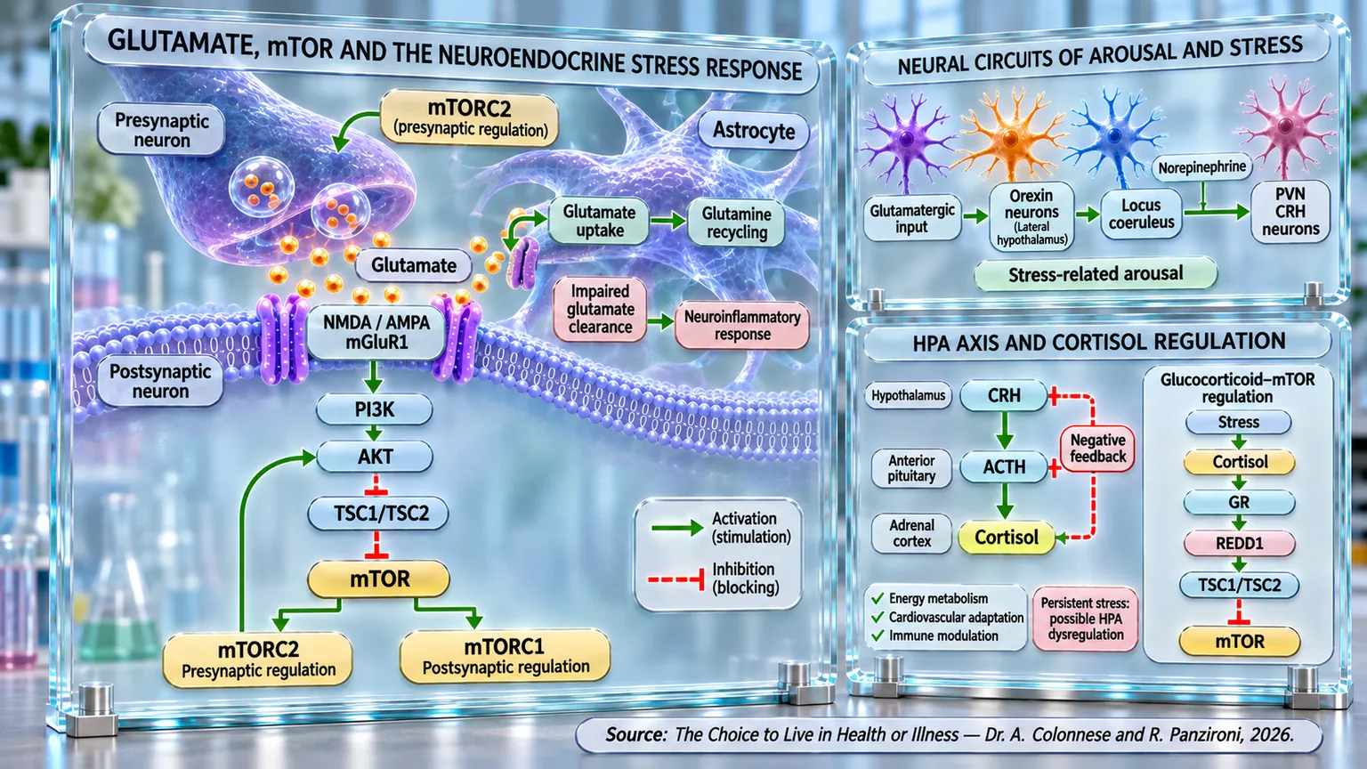
Panel diagram of glutamatergic synapses, orexin-norepinephrine circuits, and the HPA axis with green activating and red inhibitory arrows.
Infographic of the neuroendocrine response to stress: glutamate activates NMDA/AMPA-mGluR1 receptors, the PI3K-AKT-mTOR pathway, and astrocytic recycling; orexin and locus coeruleus circuits converge on CRH neurons of the PVN, while the hypothalamic-pituitary-adrenal axis coordinates CRH, ACTH, and cortisol with negative feedback and glucocorticoid-mTOR regulation.

ABSTRACT

The stress response arises from the integration of neuronal communication and endocrine regulation, which is necessary to adapt to real or perceived threats. In the book "La scelta di Vivere in Salute o in Malattia" (The Choice to Live in Health or Disease), Dr. A. Colonnese and R. Panzironi analyze the circuits that connect glutamatergic transmission, alertness, and cortisol release, highlighting how the duration of activation determines different physiological consequences, ranging from the immediate mobilization of energy resources to the changes that accompany conditions of persistent stress.

Glutamate, the main excitatory neurotransmitter in the brain, supports synaptic communication modulated by mTOR: mTORC1 contributes to postsynaptic responses, while mTORC2 is involved in presynaptic release. Astrocytes remove extracellular glutamate and convert it into glutamine, allowing for its recycling. According to the evidence examined, persistent hyperactivation of mTOR can compromise this function, promoting glutamatergic accumulation, excitability, and neuroinflammation. Glutamatergic signals also activate orexinergic neurons in the lateral hypothalamus, which are involved in maintaining wakefulness and amplifying alertness circuits through local glutamatergic connections.

Orexin reaches the locus coeruleus, which also receives glutamatergic afferents from brain regions. The resulting noradrenergic activity supports attention and behavioral preparation. Noradrenaline also contributes to the activation of neurons in the paraventricular nucleus that produce CRH. The latter stimulates the release of ACTH in the anterior pituitary, inducing the adrenal secretion of cortisol. This hormone regulates energy availability, cardiovascular function, and immune activity; the circadian rhythm and negative feedback help limit the response. During prolonged stress, noradrenergic, hypothalamic, and adrenal adaptations may occur, with changes in their sensitivity.

Overall, the glutamate–orexin–noradrenaline–CRH–ACTH–cortisol sequence describes a functional integration between synapses, brain networks, and the hypothalamic-pituitary-adrenal axis, without exhausting the complexity of the stress response. When activation is transient, it promotes adaptation; when it persists, it can be associated with alterations in hormonal rhythmicity, glucocorticoid sensitivity, and immunometabolic regulation. Understanding these connections allows us to distinguish protective mechanisms from the possible consequences of their chronic dysregulation.

mTOR and glutamate

In the previous chapter, we discovered how chronic stress, through cortisol, can alter insulin sensitivity and cellular metabolism. We followed this process down to the mechanisms involving REDD1, mTOR, AKT, and GLUT4, understanding how an initially protective response can compromise energy homeostasis. But what happens in the brain before cortisol is released? What signals transform a perceived threat into a hormonal response? To answer this, we must explore the neural circuits that regulate stress, starting from the role of AMPK and mTOR in glutamate transmission, up to the activation of orexin, noradrenaline, and the hypothalamic-pituitary-adrenal axis.

To understand this sequence, we must trace the origin of the stress response. Before cortisol reaches the blood and influences metabolism, the brain must recognize the stimulus and activate the involved neural circuits. This communication depends on a network of neurons that exchange chemical signals, regulating the intensity and duration of the response. It is precisely at this stage that a fundamental link between metabolism and brain activity emerges: the AMPK and mTOR pathways can influence synaptic function. To understand how this communication occurs, we must start with the brain's primary excitatory messenger: glutamate.

If noradrenaline increases the state of vigilance and directs attention toward potentially relevant stimuli, it is indeed glutamate that allows this activation to propagate within neural networks, transforming an alert signal into a coordinated response of the entire brain. At the base of this process lies mTOR once again, which directly regulates the functioning of glutamatergic synapses by modulating neurotransmitter release, the sensitivity of postsynaptic neurons, and the plasticity of neuronal connections. In this way, the metabolic state of the cell is translated into changes in the efficiency of synaptic communication, allowing the brain to adapt its activity to energy and environmental demands (McCabe et al., 2020) [1].

Recent research has shown that the two complexes through which mTOR acts, mTORC1 and mTORC2, perform different but complementary functions in the regulation of glutamatergic transmission. mTORC1 primarily exerts postsynaptic control (the part of the neuron that receives chemical signals transmitted by another neuron through the synapse, the point of communication between nerve cells), maintaining the number of excitatory synapses and modulating the expression of AMPA receptors responsible for the response to glutamate. When this complex is inactivated, the number of synapses and the amplitude of excitatory postsynaptic currents decrease, indicating a reduced capacity of the neuron to respond to the glutamatergic stimulus. Conversely, mTORC2 acts mainly at the presynaptic level (the part of the neuron that sends signals to other cells, releasing chemicals called neurotransmitters), regulating the probability of release of vesicles containing glutamate and the sensitivity of the entire process to intracellular calcium signals. The loss of mTORC2, in fact, reduces the evoked release of the neurotransmitter and compromises the efficiency of synaptic communication. The two complexes, therefore, operate like two sides of the same conversation: one ensures that the message is received correctly, the other ensures that it is transmitted with the right intensity and at the appropriate time (McCabe et al., 2020) [1].

In addition to neurons, astrocytes (cells of the nervous system that nourish, protect, and support neurons) also play a decisive role in controlling glutamatergic neurotransmission. For many years, these cells were considered simple structural support elements; today, however, it is known that they constitute an essential component of the so-called tripartite synapse, in which the presynaptic neuron, postsynaptic neuron, and astrocyte collaborate continuously to maintain the balance of nerve transmission. One of the most important functions of astrocytes consists of the rapid removal of glutamate from the synaptic cleft via specific transporters, which are responsible for almost all extracellular glutamate uptake. Once internalized, glutamate is converted into glutamine by glutamine synthetase (an enzyme that transforms glutamate and ammonia into glutamine) and returned to the neurons, which can then transform it back into glutamate and use it for a new synaptic transmission. This continuous recycling prevents the accumulation of the neurotransmitter in the extracellular space and protects neurons from the phenomenon of excitotoxicity, i.e., excessive stimulation of glutamatergic receptors that can culminate in cellular damage (Peterson and Binder, 2020) [2].

The function of astrocytes is in turn strictly regulated by mTOR. Under physiological conditions, this signaling pathway coordinates astrocytic energy metabolism, controls glucose utilization, the production of NADPH (a molecule that provides cells with the reducing power necessary to counteract oxidative stress), mitochondrial metabolism, the glutamate-glutamine cycle, and the maintenance of ion homeostasis (the ability of cells to keep ion concentrations in balance). However, when mTOR becomes chronically hyperactive, as occurs in various neurological pathologies, astrocytes progressively lose the ability to maintain a stable extracellular environment. The expression of specific glutamate transporters decreases, glutamine synthetase is reduced, glutamate tends to accumulate in the synaptic space, and susceptibility to neuronal hyperexcitability increases. In parallel, extracellular potassium buffering and mitochondrial function are also compromised, with alterations that further amplify the excitability of neural networks. In this way, mTOR does not only control the metabolism of the individual cell, but becomes a regulator of the entire functional balance of the brain network, determining whether glutamate will be rapidly eliminated or will continue to stimulate neurons persistently (Hochmuth and Hirrlinger, 2025) [3].

Continuing along this physiological cascade, it emerges that glutamate regulation depends not only on the activity of astrocytic transporters but also on the inflammatory state of the nervous tissue. In conditions of chronic stress, persistent pain, or neuronal damage, the hyperactivation of mTOR in astrocytes favors their transformation toward the A1 reactive phenotype, characterized by the production of pro-inflammatory mediators and the progressive loss of normal metabolic support functions. The consequence is marked neuroinflammation accompanied by a reduction in the expression of GLT-1 (a glutamate transporter protein) and glutamine synthetase, with a consequent decrease in the ability of astrocytes to remove extracellular glutamate.

The accumulation of the neurotransmitter thus causes persistent neuronal hyperexcitability, promotes central sensitization, and keeps the circuits responsible for the stress response active. The observation that pharmacological inhibition of mTOR using rapamycin restores the expression of the main glutamate clearance systems and simultaneously attenuates neuroinflammation and neuronal activity demonstrates how this pathway represents one of the main points of convergence between cellular metabolism, inflammation, and brain excitability (Dong et al., 2025) [4].

Another study suggests that glutamate does not simply represent an excitatory neurotransmitter, but also constitutes a metabolic signal through which the cell informs mTOR of nutrient availability and consequently adapts protein synthesis, energy production, and synaptic activity. In other words, the brain uses glutamate as a true common language between metabolism and neurotransmission: the amount of glutamate available simultaneously reflects the energy state of the cell and its capacity to sustain high neuronal activity (Tripathi et al., 2022) [5].

The evidence analyzed shows how mTOR represents a fundamental link between energy metabolism and neuronal communication. By regulating glutamate release, receptor sensitivity, and the ability of astrocytes to remove it, this pathway contributes to the balance of brain networks. When this regulation is altered, the accumulation of glutamate can promote neuroinflammation and hyperexcitability, modifying the response to stimuli. But how is this excitatory activity transformed into a state of vigilance and alertness? To understand this, we must follow glutamate toward the orexin neurons, which link metabolic signals to the activation of the locus coeruleus (a small area of the brainstem that produces most of the brain's noradrenaline) and the noradrenergic system during stress.

Glutamate, orexin, and locus coeruleus

To understand how glutamate transforms the activity of neuronal networks into a response of vigilance and alertness, we must follow its effect on orexin neurons. These cells, located in the lateral hypothalamus, represent an important link between energy metabolism, brain activity, and the stress response. In fact, they receive numerous glutamatergic signals that constantly regulate their excitability. Variations in glutamate transmission can therefore rapidly modify the activity of orexinergic neurons, adapting the state of vigilance to metabolic and environmental needs. In this process, glutamate transmits the excitatory signal, while orexin (a neuropeptide, a chemical messenger produced in the hypothalamus, a region of the brain) contributes to amplifying and distributing it to other brain regions, including the locus coeruleus. A fundamental step is thus outlined: metabolic signals that influence neuronal communication can contribute to the activation of circuits responsible for vigilance and the stress response.

Electrophysiological evidence has shown that orexin neurons possess spontaneous tonic activity but, above all, are capable of rapidly amplifying their activation through a sophisticated local positive feedback circuit. Contrary to what was initially hypothesized, orexin does not actually exert marked direct self-excitation on the neurons that produce it. Its main effect consists rather in activating local glutamatergic neurons, which increase the release of glutamate onto orexinergic cells, synchronizing their electrical discharge. This circuit determines a robust depolarization of orexin neurons, accompanied by an increase in the frequency of action potentials (brief electrical impulses that allow neurons to rapidly transmit information through the nervous system) that can exceed basal values by more than three times. Thanks to this mechanism, a relatively small population of neurons manages to rapidly recruit the entire orexinergic network, coordinating the simultaneous activation of the numerous brain regions responsible for wakefulness, attention, eating behavior, and the stress response. Glutamate therefore represents not only an initial excitatory stimulus, but becomes the main amplifier of orexinergic transmission, allowing the system to maintain a sufficiently intense and prolonged state of activation (Li et al., 2002) [6].

The organization of this circuit also highlights how the brain simultaneously uses amplification and control mechanisms. While glutamate synchronizes and strengthens the activity of orexinergic neurons, other neuromodulatory systems exert an opposite action to prevent their excessive activation. Among these, norepinephrine and serotonin are of particular importance, as they induce hyperpolarization of orexin neurons and significantly reduce their firing frequency. An extremely refined self-regulation system is thus realized: glutamate acts as an accelerator of orexinergic activity, while monoaminergic systems contribute to limiting its intensity, preventing the activation of wakefulness circuits from becoming excessive or persistent (Li et al., 2002) [6].

Further confirmation of the central role of glutamate comes from pharmacological studies on non-NMDA ionotropic receptors (proteins present on the surface of neurons that, when activated by glutamate, open ion channels). Experimental stimulation of these receptors using kainic acid indeed results in a marked increase in the activation of orexin neurons, documented by the increased expression of the c-Fos protein, one of the main markers of neuronal activity. Conversely, the preventive administration of the selective antagonist CNQX significantly reduces this response, demonstrating that a substantial part of orexinergic activity depends precisely on glutamatergic transmission mediated by non-NMDA receptors. The predominant role of glutamatergic transmission emerges with extreme clarity: without an adequate glutamate input, the activity of orexinergic neurons is significantly reduced, limiting the brain's ability to maintain a state of vigilance and behavioral readiness (Eyigor et al., 2010) [7].

The physiological importance of this circuit emerges with particular evidence in conditions where glutamatergic transmission is compromised. An example is represented by traumatic brain injury, in which many patients develop daytime sleepiness, reduced vigilance, and fragmentation of the sleep-wake cycle. Ultrastructural analyses have shown that these alterations do not depend on a generalized loss of orexin neurons, but rather on a marked reduction of glutamate contained in the presynaptic terminals that establish excitatory contacts with these neurons. In other words, the orexinergic system remains structurally present, but receives an insufficient number of excitatory signals to maintain the normal level of activity. It is as if the engine were perfectly functional, but receiving an insufficient amount of fuel to develop its full power. The consequence is a reduction in the activation of orexin neurons and, consequently, of the entire arousal system (state of activation of the brain and the organism that increases vigilance, attention, and the ability to react to stimuli) that depends on them (Elliott et al., 2018) [8].

Particularly interesting is the observation that this alteration can be at least partially reversible. Supplementation with branched-chain amino acids (BCAA), started after the trauma, has in fact been shown to be capable of selectively restoring the glutamate content in the nerve terminals that innervate the orexinergic neurons of the lateral hypothalamus. This result suggests that the main functional deficit resulting from trauma is not represented by an excess of inhibition, but by a loss of excitatory glutamatergic input. Since leucine, isoleucine, and valine constitute important metabolic precursors for the synthesis of glutamate, their availability allows for the progressive replenishment of the presynaptic stores of the neurotransmitter, restoring the normal excitation of orexin neurons. The effect also appears highly selective, as the restoration of glutamate was observed especially in synapses directed to orexinergic neurons, while in the cortical areas most damaged by the lesion, the neurotransmitter content remains substantially reduced. This highlights how the orexin system represents one of the most sensitive targets of metabolic changes affecting glutamate and confirms the central role of this neurotransmitter in maintaining the state of vigilance (Elliott et al., 2018) [8].

Taken together, these findings outline an extremely coherent circuit. Glutamate does not simply perform the function of an excitatory neurotransmitter, but constitutes the main regulator of the activity of orexinergic neurons. Through the activation of non-NMDA receptors (mainly AMPA receptors) and the recruitment of local glutamatergic circuits, it synchronizes and amplifies the discharge of the orexinergic population, allowing a relatively limited number of neurons to coordinate the activity of vast brain networks. When, instead, glutamatergic input is reduced, as happens after a traumatic brain injury, the entire orexin system loses efficiency, with a consequent decrease in the state of vigilance. Conversely, when the availability of glutamate increases, the activation of orexinergic neurons becomes more intense and propagates rapidly toward the main target structures of the arousal system.

The activation of orexinergic neurons represents only part of the brain's response to stress. The locus coeruleus, the main center for norepinephrine production in the brain, also receives direct glutamatergic signals from other nerve regions. This means that glutamate can increase the state of vigilance through two complementary pathways: by stimulating orexin neurons and by directly activating noradrenergic ones. But from which regions do these signals come? And how are they integrated to transform a threat into a coordinated alert response? To understand this, we must examine the glutamatergic connections that converge on the locus coeruleus.

Glutamate and locus coeruleus

The orexin system, however, is not the only source of activation for the locus coeruleus. For this nucleus to rapidly modulate the state of alertness and prepare the body to face a threat, it receives an extensive network of glutamatergic afferents from various regions of the central nervous system. Glutamate is, in fact, the primary excitatory neurotransmitter capable of causing the rapid depolarization of noradrenergic neurons in the locus coeruleus, increasing their firing rate and promoting a widespread release of noradrenaline in numerous brain areas. In this way, the activation of the locus coeruleus does not depend on a single circuit, but represents the result of the integration of multiple pieces of information regarding the body's energy, cardiovascular, metabolic, and emotional state. When these signals converge simultaneously, glutamate acts as a true switch capable of transforming local stimuli into a global alert response.

A 2013 study demonstrates that C1 neurons, located in the medulla oblongata, contribute to activating the noradrenergic system during stress. These cells respond to stimuli such as pain, hypoglycemia, hypoxia, and psychological threats. Through the release of glutamate, they directly stimulate neurons of the locus coeruleus and other brain noradrenergic nuclei, potentially increasing the production of noradrenaline. In parallel, they can activate the sympathetic nervous system, promoting the peripheral release of catecholamines (substances produced by the nervous system and adrenal glands, such as adrenaline, noradrenaline, and dopamine). Experiments conducted on mice therefore highlight a link between danger signals, glutamatergic transmission, and the coordinated activation of central and peripheral stress responses (Holloway et al., 2013) [9].

This organization highlights how glutamate represents the primary language through which different sensory and autonomic systems converge on the locus coeruleus. Noradrenergic neurons do not merely receive these signals passively; they possess a unique set of receptors that determine their sensitivity to excitatory stimuli. Laser microdissection analyses conducted on human noradrenergic neurons have shown high expression of glutamate receptors, confirming that this is the main physiological regulator of their activity. Even more interesting is the observation that, in subjects suffering from major depressive disorder, the expression of specific mGluR glutamate receptors is significantly increased precisely in the neurons of the locus coeruleus, while similar changes are not observed in the prefrontal cortex.

This data suggests that, in conditions of chronic stress and depression, it is not simply the amount of available glutamate that increases, but the very sensitivity of noradrenergic neurons to the excitatory signal that changes. In other words, the locus coeruleus becomes progressively more reactive to glutamatergic stimuli, favoring a more intense and prolonged alert response. This mechanism could contribute both to the hyperactivation of the noradrenergic system observed in depression and to the speed with which glutamatergic-acting drugs, such as ketamine, are able to profoundly modify depressive symptoms and suicidal ideation (Chandley et al., 2014) [10].

The regulation of glutamatergic transmission in the locus coeruleus also involves a sophisticated dialogue between neurons and astrocytes. Traditionally, glial glutamate transporters, particularly GLT-1 and GLAST, have been considered simple systems for removing the neurotransmitter from the synaptic cleft, with the function of limiting neuronal excitability and preventing excitotoxicity. However, in the locus coeruleus, this paradigm is surprisingly different. It has been demonstrated that the pharmacological activation of glutamate transporters using riluzole (a drug used primarily to slow the progression of ALS) causes an increase, rather than a reduction, in the activity of noradrenergic neurons. The mechanism involves astrocytes: the entry of glutamate through the transporters increases the intracellular sodium concentration, promotes the activation of the sodium-calcium exchanger, increases cytoplasmic calcium, and induces further release of glutamate by glial cells (cells of the nervous system that protect, nourish, and support neurons). This creates a positive amplification circuit that increases the activation of AMPA receptors present on the neurons of the locus coeruleus. In parallel, the propagation of the signal through gap junctions (small channels that directly connect neighboring cells) synchronizes the activity of the neuro-glial network, making the activation of the entire noradrenergic system more efficient. Under physiological conditions, this mechanism contributes to increasing alertness and activating the descending noradrenergic pathways involved in pain control; under conditions of persistent stress, however, it could contribute to maintaining a high tone in the locus coeruleus and prolonging the state of hyperarousal (Hayashida et al., 2010) [11].

Continuing along this network of afferents, it emerges that the locus coeruleus is not a simple nucleus that receives indistinct excitatory signals, but a true integration center capable of attributing a different functional meaning to each glutamatergic input. A study on mice demonstrated that glutamate activates the noradrenergic neurons of the locus coeruleus, which are involved in alertness and the stress response. The excitatory signals come mainly from three brain regions: the prefrontal cortex, the lateral hypothalamus, and the periaqueductal gray (a region of the brainstem that surrounds a small channel in the brain, helping to regulate pain, fear responses, and stress). The lateral hypothalamus presents the strongest connections, while the other regions show different characteristics. These results highlight how distinct glutamatergic circuits regulate noradrenergic activity, helping to modulate behavioral responses to environmental stimuli (Barcomb et al., 2022) [12].

Among all the glutamatergic pathways directed to the locus coeruleus, one of the most relevant in the stress response is represented by the projection coming from the ventrolateral periaqueductal gray. This region, already known for its role in the modulation of pain and defensive behaviors, sends a robust excitatory projection to the noradrenergic neurons of the locus coeruleus, whose activity increases rapidly during exposure to threatening stimuli. Through calcium recordings, optogenetic, and electrophysiological techniques, it has been demonstrated that the intensity of the activation of this pathway faithfully reflects the degree of threat perceived by the body. Glutamatergic stimulation, in fact, induces direct excitatory currents in noradrenergic neurons, leading to an increase in the release of noradrenaline and a progressive modulation of the state of arousal. Experimental inhibition of the same pathway, on the other hand, reduces pupil dilation, attenuates anxious behaviors, and decreases the overall level of arousal. These observations demonstrate how glutamate does not merely activate the locus coeruleus, but finely modulates its degree of activation, allowing for a gradual transition from a state of normal alertness to conditions of anxiety, up to the extreme levels of hyperarousal typical of panic responses (Wang et al., 2026) [13].

Taken together, these findings outline an extremely refined hierarchical organization. Glutamate collects information from metabolic, autonomic, cognitive, and defensive systems and channels it toward the locus coeruleus through specialized afferents, each endowed with its own synaptic properties and regulatory mechanisms. The locus coeruleus integrates these signals, transforming them into a coordinated activation of the central noradrenergic system, upon which alertness, attention, emotional memory, and behavioral preparation depend. When this activation exceeds a certain threshold, the noradrenaline released in the hypothalamic structures contributes to stimulating neurons secreting CRH (corticotropin-releasing hormone), creating the functional link between the neuronal circuits of arousal and the subsequent endocrine activation of the hypothalamic-pituitary-adrenal axis, which is responsible for the release of cortisol.

Norepinephrine and CRH

Intense activation of the locus coeruleus represents the moment when the stress response, which until this point has been predominantly neuronal, begins to transform into a neuroendocrine response. The noradrenaline released by noradrenergic neurons does not remain confined to the networks of vigilance and attention, but reaches the paraventricular nucleus of the hypothalamus (PVN), the main control center of the hypothalamic-pituitary-adrenal axis. It is in this small region of the hypothalamus that information from metabolic, autonomic, and emotional systems is integrated and converted into the release of corticotropin-releasing hormone (CRH), the signal that initiates the endocrine response to stress. In this sense, the PVN represents a true command center: up to this point, the brain has processed and amplified the danger signal through glutamate, orexin, and noradrenaline; from here on, this information will be transformed into a hormonal response capable of involving the entire organism.

That noradrenaline constitutes one of the main activators of the PVN was demonstrated as early as the first in vivo microdialysis studies. During acute immobilization stress, a rapid and marked increase in the extracellular concentration of noradrenaline is observed within the paraventricular nucleus, accompanied by an increase in the main catecholaminergic metabolites, an index of intense release, reuptake, and turnover of the neurotransmitter. It is interesting to observe that, after repeated exposures to the same stressor, the evoked release peak remains substantially preserved, while the baseline conditions change profoundly: the extracellular availability of noradrenaline decreases, intraneuronal metabolism increases, and the ratio between noradrenaline and dihydroxyphenylglycol (DHPG, a substance produced by the degradation of noradrenaline) is reduced. These results indicate that chronic stress does not shut down the noradrenergic response, but induces a functional remodeling of the catecholaminergic system aimed at continuously modifying its level of activity. In other words, the brain does not lose the ability to respond to stress, but rather redefines its own equilibrium point, preparing itself to react more rapidly to subsequent stimuli (Pacak et al., 1992) [14].

For noradrenaline to transform this signal into an endocrine response, however, it is necessary for the PVN neurons to be able to recognize and interpret it. Anatomical and autoradiographic analyses have shown that α1 and α2 adrenergic receptors are not uniformly distributed within the paraventricular nucleus, but are particularly concentrated in the medial parvocellular region, the site of most neurons that synthesize CRH. This organization suggests that catecholamines are structurally predisposed to exert preferential control precisely over the cells responsible for the activation of the hypothalamic-pituitary-adrenal axis, aimed at avoiding excessive catecholaminergic stimulation of CRH neurons during conditions of prolonged activation of the HPA axis, highlighting how the receptor system also actively participates in the processes of adaptation to chronic stress (Cummings and Seybold, 1988) [15].

The activation of the neuronal-glial circuit, however, represents only one of the mechanisms through which noradrenaline promotes the release of CRH. The study shows that noradrenaline can directly activate the neurons that produce CRH, which are involved in the hormonal response to stress. Through α1-adrenergic receptors, it promotes the opening of calcium channels, increasing neuronal excitability. This process occurs even without the intervention of glutamate and GABA circuits, indicating an independent mechanism of activation. In experimental models, prolonged exposure to corticosterone (a hormone produced by the adrenal glands) reduces this response, highlighting a control system that limits the excessive activation of CRH neurons. The results therefore suggest that noradrenaline directly regulates the activity of the hypothalamic circuits responsible for the endocrine response to stress (Gouws et al., 2022) [16].

Direct noradrenergic transmission to the paraventricular nucleus takes on even greater importance when analyzing the organization of the entire stress axis. The main catecholaminergic afferents that reach the PVN originate from the locus coeruleus and the nucleus of the solitary tract, forming an integrated network of noradrenergic neurons that conveys information regarding the metabolic, cardiovascular, immune, and behavioral state of the organism toward the hypothalamus. Experiments involving selective denervation of these projections have shown that their elimination significantly reduces the secretion of ACTH and corticosterone in response to acute stress, confirming that catecholaminergic afferents represent one of the main excitatory systems for CRH neurons. Even more interesting is what was observed during variable chronic stress. In this condition, the removal of noradrenergic afferents continues to attenuate the release of ACTH, but does not prevent the maintenance of corticosterone secretion, as the adrenal cortex develops greater sensitivity to the pituitary hormone. This demonstrates that the organism does not limit itself to modifying the activity of central nervous circuits, but also implements adaptations at the peripheral level, making the adrenal glands capable of producing adequate amounts of glucocorticoids even in the presence of reduced central stimulation. Chronic stress, therefore, does not depend exclusively on an overactivation of the brain, but on a profound reorganization of the entire hypothalamic-pituitary-adrenal axis, in which neuronal plasticity and endocrine adaptations cooperate to maintain the stress response (Flak et al., 2014) [17].

The evidence analyzed shows how noradrenaline represents a fundamental link between the activation of brain circuits and the endocrine response to stress. By reaching the paraventricular nucleus of the hypothalamus, this neurotransmitter stimulates the neurons that produce CRH, both directly and through local neuronal circuits. When stress persists, however, the organism progressively modifies its response. Noradrenergic activity, the sensitivity of hypothalamic neurons, and the ability of the adrenal glands to respond to stimuli all change. These adaptations demonstrate that chronic stress involves the entire neuroendocrine system, not just the brain. But what happens when hypothalamic neurons release CRH? Through what steps does this signal reach the adrenal glands and determine the production of cortisol? To understand this, we must follow the activation of the hypothalamic-pituitary-adrenal axis, in which CRH stimulates the release of ACTH (a hormone produced by the pituitary gland that stimulates the adrenal glands to release cortisol) and initiates a hormonal response capable of modifying metabolism, immune activity, and the balance of the entire organism during stress.

CRH and cortisol

The release of CRH by neurons in the paraventricular nucleus represents the transition point through which a response, until then confined to the central nervous system, transforms into an endocrine adaptation of the entire organism. Once released into the hypothalamic-hypophyseal portal circulation, CRH reaches the adenohypophysis (the anterior part of the pituitary gland, a small gland located at the base of the brain), where it stimulates corticotrope cells to synthesize and release adrenocorticotropic hormone (ACTH). The latter enters the systemic circulation and reaches the adrenal cortex, inducing the synthesis and secretion of cortisol, the primary human glucocorticoid. Through the blood, cortisol reaches virtually all organs and tissues, modulating energy metabolism, cardiovascular function, the immune system, and brain activity. The stress response, therefore, does not remain confined to the neuronal circuits of the hypothalamus and brainstem, but is transformed into an endocrine signal capable of coordinating the activity of the entire organism.

Contrary to what one might imagine, the secretion of ACTH does not depend exclusively on the amount of available CRH, but is organized according to a precise circadian rhythm generated by central mechanisms. Studies conducted through continuous infusion of human CRH for twenty-four hours have shown that, even in the presence of constant and maximal stimulation, ACTH secretion continues to follow its characteristic daily pattern, with minimum values during the night and a progressive increase in the early morning hours. When the negative feedback of cortisol is pharmacologically abolished using metyrapone (a drug that reduces the production of cortisol by the adrenal glands), the concentration of ACTH increases significantly throughout the day, but the circadian rhythmicity remains surprisingly preserved. This demonstrates that the rhythm of the hypothalamic-pituitary-adrenal axis is not generated by cortisol itself, but by central circuits, likely linked to a small area of the hypothalamus that acts as the main biological clock, while cortisol primarily performs the task of limiting its intensity through negative feedback. In other words, CRH represents the accelerator of the HPA axis, while cortisol acts as the braking system that prevents an excessive and prolonged response (Ur et al., 1995) [18].

The importance of glucocorticoid feedback emerges with even greater clarity in conditions of prolonged stress. During the initial phases of a critical illness, for example, the ACTH response to CRH administration remains substantially comparable to that observed in healthy subjects, indicating that the hypothalamus and pituitary gland retain a normal response capacity. As stress persists, however, the situation changes progressively. In the subacute and prolonged phases of critical illness, the ACTH response to CRH is reduced by more than half, despite the maintenance of high concentrations of circulating free cortisol. This temporal trend suggests that the problem does not lie in structural damage to hypothalamic or pituitary cells, but rather in a functional suppression exerted by the persistent negative feedback of cortisol. Chronic exposure to glucocorticoids, in fact, progressively reduces the sensitivity of the pituitary gland to the CRH stimulus and limits the secretion of ACTH, configuring a form of reversible central adaptation. This mechanism appears analogous to what is observed in patients treated for long periods with exogenous glucocorticoids or after the remission of Cushing's syndrome, conditions in which the hypothalamic-pituitary-adrenal axis requires time to recover its normal responsiveness to physiological stimuli (Peeters et al., 2018) [19].

Continuing along this cascade, however, a fundamental aspect emerges: cortisol is not simply the final product of the hypothalamic-pituitary-adrenal axis, but also the main regulator of its own activity. In physiological conditions, in fact, the increase in plasma cortisol concentrations activates an effective negative feedback mechanism at the level of the hypothalamus and the adenohypophysis, progressively reducing the secretion of CRH and ACTH and returning the organism toward homeostasis once the stressful stimulus has ended. This self-regulation system prevents the endocrine response from remaining active longer than necessary, limiting the exposure of tissues to glucocorticoids.

When stress becomes persistent, the continuous recruitment of the HPA axis progressively alters this balance. In the initial phases, a state of hypercortisolism generally prevails, supported by the chronic activation of the CRH-ACTH-cortisol circuit; over time, however, persistent exposure to glucocorticoids modifies the sensitivity of glucocorticoid receptors, alters the effectiveness of negative feedback, and determines a progressive loss of the physiological circadian rhythm of cortisol. In parallel, structural and functional changes occur in the hippocampus and prefrontal cortex, regions fundamental for the inhibitory control of the HPA axis. In some subjects, this persistent stimulation can evolve toward a paradoxical condition of reduced glucocorticoid responsiveness, in which cortisol, despite being present in high or normal quantities, progressively loses the ability to exert its regulatory effects on target tissues. Chronic stress thus transforms a system created to protect the organism into a mechanism capable of perpetuating neuroendocrine dysfunction and inflammation (Núñez et al., 2025) [20].

The consequences of this dysregulation extend far beyond the endocrine system. Cortisol is, in fact, one of the main modulators of the immune system and, in physiological conditions, limits the production of pro-inflammatory cytokines, reduces lymphocyte proliferation, promotes the maintenance of regulatory T cells, and contributes to the preservation of immunological tolerance. However, when sensitivity to glucocorticoids decreases, these functions are progressively compromised. This results in an increase in the production of inflammatory mediators such as IL-6, TNF-α, and IL-17, a reduction in anti-inflammatory cytokines such as IL-10, lower activity of Natural Killer cells, and a progressive loss of immune regulation. This state of chronic inflammation favors the onset or progression of numerous autoimmune diseases, including rheumatoid arthritis, systemic lupus erythematosus, multiple sclerosis, autoimmune thyroiditis, and inflammatory bowel diseases. In parallel, persistent exposure to glucocorticoids contributes to the development of metabolic, cardiovascular, gastrointestinal, musculoskeletal, and neuropsychiatric alterations, confirming how the stress response simultaneously involves virtually all the body's systems (Núñez et al., 2025) [20].

The knowledge gained in recent years has also found important applications in clinical practice. The CRH stimulation test is now one of the most widely used tools to assess the functional integrity of the hypothalamic-pituitary-adrenal axis in patients with suspected ACTH deficiency. Thanks to the use of modern immunoassays based on monoclonal antibodies, it has recently been demonstrated that a serum cortisol value of 12.6 μg/dL, measured 30 minutes after CRH administration, identifies with high accuracy patients who require replacement therapy with hydrocortisone, proving more precise than the traditional 18 μg/dL cutoff used in the past. This result highlights how an increasingly deep understanding of the physiology of the HPA axis not only produces theoretical advancements but also allows for the improvement of the diagnosis and clinical management of stress-related endocrine pathologies (Izuchi et al., 2026) [21].

The journey covered in this section has allowed us to understand how stress involves the entire organism. Everything originates in the brain, where nerve signals activate a response that progressively involves glutamate, orexin, norepinephrine, and the hypothalamic-pituitary-adrenal axis. Through the release of CRH and ACTH, this activity translates into the production of cortisol, preparing the organism to face dangerous situations. However, when stress persists, the same protective mechanisms can turn into factors of imbalance. Alterations in the circadian rhythm and negative feedback modify hormonal, metabolic, and immune regulation, favoring inflammatory processes and cellular dysfunction. Thus, a fundamental principle emerges: health depends not only on the ability to activate a stress response, but also on the ability to interrupt it when it is no longer necessary. It is this balance between activation and recovery that allows the organism to maintain its physiological stability over time.

Conclusions

The journey covered in this chapter has allowed us to understand how the brain transforms a threat into a hormonal response. Before cortisol reaches the bloodstream, numerous neural circuits must recognize, transmit, and coordinate danger signals. At the foundation of this communication is glutamate, the primary excitatory neurotransmitter of the central nervous system. Its activity is also regulated by mTOR, a metabolic pathway that influences synaptic efficiency. Specifically, mTORC1 contributes to the postsynaptic response, while mTORC2 participates in controlling the presynaptic release of glutamate.

This regulation also involves astrocytes, cells that are fundamental for maintaining a stable brain environment. Through specific transporters, they remove excess glutamate and convert it into glutamine, making its reuse possible. When mTOR becomes chronically overactive, this balance can deteriorate. Astrocytes reduce their ability to clear glutamate and may take on pro-inflammatory characteristics. The neurotransmitter thus tends to accumulate, increasing neuronal excitability and promoting neuroinflammation. It is therefore clear how a metabolic alteration can directly modify the functioning of neural networks.

But how is this increased excitability transformed into a state of vigilance? The next step involves orexin neurons, located in the lateral hypothalamus. These cells receive glutamatergic signals that increase their activity and promote the maintenance of wakefulness. Orexin can also stimulate local glutamatergic circuits, creating an amplification mechanism for the response. Research shows that a reduction in these signals, also observed after head trauma, compromises orexinergic activation. Glutamate therefore represents an essential link between energy availability, neuronal communication, and vigilance.

The activity of orexinergic neurons reaches numerous brain regions, including the locus coeruleus. This nucleus also receives direct glutamatergic signals from the prefrontal cortex, the lateral hypothalamus, and the periaqueductal gray matter. Their convergence increases the activity of noradrenergic neurons, promoting the release of norepinephrine. The body thus transitions from the simple perception of a stimulus to a coordinated state of attention, alertness, and behavioral preparation.

However, the stress response cannot stop at vigilance. Norepinephrine, in fact, contributes to activating the paraventricular nucleus of the hypothalamus, where CRH-producing neurons are located. Through specific adrenergic receptors, this neurotransmitter increases the excitability of the cells involved in the endocrine response. When stress persists, both the availability of norepinephrine and the sensitivity of hypothalamic circuits change. Research also shows adaptations in the adrenal glands, indicating that the chronic response involves both nervous and peripheral structures simultaneously.

The release of CRH marks the definitive transition from a neuronal response to a hormonal one. Upon reaching the anterior pituitary, it stimulates the secretion of ACTH, which induces the adrenal production of cortisol. The latter modifies energy metabolism, cardiovascular function, and immune system activity. Its secretion follows a circadian rhythm and is regulated by a negative feedback system. When the stress ends, this control helps to terminate the response and restore physiological homeostasis.

If, however, stress becomes persistent, hormonal rhythmicity, glucocorticoid sensitivity, and immune regulation can be altered. This leads to potential metabolic, inflammatory, and neuroendocrine consequences. Thus, the central principle of the chapter emerges: the stress response protects the body when it is proportionate and temporary, but it can compromise its balance when it becomes persistent. Health therefore depends on the ability to activate these circuits and, above all, to return them to normal.

References

[1] Genetic Inactivation of mTORC1 or mTORC2 in Neurons Reveals Distinct Functions in Glutamatergic Synaptic Transmission

McCabe et al., 2020

eLife. 2020;9:e51440.

[2] Astrocyte Glutamate Uptake and Signaling as Novel Targets for Antiepileptogenic Therapy

Peterson e Binder, 2020

Frontiers in Neurology. 2020;11:1006.

[3] Physiological and Pathological Role of mTOR Signaling in Astrocytes

Hochmuth e Hirrlinger, 2025

Neurochemical Research. 2025;50:53.

[4] mTOR Promotes Astrocyte Activation and Participates in Neuropathic Pain Through an Upregulation of RIP3

Dong et al., 2025

Neurochemical Research. 2025;50(2):93.

[5] SLC38A10 Regulate Glutamate Homeostasis and Modulate the AKT/TSC2/mTOR Pathway in Mouse Primary Cortex Cells

Tripathi et al., 2022

Frontiers in Cell and Developmental Biology. 2022;10:854397.

[6] Hypocretin/Orexin Excites Hypocretin Neurons via a Local Glutamate Neuron: A Potential Mechanism for Orchestrating the Hypothalamic Arousal System

Li et al., 2002

Neuron. 2002;36(6):1169-1181.

[7] Activation of orexin neurons through Non-NMDA Glutamate Receptors Evidenced by c-Fos Immunohistochemistry

Eyigor et al., 2010

Endocrine. 2010;37(1):167-172.

[8] Dietary Therapy Restores Glutamatergic Input to Orexin/Hypocretin Neurons After Traumatic Brain Injury in Mice

Elliott et al., 2018

Sleep. 2018;41(3):zsx212.

[9] Monosynaptic Glutamatergic Activation of Locus Coeruleus and Other Lower Brainstem Noradrenergic Neurons by the C1 Cells in Mice

Holloway et al., 2013

Journal of Neuroscience. 2013;33(48):18792-18805.

[10] Elevated Gene Expression of Glutamate Receptors in Noradrenergic Neurons of the Locus Coeruleus in Major Depression

Chandley et al., 2014

International Journal of Neuropsychopharmacology. 2014;17(10):1569-1578.

[11] Activation of Glutamate Transporters in the Locus Coeruleus Paradoxically Activates Descending Inhibition in Rats

Hayashida et al., 2010

Brain Research. 2010;1317:80-86.

[12] Properties and Modulation of Excitatory Inputs to the Locus Coeruleus

Barcomb et al., 2022

The Journal of Physiology. 2022;600(22):4897-4916.

[13] Glutamatergic Periaqueductal Gray Projections to the Locus Coeruleus Orchestrate Adaptive Arousal States in Threatening Contexts

Wang et al., 2026

Neuroscience Bulletin. 2026;42:255-269.

[14] Noradrenergic activation in the paraventricular nucleus during acute and chronic immobilization stress in rats: an in vivo microdialysis study

Pacak et al., 1992

Brain Research. 1992;589(1):91-96.

[15] Relationship of Alpha-1 and Alpha-2 Adrenergic Binding Sites to Regions of the Paraventricular Nucleus of the Hypothalamus Containing Corticotropin-Releasing Factor and Vasopressin Neurons

Cummings e Seybold, 1988

Neuroendocrinology. 1988;47(6):523-532.

[16] Regulation of corticotropin-releasing hormone neuronal network activity by noradrenergic stress signals

Gouws et al., 2022

The Journal of Physiology. 2022;600(19):4347-4359.

[17] Role of paraventricular nucleus-projecting norepinephrine/epinephrine neurons in acute and chronic stress

Flak et al., 2014

European Journal of Neuroscience. 2014;39(11):1903-1911.

[18] Continuous Administration of Human Corticotropin-Releasing Hormone in the Absence of Glucocorticoid Feedback in Man

Ur et al., 1995

Neuroendocrinology. 1995;61(2):191-197.

[19] ACTH and Cortisol Responses to CRH in Acute, Subacute and Prolonged Critical Illness: A Randomized Double-Blind Placebo-Controlled Crossover Cohort Study

Peeters et al., 2018

Intensive Care Medicine. 2018;44(12):2048-2058.

[20] Chronic Stress and Autoimmunity: The Role of HPA Axis and Cortisol Dysregulation

Núñez et al., 2025

International Journal of Molecular Sciences. 2025;26(20):9994.

[21] Defining a serum cortisol cutoff level post-CRH stimulation for diagnosing ACTH deficiency: A retrospective study validated by a nationwide registry

Izuchi et al., 2026

Frontiers in Endocrinology. 2026;17:1741709.