
Scientific Book

Hashimoto and the interplay between immunity and thyroid function

Hashimoto's thyroiditis is an autoimmune thyroid disease characterized by persistent inflammation that can progressively impair the production of thyroid hormones. Autoantibodies, lymphocytes, and alterations in immune tolerance participate in the process, while the study of immunometabolism helps clarify certain cellular mechanisms. In the book "The Choice to Live in Health or Disease," Dr. A. Colonnese and R. Panzironi explore the relationship between thyroid function, metabolism, and immune system regulation.

Cite this article
Colonnese, A. & Panzironi, R. (2026). Hashimoto and the interplay between immunity and thyroid function. The Choice to Live in Health or in Disease. https://lifesciencehub.info/en/la-scelta-di-vivere-in-salute-o-in-malattia/hashimoto-and-the-interplay-between-immunity-and-thyroid-function
The Choice to Live in Health or in Disease · ISBN 979-12-82692-03-8

DOI lsh.en.libro.12

  • Published 05/11/2026
  • Updated 05/11/2026
  • HASHIMOTO'S THYROIDITIS
  • IMMUNOMETABOLISM
  • PI3K/AKT/MTOR
  • AUTOPHAGY
  • TH17/TREG LYMPHOCYTES
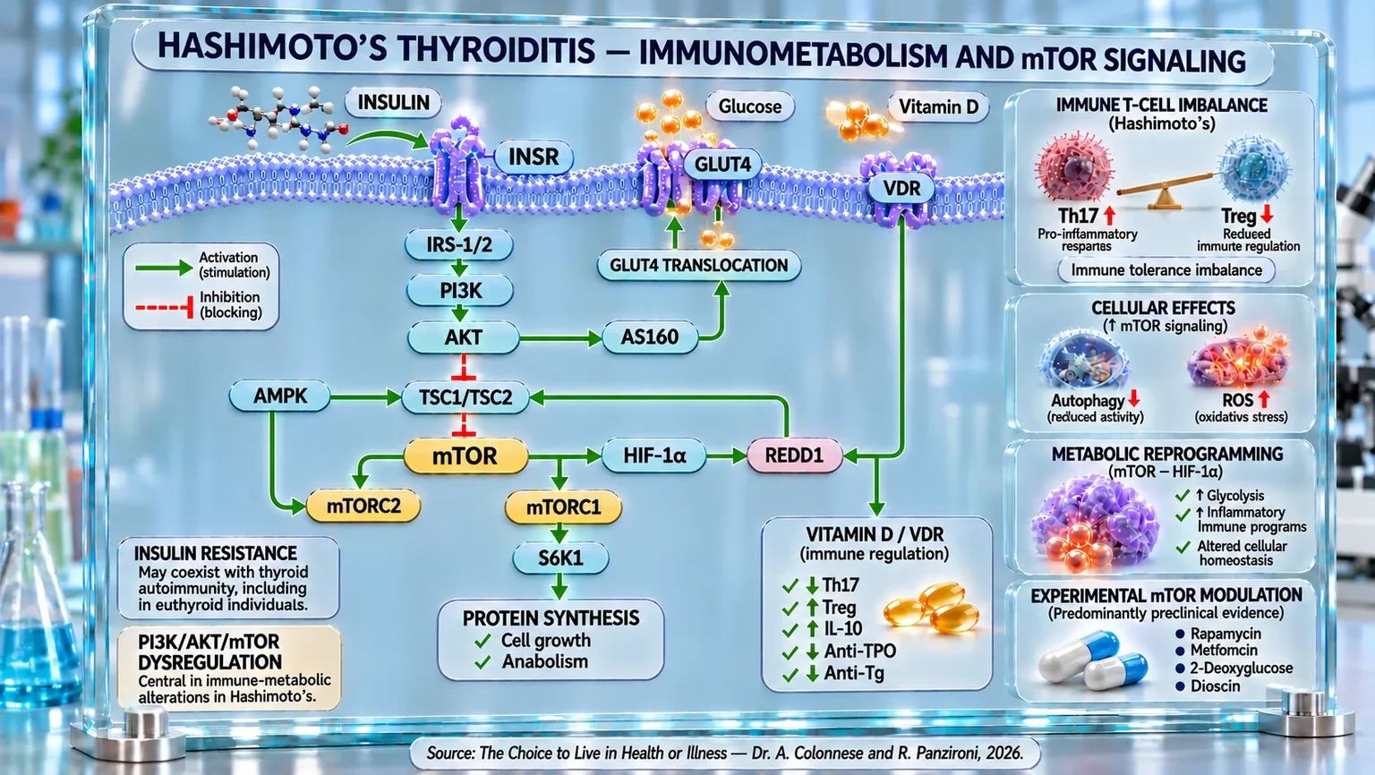
Cellular diagram showing the membrane, INSR and VDR receptors, PI3K/AKT/mTOR cascade, GLUT4, Th17/Treg balance, and indicators of autophagy and ROS.
Infographic of Hashimoto's thyroiditis: insulin/INSR and IRS-1/2–PI3K–AKT–mTOR link GLUT4 translocation, glucose uptake, and protein synthesis; vitamin D/VDR modulates immune regulation. On the right, Th17/Treg imbalance, reduced autophagy, increased ROS, and metabolic reprogramming summarize the interplay between immunity and thyroid metabolism.

ABSTRACT

Hashimoto's thyroiditis is an autoimmune condition in which the loss of tolerance to thyroid antigens is associated with immune, metabolic, and endocrine alterations. In the book "La scelta di Vivere in Salute o in Malattia" (The Choice to Live in Health or Disease), Dr. A. Colonnese and R. Panzironi analyze these interactions, delving into the mechanisms that may contribute to the maintenance of thyroid inflammation.

A central element concerns the imbalance between Th17 lymphocytes, involved in the pro-inflammatory response, and Treg cells, responsible for immunological tolerance. In Hashimoto's, Th17 cells are increased, while Treg cells may be reduced or functionally impaired. In parallel, insulin resistance has also been observed in euthyroid subjects with thyroid autoimmunity and is associated with autoantibodies. Visceral obesity and polycystic ovary syndrome can exacerbate metabolic alterations, while insulin sensitivity can influence the response to levothyroxine.

The PI3K/AKT/mTOR signaling pathway constitutes a possible link between metabolism and immunity. Excessive activation of mTOR can hinder autophagy, promote oxidative stress, and support Th17 differentiation over Treg cells. Through HIF-1α, this pathway also participates in the glycolytic reprogramming of immune cells. Predominantly preclinical studies indicate that rapamycin, metformin, 2-deoxyglucose, dioscin, and PI3K inhibitors can modulate these processes, reducing inflammatory mediators, autoantibodies, or lymphocytic infiltration in different models. However, these results do not demonstrate the clinical efficacy of mTOR inhibitors as a standard treatment.

A further element concerns vitamin D: insufficient concentrations are frequently associated with the disease, while intervention studies and meta-analysis report possible reductions in anti-TPO and anti-Tg antibodies after supplementation, especially in deficient subjects.

Overall, Hashimoto's emerges as a multifactorial condition in which insulin dysfunction, autophagy, and lymphocyte regulation may interact. Understanding these relationships helps to identify therapeutic questions, distinguishing between clinical associations, experimental results, and benefits yet to be confirmed. It remains essential to define the relative contribution of each mechanism in the different stages of the disease and in patients.

Epidemiology of Hashimoto's thyroiditis

Hashimoto's thyroiditis represents the most frequent manifestation of autoimmune thyroid diseases in settings with sufficient iodine intake. Recent studies estimate that the global prevalence is around 7.5% of the adult population, with wide geographical variations (for example, up to approximately 11% in low-to-middle-income areas).

Regarding gender, the disease shows a clear female predilection: women are affected approximately four times more frequently than men. Some sources indicate even higher ratios (4–10:1) in specific studies.

As for the age of onset, although it is possible in all age groups (including childhood), it is more common in adulthood, with a peak between 30 and 50 years of age and an increasing prevalence in middle-aged to older groups (45-65 years) according to various case series.

Overall, these data outline Hashimoto's thyroiditis as a widely prevalent autoimmune condition, characterized by a marked female predominance and a higher frequency in adulthood. However, the epidemiological distribution represents only the external manifestation of a much more complex biological process. At the root of the progressive loss of tolerance to thyroid antigens lies a profound alteration of the adaptive immune response (a specific defense that recognizes an agent and can remember it over time).

T-cell imbalance

Hashimoto's thyroiditis is one of the most emblematic autoimmune diseases, where the immune system, for reasons not yet fully understood, stops recognizing thyroid cells as its own and begins to progressively destroy them. Dominating this complex scenario are T lymphocytes, immune system cells that, under normal conditions, regulate and coordinate the body's defenses. When this balance is altered, the very cells meant for protection become the cause of damage.

In Hashimoto's thyroiditis, the protagonists of this imbalance are primarily two subtypes of T lymphocytes: Th17 and Treg cells. Th17 cells can be imagined as “impulsive soldiers,” specialized in promoting inflammation through the secretion of interleukin-17 (IL-17). Treg cells, on the other hand, represent the “diplomatic corps” of the immune system: they maintain tolerance toward the body's own tissues and limit excessive responses. When Th17 cells take over and Treg cells lose their ability to control them, a fertile ground is created for thyroid autoimmunity, a sort of cellular civil war that leads to the destruction of thyrocytes (thyroid cells that produce thyroid hormones).

Several studies have consistently described this dynamic: some researchers have shown that in patients with Hashimoto's thyroiditis, Treg cells are significantly reduced, while Th17 cells increase in proportion to the severity of the disease (Xue et al., 2015) [1]. This imbalance is closely correlated with the levels of anti-TPO and anti-Tg autoantibodies, which are markers of autoimmune activity. In other words, the more the immune “peacekeepers” decrease, the more inflammation and damage increase.

The molecular mechanism of this imbalance is complex. Some studies have highlighted how, in patients with Hashimoto's, the expression of transcription factors that control T lymphocyte differentiation is altered. FOXP3, a key regulator of Treg cells, is reduced, while genes associated with effector cells are overexpressed. This shift in the genetic center of gravity confirms the loss of tolerance and the expansion of inflammatory responses (Safdari et al., 2017) [2].

The Th17/Treg ratio, therefore, can be considered an indicator of the immunological state of the thyroid. According to some studies, it has been observed that in subjects with Hashimoto's, this ratio is strongly altered even in peripheral blood, accompanied by an alteration of the immune checkpoint system (a natural “brake” that controls the activation of the immune system), which is normally tasked with curbing the activity of T lymphocytes (Fang et al., 2022) [3].

The most interesting aspect that has emerged in recent years is the plasticity of these cells: Th17 cells, in the presence of appropriate environmental signals (cytokines such as TGF-β or IL-10), can “reprogram” themselves and acquire regulatory traits, demonstrating how the line between inflammation and tolerance is thin and dynamic (González-Amaro and Marazuela, 2016) [4].

Alongside these biological mechanisms, some experimental studies have highlighted the possibility of modulating the Th17/Treg imbalance through external interventions. In animal models, selenium supplementation has shown the ability to reduce the production of inflammatory cytokines and promote the differentiation of Treg cells, improving thyroid structure and function (Wang et al., 2023) [5].

Similarly, in an animal model of autoimmune thyroiditis, treatment with Jiayan Kangtai granules (JYKT, a traditional Chinese medicine formulation composed of a combination of nine medicinal herbs) reduced thyroid damage and lymphocyte infiltration. The treatment also resulted in a decrease in Th17 cells and an increase in regulatory T (Treg) cells, leading to a reduction in the Th17/Treg ratio. At higher doses, thyroid autoantibodies also decreased significantly. The results indicate that the protective effect of JYKT is associated with the restoration of the immunological balance between pro-inflammatory Th17 cells and regulatory Treg cells (Hou et al., 2018) [6].

Even on a genetic basis, Treg cells are confirmed as protective elements. A recent Mendelian randomization analysis (a genetic method used to obtain indications on the possible cause-effect relationship between a factor and a disease) has shown that specific genetic signatures associated with Treg cells significantly reduce the risk of developing Hashimoto's thyroiditis, confirming their causal role in the prevention of autoimmunity (Guo et al., 2024) [7].

In summary, Hashimoto's thyroiditis can be interpreted as a breakdown of the immunological harmony between pro-inflammatory and regulatory forces: Th17 cells light the fuse of inflammation, Treg cells attempt to extinguish it, but when the latter fail, due to environmental or metabolic reasons, the immune system ends up turning its weapons against itself. Restoring this balance, through genetic, nutritional, or pharmacological modulation, represents one of the most promising frontiers today for understanding and treating Hashimoto's thyroiditis.

Metabolic alterations: insulin pathway and insulin resistance

The relationship between Hashimoto's thyroiditis and insulin resistance represents a complex interplay where metabolism and immunity influence each other, resulting in a state of chronic systemic inflammation that fuels both thyroid and metabolic dysfunction.

Several studies have highlighted that insulin resistance (IR) can be present even in euthyroid subjects—those with normal thyroid function—who carry thyroid autoimmunity. In such cases, reduced insulin sensitivity appears to be associated with higher levels of anti-TPO and anti-Tg antibodies, suggesting that IR may contribute to the very genesis of the autoimmune response. Specifically, for every increase in the HOMA-IR index (an index used to estimate insulin resistance and pancreatic β-cell function), an average 18% increase in antibodies directed against a thyroid enzyme is observed, confirming a direct correlation between metabolic dysfunction and autoantibody activation (Blaslov et al., 2020) [8].

When the disease is already manifest, the presence of insulin resistance worsens the clinical course and reduces the response to conventional treatments. Some researchers have shown that in women with autoimmune subclinical hypothyroidism, levothyroxine (a medication used to compensate for or replace thyroid hormone deficiency) is less effective at reducing antibody titers if IR is present, whereas the combination with metformin, by improving insulin sensitivity, enhances the immunomodulatory effect of the thyroid hormone (Krysiak et al., 2021) [9]. In other words, glucose metabolism acts as an “amplifier or attenuator” of therapeutic efficacy, suggesting that managing IR is an integral part of treating thyroid autoimmunity.

On a pathophysiological level, endoplasmic reticulum stress (an internal network of cellular membranes) emerges as a key mechanism linking inflammation and insulin resistance: in animal models of Hashimoto's thyroiditis, chronic activation of the pathways responsible for the cellular stress response leads to impaired insulin signaling and an increase in pro-inflammatory cytokines such as TNF-α and IL-6. Pharmacological inhibition of reticulum stress with 4-phenylbutyrate, a molecule that helps cells manage internal stress, has been shown to restore insulin sensitivity and reduce thyroid damage, indicating a possible therapeutic pathway to modulate the metabolic-inflammatory component of the disease (Zhou et al., 2025) [10].

Even in hypothyroid patients treated with levothyroxine (a synthetic version of the T4 thyroid hormone), the presence of thyroid autoimmunity can be associated with an altered insulin response. Previous studies had already observed that subjects with autoimmune thyroiditis exhibited higher basal insulin levels compared to healthy controls, suggesting a subclinical defect in insulin sensitivity, even under conditions of thyroid hormonal balance (Lethagen et al., 2002) [11]. Furthermore, the presence of autoantibodies typical of autoimmune diabetes appears to reduce the secretory capacity of pancreatic β-cells, highlighting an immunological bridge between the thyroid and the pancreas.

The role of central obesity as an aggravating factor in this relationship was well illustrated in a recent study: in patients with type 2 diabetes, the triglyceride-glucose (TyG) index and its derivatives, which indirectly estimate insulin resistance, are strongly correlated with the presence of Hashimoto's thyroiditis. In particular, the TyG-BMI parameter (an index that combines glycemia, triglycerides, and BMI to indirectly estimate insulin resistance) mediates up to 74% of the effect between visceral obesity and thyroid autoimmunity, demonstrating that insulin resistance is not just a consequence, but a true mediator of autoimmune risk (Zhai et al., 2024) [12].

Another significant clinical scenario is polycystic ovary syndrome (PCOS), in which Hashimoto's thyroiditis occurs more frequently than in the general population. In these patients, the association between the two conditions amplifies insulin resistance and worsens metabolic alterations, creating a vicious cycle of inflammation, endocrine dysfunction, and hyperinsulinemia. Some studies have highlighted that women with PCOS and Hashimoto's thyroiditis show higher HOMA-IR values and reduced thyroid sensitivity, suggesting a bidirectional interaction between the thyroid system and glucose metabolism (Zhao et al., 2021) [13].

Taken together, these findings paint a unified picture: Hashimoto's thyroiditis and insulin resistance share common molecular pathways, where chronic inflammation, oxidative stress, and mitochondrial dysfunction act as connective factors. Insulin, beyond being a simple metabolic regulator, takes on the role of an “immune modulator,” influencing cytokine production and lymphocyte tolerance. Thus, Hashimoto's thyroiditis is not just an autoimmune disease, but a systemic manifestation of a broader immuno-metabolic disorder, in which the management of IR and lifestyle represents a component as essential as hormone replacement therapy.

mTOR as a central node

In the story of Hashimoto's, mTOR is the metabolic-immune "switchboard" that, when stuck on the attack signal, turns defense into damage. Certain environmental factors can turn up the volume on this switchboard: in an animal model, exposure to the plasticizer DINP (a chemical added to some plastics to increase their flexibility) aggravated autoimmune thyroiditis by activating the AKT/mTOR axis, suppressing cellular "scavenger" autophagy, and amplifying oxidative stress; restarting autophagy with rapamycin (a drug that primarily reduces mTOR activity) or buffering radicals with vitamin E attenuated thyroid lesions, proving the key role of this circuit (Duan et al., 2019) [14].

In the same vein, in thyroid follicular cells, excess IL-23 acts like a foot stuck on the accelerator: it activates the AKT/mTOR pathway, brakes autophagy, and promotes the accumulation of reactive oxygen species, fueling an inflammatory vicious cycle; blocking IL-23 or mTOR reopens the intracellular "cleaning" pathway and reduces oxidative stress (Zheng et al., 2018) [15].

In some experimental models, not only glucose but also oxidized fatty acids activated mTOR, pushing toward Th17 polarization at the expense of Tregs; inhibiting fatty acid oxidation with etomoxir (an experimental inhibitor) in human and murine models reduces pathway activation, improves thyroid histology, and restores regulatory balance, suggesting a concrete metabolic therapeutic target (Zhang et al., 2025) [16].

When endogenous "safety brakes" are missing, mTOR runs even faster: reduced serum levels of the protein sestrin-2, a modulator that normally keeps mTOR in check and supports autophagy, have been observed in patients with Hashimoto's, suggesting a lower intrinsic capacity to clear oxidative stress and contain inflammation (Ates et al., 2025) [17].

Finally, the metabolism of tryptophan (an essential amino acid) adds an additional layer of control: its systemic deficiency and the modulation of the PI3K-AKT pathway influence the balance between effector and regulatory T cells; in animal models, supplementing tryptophan reduces lesions and inflammatory mediators, while inhibiting its metabolism worsens the disease, signaling a dialogue between different metabolic pathways and organs with the upstream nodes of mTOR (Zhang et al., 2025) [18].

Overall, these findings converge on a central point: in Hashimoto's thyroiditis, mTOR can represent a connecting node between metabolic alterations, oxidative stress, autophagy, and immune dysregulation. Its excessive activation can, in fact, reduce cellular "cleaning" mechanisms, promote inflammation, and contribute to the imbalance between Th17 and Treg cells, creating conditions favorable to the persistence of thyroid damage. Conversely, containing its activity emerges, in experimental models, as a possible strategy to interrupt this circuit. These very observations raise a fundamental question: what happens when mTOR is inhibited directly? Studies conducted with specific mTOR inhibitors allow us to understand more clearly what effects the pharmacological control of this node can produce in autoimmune thyroiditis.

mTOR inhibitors

In Hashimoto's thyroiditis, the overactivation of mTOR may contribute to maintaining autoimmunity by altering the behavior of both immune cells and thyroid follicular cells. mTOR activity promotes anabolic and glycolytic programs compatible with the expansion of Th17 and Th1 cells, while simultaneously limiting autophagy, an important mechanism for cell protection and renewal. Consequently, the direct or indirect inhibition of this pathway can address various aspects of the disease: lymphocyte metabolism, IL-17 production, oxidative stress, autophagy, and inflammatory infiltration of the thyroid.

Particularly direct evidence was provided in 2018: in thyroid follicular cells from patients with Hashimoto's, the study authors observed an increase in IL-23 associated with the activation of the AKT–mTOR pathway, the suppression of autophagy, and the accumulation of reactive oxygen species. The use of rapamycin, a direct mTOR inhibitor, significantly restored autophagy and reduced ROS accumulation. This result demonstrates that mTOR inhibition can act directly on thyroid cells, interrupting a circuit that links inflammation, impaired cellular degradation, and oxidative stress in Hashimoto's (Zheng et al., 2018) [15].

In a 2021 study on CD4+ lymphocytes from patients with Hashimoto's, glycolysis, GLUT1 (a transporter that allows glucose to enter cells), HK2, PKM2, and LDHA (enzymes involved in glucose metabolism) were found to be increased, while the mTOR–HIF-1α–glycolysis axis was activated in the mouse model used in the same study. Treatment with metformin and 2-deoxyglucose (a glucose-like molecule that enters cells but hinders their normal energy metabolism) reduced the activity of this metabolic pathway and decreased Th1 and Th17 populations. Metformin is not a direct mTOR inhibitor, but it can limit its activity through cellular energy regulation, showing how the functional reduction of the mTOR–HIF-1α axis can rebalance the immune response in autoimmune thyroiditis (Zhao et al., 2021) [19].

Further confirmation arrived in 2023, when researchers studied dioscin (a natural compound belonging to steroidal saponins) in an autoimmune thyroiditis model. In diseased rats, mTOR, TLR4, and NF-κB (two important components of the immune and inflammatory response) were increased in thyroid tissue. Treatment with dioscin significantly reduced mTOR expression at both the mRNA and protein levels, decreased TgAb, TPOAb, and TRAb (antibodies directed against specific thyroid structures), and reduced lymphocyte infiltration and follicular destruction. The authors therefore proposed that part of the protective effect of dioscin depends on the suppression of the mTOR pathway, with a consequent attenuation of the thyroid inflammatory response (Zhang et al., 2023) [20].

Finally, a recent 2024 study analyzed blocking the cascade located immediately upstream of mTOR using LY294002, a PI3K inhibitor. In mice with experimental autoimmune thyroiditis, p-AKT, p-mTOR, S6K1, S6K2, Th17 cells, and IL-17A were increased. Treatment with LY294002 dose-dependently reduced AKT/mTORC1 activation, the number of Th17 cells, IL-17A production, and the histological severity of the thyroiditis. The study therefore shows that interrupting the PI3K–AKT–mTORC1 signal can directly reduce the Th17 program responsible for part of the autoimmune thyroid damage (He et al., 2024) [21].

Overall, this evidence indicates that mTOR inhibition can intervene in Hashimoto's thyroiditis through at least two major complementary mechanisms. In thyroid follicular cells, blocking mTOR can promote autophagy; in immune cells, it can limit the metabolic reprogramming that supports Th1 and especially Th17, reducing IL-17 and the immune system's ability to maintain the attack against thyroid tissue. The sequence can therefore be outlined as: PI3K/AKT/mTOR inhibition → glycolysis → reduction of Th17/IL-17 plus recovery of autophagy → decrease in oxidative stress and thyroid inflammation. These results are promising, but they derive primarily from cellular and animal models and do not yet constitute a demonstration of the clinical efficacy of mTOR inhibitors as a standard therapy for Hashimoto's thyroiditis.

Vitamin D as an immuno-metabolic modulator

In recent years, vitamin D has progressively taken on a central role in the understanding of Hashimoto's thyroiditis, shifting from a simple regulator of bone metabolism to a true immune modulator. Numerous clinical and experimental findings have clarified its impact on the balance between inflammation and tolerance, demonstrating that its deficiency is an active factor in promoting thyroid autoimmunity.

Several clinical studies have documented that vitamin D deficiency is extremely common in patients with Hashimoto's, often exceeding 80% of cases, and is accompanied by elevated levels of anti-TPO antibodies, a direct sign of autoimmune activity. In a study conducted on the island of Crete, researchers observed that after just four months of supplementation with cholecalciferol (1,200–4,000 IU/day), there was an average 20% reduction in anti-TPO titers, with an overall improvement in immune status (Mazokopakis et al., 2015) [22].

These results were subsequently confirmed and expanded by a large intervention program involving over 11,000 subjects, which showed that achieving serum 25(OH)D levels ≥ 50 ng/mL was associated with a 30% decrease in the risk of hypothyroidism and a significant reduction in thyroid autoantibodies. Adequate supplementation, therefore, not only supports endocrine function but also exerts a protective effect on the progression of the disease (Mirhosseini et al., 2017) [23].

The mechanism through which vitamin D exerts this effect appears to be twofold: on one hand, it directly modulates lymphocyte activity, promoting the differentiation of regulatory T cells (Treg) and reducing the pro-inflammatory Th17 fraction; on the other, it acts on dendritic cells (cells that recognize antigens and present them to T lymphocytes), reducing the expression of a specific class of molecules and attenuating antigen presentation. In this way, vitamin D helps to restore the immune system to a state of tolerance (Lebiedziński and Lisowska, 2023) [24].

Clinically, controlled studies on one hundred patients with autoimmune thyroid diseases have shown that vitamin D supplementation can significantly reduce thyroid autoimmunity. After treatment, patients receiving supplementation showed a 46.73% reduction in anti-thyroperoxidase (TPO-Ab) antibodies, compared to 16.6% in the control group. Furthermore, 68% of treated subjects responded to therapy compared to 44% of controls. The results therefore indicate a potential immunomodulatory effect of vitamin D in autoimmune thyroid diseases (Chaudhary et al., 2016) [25].

In 2019, other researchers highlighted that vitamin D supplementation can rebalance CD4+ T-cell subpopulations, reducing the Th17/Tr1 ratio and increasing the expression of the anti-inflammatory cytokine IL-10, elements consistent with an immunoregulation effect sustained over time (Nodehi et al., 2019) [26].

Meta-analyses published in subsequent years have consolidated these data: some researchers analyzed a total of more than 1,500 patients, highlighting a significant reduction in anti-TPO and anti-Tg titers after vitamin D supplementation, accompanied by a slight improvement in thyroid function (increase in FT3 and FT4 hormones, reduction in TSH). The effects are more marked in protocols lasting longer than 12 weeks and with adequate dosages of vitamin D3 or calcitriol (Tang et al., 2023) [27]; (Zhang et al., 2021) [28].

Taken together, these findings paint vitamin D as a silent but powerful modulator of thyroid immunity; in fact, as we saw in Chapter 6 dedicated to the immune system, through the modulation of mTOR, it corrects the imbalance between effector and regulatory cells, dampens systemic inflammation, and improves the sensitivity of thyroid cells to endogenous hormones, which are central mechanisms in the genesis of the disease. Metaphorically, it is as if vitamin D acts as a "conductor" that restores rhythm to a dissonant immune system.

In summary, research converges on a clear concept: vitamin D is not a simple adjuvant, but a key element in the prevention and control of Hashimoto's thyroiditis, especially in deficient individuals. Adequate supplementation, calibrated on an individual basis and continued over time, can help reduce autoimmune activity, stabilize thyroid function, and improve the response to conventional therapies.

Conclusions

Taken together, the scientific evidence analyzed outlines Hashimoto's thyroiditis as a condition in which autoimmunity, cellular metabolism, and endocrine regulation are deeply interconnected. The disease, which is particularly frequent in females and in adulthood, stems from the progressive loss of tolerance to thyroid antigens, but the path developed in this chapter shows how this process involves a much broader biological network.

The first step concerns the imbalance of T lymphocytes. In patients with Hashimoto's, Th17 cells are increased, while Tregs, which are responsible for maintaining tolerance, are reduced or functionally compromised. Experimental and genetic studies reinforce the protective role of Tregs, showing how interventions capable of rebalancing the Th17/Treg ratio can be accompanied by a reduction in autoimmune thyroid damage.

This immunological alteration is accompanied by significant insulin metabolism dysfunction. Insulin resistance has been documented even in euthyroid subjects with thyroid autoimmunity and is associated with autoantibody levels. The presence of IR can also influence the response to levothyroxine, while visceral obesity and conditions such as PCOS amplify the interaction between hyperinsulinemia, metabolic alterations, and autoimmunity.

In this interplay, mTOR emerges as a central node. The activation of AKT/mTOR can reduce autophagy, increase oxidative stress, and promote pro-inflammatory immune programs; furthermore, excessive mTOR activity favors Th17 polarization at the expense of Tregs. mTOR therefore represents a possible point of convergence between metabolic signals, autophagy, cellular stress, and the loss of immunological tolerance.

Consistently, studies on mTOR inhibitors show that rapamycin can restore autophagy and reduce ROS in thyroid follicular cells; metformin and 2-deoxyglucose can attenuate the mTOR–HIF-1α–glycolysis axis and reduce Th1 and Th17; dioscin reduces mTOR, autoantibodies, and lymphocyte infiltration; finally, upstream PI3K inhibition reduces AKT/mTORC1, Th17, and IL-17A. A sequence is therefore outlined in which the reduction of PI3K/AKT/mTOR → containment of glycolytic reprogramming and Th17/IL-17 + recovery of autophagy → reduction of oxidative stress and thyroid inflammation. It is, however, essential to remember that this evidence is derived primarily from preclinical studies and does not yet demonstrate the efficacy of mTOR inhibitors as a standard therapy for Hashimoto's.

Finally, vitamin D completes this immunometabolic picture. Its deficiency is frequently associated with Hashimoto's, while various intervention studies and meta-analysis show that supplementation, especially in deficient subjects, can reduce anti-TPO and anti-Tg and promote a more regulatory immune profile, with an increase in IL-10 and rebalancing of T cell populations (Th17 and Treg). Overall, a consistent common thread emerges: metabolic alteration → PI3K/AKT/mTOR dysregulation → impairment of autophagy and immunometabolic reprogramming → Th17/Treg imbalance → inflammation and thyroid autoimmunity, configuring Hashimoto's as a pathology in which metabolism and immunity closely contribute to the maintenance of the autoimmune process.

References

[1] The possible role of CD4+CD25(high)Foxp3+/CD4+IL-17A+ cell imbalance in the autoimmunity of patients with Hashimoto thyroiditis

Xue et al., 2015

Endocrine. 2015 Dec;50(3):665-73.

[2] Imbalances in T Cell-Related Transcription Factors Among Patients with Hashimoto's Thyroiditis

Safdari et al., 2017

Sultan Qaboos Univ Med J. 2017 May;17(2):e174-e180.

[3] The changes in peripheral blood Th17 and Treg ratios in Hashimoto's thyroiditis are accompanied by differential PD-1/PD-L1 expression

Fang et al., 2022

Front Endocrinol (Lausanne). 2022 Aug 25:13:959477.

[4] T regulatory (Treg) and T helper 17 (Th17) lymphocytes in thyroid autoimmunity

González-Amaro e Marazuela, 2016

Review Endocrine. 2016 Apr;52(1):30-8.

[5] Selenium regulates T cell differentiation in experimental autoimmune thyroiditis in mice

Wang et al., 2023

Int Immunopharmacol. 2023 Nov;124(Pt B):110993.

[6] Protective effects of Jiayan Kangtai granules on autoimmune thyroiditis in a rat model by modulating Th17/Treg cell balance

Hou et al., 2018

J Tradit Chin Med. 2018 Jun;38(3):380-390.

[7] Treg cells as a protective factor for Hashimoto`s thyroiditis: a Mendelian randomization study

Guo et al., 2024

Front Endocrinol (Lausanne). 2024 Mar 8:15:1347695.

[8] The association of subclinical insulin resistance with thyroid autoimmunity in euthyroid individuals

Blaslov et al., 2020

Acta Clin Croat. 2020 Dec;59(4):696-702.

[9] Insulin resistance attenuates the impact of levothyroxine on thyroid autoimmunity and hypothalamic-pituitary-thyroid axis activity in women with autoimmune subclinical hypothyroidism

Krysiak et al., 2021

Clin Exp Pharmacol Physiol. 2021 Sep;48(9):1215-1223. Epub 2021 Jun 21.

[10] Endoplasmic reticulum stress contributes to insulin resistance in Hashimoto's thyroiditis

Zhou et al., 2025

Endocr Connect. 2025 Aug 11;14(8):e250158.

[11] Glutamic Acid Decarboxylase Antibody Positivity Is Associated with an Impaired Insulin Response to Glucose and Arginine in Nondiabetic Patients with Autoimmune Thyroiditis

Lethagen et al., 2002

J Clinic Endocr & Metab, Volume 87, Issue 3, 1 March 2002, Pages 1177–1183.

[12] Mediating role of triglyceride-glucose index and its derivatives in the relationship between central obesity and Hashimoto thyroiditis in type 2 diabetes

Zhai et al., 2024

Lipids Health Dis. 2024 Dec 19;23(1):411.

[13] A Comparative Study on Insulin Secretion, Insulin Resistance and Thyroid Function in Patients with Polycystic Ovary Syndrome with and without Hashimoto's Thyroiditis

Zhao et al., 2021

Diabetes Metab Syndr Obes. 2021 Apr 28:14:1817-1821.

[14] DINP aggravates autoimmune thyroid disease through activation of the Akt/mTOR pathway and suppression of autophagy in Wistar rats

Duan et al., 2019

Environ Pollut. 2019 Feb:245:316-324.

[15] Increased Interleukin-23 in Hashimoto's Thyroiditis Disease Induces Autophagy Suppression and Reactive Oxygen Species Accumulation

Zheng et al., 2018

Front Immunol. 2018 Jan 29:9:96.

[16] The immune mechanism of the mTOR/ACC1/CPT1A fatty acid oxidation signaling pathway in Hashimoto's thyroiditis

Zhang et al., 2025

J Endocrinol Invest. 2025 Apr;48(4):845-859.

[17] Reduced serum sestrin 2 levels in Hashimoto's disease: a cross-sectional study on a potential pathophysiological and diagnostic role

Ates et al., 2025

Endocr Connect. 2025 Sep 29;14(9):e250546.

[18] Dysregulated tryptophan metabolism: driving T cell subsets and PI3K-Akt pathway alterations in Hashimoto's thyroiditis

Zhang et al., 2025

Front Immunol. 2025 Sep 2:16:1605739.

[19] Reversal of Abnormal CD4+ T Cell Metabolism Alleviates Thyroiditis by Deactivating the mTOR/HIF1a/Glycolysis Pathway

Zhao et al., 2021

Front Endocrinol (Lausanne). 2021 Jun 4:12:659738.

[20] Dioscin Ameliorates Experimental Autoimmune Thyroiditis via the mTOR and TLR4/NF-κB Signaling.

Zhang et al. Drug Design, 2023

Development and Therapy, vol. 17, 2023, pp. 2273–2285.

[21] Notch Signaling Regulates Th17 Cells Differentiation through PI3K/AKT/mTORC1 Pathway and Involves in the Thyroid Injury of Autoimmune Thyroiditis.

He et al. Journal of Endocrinological Investigation, 2024

vol. 47, no. 8, 2024, pp. 1971–1986.

[22] Is vitamin D related to pathogenesis and treatment of Hashimoto's thyroiditis?

Mazokopakis et al., 2015

Clinical Trial Hell J Nucl Med. 2015 Sep-Dec;18(3):222-7.

[23] Physiological serum 25-hydroxyvitamin D concentrations are associated with improved thyroid function-observations from a community-based program

Mirhosseini et al., 2017

Endocrine. 2017 Dec;58(3):563-573.

[24] Impact of Vitamin D on Immunopathology of Hashimoto's Thyroiditis: From Theory to Practice

Lebiedziński e Lisowska, 2023

Review Nutrients. 2023 Jul 17;15(14):3174.

[25] Vitamin D supplementation reduces thyroid peroxidase antibody levels in patients with autoimmune thyroid disease: An open-labeled randomized controlled trial

Chaudhary et al., 2016

Indian J Endocrinol Metab. 2016 May-Jun;20(3):391-8.

[26] Effects of vitamin D supplements on frequency of CD4+ T-cell subsets in women with Hashimoto's thyroiditis: a double-blind placebo-controlled study

Nodehi et al., 2019

Eur J Clin Nutr. 2019 Sep;73(9):1236-1243.

[27] Effects of vitamin D supplementation on autoantibodies and thyroid function in patients with Hashimoto's thyroiditis: A systematic review and meta-analysis

Tang et al., 2023

Medicine (Baltimore). 2023 Dec 29;102(52):e36759.

[28] Effects of vitamin D on thyroid autoimmunity markers in Hashimoto's thyroiditis: systematic review and meta-analysis

Zhang et al., 2021

J Int Med Res. 2021 Dec;49(12):3000605211060675.