
Scientific Book

Vitamin D and the regulation of cellular functions

Vitamin D plays an essential role in calcium metabolism and bone health, but it also participates in the regulation of numerous cellular processes through the VDR receptor. Research is exploring its interactions with the immune system, inflammation, and insulin sensitivity, with clinical results that are not always consistent. In the book "The Choice to Live in Health or Disease," Dr. A. Colonnese and R. Panzironi delve into these mechanisms, distinguishing documented biological functions from the potential applications of supplementation.

Cite this article
Colonnese, A. & Panzironi, R. (2026). Vitamin D and the regulation of cellular functions. The Choice to Live in Health or in Disease. https://lifesciencehub.info/en/la-scelta-di-vivere-in-salute-o-in-malattia/vitamin-d-and-the-regulation-of-cellular-functions
The Choice to Live in Health or in Disease · ISBN 979-12-82692-03-8

DOI lsh.en.libro.25

  • Published 05/11/2026
  • Updated 05/11/2026
  • VITAMIN D
  • VDR RECEPTOR
  • INSULIN RESISTANCE
  • PI3K/AKT/MTOR
  • REDD1
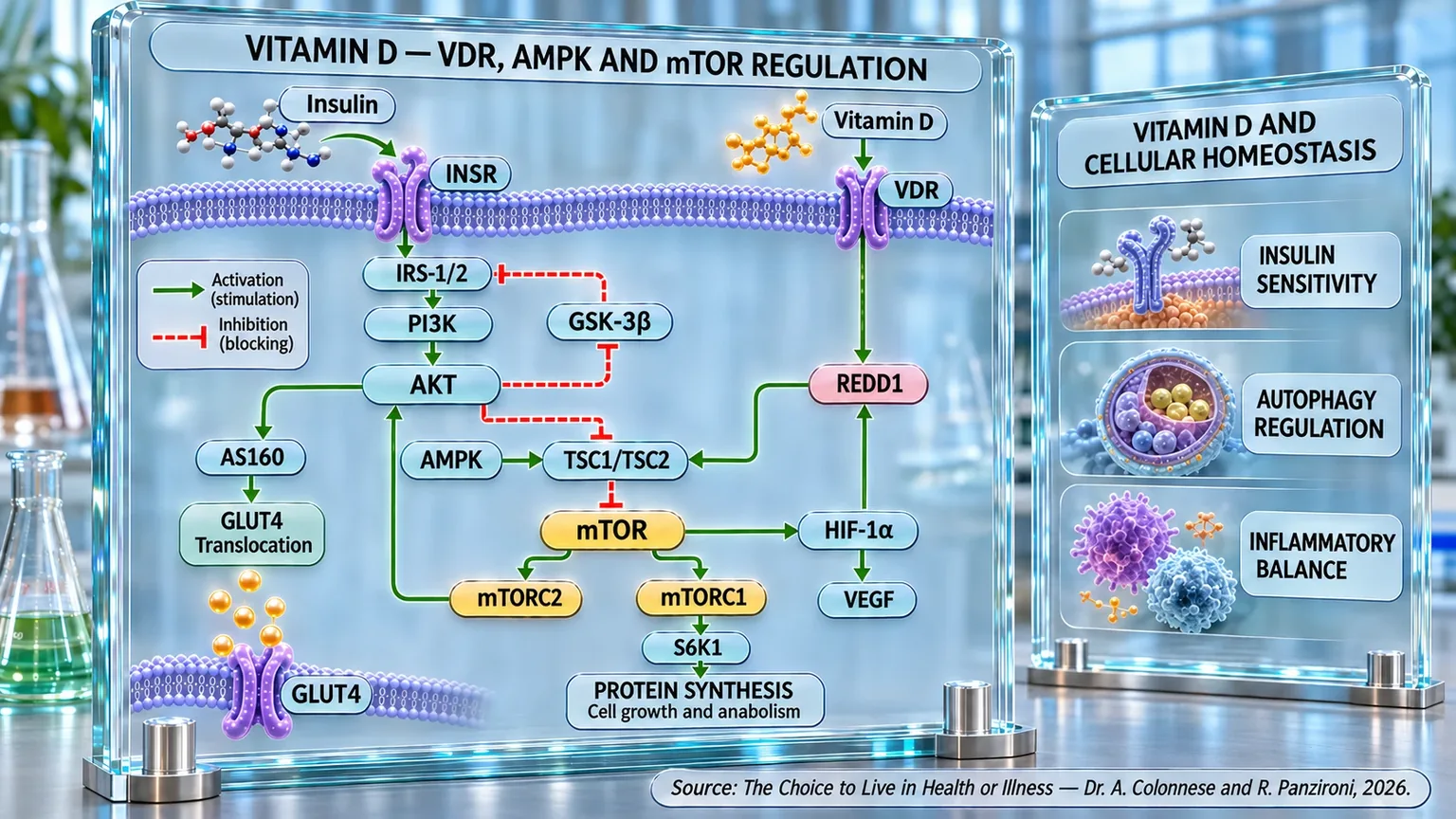
Scientific panel showing cell membranes, INSR and VDR receptors, and activation/inhibition arrows between PI3K/AKT, AMPK, and mTOR pathways.
Diagram of cellular regulation associated with vitamin D and insulin: VDR and INSR receptors connect IRS-1/2, PI3K, AKT, AMPK, TSC1/TSC2, mTORC1/2, GLUT4, and HIF-1α/VEGF, with indicated effects on protein synthesis, insulin sensitivity, autophagy, cell growth, and inflammatory balance in immunometabolic homeostasis.

ABSTRACT

Vitamin D performs functions that go beyond bone metabolism, playing a role in immune, energy, and cellular regulation. In the book "La scelta di Vivere in Salute o in Malattia" (The Choice to Live in Health or Disease), Dr. A. Colonnese and R. Panzironi explore the role of the VDR receptor in inflammatory processes, cardiovascular function, and the integrity of intestinal and respiratory barriers. The modulation of immune cells contributes to the balance between defense and tolerance, offering a possible interpretation of the associations between vitamin deficiency, chronic inflammation, and autoimmunity.

On a metabolic level, vitamin D can influence pancreatic β-cell function, insulin sensitivity, and the inflammatory response of tissues. Experimental mechanisms involve PI3K/AKT/mTOR, a pathway that coordinates cell growth and survival. VDR activation can increase REDD1, promoting the limitation of mTOR activity and the regulation of autophagy through TSC1/TSC2. In parallel, AMPK participates in energy adaptation, glucose utilization, and lipid oxidation, in interaction with SIRT1 and mTOR.

Studies on supplementation report improvements in certain glycemic parameters, insulin sensitivity, and inflammatory markers, especially in the presence of an initial deficiency. Oncological meta-analysis suggests a possible reduction in mortality, without a corresponding significant decrease in cancer incidence. Experimental results concern diabetic nephropathy, osteoarthritis, hepatic steatosis, and neoplastic models, in which effects on inflammation, autophagy, and proliferation emerge. Further investigations are exploring possible relationships with Parkinson's, autism, and ADHD, which still require clinical confirmation.

Overall, vitamin D appears to be involved in molecular networks that link immunity, metabolism, and cellular maintenance. However, the efficacy of supplementation depends on the initial status, dosage, duration, and individual conditions. Correcting documented deficiencies therefore represents a distinct goal from attributing generalized therapeutic benefits to the vitamin. Research perspectives need to clarify which experimental effects are transferable to clinical practice, without replacing established treatments. Particular attention should be paid to the distinction between pathway modulation and verifiable clinical outcomes.

Its properties

For many years, vitamin D was considered almost exclusively a vitamin involved in skeletal health. Its role in promoting the intestinal absorption of calcium and phosphorus and in maintaining proper bone mineralization is indisputable. As scientific research has progressed, however, a much broader picture has emerged. Today, vitamin D is recognized as a regulatory molecule capable of influencing numerous organs and systems in the body. The discovery of the Vitamin D Receptor (VDR) in immune, cardiac, endothelial, pancreatic, and renal cells has profoundly changed our understanding of its biological functions, suggesting that its scope extends far beyond bone metabolism (Młynarska et al., 2025) [1].

To understand the importance of this molecule, one can imagine the body as a large orchestra. For a long time, it was thought that vitamin D was simply one of the musicians assigned to a single section. More recent evidence shows that it is more like a conductor, capable of coordinating numerous instruments simultaneously so that the final result is functional.

One of the areas where vitamin D exerts its most important actions is the immune system. Numerous cells involved in the body's defense express the VDR and are able to respond directly to signals mediated by vitamin D. This allows for an extremely refined regulation of immune processes, enabling the body to defend itself against pathogens without generating excessive inflammation (Athanassiou et al., 2023) [2].

Vitamin D is involved in both innate and adaptive immunity. At the level of innate immunity, it promotes a more tolerogenic behavior (favoring the immune system's ability to recognize and tolerate harmless substances) in monocytes, macrophages, dendritic cells, and natural killer cells. In particular, it reduces the expression of molecules involved in antigen presentation and limits the production of numerous pro-inflammatory cytokines, while favoring the synthesis of anti-inflammatory mediators such as interleukin-10 (Athanassiou et al., 2023) [2]. The result is a more controlled immune response, capable of countering external threats without unnecessarily fueling inflammation.

Adaptive immunity is also significantly influenced. Evidence shows that vitamin D favors the shift from a Th1-type immune response, characterized by greater inflammatory activity, toward a more regulated Th2 response. In parallel, it promotes the differentiation of regulatory T cells (Treg), which are fundamental for maintaining immunological tolerance (Athanassiou et al., 2023) [2]. In other words, vitamin D helps prevent the immune system from losing the ability to distinguish what is useful from what should be eliminated.

This function is relevant in autoimmune diseases. Numerous clinical observations have documented that low levels of vitamin D are frequently present in patients suffering from rheumatoid arthritis, lupus erythematosus, Sjögren's syndrome, ankylosing spondylitis, psoriatic arthritis, systemic sclerosis, and inflammatory myopathies. In many of these conditions, vitamin deficiency is associated with greater disease activity and more severe clinical manifestations (Athanassiou et al., 2023) [2].

The immunological properties of vitamin D also extend to barrier tissues, which are the structures that represent the first point of contact between the body and the external environment. The intestinal and respiratory epithelium can be compared to the defensive walls of a city. When these structures maintain their integrity, viruses, bacteria, and other potentially harmful substances find it more difficult to penetrate deep tissues. Vitamin D helps preserve this function through the regulation of cell junctions, mucus production, and the balance of the resident gut microbiota (Hamza et al., 2023) [3].

The relationship between vitamin D and the gut microbiota represents one of the most promising fields of research in recent years. Through VDR-mediated signaling, vitamin D contributes to the maintenance of microbial homeostasis at both the intestinal and respiratory levels. When this regulation fails, phenomena such as dysbiosis, increased permeability of epithelial barriers, and the activation of chronic inflammatory processes can occur (Hamza et al., 2023) [3].

Another important property of vitamin D concerns the regulation of mast cells. These immune cells, which are particularly abundant in the skin, respiratory mucosa, and gastrointestinal tract, play a fundamental role in both defense against infectious agents and in allergic reactions. Evidence shows that vitamin D helps maintain the functional stability of mast cells, reducing their tendency to release large quantities of inflammatory mediators such as histamine, tryptase, and pro-inflammatory cytokines (Mehrani et al., 2023) [4].

This mechanism becomes important during viral infections. Several studies have highlighted that subjects with low levels of vitamin D exhibit greater susceptibility to infections and a higher risk of developing excessive inflammatory responses. Conversely, adequate levels of vitamin D promote a more efficient antiviral response through the induction of antimicrobial peptides and the enhancement of immune mechanisms involved in the production of antiviral interferons (Mehrani et al., 2023) [4].

The properties of vitamin D are not limited to the immune system. The VDR receptor is also expressed in cells of the cardiovascular system, including cardiomyocytes and endothelial cells. This observation has led to the development of numerous studies investigating the role of vitamin D in cardiac and vascular health (Anilkumar et al., 2024) [5].

Preclinical evidence indicates that vitamin D is able to modulate numerous signaling pathways involved in inflammation, oxidative stress, and tissue remodeling. Through interaction with pathways such as PI3K/AKT/mTOR and AMPK, vitamin D helps limit processes associated with cardiac hypertrophy, fibrosis, and vascular dysfunction; in experimental models, vitamin D deficiency has also been associated with endothelial dysfunction, increased blood pressure, and impaired cardiac function. Conversely, supplementation has shown the ability to reduce inflammation, attenuate fibrosis, limit the formation of atherosclerotic plaques, and improve overall cardiovascular functionality (Anilkumar et al., 2024) [5].

Energy metabolism also appears to be influenced by vitamin D. Numerous epidemiological studies have highlighted an inverse relationship between circulating levels of 25-hydroxyvitamin D and the risk of developing type 2 diabetes. This effect appears to be related to both an improvement in insulin sensitivity and better pancreatic β-cell functionality, as well as the ability of vitamin D to reduce low-grade chronic inflammation and oxidative stress (Młynarska et al., 2025) [1].

Taken together, these observations describe an extremely versatile molecule involved in maintaining immune balance, the integrity of mucosal barriers, cardiovascular health, and proper energy metabolism. Vitamin D therefore emerges not only as an essential nutrient for bone health, but as one of the most important biological regulators of the human body.

Supplementation

After understanding the numerous physiological functions of vitamin D, a question naturally arises: if this molecule participates in so many biological processes, what happens when a deficiency is corrected through supplementation?

As research has progressed, it has emerged that the benefits of supplementation appear to depend on multiple factors, including initial vitamin status, the presence of specific pathologies, the dosage used, and the duration of the intervention. In other words, vitamin D does not appear to be a therapy that is valid for everyone indiscriminately, but rather a biological tool that can exert relevant effects when used in the appropriate context.

One of the most studied fields concerns glucose metabolism and insulin sensitivity. Interest stems from the observation that low levels of vitamin D are frequently found in subjects with obesity, metabolic syndrome, prediabetes, and type 2 diabetes. This association has prompted researchers to verify whether correcting the deficiency could concretely modify the functioning of glucose metabolism.

To understand these effects, it is useful to remember that adipose tissue is not simply a fat storage depot. Today, it is considered a true endocrine organ, capable of producing numerous molecules that influence metabolism, inflammation, and insulin sensitivity. When adipose tissue expands excessively, it can develop a chronic inflammatory state associated with fibrosis phenomena that progressively compromise the body's response to insulin.

A study conducted on overweight and obese subjects with vitamin D deficiency showed that supplementation significantly reduces the expression of genes involved in inflammatory and fibrotic processes of adipose tissue. Concurrently, a reduction in collagen accumulation and an improvement in hepatic insulin sensitivity were observed. Conversely, subjects who did not receive supplementation showed a progressive worsening of metabolic parameters over time (Lontchi-Yimagou et al., 2020) [6].

The particularly interesting aspect of this work lies in the fact that the results obtained in humans were also confirmed in genetically modified animal models. Mice lacking the vitamin D receptor in adipocytes actually developed greater inflammation, greater adipose tissue fibrosis, and more marked hepatic insulin resistance. These observations suggest that an important part of the metabolic effects of vitamin D is mediated directly by adipocytes through the VDR receptor (Lontchi-Yimagou et al., 2020) [6].

The evidence becomes even more robust when analyzing large meta-analysis studies that bring together the results of numerous randomized clinical trials. A review including over four thousand participants highlighted that the co-supplementation of vitamin D and calcium leads to a significant reduction in fasting glycemia, circulating insulin levels, and the HOMA-IR index, one of the main markers of insulin resistance (Asbaghi et al., 2019) [7].

The analysis also showed that the benefits tend to be more evident when relatively high dosages are used and when the intervention is maintained for a sufficiently long period. The results suggest that correcting the deficiency can help improve the body's ability to use glucose and respond effectively to the action of insulin (Asbaghi et al., 2019) [7].

Further confirmation comes from a recent meta-analysis that evaluated the effects of supplementation in subjects affected by diabetes and prediabetes. The results show that vitamin D supplementation is associated with a significant reduction in glycated hemoglobin, fasting glycemia, circulating insulin, and HOMA-IR. In parallel, an improvement in the functionality of pancreatic β-cells, which are responsible for the synthesis and secretion of insulin, was observed (Bruna-Mejías et al., 2025) [8].

The observed benefits do not concern only glucose metabolism. Supplementation was also associated with improvements in the lipid profile, with reductions in LDL cholesterol, total cholesterol, and triglycerides. At the same time, a decrease in C-reactive protein, one of the main markers of chronic systemic inflammation, was documented; particularly interesting is the data observed in subjects with prediabetes. In this population, vitamin D supplementation was associated with a higher probability of returning to normoglycemia, suggesting that correcting the deficiency may help slow down or even reverse some of the metabolic alterations that precede the onset of overt diabetes (Bruna-Mejías et al., 2025) [8].

A second sector that has attracted considerable attention concerns oncology. For many years, observational studies have shown that individuals with higher levels of vitamin D tend to have a better prognosis in various forms of cancer; one of the largest available meta-analyses analyzed the results of twenty-six randomized clinical trials, evaluating the effect of vitamin D supplementation on cancer incidence and mortality. The results showed that supplementation does not significantly reduce the overall number of new cancer cases diagnosed in the general population; however, the same work highlighted an extremely interesting finding: supplementation is associated with a significant reduction in total cancer mortality. In other words, vitamin D does not seem to prevent the onset of neoplasms, but it could help improve the survival of patients who develop the disease (Guo et al., 2023) [9].

Similar conclusions emerge from a second meta-analysis that delved into the role of different administration methods. In this case, too, no significant reduction in the overall incidence of tumors was observed, while a decrease in cancer mortality was confirmed; a particularly relevant element that emerged from this analysis concerns the frequency of intake. The greatest benefits seem to be associated with daily supplementation, while protocols based on large intermittent boluses show less favorable results. This suggests that maintaining constant physiological levels of vitamin D may be more important than the simple occasional administration of very high doses (Keum et al., 2022) [10].

The authors also observed that obesity could attenuate some of the favorable effects of supplementation. A possible explanation lies in the ability of adipose tissue to sequester vitamin D, reducing its bioavailability. Added to this is the persistent inflammatory state typical of obesity, which could interfere with some of the biological mechanisms through which the vitamin exerts its actions (Keum et al., 2022) [10].

Taken together, this evidence suggests that the best results seem to emerge in subjects who are truly deficient and in specific pathological contexts, particularly those characterized by metabolic alterations, chronic inflammation, and increased cardiometabolic risk. When the deficiency is corrected appropriately, vitamin D shows the ability to improve various aspects of glucose metabolism, modulate systemic inflammation, and contribute to a better prognosis in certain oncological conditions.

Conditions treated

After examining the biological properties of vitamin D and the evidence regarding its supplementation, it is natural to wonder in which clinical conditions this knowledge finds a possible practical application. In recent years, research has progressively expanded the field of interest for vitamin D, shifting it from the traditional skeletal scope toward numerous pathologies characterized by chronic inflammation, immune alterations, metabolic dysfunctions, and degenerative processes.

One of the most studied areas is oncology. The interest stems from the observation that many tumor cells express the vitamin D receptor and maintain, at least in part, the ability to respond to biological signals mediated by this molecule. This has led researchers to hypothesize that vitamin D may directly influence the behavior of neoplastic cells and, simultaneously, modulate the body's immune response against the tumor.

A particularly interesting example comes from squamous cell carcinomas of the head and neck, tumors characterized by high biological aggressiveness and an often unfavorable prognosis in advanced stages. Experimental studies have shown that vitamin D is capable of significantly reducing both tumor onset and progression. In parallel, a marked increase in the infiltration of anti-tumor immune cells, including cytotoxic T lymphocytes and natural killer cells, has been observed, accompanied by a reduction in cell populations that normally promote immunosuppression within the tumor microenvironment (Brust et al., 2024) [11].

Observations made on tumor cells have also shown that the biologically active form of vitamin D is capable of slowing down cell proliferation and migration, two processes fundamental for tumor growth and spread. At the molecular level, these effects seem to involve central pathways of carcinogenesis with various mechanisms associated with inflammation and tumor hypoxia; these results are particularly interesting because they suggest that vitamin D may act simultaneously on two fronts. On one hand, it directly influences the biology of tumor cells; on the other, it contributes to making the immune system more efficient in recognizing and countering neoplastic growth. Although further clinical studies are needed to confirm these effects in humans, such evidence helps to understand why vitamin D is now considered a potential adjuvant in oncology (Brust et al., 2024) [11].

A second area of great interest concerns type 2 diabetes. As already discussed in the previous paragraph, numerous pieces of evidence link vitamin D deficiency to insulin resistance and alterations in glucose metabolism. However, more recent knowledge now allows for a better understanding of the molecular mechanisms involved.

Vitamin D exerts its effects on the pancreas, skeletal muscle, and adipose tissue through binding with the VDR receptor. This binding influences numerous metabolic pathways, including AMPK and mTOR, two fundamental energy regulation systems—addressed extensively in previous chapters—for the correct utilization of glucose. Through these mechanisms, vitamin D promotes fatty acid oxidation, improves glucose utilization, and contributes to reducing insulin resistance (Fuentes-Barría et al., 2025) [12].

In parallel, vitamin D exerts an important anti-inflammatory action. Chronic low-grade inflammation represents one of the central elements in the pathophysiology of type 2 diabetes. By reducing the production of pro-inflammatory cytokines and improving the function of adipose tissue, vitamin D contributes to creating a more favorable metabolic environment. The clinical implications of these mechanisms appear particularly relevant in the prediabetes phase; several studies suggest that adequate levels of vitamin D are associated with a lower probability of progression toward overt diabetes and a greater possibility of returning to a normoglycemic state. Although vitamin D cannot be considered a substitute therapy for lifestyle changes, its role as a possible supportive factor in the prevention and management of metabolic disease is emerging ever more clearly (Fuentes-Barría et al., 2025) [12].

When diabetes persists for many years, chronic complications involving various organs can develop. Among these, one of the most serious is diabetic nephropathy, currently considered one of the leading causes of end-stage chronic kidney disease worldwide. The progression of kidney damage is sustained by multiple factors, including persistent hyperglycemia, chronic inflammation, oxidative stress, and alterations in intracellular energy regulation systems. In this context, vitamin D seems to exert particularly interesting effects. Experimental studies have shown that treatment with vitamin D improves numerous parameters associated with diabetic nephropathy, including albuminuria, kidney function, blood pressure, and inflammatory markers (Khodir et al., 2020) [13].

One of the most fascinating aspects concerns the involvement of autophagy, a process through which the cell eliminates damaged components and maintains its functional efficiency. In kidneys affected by diabetic nephropathy, this mechanism tends to be compromised. Vitamin D seems to promote the restoration of autophagy through the modulation of the mTOR pathway, thus contributing to the protection of renal cells from chronic metabolic damage (Khodir et al., 2020) [13].

In addition to metabolic pathologies, vitamin D is also attracting increasing attention in the field of neurodegenerative diseases. One of the most studied examples is Parkinson's disease, a condition characterized by the progressive loss of dopaminergic neurons and the appearance of motor symptoms such as rigidity, slowness of movement, and tremor. In recent years, it has emerged that neurodegeneration does not depend exclusively on neuronal loss but also involves significant immune alterations. In particular, many patients present an imbalance between pro-inflammatory Th17 cells and regulatory T cells (Treg), a condition that promotes chronic neuroinflammation (Li et al., 2025) [14].

Vitamin D seems to intervene precisely on this immunological balance. In patients affected by Parkinson's and simultaneously deficient in vitamin D, supplementation resulted in a reduction of Th17 cells and an increase in Treg cells, bringing the ratio between these cell populations back toward values closer to normal. In parallel, a significant improvement in motor function was observed; the authors hypothesize that these effects may be linked to the ability of vitamin D to modulate neuroinflammation, influence microglia activity, and limit some processes associated with the aggregation of α-synuclein, one of the proteins most involved in the pathogenesis of the disease (Li et al., 2025) [14].

The interest in the neurological role of vitamin D is not, however, limited to adult neurodegenerative diseases. In recent years, numerous studies have begun to explore the possible involvement of vitamin D in neurodevelopmental disorders, particularly in autism spectrum disorder and attention deficit hyperactivity disorder.

This line of research stems from the discovery that the vitamin D receptor is expressed in numerous brain regions involved in neuronal development, synaptic plasticity, and behavior regulation. Experimental studies suggest that vitamin D participates in the regulation of important neurotransmitters, including dopamine, serotonin, and glutamate, which are fundamental systems for attention, social behavior, and emotional modulation (Ye et al., 2023) [15].

In the context of autism, several studies have observed an association between low levels of vitamin D and a higher risk of developing the disorder. The available evidence suggests that vitamin D may influence neuronal circuits involved in social behaviors through the modulation of pathways such as mTOR, the endocannabinoid system, and oxytocin-mediated signaling; associations between vitamin D deficiency and alterations in the dopaminergic and serotonergic pathways that regulate attention, impulse control, and motor activity have also been observed in ADHD (Attention Deficit Hyperactivity Disorder). Although clinical evidence is still preliminary, these results are opening new research perspectives on the role of vitamin D in the development and functioning of the human brain (Ye et al., 2023) [15].

The evidence examined shows how vitamin D can intervene in biological processes common to numerous pathologies, from inflammation to the regulation of cellular metabolism. Among these, a central role belongs to insulin sensitivity, which influences the energy balance of many tissues. This relationship deserves specific in-depth analysis, as it allows for connecting the described molecular mechanisms to the most widespread metabolic alterations. But through which processes can vitamin D influence the response of cells to insulin and contribute to the maintenance of glycemic balance?

Vitamin D and insulin resistance

To understand this link, we must start with a fundamental process: the ability of cells to correctly recognize and use insulin signals. When this response weakens, the body may compensate by increasing the secretion of the hormone, which over time promotes metabolic imbalances. Vitamin D may intervene in various mechanisms involved, modulating inflammation, pancreatic function, and several cellular signaling pathways.

Insulin resistance can be imagined as a progressive loss of sensitivity in peripheral tissues to the signals sent by insulin. Under normal conditions, this hormone acts as a key capable of opening cell doors to glucose. When insulin resistance occurs, the lock becomes progressively less sensitive, and the pancreas is forced to produce ever-increasing amounts of insulin to achieve the same metabolic effect. Over the years, this compensatory mechanism can promote the onset of prediabetes, type 2 diabetes, and numerous cardiovascular and metabolic complications.

In recent years, numerous epidemiological studies have highlighted a close relationship between low serum levels of vitamin D and increased insulin resistance. One of the most comprehensive systematic reviews available has shown that subjects with lower concentrations of 25-hydroxyvitamin D exhibit significantly higher levels of fasting glycemia, fasting insulin, and HOMA-IR index, one of the main indicators used to estimate insulin sensitivity; this association appears biologically plausible considering that vitamin D acts in numerous tissues involved in glycemic homeostasis. Vitamin D receptors are, in fact, expressed in pancreatic β-cells, the liver, skeletal muscle, and adipose tissue. Through these receptors, vitamin D helps regulate insulin secretion, intracellular calcium metabolism, insulin receptor expression, and the survival of pancreatic β-cells (Lei et al., 2023) [16].

Epidemiological evidence also suggests that vitamin D deficiency is not just a consequence of the metabolic alterations associated with obesity and diabetes, but may itself constitute an independent risk factor. In a large umbrella review that synthesized the results of over seventy observational studies, subjects with low serum levels of vitamin D had a significantly higher risk of developing type 2 diabetes compared to individuals with adequate concentrations; it is particularly interesting that this association remained significant even after numerous sensitivity analyses, suggesting that vitamin D deficiency may directly contribute to the pathophysiology of diabetes (Cheng et al., 2024) [17].

When moving from observational studies to intervention studies, an equally interesting picture emerges. In patients with type 2 diabetes, vitamin D supplementation has been associated with significant improvements in fasting glycemia, glycated hemoglobin, insulinemia, and the HOMA-IR index. However, one of the most important aspects that emerged from the literature concerns the role of baseline vitamin status.

Recent analyses show that the greatest benefits are observed in subjects who have a real vitamin D deficiency before the start of supplementation. In normovitaminic subjects, however, the effects tend to be more modest or sometimes absent. This behavior has led some authors to describe vitamin D as a true "threshold nutrient," whose metabolic benefit emerges primarily when a pre-existing deficiency is corrected (Cheng et al., 2024) [17].

The importance of this concept becomes particularly evident in prediabetes, an intermediate condition characterized by glycemic alterations that are still reversible. In this phase, the pancreas generally maintains a fair capacity for compensation, and metabolic damage is not yet fully consolidated. This makes prediabetes a particularly favorable window of time for preventive interventions.

A recent systematic review including thirteen randomized clinical trials highlighted that vitamin D supplementation in subjects with prediabetes leads to a significant reduction in fasting glycemia, glycated hemoglobin, and values obtained through oral glucose tolerance tests. Taken together, these results suggest that correcting vitamin deficiency may help slow or reduce the progression toward overt diabetes; from a pathophysiological point of view, these benefits seem to derive from a combination of complementary mechanisms. Vitamin D increases the expression of insulin receptors, improves peripheral insulin sensitivity, regulates intracellular calcium metabolism, and helps reduce the low-grade chronic inflammation that frequently accompanies insulin resistance (Hassan et al., 2024) [18].

A particularly interesting aspect concerns the relationship between dosage and metabolic response. For many years, it was hypothesized that increasingly higher dosages of vitamin D would necessarily produce superior metabolic effects. More recent evidence, however, shows a more complex picture.

A network meta-analysis including forty randomized clinical trials demonstrated that very high dosages increase serum vitamin D concentrations more markedly, but do not always produce proportionally superior benefits on glycemic parameters. In some cases, low or moderate dosages showed particularly favorable effects on glycated hemoglobin and fasting insulin levels. These results suggest that reaching physiologically adequate levels of vitamin D may be more important than simply increasing serum concentrations indiscriminately. Once again, the concept emerges that vitamin D seems to act primarily as a metabolic regulator, rather than as a traditional pharmacological agent with a strictly dose-dependent response (Zhang et al., 2025) [19].

The influence of vitamin D on insulin resistance also appears particularly evident in pediatric populations affected by overweight and obesity. In these subjects, vitamin D deficiency is extremely frequent, likely due to the sequestration of the vitamin within adipose tissue and the consequent reduction of its systemic bioavailability.

A recent network meta-analysis conducted on children and adolescents with overweight or obesity highlighted that vitamin D supplementation leads to significant improvements in the HOMA-IR index and C-reactive protein, one of the main markers of systemic inflammation. The most evident benefits were observed in protocols that used high dosages, likely necessary to compensate for the lower biological availability of the vitamin in subjects with high adipose mass. These observations are particularly important because they confirm how insulin resistance, chronic inflammation, and vitamin D deficiency represent phenomena that are closely interconnected from the earliest stages of life. Intervening early on these factors could help reduce the risk of developing chronic metabolic diseases in adulthood (Zhu et al., 2024) [20].

The analyzed evidence shows how vitamin D can contribute to the regulation of insulin sensitivity through various cellular mechanisms. Among these, the control of inflammation, pancreatic function, and the balance of metabolic signals take on particular importance. However, to better understand this relationship, it is necessary to delve into one of the main systems that coordinate these processes: the PI3K/AKT/mTOR pathway. Its activity could represent a link between vitamin D deficiency, alterations in energy metabolism, and the development of insulin resistance in the various tissues of the body.

Vitamin D and the PI3K/AKT/mTOR pathway

If there is one element that emerges with particular force from the most recent scientific literature on vitamin D, it is its close relationship with the PI3K/AKT/mTOR pathway. Over the last few years, numerous studies have shown that many of the properties attributed to vitamin D seem to converge precisely on this cellular regulation system. To understand the importance of this relationship, it is useful to remember that mTOR, an acronym for Mammalian Target of Rapamycin, represents a sort of metabolic control center for the cell. Through its modulation, the cell decides whether to grow, proliferate, synthesize proteins, store energy, or activate intracellular repair and recycling mechanisms such as autophagy.

When this system functions correctly, the body is able to adapt efficiently to changes in the internal and external environment. However, when mTOR remains excessively active or excessively inhibited for long periods, numerous pathologies can appear, including type 2 diabetes, obesity, degenerative diseases, cancer, and accelerated aging processes. In this context, vitamin D emerges as one of the most interesting regulators of the pathway, capable of influencing its activity in a dynamic and biologically context-dependent manner.

One of the clearest examples of this regulatory function comes from osteoarthritis. For many years, this disease was considered a simple consequence of joint wear and tear. Today, however, we know that the progression of the pathology involves complex cellular mechanisms that directly affect the survival of chondrocytes, the cells responsible for maintaining articular cartilage.

Within chondrocytes, autophagy plays an essential role. This process can be compared to an internal system for collecting and recycling cellular waste. When autophagy functions correctly, damaged proteins and deteriorated organelles are eliminated and replaced. When the system slows down, however, cells begin to accumulate damage that promotes apoptosis and tissue degeneration.

Experimental evidence shows that vitamin D is able to activate autophagy in chondrocytes through the modulation of the AMPK/mTOR axis. Specifically, vitamin D increases the activity of AMPK, one of the main cellular energy sensors, and simultaneously reduces the activity of mTOR. This combination promotes the activation of the autophagic program, increases the formation of autophagolysosomes (cellular structures that degrade and recycle damaged or useless components), and significantly reduces the programmed cell death of chondrocytes (Liu et al., 2024) [21].

The final result is better preservation of the extracellular matrix and a reduction in cartilage degradation. In other words, vitamin D seems to contribute to keeping the cellular maintenance mechanisms active, which allows chondrocytes to survive the metabolic and inflammatory stress associated with osteoarthritis (Liu et al., 2024) [21].

The relationship between vitamin D and mTOR also emerges in adipose tissue, one of the most important metabolic organs in the body. In recent years, it has become evident that body fat is not simply an energy store, but a highly dynamic tissue capable of communicating with the brain, liver, muscles, and immune system.

In this context, vitamin D seems to influence the behavior of adipocytes (cells of the adipose tissue that store fat as an energy reserve) through the modulation of the PI3K/AKT/mTOR/p53 axis. Experimental studies have shown that vitamin D3 is able to reduce the activation of the PI3K/AKT/mTOR cascade and simultaneously increase signaling mediated by p53, a protein closely involved in the control of cellular stress and genomic integrity; in parallel, an increase in autophagic activity and a significant reduction in lipid accumulation have been observed. In animal models fed high-fat diets, vitamin D3 supplementation also limited weight gain and modified the expression of numerous genes involved in adipose metabolism (Zhao and Qin, 2022) [22].

These results suggest that vitamin D may contribute to the maintenance of energy homeostasis through fine-tuning of the metabolic processes controlled by mTOR. Once again, a fundamental concept emerges: the goal does not seem to be the indiscriminate inhibition of the pathway, but its rebalancing toward more favorable physiological conditions.

This interpretation finds further confirmation in studies on diabetes mellitus. In this context, the chronic hyperactivation of mTORC1 represents one of the main mechanisms involved in the development of insulin resistance. When mTORC1 remains constantly active, insulin signal transmission becomes progressively less efficient, promoting hyperglycemia, inflammation, and vascular damage.

According to the most recent evidence, vitamin D exerts a significant part of its metabolic effects precisely through the negative modulation of mTOR. One of the most studied mechanisms involves increasing the expression of REDD1, a protein capable of activating the TSC1/TSC2 complex, a known physiological suppressor of mTOR. Through this sequence of events, vitamin D helps to limit the excessive activity of mTORC1 and restore better insulin sensitivity; in parallel, improvements in autophagy, reduction of oxidative stress, and attenuation of inflammatory processes have been observed. The authors also suggest that the co-supplementation of vitamin D and L-cysteine could amplify these effects thanks to a synergistic action on the reduction of ROS and the down-regulation of the mTOR pathway (Stevens et al., 2025) [23].

If in metabolic diseases vitamin D contributes mainly to containing the pathological hyperactivation of mTOR, in oncology this function takes on even deeper implications. Tumor cells, in fact, depend on pathways such as PI3K/AKT/mTOR to support their growth, energy metabolism, and ability to survive in unfavorable environmental conditions.

One of the most significant examples comes from non-small cell lung cancer. In this tumor, vitamin D has demonstrated the ability to reduce the activity of the PI3K/AKT/mTOR pathway both in vitro and in animal models. This effect is accompanied by a marked reduction in tumor glycolysis, a phenomenon known as the Warburg effect, which represents one of the distinctive characteristics of neoplastic cells. Instead of primarily using mitochondrial respiration, these cells consume enormous amounts of glucose, rapidly converting it into lactate, thus obtaining the substrates necessary to support accelerated proliferation. Vitamin D reduces the expression of key enzymes in this process, including GLUT1 and other glycolytic enzymes, limiting the energy availability necessary for tumor growth (Yiyan et al., 2022) [24].

A similar picture emerges in bladder cancer. In this case, too, vitamin D exerts significant effects on tumor metabolism through the modulation of the AKT/mTOR pathway. Cells treated with vitamin D show a reduction in glucose consumption, lactate production, and intracellular ATP levels, clear signs of a suppression of the Warburg effect; even more interesting is the fact that these metabolic changes are accompanied by greater sensitivity to cisplatin, one of the main drugs used in the treatment of bladder cancer. Vitamin D, in fact, increases cellular apoptosis and reduces the expression of proteins associated with chemoresistance, suggesting a possible role as an adjuvant agent in oncological therapies (Zhou et al., 2026) [25].

Observing this evidence as a whole, an extremely coherent picture emerges. In different tissues and in very different pathological conditions, vitamin D seems to act as a sophisticated regulator of mTOR signaling. When the pathway is excessively active, as in diabetes or many neoplasms, vitamin D tends to reduce its activity. When, on the other hand, it is necessary to promote cellular repair and homeostasis maintenance processes, its action on the AMPK/mTOR axis promotes autophagy and intracellular protection mechanisms.

More than a simple mTOR inhibitor, vitamin D therefore appears as a physiological modulator of the entire system. Its function seems to be to bring cellular metabolism back toward a state of balance, favoring the survival of healthy cells and, at the same time, limiting the metabolic programs that support inflammation, tissue degeneration, and tumor growth.

Vitamin D, REDD1 and mTOR

If the relationship between vitamin D and mTOR represents one of the most fascinating topics to emerge from research in recent years, the discovery of the role of REDD1, also known as DDIT4 (DNA Damage-Inducible Transcript 4), has allowed us to understand with greater precision the molecular mechanism that connects these two protagonists of cellular physiology. For a long time, the antiproliferative, immunomodulatory, and metabolic effects of vitamin D were observed in numerous experimental models without it being entirely clear what the intracellular link responsible for these actions was. Today, we know that a significant portion of these effects converges precisely on the vitamin D–VDR–REDD1–mTOR axis.

To understand the importance of this discovery, it is useful to remember that REDD1, as we have already seen in Chapter 4 dedicated to the insulin pathway, is a protein produced by the cell in response to stress conditions. Its main task is to limit the activity of mTORC1 when cell growth could be harmful or energetically disadvantageous. In other words, if mTOR can be compared to the cell's metabolic accelerator, REDD1 represents one of the main braking systems tasked with preventing that acceleration from becoming excessive.

The first evidence directly linking vitamin D to this mechanism showed that the biologically active form of vitamin D, 1,25-dihydroxyvitamin D3, is able to increase the expression of REDD1 and consequently reduce the activity of mTOR. This effect proved so important that the experimental silencing of REDD1 was sufficient to completely abolish the antiproliferative effects of vitamin D, indicating that this protein constitutes one of the main biological mediators through which the hormone exerts its regulatory functions (Lisse and Hewison, 2011) [26].

Subsequently, more in-depth studies demonstrated that REDD1 is not simply a protein indirectly influenced by vitamin D, but constitutes a true direct genomic target of the vitamin D receptor (VDR). When vitamin D enters the cell and binds to the VDR, the activated complex reaches the nucleus and binds directly to the regulatory regions of the REDD1 gene, inducing its transcription. The increase in REDD1 then determines the activation of the TSC1/TSC2 complex, which in turn blocks Rheb, the main protein responsible for the activation of mTORC1. The final result is a reduction in the phosphorylation of S6K1 (an enzyme activated by mTORC1) and a decrease in cell proliferation (Lisse et al., 2011) [27].

This discovery had a considerable impact because it provided a unifying explanation for numerous biological effects of vitamin D previously considered independent of each other. Instead of acting through multiple separate pathways, vitamin D appears to use REDD1 as a sort of central hub capable of coordinating cell growth, energy metabolism, autophagy, and stress response (Lisse et al., 2011) [27].

One of the areas in which this mechanism appears particularly relevant is diabetes mellitus. Chronic hyperglycemia, in fact, tends to profoundly alter the balance of the mTOR pathway, favoring its persistent activation. In renal mesangial cells exposed to high concentrations of glucose, a significant reduction in REDD1 was observed, accompanied by an increase in the activity of Rheb and mTOR. This alteration promotes cell proliferation and contributes to the progression of diabetic nephropathy.

The administration of active vitamin D proved capable of reversing these modifications. The increase in REDD1 induced by the VDR determines, in fact, the reactivation of the TSC2-mTOR axis, reducing the proliferation of mesangial cells and improving numerous structural and functional parameters of the diabetic kidney. These include the reduction of albuminuria, the improvement of glomerular architecture, and the decrease in the thickness of the glomerular basement membrane (Wang et al., 2016) [28].

The importance of REDD1 in this context was further confirmed by studies that used overexpression and gene silencing techniques. When REDD1 is artificially increased, cell proliferation is reduced even in the absence of vitamin D. Conversely, when this protein is silenced, the beneficial effects of vitamin D on the mTOR pathway disappear almost completely. This demonstrates that REDD1 does not represent a simple biological marker of the response to vitamin D, but a functionally indispensable element for its antiproliferative action (Chen et al., 2018) [29].

An analogous mechanism was also observed in pancreatic β-cells, which are responsible for insulin production. Chronic hyperglycemia induces a reduction in REDD1 and TSC1/TSC2, accompanied by marked activation of the mTOR axis. This imbalance favors the activation of pro-apoptotic proteins and accelerates the loss of β-cells, one of the fundamental events in the progression of diabetes mellitus.

Active vitamin D proved capable of restoring the expression of REDD1 and the TSC complexes, reducing the activity of mTOR and limiting the activation of apoptotic signals. In this way, vitamin D contributes to preserving the survival of β-cells and counteracting the toxic effects of chronic hyperglycemia (Yang et al., 2015) [30].

The importance of the vitamin D–REDD1–mTOR axis also emerges with particular clarity in oncology. Tumor cells, in fact, depend on constant mTOR activation to support growth, protein synthesis, and metabolic adaptation. In numerous neoplasms, a reduction in REDD1 expression has been observed, suggesting the loss of an important physiological mechanism for controlling proliferation.

In cutaneous squamous cell carcinoma, for example, vitamin D directly induces the expression of REDD1 through the VDR receptor. The increase in this protein determines a reduction in mTORC1 activity, promotes autophagy, and limits cell proliferation. This mechanism also appears to improve the response to DNA damage caused by ultraviolet radiation, contributing to the protection of genomic stability (Vishlaghi and Lisse, 2020) [31].

Further studies have confirmed that REDD1 acts as a true tumor suppressor in squamous cell carcinoma. Tumor cells that express low levels of REDD1 show, in fact, greater mTORC1 activity, reduced autophagy, and faster growth. Conversely, the induction of REDD1 by vitamin D leads to a marked reduction in cell proliferation and a significant decrease in tumor growth in animal models (Zhang et al., 2019) [32].

The VDR-REDD1-mTOR axis, however, does not appear to be limited to tumors and glucose metabolism. Studies conducted on the skin and hair follicles have shown that REDD1 represents a direct target of the VDR even during processes of epidermal differentiation and tissue repair. In animal models lacking the vitamin D receptor, the deregulation of REDD1 is associated with defects in the follicular cycle, alopecia, and slowed wound healing, highlighting the fundamental role of this pathway in maintaining skin homeostasis (Zhao et al., 2016) [33].

The most recent research has finally further expanded the biological picture of REDD1. In human osteosarcoma cells, it was observed that vitamin D profoundly modifies mitochondrial metabolism, reducing the production of reactive oxygen species and promoting mechanisms of adaptation to stress. In this context, REDD1 assumes a particularly interesting role because its intracellular localization appears to be regulated directly by the functional state of the mitochondria. When vitamin D induces a controlled depolarization of the mitochondrial membrane, REDD1 is released into the cytoplasm, where it can fully exert its inhibitory action on mTORC1 (Quigley et al., 2022) [34].

This phenomenon suggests that REDD1 represents not only a regulator of cell growth but also an important metabolic sensor capable of integrating information coming from energy metabolism, oxidative stress, and mitochondrial function. Through this system, vitamin D appears to promote a metabolic condition characterized by lower production of free radicals, greater efficiency of cellular repair mechanisms, and reduction of proliferative signals associated with neoplastic transformation (Quigley et al., 2022) [34].

Considering all this evidence together, a surprisingly coherent picture emerges. REDD1 appears as the main molecular bridge through which vitamin D exerts most of its deepest biological actions. Through the activation of the VDR receptor, vitamin D increases the expression of REDD1, which in turn activates the TSC1/TSC2 complex and reduces the activity of mTOR. This sequence of events influences fundamental processes such as cell proliferation, autophagy, energy metabolism, stress response, cell survival, and tumor control. More than a simple nutrient involved in bone metabolism, vitamin D therefore emerges as a sophisticated regulator of cellular homeostasis, capable of using REDD1 as a central hub to coordinate some of the most important metabolic decisions of the cell.

Vitamin D, AMPK and mTOR

If REDD1 represents one of the primary systems through which vitamin D directly regulates mTOR activity, there is a second major biological axis that helps explain much of its metabolic effects: the signaling pathway mediated by AMPK. In recent years, in fact, numerous studies have highlighted how vitamin D is capable of profoundly influencing this cellular energy sensor, creating a direct link between energy availability, metabolism, autophagy, and cell survival.

To understand the importance of this relationship, it is useful to remember that AMPK, an acronym for Adenosine Monophosphate-Activated Protein Kinase, acts as the cell's true energy gauge. When energy reserves decrease, AMPK is activated and induces a series of adaptations aimed at energy conservation and the production of new energy. At the same time, it tends to limit high-energy-consuming anabolic processes, many of which are directly controlled by mTOR.

In simple terms, if mTOR can be compared to the accelerator pedal that promotes growth, protein synthesis, and cell proliferation, AMPK represents the system that evaluates whether the cell actually has the fuel necessary to sustain such acceleration. Vitamin D seems to fit right into this delicate balance, helping to coordinate the activities of both systems.

The most recent evidence indicates that the activation of the vitamin D receptor is capable of directly stimulating AMPK in numerous metabolically active tissues, including the liver, skeletal muscle, adipose tissue, and pancreas. Through this activation, vitamin D promotes fatty acid oxidation, improves glucose utilization, and reduces intracellular lipid accumulation. At the same time, increased AMPK activity helps limit excessive mTOR activation, improving insulin sensitivity and countering insulin resistance; this mechanism is particularly relevant in type 2 diabetes, a condition characterized by a profound alteration of cellular energy homeostasis. In this context, the activation of AMPK by vitamin D seems to contribute to the improvement of glucose metabolism not only through increased glucose uptake, but also through more efficient utilization of fatty acids and a reduction in chronic systemic inflammation (Fuentes-Barría et al., 2025) [12].

The importance of this axis emerges even more clearly by observing what happens in the liver during non-alcoholic fatty liver disease. This pathology, now considered one of the most frequent manifestations of metabolic syndrome, is characterized by the progressive accumulation of fat within hepatocytes, accompanied by oxidative stress, inflammation, and alterations in mitochondrial function.

In experimental models of fatty liver disease, the active form of vitamin D has been shown to significantly reduce intracellular triglyceride accumulation, the production of reactive oxygen species, and cell damage induced by lipotoxicity. At the same time, a marked increase in the expression of SIRT1 (an enzyme that regulates energy metabolism) and the phosphorylation of AMPK was observed, suggesting the activation of a coordinated response aimed at restoring cellular metabolic balance (Chang, 2024) [35].

However, the influence of vitamin D on the AMPK axis is not limited to hepatic metabolism. It has recently emerged that this signaling pathway also plays a fundamental role in the regulation of the intestinal ecosystem. The gut is, in fact, one of the main interfaces between the organism and the external environment, hosting trillions of microorganisms that influence metabolism, immunity, and systemic inflammation.

In animal models of type 2 diabetes, vitamin D deficiency is associated with profound alterations of the gut microbiota, increased permeability of the intestinal barrier, endotoxemia (the presence in the blood of endotoxins, substances produced by certain bacteria), and increased oxidative stress. Treatment with activated vitamin D was found to be capable of significantly improving these parameters, reducing systemic inflammation and favoring the restoration of the integrity of the intestinal barrier; in this case as well, one of the main mechanisms involved seems to be the coordinated activation of SIRT1 and AMPK. The increase in AMPK phosphorylation observed in treated animals is, in fact, associated with a reduction in oxidative stress, better antioxidant function, and a significant improvement in glycemic control. These results suggest that vitamin D may exert part of its metabolic effects through complex communication between the microbiota, the gut, and cellular energy metabolism (Zhang et al., 2025) [36].

The interaction between vitamin D, AMPK, and mTOR also takes on particular relevance in autophagy processes. As previously discussed, autophagy represents one of the main cellular maintenance systems. Through this mechanism, the cell eliminates damaged proteins, defective organelles, and non-functional components, preserving its biological integrity. In osteoarthritis, a pathology characterized by the progressive degeneration of cartilage, the reduction of autophagy contributes directly to chondrocyte death and the loss of the extracellular matrix. Recent studies have shown that vitamin D is able to reactivate autophagy through the activation of AMPK and the simultaneous inhibition of mTOR. This leads to an increase in the formation of functional autophagosomes and lysosomes, reducing chondrocyte apoptosis and slowing joint degeneration; these results are particularly interesting because they show how vitamin D does not act simply as a regulator of energy metabolism, but also as a promoter of cellular repair and maintenance processes. Through the AMPK/mTOR axis, vitamin D in fact promotes a physiological condition that allows cells to better cope with the metabolic and inflammatory stresses associated with aging and chronic diseases (Liu et al., 2024) [21].

The importance of this mechanism also emerges strongly in oncology. Tumor cells frequently depend on high mTOR activity and a profound reorganization of energy metabolism to support uncontrolled growth. In this context, vitamin D seems to exert particularly interesting effects through the modulation of the p53–AMPK–mTOR axis.

In gastric carcinoma, vitamin D3 promotes cellular autophagy through the activation of p53 and AMPK and the consequent inhibition of mTOR. This process is associated with a reduction in cell proliferation and a greater capacity of tumor cells to activate programmed cell death; the activation of autophagy in this context takes on a particularly relevant meaning. Unlike healthy cells, which use autophagy to maintain their integrity, many tumor cells develop alterations of this process to favor survival and uncontrolled growth. The ability of vitamin D to restore proper regulation of the p53–AMPK–mTOR axis could therefore represent one of the mechanisms through which it exerts part of its antitumor properties (Wang et al., 2024) [37].

Looking at this evidence as a whole, an extremely coherent picture emerges. If REDD1 represents one of the main genomic mediators through which vitamin D controls mTOR activity, AMPK instead appears as the great energy sensor that translates this signal into concrete metabolic adaptations. Through the activation of SIRT1, AMPK, and autophagic programs, vitamin D coordinates a vast network of processes involved in glucose metabolism, lipid oxidation, antioxidant response, intestinal function, cell survival, and the control of tumor proliferation. Rather than acting on a single molecular target, vitamin D therefore seems to orchestrate a complex energy regulation system capable of maintaining the cell in a state of metabolic balance compatible with health and longevity.

Conclusions

Throughout this chapter, we have discovered how vitamin D performs functions that go far beyond bone health. Through the VDR receptor, this molecule participates in the regulation of the immune system, energy metabolism, and cell survival. The research examined shows its involvement in the inflammatory response, cardiovascular function, and the protection of intestinal and respiratory barriers. In particular, vitamin D helps regulate the activity of immune cells, promoting a balance between defense and tolerance. This role helps us understand the associations observed between vitamin deficiency, chronic inflammation, and various autoimmune diseases.

These properties lead to a fundamental question: can correcting a deficiency concretely improve health? Studies on supplementation suggest potential benefits, especially in individuals with insufficient initial levels. Research has documented improvements in certain glycemic parameters, insulin sensitivity, and inflammatory markers. In oncology, some meta-analysis studies have also noted a reduction in mortality, without demonstrating a significant decrease in new cancer cases. However, the results depend on individual conditions, dosages, and the duration of the intervention.

This distinction becomes even more important when observing the pathologies examined. In type 2 diabetes, vitamin D could contribute to the regulation of glucose metabolism and pancreatic function. In diabetic nephropathy, experimental studies suggest protective effects through the control of inflammation and autophagy. Further research has explored its role in Parkinson's, autism, and ADHD, highlighting possible links with immune balance and brain function. Although many applications require clinical confirmation, a common element emerges: the ability of vitamin D to influence the mechanisms that regulate cellular activity.

To understand this connection, we have delved into the relationship between vitamin D and insulin resistance. When tissues respond less effectively to insulin, the body may increase the production of the hormone, promoting further metabolic imbalances. Research indicates that vitamin D could intervene in the function of pancreatic β-cells, inflammation, and the response of peripheral tissues. The benefits of supplementation seem particularly relevant in deficient subjects and in the early stages of glycemic alterations. But to understand how these effects develop, we must observe the signals that govern intracellular metabolism.

At the center of these processes, we find the PI3K/AKT/mTOR pathway, which is fundamental for coordinating growth, energy consumption, and cellular repair. Experimental studies show that vitamin D can limit its excessive activation, promoting autophagy and reducing certain proliferative signals. This mechanism has been observed in osteoarthritis, adipose tissue, and various tumor models. A decisive role belongs to REDD1, whose expression can increase through the VDR receptor. REDD1 therefore promotes the action of the TSC1/TSC2 complex, helping to contain mTOR activity and regulate cell growth.

Alongside REDD1, AMPK intervenes as the sensor that helps the cell adapt to energy availability. Through this pathway, vitamin D can promote glucose utilization, fat oxidation, and cellular maintenance processes. Studies on hepatic steatosis, the gut, and osteoarthritis show the involvement of AMPK, SIRT1, and mTOR in the control of metabolic stress. Some oncological models also suggest possible effects on cell proliferation. Taken together, this evidence describes vitamin D as a regulator of biological balance, whose value lies in its ability to support normal cellular functions without replacing established therapies.

References

[1] Vitamin D and Chronic Disorders: A Review of Metabolic and Cardiovascular Diseases

Młynarska et al., 2025

Pharmaceuticals (Basel), 2025;18(10):1467.

[2] Vitamin D and Autoimmune Rheumatic Diseases

Athanassiou et al., 2023

Biomolecules, 2023;13(4):709.

[3] Immunomodulatory Properties of Vitamin D in the Intestinal and Respiratory Systems

Hamza et al., 2023

Nutrients, 2023;15(7):1696.

[4] Vitamin D Influences the Activity of Mast Cells in Allergic Manifestations and Potentiates Their Effector Functions against Pathogens

Mehrani et al., 2023

Cells, 2023;12(18):2271.

[5] Vitamin D as a Modulator of Molecular Pathways Involved in Cardiovascular Diseases: Evidence from Preclinical Studies

Anilkumar et al., 2024

Life Sciences, 2024;357:123062.

[6] Insulin-sensitizing effects of vitamin D repletion mediated by adipocyte vitamin D receptor: Studies in humans and mice

Lontchi-Yimagou et al., 2020

Molecular Metabolism, 2020;42:101095.

[7] Effect of Calcium–Vitamin D Co-Supplementation on Insulin, Insulin Sensitivity, and Glycemia: A Systematic Review and Meta-Analysis of Randomized Clinical Trials

Asbaghi et al., 2019

Hormone and Metabolic Research, 2019;51(5):288-295.

[8] Effectiveness of Vitamin D Supplementation on Biochemical, Clinical and Inflammatory Parameters in Patients with Different Types of Diabetes: A Systematic Review and Meta-Analysis

Bruna-Mejías et al., 2025

Nutrients, 2025;17(18):2991.

[9] Association Between Vitamin D Supplementation and Cancer Incidence and Mortality: A Trial Sequential Meta-Analysis of Randomized Controlled Trials

Guo et al., 2023

Critical Reviews in Food Science and Nutrition, 2023;63(26):8428-8442.

[10] Vitamin D supplementation and total cancer incidence and mortality by daily vs. infrequent large-bolus dosing strategies: a meta-analysis of randomised controlled trials

Keum et al., 2022

British Journal of Cancer, 2022;127(5):872-878.

[11] Effects of Vitamin D on Tumor Cell Proliferation and Migration, Tumor Initiation and Anti-Tumor Immune Response in Head and Neck Squamous Cell Carcinomas

Brust et al., 2024

Biomedicine & Pharmacotherapy, 2024;180:117497.

[12] Vitamin D and Type 2 Diabetes Mellitus: Molecular Mechanisms and Clinical Implications – A Narrative Review

Fuentes-Barría et al., 2025

International Journal of Molecular Sciences, 2025;26(5):2153.

[13] Autophagy and mTOR Pathways Mediate the Potential Renoprotective Effects of Vitamin D on Diabetic Nephropathy

Khodir et al., 2020

International Journal of Nephrology, 2020;2020:7941861.

[14] Impact of Vitamin D3 Supplementation on Motor Functionality and the Immune Response in Parkinson's Disease Patients with Vitamin D Deficiency

Li et al., 2025

Scientific Reports, 2025;15(1):25154.

[15] The Synaptic and Circuit Functions of Vitamin D in Neurodevelopment Disorders

Ye et al., 2023

Neuropsychiatric Disease and Treatment, 2023;19:1515-1530.

[16] Serum and Supplemental Vitamin D Levels and Insulin Resistance in Type 2 Diabetes Populations: A Systematic Review and Meta-Analysis

Lei et al., 2023

Scientific Reports. 2023;13:12343.

[17] Preventive and Improving Effects of Vitamin D on Type 2 Diabetes Mellitus: Evidence from an Umbrella Review of Cohort Studies and Randomized Controlled Trials

Cheng et al., 2024

Frontiers in Nutrition. 2024;11:1462535.

[18] The Effect of Vitamin D Supplementation on Insulin Resistance and Progression from Prediabetes to Diabetes Mellitus: A Systematic Review and Meta-Analysis

Hassan et al., 2024

QJM: An Int Journal of Medicine. 2024;117(Suppl 2):hcae175.249.

[19] Effects of Oral Vitamin D Supplementation on Vitamin D Levels and Glycemic Parameters in Patients with Type 2 Diabetes Mellitus: A Network Meta-Analysis

Zhang et al., 2025

Biomedical and Environmental Sciences. 2025;38(6):716-726.

[20] Optimal Dosage of Vitamin D Supplements to Improve Insulin Resistance in Overweight/Obese Children and Adolescents: A Systematic Review and Network Meta-Analysis

Zhu et al., 2024

European Journal of Nutrition. 2024;63:763-775.

[21] Vitamin D Plays a Protective Role in Osteoarthritis by Regulating AMPK/mTOR Signalling Pathway to Activate Chondrocyte Autophagy

Liu et al., 2024

Clinical and Experimental Rheumatology, 2024;42(3):736-745.

[22] Vitamin D3 affects Browning of White Adipocytes by Regulating Autophagy via PI3K/Akt/mTOR/p53 Signaling In Vitro and In Vivo

Zhao e Qin, 2022

Apoptosis, 2022;27(11-12):992-1003.

[23] Potential of Vitamin D and L-Cysteine Co-Supplementation for Downregulation of Mammalian Target of Rapamycin (mTOR): A Novel Therapeutic Approach to Diabetes

Stevens et al., 2025

Metabolic Syndrome and Related Disorders, 2025;23(1):13-22.

[24] Vitamin D Affects the Warburg Effect and Stemness Maintenance in Non-Small Cell Lung Cancer Cells by Regulating the PI3K/AKT/mTOR Signaling Pathway

Yiyan et al., 2022

Current Cancer Drug Targets, 2022;22(1):86-95.

[25] Vitamin D Promotes Apoptosis and Enhances Cisplatin Sensitivity in Bladder Cancer Cells by Inhibiting the Warburg Effect Through AKT/mTOR Pathway

Zhou et al., 2026

BMC Urology, 2026;26(1):11.

[26] Vitamin D: A New Player in the World of mTOR Signaling

Lisse e Hewison, 2011

Cell Cycle. 2011;10(12):1888-1889.

[27] Gene Targeting by the Vitamin D Response Element Binding Protein Reveals a Role for Vitamin D in Osteoblast mTOR Signaling

Lisse et al., 2011

FASEB Journal. 2011;25(3):937-947.

[28] In Vitro and In Vivo Inhibition of mTOR by 1,25-Dihydroxyvitamin D3 to Improve Early Diabetic Nephropathy via the DDIT4/TSC2/mTOR Pathway

Wang et al., 2016

Endocrine. 2016;54(2):348-359.

[29] 1,25-Dihydroxyvitamin D3 inhibits the proliferation of rat mesangial cells induced by high glucose via DDIT4

Chen et al., 2018

Oncotarget. 2018;9(1):418-427.

[30] 1,25(OH)2D3 Protects β Cell Against High Glucose-Induced Apoptosis Through mTOR Suppressing

Yang et al., 2015

Molecular and Cellular Endocrinology. 2015;414:111-119.

[31] Exploring vitamin D signalling within skin cancer

Vishlaghi e Lisse, 2020

Clinical Endocrinology. 2020;92(4):273-281.

[32] DNA Damage-Inducible Transcript 4 Is an Innate Guardian for Human Squamous Cell Carcinoma and a Molecular Vector for the Antitumor Effect of 1,25(OH)2D3

Zhang et al., 2019

Experimental Dermatology. 2019;28(1):45-52.

[33] DNA Damage-Inducible Transcript 4 Is an Innate Surveillance System of Hair Follicular Stress in Vitamin D Receptor Knockout Mice and a Regulator of Wound Re-Epithelialization

Zhao et al., 2016

International Journal of Molecular Sciences. 2016;17(12):1984.

[34] Vitamin D Modulation of Mitochondrial Oxidative Metabolism and mTOR Enforces Stress Adaptations and Anticancer Responses

Quigley et al., 2022

JBMR Plus. 2022;6(1):e10572.

[35] Vitamin D Mitigates Hepatic Fat Accumulation and Inflammation and Increases SIRT1/AMPK Expression in AML-12 Hepatocytes

Chang, 2024

Molecules. 2024;29(6):1401.

[36] Activated Vitamin D3 Improves Gut Microbiota and Intestinal Barrier in Type 2 Diabetic Mice Through Modulation of AMPK Activation

Zhang et al., 2025

Scientific Reports. 2025;15:43579.

[37] Vitamin D3 Promotes Gastric Cancer Cell Autophagy by Mediating p53/AMPK/mTOR Signaling

Wang et al., 2024

Frontiers in Pharmacology. 2024;14:1338260.