Scientific Book

Alzheimer and cerebral metabolic fragility

Alzheimer's disease is a neurodegenerative condition characterized by a progressive deterioration of cognitive functions. In addition to the accumulation of beta-amyloid and alterations in the tau protein, research is exploring the contribution of neuroinflammation, mitochondrial dysfunction, and alterations in cerebral insulin signaling. In the book "The Choice to Live in Health or Disease," Dr. A. Colonnese and R. Panzironi delve into the relationship between energy metabolism, PI3K/AKT/mTOR pathways, and neurodegenerative processes.

Cite this article
Colonnese, A. & Panzironi, R. (2026). Alzheimer and cerebral metabolic fragility. The Choice to Live in Health or in Disease. https://lifesciencehub.info/en/la-scelta-di-vivere-in-salute-o-in-malattia/alzheimer-and-cerebral-metabolic-fragility
The Choice to Live in Health or in Disease · ISBN 979-12-82692-03-8

DOI lsh.en.libro.15

  • Published 05/11/2026
  • Updated 05/11/2026
  • ALZHEIMER'S DISEASE
  • CEREBRAL INSULIN
  • PI3K/AKT/MTOR
  • HIF-1Α
  • BETA-AMYLOID
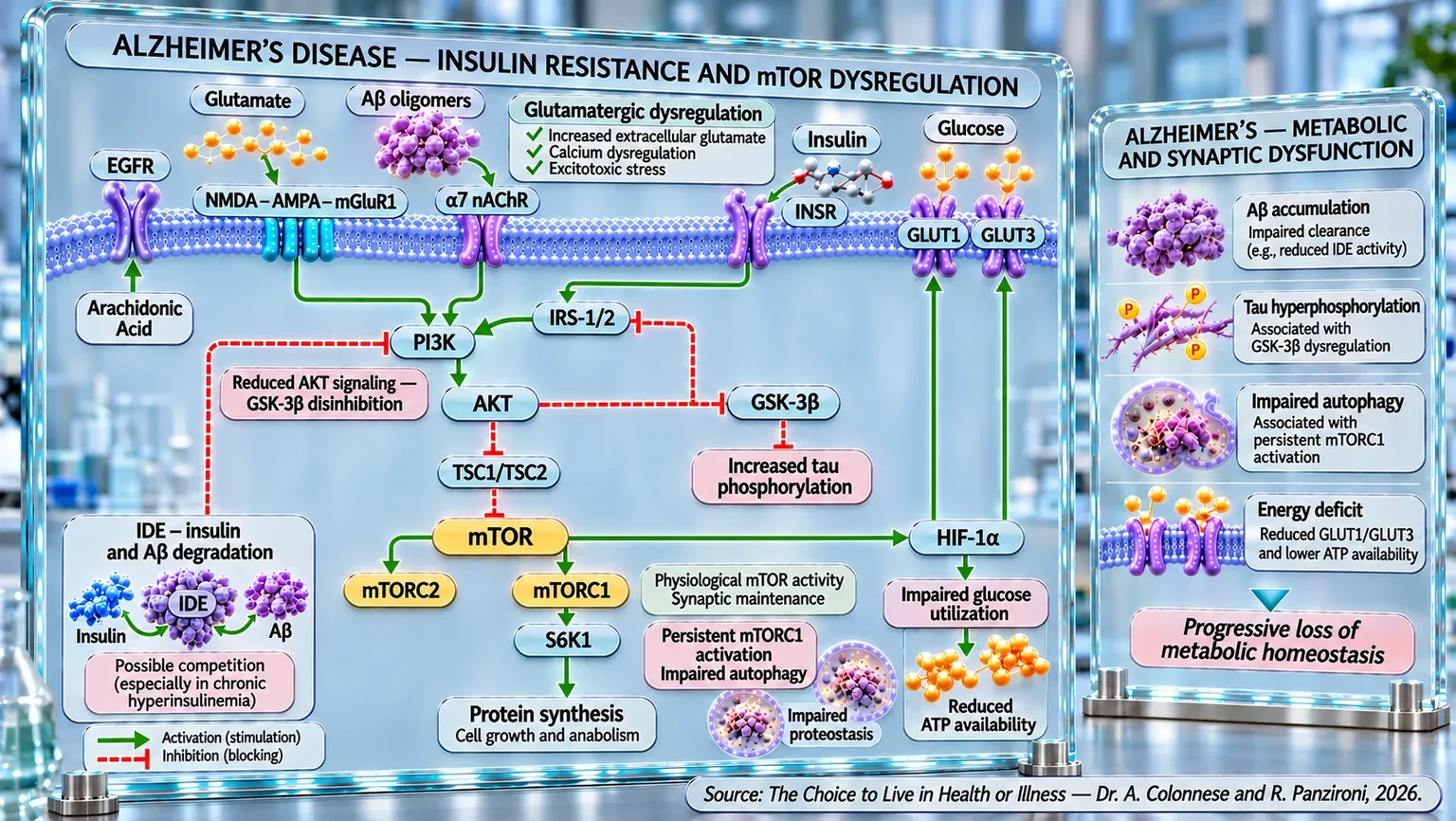
Molecular diagram with membrane receptors, activation and inhibition arrows, amyloid aggregates, tau protein, and autophagic vesicles.
Infographic of metabolic and synaptic dysfunction in Alzheimer's: insulin resistance, Aβ oligomers, and glutamatergic imbalance alter IRS-1/2 and PI3K/AKT/mTOR, promoting tau hyperphosphorylation, impaired autophagy, and reduced proteostasis; alterations in GLUT1/GLUT3 and HIF-1α lead to poor glucose utilization and ATP deficit.

ABSTRACT

Alzheimer's disease involves interconnected alterations in insulin signaling, energy metabolism, and neuronal function, in addition to processes associated with beta-amyloid and tau. In the book "La scelta di Vivere in Salute o in Malattia" (The Choice to Live in Health or Disease), Dr. A. Colonnese and R. Panzironi explore these relationships in depth, highlighting the potential contribution of cerebral insulin resistance to cognitive decline even in the absence of diabetes. This perspective integrates neuropathological markers with the conditions necessary for synaptic function.

A reduced response to insulin can alter the processing of the APP protein, compromise autophagy, and hinder the clearance of beta-amyloid oligomers. The IDE enzyme participates in the degradation of both insulin and beta-amyloid: its insufficient activity could reduce amyloid clearance, while hyperinsulinemia could accentuate the competition between substrates. The PI3K/AKT/mTOR pathway links these processes to the regulation of protein synthesis and neuronal survival.

Persistent activation of mTOR is associated with amyloid accumulation, tau alterations, and reduced autophagy, although its physiological activity remains necessary for synaptic plasticity. Amyloid oligomers can also interfere with glutamate release and reuptake, promoting excitotoxicity, calcium overload, and oxidative stress. In parallel, the feedback of S6K1 on IRS-1 and IRS-2 can attenuate insulin signaling; the resulting reduction in AKT limits the inhibition of GSK3β, potentially promoting tau hyperphosphorylation.

Energy deficit includes alterations in glycolysis, mitochondrial function, and ATP production, along with a possible reduction in GLUT1 and GLUT3 transporters. HIF-1α, also regulated through PI3K/AKT/mTOR, participates in the control of these transporters; their simultaneous decrease suggests further metabolic connections. Overall, the evidence outlines potentially self-reinforcing circuits between insulin resistance, pathological proteins, and excitotoxicity. Understanding their regulation could guide new research, without attributing exclusive causality to single molecular pathways or anticipating unproven clinical benefits. The distinction between associations and experimental mechanisms remains fundamental for interpreting these results.

Alzheimer's, the latest research

Alzheimer's disease is now the most frequent form of dementia and one of the greatest health challenges of the 21st century. The increase in life expectancy has led to a constant rise in the number of people affected by this condition, transforming it from a primarily clinical problem into a global social, economic, and care-related issue. It is currently estimated that over 55 million people worldwide live with some form of dementia, and that approximately 60-70% of these cases are attributable to Alzheimer's disease. Incidence increases significantly after age 65, approximately doubling every five years of age, while prevalence reaches its highest levels in those over 80. Women are affected more frequently than men, not only due to their greater longevity but also because of biological and hormonal factors that appear to influence susceptibility to the disease.

For many years, Alzheimer's was considered almost exclusively a disease characterized by the accumulation of two abnormal proteins: beta-amyloid, which deposits in the extracellular space forming characteristic senile plaques, and the tau protein, which undergoes pathological changes inside neurons, giving rise to neurofibrillary tangles. This interpretation dominated research for decades, orienting much of the studies and pharmacological development toward the removal of these protein aggregates. Today, however, the view of the disease has changed profoundly. The most recent evidence shows that Alzheimer's cannot be explained by a single biological mechanism, but represents the result of continuous interaction between multiple pathological processes that fuel each other, including metabolic alterations, neuroinflammation, mitochondrial dysfunction, impaired synaptic communication, vascular factors, and genetic predisposition (Zhang et al., 2025) [1]; (Barnwal et al., 2025) [2].

This new perspective profoundly changes the way the disease is interpreted. A comparison can help understand its significance. For a long time, it was thought that Alzheimer's was similar to a building collapsing due to the failure of two main pillars, represented by beta-amyloid and the tau protein. Today, it seems more accurate to imagine an entire city in which the electrical grid, water system, communications, transport, and maintenance services begin to fail simultaneously. Each individual failure makes the functioning of the others more difficult, eventually leading to a progressive collapse of the entire system. Similarly, in a brain affected by Alzheimer's, no pathological process acts in isolation; rather, each amplifies the effects of the others, accelerating neurodegeneration (Barnwal et al., 2025) [2]; (Yuan et al., 2025) [3].

One of the most interesting aspects to emerge in recent years concerns the role of cerebral energy metabolism. The brain accounts for only a small part of body weight, about 2%, but consumes approximately a quarter of the glucose available in the body to support the enormous activity of neurons. Every thought, every memory, every movement, and every emotion depends on a continuous production of energy. When this energy supply begins to decrease, nerve cells progressively lose the ability to communicate with each other and become increasingly vulnerable to degenerative processes (Yuan et al., 2025) [3].

The most recent research suggests that these metabolic alterations are not a simple consequence of the disease, but one of its central elements. In the brains of patients with Alzheimer's, reduced glucose utilization, progressive cerebral insulin resistance, mitochondrial function alterations, increased oxidative stress, and significant modifications in lipid metabolism are observed. All these phenomena compromise the energy availability of neurons, reduce synaptic efficiency, and promote the progression of neurodegeneration (Barnwal et al., 2025) [2]; (Yuan et al., 2025) [3].

The relationship between diabetes mellitus and Alzheimer's has also been deeply re-evaluated. For many years, diabetes was considered exclusively an epidemiological risk factor (an element associated with an increased risk of disease in a population). Today, however, numerous pieces of evidence suggest that glucose metabolism alterations may directly participate in the biological processes of neurodegeneration. A recent meta-analysis that included 37 studies and nearly twelve thousand participants highlighted how diabetes mellitus and altered glucose metabolism are associated primarily with an increase in tau protein biomarkers, while the association with beta-amyloid appears much less consistent. This result suggests that cognitive deterioration related to diabetes may follow pathological pathways at least partially different from the traditional amyloid cascade (Van Gils et al., 2024) [4].

Further confirmation comes from fluorodeoxyglucose positron emission tomography (FDG-PET) studies, a method capable of visualizing glucose consumption in different brain areas. The systematic review by Soltani and colleagues showed that diabetes, hyperglycemia, and insulin resistance are associated with a reduction in cerebral metabolism in the same regions most involved in the early stages of Alzheimer's disease, such as the parietotemporal cortex, the precuneus, the posterior cingulate cortex, and the frontal cortex. A particularly interesting finding is that this metabolic pattern was also observed in subjects who were still cognitively normal, suggesting that energy alterations may precede the appearance of clinical symptoms by many years (Soltani et al., 2025) [5].

Continuing along this line of research, it has become increasingly evident that Alzheimer's should not be interpreted as a disease limited to the accumulation of abnormal proteins, but as a progressive loss of the brain's ability to maintain its biological homeostasis. Each neuron can be compared to a small operations center that, to function correctly, requires energy, efficient communication, repair systems, and waste disposal mechanisms. When just one of these systems begins to deteriorate, the others initially manage to compensate for the deficit; over time, however, the ability to adapt decreases and the damage progressively extends to the entire brain network (Barnwal et al., 2025) [2]; (Zhang et al., 2025) [1].

Among the protagonists of this progressive loss of balance, the tau protein certainly remains. Under physiological conditions, it plays a fundamental role in maintaining the internal scaffolding of neurons stable. If we imagine the neuron as a long railway line, the microtubules represent the tracks along which nutrients, organelles, and molecules essential for cell survival are transported. Tau performs the task of the sleepers that keep these tracks perfectly aligned. When the protein is hyperphosphorylated, it progressively loses the ability to stabilize the microtubules, promoting their disintegration. Intracellular transport slows down, neuronal communication becomes inefficient, and, over time, the characteristic neurofibrillary tangles observed in the brains of patients with Alzheimer's form (Medeiros et al., 2011) [6].

Recent knowledge has, however, also changed the way tau toxicity is interpreted. For a long time, it was believed that neurofibrillary tangles were the main culprit for cognitive deterioration. Today, it is emerging that soluble and hyperphosphorylated forms of the protein could be even more harmful. These alterations, in fact, begin to compromise neuronal functioning long before the formation of large aggregates visible under a microscope. In other words, the tangles seem to represent the final result of a pathological process that began many years earlier, when tau function was already deeply compromised (Medeiros et al., 2011) [6].

This interpretation is also confirmed by molecular imaging studies. Using PET tracers specific for the tau protein, Bejanin and colleagues demonstrated that the distribution of pathological tau correlates closely with the deterioration of different cognitive functions. The impairment of episodic memory, language, executive functions, and visuospatial skills follows the anatomical localization of tau accumulation in the respective brain areas. A particularly relevant aspect that emerged from this study is that this relationship remains evident even after statistically correcting for the contribution of brain atrophy and beta-amyloid deposition. This suggests that tau exerts direct effects on neuronal function, regardless of the loss of brain tissue or the amount of amyloid plaques present (Bejanin et al., 2017) [7].

These observations have profoundly changed the way the two main neuropathological markers of Alzheimer's are interpreted. Beta-amyloid continues to represent a fundamental event in the early stages of the disease, but the progression of cognitive deterioration seems to depend to a greater extent on the spread of tau pathology. It is therefore not sufficient to know the amount of amyloid present in the brain to predict the severity of the disease; instead, it becomes essential to understand how tau progressively alters the organization of neuronal networks and compromises the ability of different brain regions to communicate with each other (Bejanin et al., 2017) [7]; (Medeiros et al., 2011) [6].

In parallel, the diagnosis of Alzheimer's is also undergoing a profound evolution. In the past, definitive confirmation of the disease was possible only through neuropathological examination of the brain after death. Today, thanks to the development of cerebrospinal fluid and plasma biomarkers and sophisticated neuroimaging techniques, it is possible to identify many of the biological alterations already in the early stages, when symptoms are still mild or even absent. This possibility opens up extremely important prospects, since any therapeutic strategy has a better chance of success when applied before neuronal loss becomes irreversible (Zhang et al., 2025) [1].

The treatment of the disease is also experiencing a phase of profound transformation. Recent immunotherapies directed against beta-amyloid have shown that it is possible to modify at least in part some of the fundamental pathological processes of Alzheimer's. However, the observed clinical benefits are still modest and are not sufficient to stop the progression of neurodegeneration. This result confirms a concept now shared by the scientific community: a disease supported by numerous biological mechanisms will hardly be controlled by intervening on a single molecular target. For this reason, research is moving toward integrated strategies that combine pharmacological interventions, lifestyle modifications, improvement of energy metabolism, inflammation control, and personalized medicine, adapting the treatment to the biological characteristics of each patient (Barnwal et al., 2025) [2]; (Zhang et al., 2025) [1].

In light of this knowledge, a new interpretation of Alzheimer's emerges. More than a simple disease characterized by the accumulation of plaques and tangles, it appears as the result of a progressive crisis of cerebral homeostasis, in which energy metabolism, insulin signaling, mitochondrial function, neuroinflammation, proteostasis (control of the quality and balance of proteins in the cell), and synaptic communication gradually lose the ability to cooperate. Understanding this network of interactions is not just a theoretical exercise, but constitutes the essential prerequisite for developing truly effective preventive and therapeutic strategies. Precisely for this reason, the following paragraphs will analyze in detail the mechanisms that link insulin metabolism to the pathophysiology of Alzheimer's, starting from cerebral insulin resistance, which is now considered one of the most promising elements for understanding the origin and progression of the disease.

Insulin resistance and Alzheimer's

In the previous paragraph, we saw how Alzheimer's disease can no longer be interpreted solely as a pathology characterized by the accumulation of beta-amyloid and tau protein. Among the processes that have attracted the most research attention in recent years is a progressive loss of the brain's ability to respond correctly to insulin. Speaking of cerebral insulin resistance means describing a condition in which insulin is present, but the cells of the central nervous system respond inadequately to its signal. It is a concept similar to that observed in peripheral tissues during type 2 diabetes, but with a fundamental difference: in the brain, insulin does not only play a role in energy metabolism; it participates in the regulation of synaptic plasticity, learning, memory, neuronal survival, cerebral perfusion, and numerous neuroendocrine processes. For this reason, a reduction in the insulin response can have consequences that go far beyond simple glucose management (Bedse et al., 2015) [8]; (Rhea et al., 2024) [17].

This observation has contributed to the spread of the expression “type 3 diabetes,” used by some researchers to describe Alzheimer's as a condition characterized by marked insulin resistance of the brain. The definition is evocative because it highlights the numerous pathophysiological similarities between type 2 diabetes and Alzheimer's: insulin resistance, neuroinflammation, oxidative stress, mitochondrial dysfunction, advanced glycation end-products, and alterations in energy metabolism appear in both conditions (Michailidis et al., 2022) [10]. However, the term should not be interpreted as the formal existence of a new category of diabetes. Evidence obtained directly from human brain tissue shows that cerebral insulin resistance has its own characteristics and can also appear in subjects who do not have systemic diabetes (Talbot et al., 2012) [13].

One of the decisive steps in understanding this phenomenon came precisely from the study of the human brain. By analyzing samples of the hippocampal formation, Talbot and colleagues demonstrated that in patients affected by Alzheimer's, even in the absence of diabetes, the response to insulin stimulation was significantly reduced. The impairment became progressively more evident when moving from cognitively normal subjects to those with mild cognitive impairment, and finally to patients with full-blown Alzheimer's. The same study highlighted a simultaneous resistance to the IGF-1 signal (a hormone that promotes the growth and development of cells and tissues), indicating that the deficit involved a broader system of metabolic and trophic regulation. A particularly important finding was the relationship with cognitive performance: the more altered the insulin signaling markers were, the worse the episodic memory and working memory (Talbot et al., 2012) [13].

Perhaps the most interesting aspect is that this condition does not necessarily appear as a late phenomenon, produced by a brain that has already severely degenerated. On the contrary, several studies suggest that it can appear very early. Mullins and colleagues observed alterations in specific neuronal insulin signaling mediators many years before the clinical onset of Alzheimer's. The modifications were also associated with particular patterns of brain atrophy, while traditional indicators of peripheral insulin resistance did not show the same anatomical correspondence. This data suggests that measuring only the body's insulin sensitivity does not necessarily allow us to understand what is happening in the brain (Mullins et al., 2017) [12].

It is therefore useful to imagine the brain and the periphery as two connected rooms in the same building. A problem with the electrical system can affect both, but not necessarily with the same intensity or at the same time. The presence of peripheral insulin resistance can increase the probability of brain alterations, but the two phenomena are not perfectly overlapping. This distinction is also confirmed by simultaneous analyses of blood and cerebrospinal fluid performed in the early stages of the disease. In subjects with mild cognitive impairment or early Alzheimer's, metabolic alterations were observed in both the periphery and the center, but with different profiles: in the cerebrospinal fluid, insulin was reduced, while in the blood, levels did not show the same decrease (de la Monte et al., 2019) [11].

These observations reinforce the idea that insulin dysregulation in Alzheimer's is not confined to a single organ but may be part of a broader metabolic alteration. At the same time, however, they show that the central nervous system maintains its own physiological autonomy. Reduced availability of insulin in the brain can compromise neuronal plasticity, the maintenance of myelin (the protective coating of nerves that facilitates the transmission of nerve signals), and cell survival even when peripheral concentrations of the hormone do not appear particularly altered (de la Monte et al., 2019) [11].

Experimental models also provide an important temporal element. In APP/PS1 transgenic mice, predisposed to the development of an Alzheimer's-like pathology, alterations in glucose tolerance, a tendency toward hyperinsulinemia, and a decrease in insulin sensitivity were already present at two months of age, before significant beta-amyloid deposition and well before cognitive impairment, which only became evident several months later (Macklin et al., 2017) [14]. Although this is an animal model and it is therefore not possible to automatically transfer this timeline to humans, the data suggests that metabolic dysregulation may precede some of the classic neuropathological manifestations rather than simply being a consequence of them.

Research has also shown that the relationship between insulin resistance and Alzheimer's appears to be bidirectional. Compromised insulin signaling creates favorable conditions for neurodegeneration, while some of the pathological processes of Alzheimer's can in turn worsen the response to insulin. In particular, beta-amyloid oligomers have been associated with the desensitization of insulin receptors and alterations in intracellular mediators of the insulin response, helping to establish a self-sustaining circuit (Bedse et al., 2015) [8], (Mullins et al., 2017) [12]. The molecular detail of this interaction will be addressed in the following paragraphs; what is important to emphasize at this stage is that insulin metabolism and Alzheimer's neuropathology seem to influence each other.

This view is consistent with the interpretation of Alzheimer's as a complex metabolic disease. Resistance to insulin and IGF-1 can contribute simultaneously to the reduction of neuronal energy efficiency, oxidative stress, neuroinflammation, and the progressive loss of cellular functions (de la Monte, 2012) [15].

Zheng's research also highlights that in Alzheimer's disease, cerebral insulin resistance can compromise the IRS-1/PI3K/AKT pathway, favoring cognitive decline. Alterations in the phosphorylation of IRS-1, including through effectors such as mTOR and S6K1, are associated with β-amyloid deposition and tau protein hyperphosphorylation. Antidiabetic drugs, such as metformin, thiazolidinediones, and GLP-1 analogs, could modulate these mechanisms, improving cerebral insulin signaling and representing potential therapeutic strategies to slow the progression of Alzheimer's (Zheng and Wang, 2021) [16].

A further clue in favor of the biological importance of this system comes from studies that have attempted to directly restore insulin availability in the central nervous system. In the experimental model of Alzheimer's induced by streptozotocin, prolonged intranasal insulin administration improved spatial memory, reduced tau protein hyperphosphorylation, attenuated microglial activation (the response of the brain's immune cells to a possible injury or stimulus), and promoted hippocampal neurogenesis (Guo et al., 2017) [9]. These results do not prove that intranasal insulin represents a definitive therapy for Alzheimer's, especially because they derive from an animal model that does not fully reproduce the human disease, but they provide important experimental evidence: modifying the cerebral insulin signal can simultaneously influence cognition, neuroinflammation, and neurodegenerative processes.

Overall, the available evidence therefore indicates that cerebral insulin resistance cannot be considered a simple side effect of diabetes nor an inevitable consequence of advanced neuronal degeneration. It can manifest in the absence of diabetes, appear in the early stages of cognitive impairment, be directly associated with the loss of mnemonic functions, and interact with the main biological alterations of Alzheimer's. The next question then becomes inevitable: how can a reduced response to insulin concretely favor the formation and accumulation of beta-amyloid? It is precisely this step that allows us to enter the heart of the metabolic circuit of Alzheimer's and which will constitute the starting point of the next paragraph.

Insulin resistance and beta-amyloid

In the previous paragraph, we saw how cerebral insulin resistance can appear early in Alzheimer's disease and compromise functions that go far beyond simple glucose utilization. At this point, it becomes necessary to understand how this alteration can concretely enter the pathophysiology of beta-amyloid. The relationship, in fact, does not seem to depend on a single mechanism. An inefficient response to insulin can simultaneously influence the production of beta-amyloid (Aβ), its tendency to aggregate, and the ability of nerve cells to eliminate it. In other words, insulin resistance seems to act on both sides of the scale: it can increase what is produced and, at the same time, make what should be removed less efficient.

To understand this step, we must start from the amyloid precursor protein, or APP. Beta-amyloid does not actually originate as an autonomous protein, but is derived through the enzymatic cleavage of this membrane protein. The way APP is modified and subsequently processed can therefore determine how much Aβ will be produced. Under physiological conditions, insulin seems to help contain this process: in cortical neurons, it reduces the phosphorylation of APP and can therefore exert a potentially protective effect against the formation of beta-amyloid (de la Monte, 2012) [18]; (Kim et al., 2019) [19].

The situation changes profoundly when the neuron becomes insulin-resistant. In experimental models, the induction of this condition leads to an increase in the basal phosphorylation of APP and renders insulin unable to exert its previous protective effect. The same phenomenon has been observed in vivo in animals with metabolic syndrome induced by a high-fat diet, in which the cerebral cortex showed greater phosphorylation of APP. These observations suggest an important step: insulin resistance does not merely deprive the neuron of a metabolic signal, but directly modifies the conditions under which APP is processed, creating an environment more favorable to the production of Aβ (de la Monte, 2012) [18]; (Kim et al., 2019) [19].

A second mechanism concerns what we could define as the cell's internal recycling system. Neurons possess structures called autophagosomes, involved in the collection and transfer of cellular material destined for degradation. Under normal conditions, this system contributes to maintaining intracellular order; the material is engulfed, transported, and subsequently eliminated. In insulin resistance, however, this flow can become inefficient. Autophagosomes accumulate and, instead of representing only temporary containers destined for disposal, they can transform into compartments in which APP and the enzymes necessary for its processing concentrate (Son et al., 2012) [22].

Son and collaborators observed this phenomenon both in cellular models and in animals characterized by insulin resistance. The alteration of insulin signaling was accompanied by the accumulation of autophagosomes and an increase in the activity of beta- and gamma-secretases, the enzymes that participate in the production of Aβ. These intracellular compartments were particularly rich in the proteins involved in APP processing, as if a system normally destined for cellular recycling became, under certain metabolic conditions, a place favorable to the production of beta-amyloid. When the accumulation of autophagosomes was reduced experimentally, the production of Aβ also decreased; similarly, improving insulin sensitivity attenuated both alterations (Son et al., 2012) [22].

Insulin resistance, however, also seems to influence what happens after the formation of the peptide. The newly produced beta-amyloid does not necessarily possess the same toxicity as its aggregated forms. Particular importance is attributed to soluble oligomers, small aggregates capable of interfering early with the functioning of synapses. Under physiological conditions, signaling through insulin and IGF-1 receptors participates in the regulation of their elimination. Zhao and collaborators showed that cells equipped with functioning receptors were more capable of reducing Aβ oligomers, favoring their conversion toward less aggregated forms and their subsequent removal. Conversely, when receptor function was compromised pharmacologically or through inactivating mutations, the oligomers tended to accumulate and aggregate further (Zhao et al., 2009) [20].

The biological significance is particularly interesting because it highlights that insulin resistance can modify not only the quantity of beta-amyloid present but also its aggregation state. It is as if a normal disposal service could no longer remove small accumulations of material: these remain in the environment, progressively meet each other, and form increasingly larger aggregates. In the neuron, the loss of proper insulin receptor function reduces the clearance of oligomers and, simultaneously, favors the appearance of more aggregated amyloid species (Zhao et al., 2009) [20].

To this process are added modifications of the neuronal membrane itself. Yamamoto and collaborators identified a possible link between insulin resistance and Aβ aggregation through the ganglioside GM1, a component of lipid microdomains present on the membrane of nerve cells. When insulin signaling was reduced experimentally, the concentration and aggregation of GM1 increased in specific areas of the presynaptic membrane. These modifications created conditions particularly favorable to the assembly of beta-amyloid on the neuronal surface. The same pattern was observed in the brains of diabetic and insulin-resistant mice, in which the reduction of insulin receptor function was accompanied by greater GM1 aggregation and an acceleration of Aβ assembly (Yamamoto et al., 2012) [21].

This finding adds a further piece to the puzzle: insulin resistance does not only modify the enzymes or intracellular pathways that regulate Aβ, but can physically change the membrane environment in which the peptide encounters other molecules and begins to aggregate. The surface of the neuron, therefore, should not be imagined as a passive wall. It is rather a dynamic environment, composed of microdomains that can become more or less favorable to the formation of protein aggregates. By altering this organization, the loss of insulin signaling can create a sort of "collection point" where beta-amyloid finds conditions more favorable to fibrillogenesis (the process of fibril formation through protein aggregation) (Yamamoto et al., 2012) [21].

The various pieces of evidence, therefore, begin to converge. On one hand, insulin resistance favors modifications of APP associated with greater amyloidogenic processing; on the other, it can lead to the accumulation of intracellular compartments rich in the enzymes necessary for Aβ production; simultaneously, it modifies the neuronal membrane, making it more favorable to the aggregation of the peptide, and reduces the efficiency of the systems responsible for its elimination. In models of insulin resistance, reductions in the insulin-degrading enzyme, IDE, have also been observed, accompanied by a further decrease in the degradation of beta-amyloid (Son et al., 2012) [22]. This particular mechanism will be explored separately in the next paragraph, because it represents one of the most important points of connection between insulin metabolism and peptide clearance.

The relationship is made even more complex by the fact that beta-amyloid does not seem to be simply the consequence of insulin resistance. Once accumulated, the oligomers can in fact further compromise insulin signaling, reducing the availability and functionality of receptors on the neuronal surface. This generates a pathological circuit in which lower insulin sensitivity favors the accumulation of Aβ and the peptide, in turn, makes the neuron even less sensitive to insulin (Zhao et al., 2009) [20]. It is precisely this mechanism of mutual reinforcement that explains why a small metabolic alteration, if maintained over time, could theoretically amplify progressively.

Reviews that have analyzed the relationship between type 2 diabetes and Alzheimer's as a whole confirm this interpretation, placing insulin dysfunction within a broader network that includes glucose metabolism, mitochondrial function, oxidative stress, inflammation, and altered APP metabolism (Rad et al., 2018) [23]. However, it is important to maintain a distinction between biological plausibility and clinical demonstration: a considerable part of the described mechanisms derives from cell cultures or animal models and does not yet allow us to establish how much each pathway contributes quantitatively to the formation of amyloid in the human brain.

Evidence on physical activity also provides an interesting element of support, but invites the same caution. A recent systematic review showed that in animal models, physical activity improves insulin sensitivity and, in parallel, reduces levels of Aβ and phosphorylated tau. In humans, however, the picture is still incomplete: higher levels of physical activity are generally associated with better insulin sensitivity, but the relationships with cerebral beta-amyloid load are less consistent. Above all, none of the analyzed studies has formally demonstrated that the improvement of insulin resistance represents the causal mediator through which exercise reduces Aβ (Slee et al., 2026) [24].

Overall, a particularly coherent pathophysiological model emerges. When the brain progressively loses the ability to respond to insulin, APP can be directed more toward processes favorable to the formation of Aβ; autophagosomes can accumulate and transform into environments favorable to its production; the neuronal membrane can become more predisposed to its aggregation; and the systems responsible for the removal of oligomers can lose efficiency. The beta-amyloid that accumulates can finally further damage insulin signaling, closing the circuit. Within this system, a particularly interesting role is played by the insulin-degrading enzyme, IDE, a molecule capable of interacting with both insulin and beta-amyloid. Understanding what happens to this enzyme when insulin levels and insulin resistance increase will therefore allow us to add the next piece to our reconstruction.

IDE and beta-amyloid

The role of the insulin-degrading enzyme, known as IDE, allows us to understand one of the most interesting links between insulin metabolism and beta-amyloid accumulation. IDE (Insulin Degrading Enzyme) is a metalloprotease (enzymes that cleave proteins thanks to the presence of a metal) capable of degrading numerous biologically active peptides, but two of its substrates are of particular importance in the pathophysiology of Alzheimer’s: insulin and beta-amyloid. In other words, the same enzymatic system that helps remove insulin also participates in the clearance of Aβ. This characteristic turns IDE into a sort of metabolic crossroads where type 2 diabetes and Alzheimer’s disease can meet. Its importance was demonstrated quite clearly by experimentally deleting the IDE gene in mice: the loss of the enzyme reduced cerebral degradation of beta-amyloid by more than 50% and, simultaneously, compromised hepatic insulin degradation to a similar extent. The result was the simultaneous appearance of cerebral Aβ accumulation, hyperinsulinemia, and glucose intolerance, showing how the impairment of a single enzyme can produce alterations attributable to both amyloid pathology and diabetic metabolism (Farris et al., 2003) [25].

This finding is particularly important because it demonstrates that IDE is not simply one of many proteases capable of degrading beta-amyloid. Even in the presence of alternative Aβ elimination systems, its absence was sufficient to cause a chronic increase in the brain. The same study also suggested a second potentially decisive mechanism: when insulin remains chronically elevated, it can compete with beta-amyloid for the available enzyme. In this situation, IDE must manage two substrates simultaneously, and the excess insulin can theoretically reduce the amount of degradative activity available for Aβ, progressively favoring its accumulation (Farris et al., 2003) [25].

We can imagine IDE as a disposal team that must eliminate two different types of material. As long as the load remains physiological, both operations can proceed regularly. However, if one of the two substrates increases persistently, as could happen during chronic hyperinsulinemia, an increasing portion of the degradative capacity is occupied by managing insulin. Beta-amyloid can therefore remain in the extracellular environment longer, increasing the probability of encountering other Aβ molecules and aggregating. This interpretation is also echoed in more recent reviews, which identify the competition between insulin and beta-amyloid as one of the central biological critical points in the relationship between diabetes and cognitive decline (Tian et al., 2023) [29].

IDE, however, is not regulated independently of insulin signaling. There is a more complex relationship in which insulin itself can help modulate the amount of the enzyme. In hippocampal neurons, insulin stimulation led to an increase of approximately 25% in IDE levels. When the intracellular cascade responsible for signal transmission was blocked, this increase disappeared. Furthermore, in the brains of patients with Alzheimer’s and in mouse models of the disease, an association has been observed between reduced insulin signaling, decreased IDE, and increased beta-amyloid monomer (Zhao et al., 2004) [26].

An apparently paradoxical mechanism thus emerges. A physiological insulin signal can contribute to the maintenance of IDE, indirectly favoring the degradation of Aβ. When insulin resistance appears, however, the cell’s ability to respond correctly to insulin decreases, and the regulation of the enzyme can also be compromised. At the same time, if circulating insulin remains chronically elevated, the hormone can compete with beta-amyloid for IDE. This means that it is not simply the absolute amount of insulin that determines a favorable or unfavorable effect, but the balance between hormone availability, receptor sensitivity, and the degradative capacity of the enzyme.

Evidence obtained directly from human neurons further strengthens this model. Using cortical neurons derived from induced pluripotent stem cells, Rowland and colleagues compared the role of IDE with that of neprilysin (an enzyme that helps eliminate certain peptides, including beta-amyloid in the brain) in the degradation of beta-amyloid. Selective inhibition of IDE significantly reduced Aβ degradation and, in a three-dimensional system, led to a marked increase in amyloid deposits. Inhibition of neprilysin, on the other hand, did not produce the same result. Even more interesting is the fact that these neurons came from subjects without Alzheimer’s and without mutations in the proteins responsible for the familial forms of the disease: simply by reducing IDE activity, it became possible to promote beta-amyloid deposition (Rowland et al., 2023) [30].

This result supports a fundamental concept, especially for sporadic Alzheimer’s: beta-amyloid accumulation does not necessarily have to stem from excessive production. It can also appear because the normal removal system becomes inefficient. It is the same difference that exists between a faucet left open too wide and a partially clogged drain: in both cases, the water level rises. In sporadic forms of Alzheimer’s, a reduction in Aβ clearance could represent a particularly relevant phenomenon, and IDE seems to be one of the main players in this process (Rowland et al., 2023) [30].

Interventions capable of modifying IDE expression also provide interesting experimental confirmation. In APP/PS1 transgenic mice, treatment with metformin reduced cerebral beta-amyloid content and improved cognitive performance, without causing substantial changes to the main systems responsible for Aβ production or transport. The most evident variation concerned the increase in IDE expression, accompanied by the activation of AMPK. The authors therefore proposed that the increased degradation of beta-amyloid represented one of the main mechanisms associated with the reduction in amyloid load observed in the experimental model (Lu et al., 2020) [27].

The regulation of IDE, however, appears much broader and involves numerous intracellular systems. Evidence collected by Tian and colleagues indicates that different metabolic and inflammatory pathways can modify the enzyme’s expression; despite the diversity of the mechanisms, in experimental models, the increase in IDE tends to be associated with a reduction in beta-amyloid deposition and an improvement in cognitive performance (Tian et al., 2023) [29]. This confirms how deeply IDE is embedded within a much larger metabolic network, the individual steps of which will be explored progressively in the following paragraphs.

Reviews dedicated to the relationship between insulin resistance and Alzheimer’s, in fact, place IDE among the mechanisms capable of amplifying the disease’s metabolic vicious cycle: reduced enzyme function decreases beta-amyloid clearance and can simultaneously alter insulin metabolism, while insulin resistance, oxidative stress, and neuroinflammation further contribute to the progression of neurodegeneration (Wei et al., 2021) [28].

IDE thus emerges as a true point of convergence between two worlds that have been studied separately for many years. On one hand, it regulates insulin and participates in metabolic homeostasis; on the other, it contributes to the removal of beta-amyloid and limits its deposition. When this balance is compromised, hyperinsulinemia, insulin resistance, and reduced Aβ degradation can begin to fuel each other. Understanding this crossroads now allows us to take a further step: observing what happens when the metabolic signaling that normally regulates growth, protein synthesis, energy availability, and autophagy remains chronically active. This is where one of the cell’s main metabolic regulators, mTOR, comes into play.

mTOR activation and Alzheimer's

After observing how chronic hyperinsulinemia and reduced activity of the Insulin Degrading Enzyme promote the accumulation of β-amyloid, the need arises to understand the intracellular mechanism capable of transforming these metabolic alterations into progressive neuronal damage. In recent years, research has focused on the mTOR (mammalian Target Of Rapamycin) complex, a protein that represents, as we have already seen in previous chapters, one of the main regulators of cellular homeostasis. Under physiological conditions, mTOR coordinates processes essential for neuronal survival, including protein synthesis, energy metabolism, cell growth, synaptic plasticity, memory, and autophagy. Its role can be compared to that of an orchestra conductor: it does not directly produce proteins, nor does it eliminate damaged ones, but it coordinates the work of numerous systems so that every process occurs at the right time and with the appropriate intensity. When this balance is maintained, the neuron preserves its functionality; when, instead, mTOR activity remains chronically elevated, the entire cellular regulatory network progressively loses its adaptive capacity, favoring the onset of neurodegeneration (Cai et al., 2015) [31]; (Perluigi et al., 2021) [35].

For many years, it was believed that mTOR was simply a protein involved in cell growth processes. More recent research has shown that this kinase occupies a central position in the pathophysiology of Alzheimer's, as it receives information from numerous metabolic pathways and, simultaneously, controls various processes responsible for the accumulation of pathological proteins. Persistent hyperactivation of mTOR, in fact, increases the production of β-amyloid through the modulation of amyloid precursor protein (APP) metabolism and the activity of β- and γ-secretases (enzymes that cleave specific proteins within or near the cell membrane), while simultaneously reducing its clearance by inhibiting autophagy. In parallel, it promotes the formation of hyperphosphorylated tau protein, contributing to the development of the neurofibrillary tangles that characterize the disease. In this way, a single signaling pathway is able to influence both of the main neuropathological alterations of Alzheimer's simultaneously (Cai et al., 2015) [31].

One of the most interesting aspects emerging from recent reviews concerns the fact that mTOR does not exert its effects exclusively on β-amyloid and tau, but coordinates numerous systems essential for the maintenance of neuronal homeostasis. These include mitochondrial function, energy production, response to oxidative stress, proteostasis, and autophagic mechanisms. When the activity of the pathway remains persistently elevated, autophagy is progressively suppressed, limiting the neuron's ability to eliminate aggregated proteins and damaged organelles. Simultaneously, the production of reactive oxygen species increases, mitochondrial dynamics are altered, and ATP production (the main immediate source of energy usable by the cell) decreases, establishing a vicious cycle in which energy dysfunction, protein accumulation, and oxidative stress feed each other (Perluigi et al., 2021) [35].

Important confirmation of mTOR's involvement comes directly from the human brain. By analyzing hippocampal samples obtained from patients affected by Alzheimer's at different stages of severity, Sun and colleagues observed a progressive increase in the activity of the mTORC1 complex, evidenced by the increase in phosphorylated mTOR, the S6K1 protein, and their respective activated forms. The intensity of mTORC1 activation was also closely correlated with the severity of cognitive impairment, indicating that the clinical progression of Alzheimer's proceeds in parallel with the increase in the activity of this signaling pathway (Sun et al., 2014) [32].

Experimental evidence further strengthens this hypothesis. In a murine model of postoperative cognitive dysfunction, characterized by numerous mechanisms common to Alzheimer's disease, activation of the mTOR pathway was associated with the accumulation of β-amyloid, hyperphosphorylation of the tau protein, and memory deterioration. Preventive treatment with rapamycin blocked the activation of the cascade, simultaneously reduced Aβ and pathological tau, and significantly improved the animals' cognitive performance. These results indicate that the hyperactivation of the mTOR pathway does not merely represent a marker of neurodegeneration, but directly contributes to the development of the alterations responsible for cognitive deficit (Shen et al., 2016) [33].

Further confirmation comes from the PDAPP transgenic mouse model (a mouse genetically modified to develop beta-amyloid accumulations), in which chronic administration of rapamycin prevented the onset of cognitive deficits, selectively reduced cerebral levels of Aβ, and increased autophagic activity without modifying the production of the APP precursor protein. This suggests that the reduction of the amyloid load does not necessarily depend on a lower production of β-amyloid, but rather on the restoration of the neuron's ability to eliminate it through autophagy. In other words, by inhibiting mTOR hyperactivity, the cellular system recovers one of the main physiological mechanisms dedicated to the removal of aggregated proteins (Spilman et al., 2010) [34].

The centrality of mTOR also emerges in other conditions characterized by early Alzheimer-type neuropathology. In Down syndrome, for example, hyperactivation of the PI3K/AKT/mTOR pathway appears many years before the clinical onset of dementia and is associated with the progressive deposition of β-amyloid, hyperphosphorylation of tau, impairment of autophagy, and cognitive decline. Oxidative stress, mitochondrial dysfunction, and alterations in proteostasis further amplify these phenomena, highlighting how the dysregulation of mTOR represents a convergence point for multiple pathological processes (Di Domenico et al., 2018) [36]; (Azimzadeh et al., 2024) [38].

However, the available evidence suggests that the role of mTOR is more complex than it may appear. This pathway should not, in fact, be considered simply "active" or "inhibited." As highlighted by Maiese, the biological function of mTOR depends on the intensity of the signal, the type of complex involved, the stage of the disease, and the cellular context. Indiscriminate inhibition could compromise essential processes such as memory, synaptic plasticity, and neuronal survival, while its persistent hyperactivation favors neurodegeneration. The therapeutic goal, therefore, is not to completely eliminate the activity of the pathway, but to restore the correct physiological balance between activation and deactivation (Maiese, 2014) [37]; (O'Neill, 2013) [39].

All these observations lead to an important conclusion: mTOR does not represent a simple participant in the pathophysiology of Alzheimer's, but a true integration node capable of connecting energy metabolism, autophagy, proteostasis, mitochondrial function, insulin signaling, and neuronal survival. Understanding how this pathway is progressively hyperactivated therefore constitutes the next step in reconstructing the sequence of molecular events that leads to neurodegeneration. For this very reason, it is necessary to analyze one of the most studied mechanisms of recent years: the ability of β-amyloid oligomers to directly activate the PI3K–AKT–mTOR cascade, transforming the accumulation of the amyloid protein into a powerful stimulus for intracellular metabolic alteration.

Oligomeric Aβ and activation of the PI3K–AKT–mTOR pathway

After seeing how mTOR hyperactivation constitutes one of the central nodes of Alzheimer's disease, the next step is to understand where this activation might originate. An important part of the answer seems to come from β-amyloid itself. For many years, this peptide was considered almost exclusively as pathological material destined to accumulate in the brain; however, evidence shows that Aβ is also present under physiological conditions and, at low concentrations, can participate in normal neuronal signal transduction. Indeed, Luo and colleagues demonstrated that extremely small amounts of Aβ were able to activate PI3K. Inhibition with wortmannin (an experimental substance that blocks PI3K) completely abolished this effect, confirming that the response was truly dependent on PI3K. This finding introduces a fundamental concept: β-amyloid is not necessarily toxic in itself, but it can become so when its concentration, conformation, and duration of exposure change (Luo et al., 1996) [40].

The transition from physiology to pathology seems to depend primarily on the formation of oligomers. Bhaskar and colleagues directly compared Aβ monomers, fibrils, and oligomers, observing that it was primarily the latter that caused significant neuronal alterations. Indeed, oligomers induced retraction of neuronal processes, reentry into the cell cycle, and increased phosphorylation of AKT and mTOR. The use of PI3K, AKT, or mTOR inhibitors blocked cell cycle events, demonstrating that the PI3K–AKT–mTOR cascade represented an essential step in the pathological response. Monomers and fibrils, under the same experimental conditions, showed much more limited effects (Bhaskar et al., 2009) [45].

AKT also responds to Aβ in a dynamic manner. Abbott and colleagues showed that acute exposure to Aβ leads to a rapid increase in AKT phosphorylation through the coordinated involvement of α7 nicotinic receptors and NMDA receptors (glutamate-activated receptors). This initial response could represent an adaptive attempt by the neuron. However, when exposure becomes chronic, the picture changes: in the TAS10 mouse model (genetically modified mice to develop some characteristics similar to those observed in human Alzheimer's), hippocampal AKT phosphorylation was reduced precisely in coincidence with the appearance of amyloid plaques and memory deficits. Aβ therefore seems to exert a biphasic effect, with an initial phase of cascade activation and a subsequent loss of its normal regulation (Abbott et al., 2008) [47].

The connection between Aβ and mTOR was further clarified in studies by Caccamo. In cells producing natural β-amyloid oligomers and in 3xTg-AD mice (a genetically modified animal model used to study the mechanisms of Alzheimer's), mTOR activity was significantly increased. When Aβ accumulation was prevented genetically or reduced via antibodies, mTOR signaling returned toward physiological levels. Conversely, the injection of naturally secreted Aβ into the hippocampus of healthy animals was sufficient to rapidly increase the activity of the pathway. These experiments provided one of the most direct demonstrations that Aβ is not simply associated with mTOR hyperactivation, but can be a cause of it (Caccamo et al., 2011) [44].

Previous work by Caccamo and colleagues had already shown the functional consequences of this relationship. In 3xTg-AD mice and in cell systems producing oligomers, Aβ accumulation was accompanied by an increase in mTOR signaling and a reduction in autophagy. When mTOR was inhibited with rapamycin, learning and memory performance improved, while both Aβ and pathological tau decreased. Crucially, when autophagy was experimentally prevented, rapamycin lost the ability to reduce β-amyloid. This demonstrates that mTOR hyperactivation can turn into an amplifier of the pathology: Aβ activates mTOR, mTOR suppresses autophagy, and reduced autophagy favors further Aβ accumulation (Caccamo et al., 2010) [42].

The review by Oddo integrates these results by showing how this circuit does not exclusively concern the clearance of pathological proteins. Soluble forms of Aβ can activate PI3K/AKT, increase mTOR, and simultaneously alter protein synthesis, synaptic plasticity, autophagy, and tau metabolism. However, the same data highlight the existence of an optimal physiological window: insufficient mTOR activity compromises memory consolidation, but excessive activation also produces cognitive deficits. In Alzheimer's disease, the problem therefore seems to be primarily the loss of the normal ability to turn the pathway on and off appropriately (Oddo, 2012) [41].

Lee and colleagues added a further level of detail, showing how Aβ oligomers can activate PI3K/AKT/mTORC1 through an increase in calcium influx and the production of reactive oxygen species. mTORC1 activation also increased the synthesis of HIF-1α (a protein that helps cells adapt to oxygen deficiency) and simultaneously suppressed autophagy. This combination favored the expression of cell cycle proteins and the activation of CDK2 (an enzyme that helps regulate when a cell grows and divides), which in turn promoted pathological tau phosphorylation, microtubule destabilization, and neuronal death (Lee et al., 2017) [43].

Hyperactivation of the cascade also produces very early functional consequences. Wang and colleagues demonstrated that Aβ oligomers suppress hippocampal gamma oscillations (rhythmic electrical activity of the hippocampus) through the mTOR/S6K1 axis. Rapamycin prevented this alteration, while pharmacological activation of mTOR via leucine reproduced it. Selective inhibition of S6K1 also restored neuronal synchronization in both hippocampal preparations and APP/PS1 mice, indicating that the Aβ–mTOR–S6K1 cascade can compromise communication between brain networks even before extensive neuronal loss occurs (Wang et al., 2023) [46].

Overall, these findings describe a coherent sequence: physiological Aβ can participate in normal PI3K signaling, but when the peptide accumulates in the form of oligomers, the same cascade is exploited in a pathological way. PI3K activates AKT, AKT contributes to removing the brake exerted on mTORC1, which remains chronically hyperactive. From this derive reduced autophagy, alterations in protein synthesis, activation of S6K1, aberrant reentry into the cell cycle, tau modifications, and impairment of neuronal networks. However, another fundamental step remains to be clarified: how β-amyloid interacts with the glutamatergic system and NMDA receptors, transforming the alteration of cellular signaling into excitotoxicity. This is the starting point of the next paragraph.

Beta-amyloid and glutamate

If in the previous chapter we saw how β-amyloid oligomers are able to activate the PI3K–AKT–mTOR cascade, it remains to be understood what the mechanism is that actually sets this cellular response in motion. A fundamental part of the explanation lies in glutamate, the main excitatory neurotransmitter of the central nervous system. Every thought, every memory, and every movement depends on the balance with which this molecule is released into the synaptic space (the space between two neurons through which the chemical signal passes) and subsequently removed. One could imagine glutamate as the accelerator of a car: slight pressure allows for smooth movement, while continuous and uncontrolled acceleration inevitably leads to losing control of the vehicle. Something very similar happens in the brain. Physiological glutamatergic stimulation is essential for learning and memory; persistent excess, however, becomes toxic to neurons.

The most recent research shows that β-amyloid alters this balance from the very early stages of the disease. Contrary to what was hypothesized in the past, the first effect of Aβ oligomers does not consist of an immediate reduction in glutamatergic transmission, but in its marked hyperactivation. Using an electrochemical biosensor capable of measuring glutamate release from individual neuronal varicosities (small swellings on neurons from which neurotransmitters can be released), Yang and colleagues observed that after just thirty minutes of exposure to Aβ oligomers, the vesicular release of the neurotransmitter increases significantly. Only later, after several hours of exposure, do the glutamate reserves begin to progressively deplete, eventually leading to a marked reduction in synaptic transmission (Yang et al., 2019) [48].

This observation was also confirmed by Brito-Moreira and colleagues, who demonstrated how soluble β-amyloid oligomers simultaneously increase the extracellular concentration of glutamate and that of D-serine, a co-agonist essential for the full activation of NMDA receptors. The increase in glutamate depends on the electrical activity of neurons, the influx of calcium, and the activation of the NMDA receptors themselves, creating an amplification circuit in which the activation of neurons promotes further release of the neurotransmitter. The authors also observed an increase in the exocytosis of presynaptic vesicles (the process by which a neuron releases neurotransmitters into the space between two nerve cells) and greater spontaneous postsynaptic activity, indicating that the neuronal network quickly enters a state of hyperexcitability (Brito-Moreira et al., 2011) [49].

The extracellular accumulation of glutamate, however, does not depend solely on an increase in release. A second, equally important mechanism consists of the reduction of its reuptake. Li and colleagues demonstrated that soluble Aβ oligomers compromise the ability of nerve endings to remove glutamate from the extracellular space. When reuptake is reduced, the neurotransmitter remains around the synapses for longer, favoring persistent activation of glutamatergic receptors. This phenomenon facilitates long-term depression (LTD) in the hippocampus, a physiological process involved in the remodeling of neuronal connections that, if excessively activated, contributes to the loss of synapses (Li et al., 2009) [50].

At this point, another fundamental protagonist of brain physiology comes into play: the astrocyte. For a long time, these cells were considered simple support elements for neurons. Today, however, we know that they represent the main "cleaners" of synapses, because they rapidly remove excess glutamate through GLT-1 and GLAST transporters. Matos and colleagues demonstrated that the β-amyloid peptide profoundly compromises this function. Exposure to Aβ actually reduces both the activity and the expression of the two main transporters; the final result is a drastic reduction in the ability of astrocytes to eliminate extracellular glutamate, creating an environment favorable to neuronal excitotoxicity (Matos et al., 2008) [51].

But astrocytes do not just lose the ability to eliminate glutamate: they themselves become a new source of the neurotransmitter. Talantova and colleagues identified a completely new pathogenetic cascade in which Aβ oligomers activate the α7 nicotinic receptors present on astrocytes, inducing a massive release of glutamate into the extracellular space. Within a few minutes, a tonic glutamatergic current, an increase in nitric oxide production, hyperphosphorylation of the tau protein, and a progressive loss of dendritic spines (small structures on neurons that receive signals from other neurons) appear. The authors also demonstrated that this process can be countered pharmacologically: while memantine (a drug that modulates the action of glutamate in the brain) provides only partial protection, Nitromemantine (an experimental compound derived from memantine, studied to protect neurons from excessive glutamate activity) almost completely prevents the synaptic loss induced by β-amyloid, confirming the central role of NMDA receptors in neurodegeneration (Talantova et al., 2013) [52].

Further confirmation comes from the study by Sanz-Blasco and colleagues, which shows how oligomeric β-amyloid (Aβ) stimulates the release of glutamate in astrocytes through a calcium-dependent vesicular mechanism. Levetiracetam (LEV), an antiepileptic drug already associated with cognitive improvements in Alzheimer's, reduces this Aβ-induced glutamate release. The effect is confirmed by sensitivity to tetanus neurotoxin, which blocks vesicular release. These results suggest that the drug levetiracetam could limit neuronal hyperexcitability in Alzheimer's by modulating the pathological communication between astrocytes and neurons (Sanz-Blasco et al., 2016) [53].

Taken together, these findings describe an extremely coherent pathogenetic sequence. β-amyloid oligomers initially cause excessive glutamate release, increase the loading of synaptic vesicles, progressively compromise the reuptake of the neurotransmitter by both neurons and astrocytes, and finally transform the astrocytes themselves into an additional source of glutamate. The result is a persistent accumulation of the neurotransmitter in the extracellular space, capable of maintaining chronic stimulation of NMDA receptors and creating the ideal conditions for the activation of the main intracellular pathways involved in the progression of Alzheimer's. The link between excess glutamate and the activation of the PI3K–AKT–mTOR cascade will be the subject of the next paragraph.

Glutamate and PI3K-AKT-mTOR activation

In the previous paragraph, we saw how β-amyloid can profoundly alter glutamate homeostasis, increasing its release, impairing its reuptake, and promoting its accumulation in the extracellular space. To understand why this alteration is so important in the progression of Alzheimer's disease, it is necessary to follow the glutamate signal beyond the neuronal membrane. Glutamate, in fact, is not just the brain's main excitatory neurotransmitter: through different receptors, it is capable of modifying the activity of numerous intracellular pathways, including PI3K, AKT, and mTOR. Under physiological conditions, this dialogue is essential to allow synapses to adapt, strengthen, or weaken in response to stimuli. However, when glutamatergic stimulation becomes persistent and uncontrolled, the same molecular network can lose its regulation and contribute to neuronal damage.

One of the clearest demonstrations of the link between glutamate and PI3K–AKT–mTOR comes from the study of metabotropic glutamate receptors (mGluR). In the CA1 area of the hippocampus (the area of the hippocampus involved primarily in the formation and consolidation of memories), the activation of mGluR1 and mGluR5 receptors rapidly determines an increase in the phosphorylation of AKT and mTOR. Researchers demonstrated that by blocking PI3K with two different inhibitors, the activation of both AKT and mTOR was prevented, identifying a precise sequence in which PI3K occupies a position upstream of the two kinases. The cascade was also active in synaptic compartments, and its inhibition prevented long-term depression mediated by mGluR receptors, showing how PI3K–AKT–mTOR represents the link through which glutamate can transform a receptor stimulus into new modifications of protein synthesis and synaptic plasticity (Hou and Klann, 2004) [54].

A second access route to the cascade is represented by AMPA receptors. In a study on sarcosine (a natural substance involved in metabolism and the regulation of certain nerve signals), a rapid increase in the phosphorylation of AKT and mTOR was observed, associated with greater insertion of AMPA receptors into the neuronal membrane. Blocking AMPAR with NBQX (a compound used in neuroscience research that blocks glutamate AMPA receptors) prevented the activation of AKT and mTOR, while rapamycin blocked mTOR without preventing the modifications of the AMPA receptor. This hierarchical relationship therefore placed AMPAR upstream of the intracellular cascade, experimentally showing that a modification of glutamatergic transmission can propagate to mTOR (Chen et al., 2015) [55].

The same principle was confirmed with echinacoside (a natural plant-derived substance studied for its possible protective effects on cells). Stimulation of transmission mediated by AMPA receptors increased the phosphorylation of AKT and mTOR in the hippocampus; when AMPAR was blocked, the activation of the entire cascade disappeared. Although the study originated in an antidepressant context and not in Alzheimer's, it provides independent confirmation that glutamatergic activation through AMPAR can reach AKT and mTOR and modify neuronal plasticity (Chuang et al., 2022) [56].

Particularly recent confirmation comes from experiments with danshensu (a natural substance from Salvia miltiorrhiza studied for its protective and antioxidant properties). In this case, too, the stimulation of AMPAR represented the initial event, followed by the activation of AKT and subsequently of mTOR. Pharmacological blocking of AMPA receptors, in fact, prevented the increase in the phosphorylation of all these proteins, while the inhibition of AKT interrupted the transduction toward mTOR. Once again, a direct communication emerges between the glutamatergic system and the intracellular machinery responsible for synaptic plasticity (Chuang et al., 2024) [57].

Vanillic acid (a natural plant substance with antioxidant properties) has allowed for further isolation of this sequence. A study, in fact, showed that vanillic acid produces antidepressant effects in mice, reducing behaviors associated with depression without modifying locomotor activity. In the prefrontal cortex, the compound increases AMPAR receptor activity and activates the AKT-mTOR pathway, without involving ERK (a protein that transmits cellular signals by regulating cell growth and activity). Blocking AMPAR, AKT, or mTOR, in fact, attenuates the antidepressant effect. The results therefore indicate that vanillic acid could exert its action through the AMPAR-AKT-mTOR pathway, suggesting a potential neurobiological mechanism relevant to depression (Chuang et al., 2020) [58].

A 2020 review allows these results to be placed in a broader framework. When the extracellular concentration of glutamate exceeds the capacity of physiological control systems, excessive receptor activation causes an increase in intracellular calcium, the production of reactive oxygen species, a reduction in glutathione (an antioxidant substance that protects cells from oxidative damage), and the accumulation of hydrogen peroxide. In parallel, the PI3K/AKT/mTOR pathway and autophagy pathways are involved (Afshari et al., 2020) [59].

The complexity of this response is also confirmed by experiments conducted with exosomes derived from mesenchymal stem cells (cells capable of generating certain types of tissue and contributing to their repair). In HT22 neuronal cells (laboratory-cultured cells used as a model), glutamate caused a marked increase in apoptosis; exosomes, on the other hand, reduced the damage and increased cell survival. When PI3K/AKT or mTOR were pharmacologically inhibited, this protection decreased significantly. The study therefore shows that the PI3K–AKT–mTOR pathway does not exclusively represent a damage mechanism: in the presence of an excitotoxic stimulus, it can also be recruited as a survival system. The significance of the activation therefore depends on the context and the duration of the signal (Rong et al., 2024) [60].

It is precisely this apparent contradiction that becomes decisive for understanding Alzheimer's. Some researchers have shown that, after excitotoxic exposure to glutamate, neurons rapidly develop a form of insulin resistance. The increase in intracellular Ca2+ and mitochondrial depolarization were, in fact, accompanied by a 48% reduction in insulin receptor activation, a 72% reduction in AKT, a 44% reduction in mTOR, and a 38% reduction in the inhibitory phosphorylation of GSK3β (an enzyme that regulates various cellular processes) in response to insulin (Pomytkin et al., 2019) [61].

A picture more complex than a simple "activation" of mTOR thus emerges. Physiological glutamate uses PI3K–AKT–mTOR to regulate plasticity and protein synthesis; glutamatergic excess can abnormally activate these same networks, alter calcium and mitochondria, and, simultaneously, make the neuron less capable of responding to insulin. In Alzheimer's, where β-amyloid promotes the very extracellular accumulation of glutamate, these mechanisms can therefore converge toward a progressive loss of normal neuronal metabolic regulation. To understand how this alteration can transform into a circuit capable of self-sustenance, we must now follow mTOR toward one of its main effectors: S6K1, and observe how the latter can interfere with IRS-1, directly linking mTOR activity to the development of insulin resistance.

mTOR, S6K1, IRS-1 and insulin resistance

In the previous paragraph, we observed how glutamate, when it shifts from a normal neuronal communication tool to an excitotoxic signal, can profoundly alter the regulation of the PI3K–AKT–mTOR pathway and, simultaneously, compromise the cell's ability to respond to insulin. However, a fundamental question remains: how can a pathway that belongs to the same insulin cascade contribute, when it remains excessively active, to the onset of insulin resistance? The answer lies in one of the main effectors of mTORC1, the ribosomal protein S6 kinase 1, or S6K1. Under physiological conditions, this system functions as a sophisticated self-regulation mechanism. When nutrients and anabolic signals are abundant, mTORC1 activates S6K1, promoting growth and protein synthesis; at the same time, however, S6K1 can exert negative feedback on insulin signaling. It is like a thermostat: once a certain temperature is reached, the system automatically reduces the signal that activated it. The problem emerges when this brake, instead of being temporary, remains constantly engaged.

A 2006 study described S6K1 as an important meeting point between nutrient availability and insulin sensitivity. mTORC1 and S6K1 can therefore continue to receive a strong nutritional signal even while the ability of insulin to transmit its message is progressively decreasing. In human studies, the infusion of amino acids indeed led to an increase in S6K1 activity associated with a reduction in insulin sensitivity of approximately 25%, the phosphorylation of IRS-1, a reduction in PI3K activation, and lower glucose uptake in skeletal muscle (Um et al., 2006) [62].

To understand this mechanism, we must focus on IRS-1. This protein can be imagined as a molecular bridge: after the activation of the insulin receptor, it allows the signal to reach PI3K and, subsequently, AKT. Its function, however, also depends on how it is phosphorylated. Even more significantly, in humans, the infusion of amino acids simultaneously produced activation of S6K1, increased phosphorylation of IRS-1 with a loss of IRS-1 functionality, and the onset of insulin resistance in skeletal muscle (Tremblay et al., 2007) [63].

It is thus understood that the hyperactivity of mTOR/S6K1 is not limited to temporarily putting a brake on the insulin signal: if it persists, it can progressively deplete the cell of the very intermediates necessary to transmit it. Some researchers have provided a particularly clear demonstration of this phenomenon. The loss of TSC1 or TSC2 produced constitutive activation of the mTOR/S6K1 cascade accompanied by a marked reduction of IRS-1 and, to a lesser extent, IRS-2. Although AKT was still present in normal quantities, insulin could no longer activate it. The problem, therefore, did not consist of a lack of AKT, but in the interruption of communication upstream: IRS-1/2 and their association with the insulin receptor and PI3K decreased (Shah et al., 2004) [64].

The same study adds an essential element. The hyperactivation of the mTOR/S6K1 pathway accelerated the degradation of IRS-1 and IRS-2 and simultaneously reduced their mRNA; both effects were reversible through the inhibition of mTOR with rapamycin. Furthermore, the overexpression of IRS-1 or a constitutively active PI3K was able to restore AKT activation. This result localizes the defect with remarkable precision: insulin resistance emerges because the signal is interrupted at the level of the IRS, before reaching PI3K and AKT adequately. The mTOR/S6K pathway, when constitutively active, thus ends up weakening the very insulin cascade that it normally contributes to regulating (Shah et al., 2004) [64].

An independent model further reinforces this principle: a study conducted in 2020 focused on murine podocytes (mouse kidney cells used in scientific research) exposed to palmitate (a form of a common saturated fatty acid used by cells in fat metabolism), therefore an experimental system related to diabetic nephropathy and not to Alzheimer's disease; the study provides a mechanistic confirmation of the relationship between mTORC1/S6K1 activation, IRS-1 modification, and loss of insulin response. Palmitate, in fact, activated mTORC1/S6K1 and increased the phosphorylation of IRS-1, accompanied by a reduction in AKT phosphorylation in response to insulin. The inhibition of mTORC1 by rapamycin reduced the phosphorylation of IRS-1 and restored that of AKT (Denhez et al., 2020) [65].

At this point, the connection with what was described in the previous paragraphs becomes particularly interesting. We have seen that β-amyloid can promote glutamate dysregulation and that glutamatergic transmission is capable of reaching PI3K–AKT–mTOR; we have also observed that excitotoxicity can compromise the neuronal response to insulin. The studies analyzed in this paragraph now add a further piece: once mTORC1/S6K1 remains excessively active, there is a molecular mechanism through which this same pathway can exert negative feedback on IRS-1 and IRS-2, hindering the propagation of the insulin signal.

The picture that emerges is therefore that of a self-regulation circuit that can turn into a vicious cycle. Excessive mTORC1 activity keeps S6K1 high; S6K1 modifies and weakens IRS-1, while chronic activation of the mTOR/S6K1 axis can also promote the loss of IRS-1 and IRS-2; by decreasing IRS, the insulin receptor becomes less and less effective at transferring its signal to PI3K and AKT. The cell can thus find itself in the apparently paradoxical condition of possessing a strongly active mTOR/S6K1 pathway and, at the same time, responding less and less to insulin. It is precisely from this separation between mTOR activity and insulin signal functionality that we can proceed to the next step of our molecular story: understanding what happens to AKT and GSK3β, and why their alteration becomes particularly important in Alzheimer's disease.

The AKT-GSK3β pathway and Alzheimer's

In the previous paragraph, we saw how the persistent overactivation of the mTORC1–S6K1 axis can transform a normal regulatory system into a mechanism capable of progressively weakening the cellular response to insulin. When IRS-1 and IRS-2 lose efficiency, the signal that should reach PI3K and subsequently AKT is attenuated. In the brain, this alteration takes on particular significance because AKT does not just represent a step in insulin signaling: it also acts as one of the main control systems for glycogen synthase kinase-3β, or GSK-3β, a protein that occupies a central position in the pathophysiology of Alzheimer's disease. To understand this relationship, it can be helpful to imagine AKT as a molecular brake. When the pathway is functioning correctly, AKT phosphorylates GSK-3β, reducing its activity. When the PI3K–AKT signal weakens, however, this brake is progressively released, and GSK-3β can become excessively active (Limantoro et al., 2023) [66].

This step helps us understand why cerebral insulin resistance can have consequences that go far beyond a simple difficulty in glucose utilization. In an experimental model of sporadic Alzheimer's obtained through intracerebroventricular administration of streptozotocin (a substance used in research to damage insulin-producing pancreatic cells), some researchers observed an impairment of cerebral insulin signaling accompanied by a reduction in the activity of PI3K and AKT-dependent pathways. Simultaneously, the inhibitory phosphorylation of GSK-3β decreased, thus leaving the kinase more active (Deng et al., 2009) [67]. This is an important result because it builds a very precise molecular bridge: if the insulin signal loses strength upstream, AKT exerts its control less effectively, and GSK-3β can increase its activity.

The problem becomes evident when considering one of the main targets of GSK-3β: the tau protein. Under physiological conditions, tau contributes to the stabilization of microtubules, structures that can be compared to tracks along which essential molecules are transported from one region of the neuron to another. Tau phosphorylation is a normal regulatory process, but when it becomes excessive, the protein progressively loses its ability to interact correctly with microtubules. GSK-3β is one of the main kinases involved in this process, and its hyperactivity promotes the accumulation of hyperphosphorylated tau and the formation of neurofibrillary tangles (Zhang et al., 2018) [68].

In the 2009 study mentioned earlier, this sequence is observable with particular clarity. The reduction in PI3K–AKT activity and the consequent greater activity of GSK-3β were associated with an increase in tau phosphorylation. This was not just a biochemical modification: the tau isolated from the animals' brains significantly lost the ability to bind to microtubules (Deng et al., 2009) [67]. The altered metabolic signal therefore translates into a concrete modification of neuronal architecture: less AKT activity means less inhibition of GSK-3β; a more active GSK-3β promotes the pathological phosphorylation of tau; and the modified tau loses part of its stabilizing function.

The relevance of GSK-3β in Alzheimer's, however, does not end with tau. Some researchers describe this kinase as a node capable of simultaneously influencing different aspects of the disease. Its activity increases with age and is further elevated in the central nervous system in Alzheimer's; in addition to tau phosphorylation, GSK-3β participates in the regulation of β-amyloid production, interferes with synaptic plasticity and long-term potentiation, influences neurogenesis, and contributes to the neuroinflammatory response (Lauretti et al., 2020) [69]. In other words, GSK-3β seems to be in a position where its dysregulation can propagate simultaneously toward many of the biological characteristics of Alzheimer's.

The link with β-amyloid also deserves attention. The evidence collected indicates that GSK-3β can therefore influence the amyloidogenic processing of APP (the transformation of APP that leads to the production of beta-amyloid) (Lauretti et al., 2020) [69]. An interesting experimental confirmation comes from a 2007 study: in transgenic mice expressing human APP, the reduction of GSK-3β activity—obtained pharmacologically with lithium or genetically through a dominant-negative form of the kinase—resulted in a decrease in tau phosphorylation, but also in β-amyloid production and plaque burden. These molecular changes were associated with better preservation of the dendritic structure (the neuron's network of branches) in the hippocampus and frontal cortex, as well as an improvement in cognitive performance (Rockenstein et al., 2007) [70].

The study also suggests that GSK-3β may intervene further upstream in amyloid formation by modifying the phosphorylation and maturation of APP. Reducing the kinase's activity, in fact, decreased APP phosphorylation and the availability of the mature protein in intracellular compartments favorable to its amyloidogenic processing. The authors thus hypothesize that the inhibition of GSK-3β could simultaneously limit two characteristic processes of Alzheimer's: tau pathology and β-amyloid pathology (Rockenstein et al., 2007) [70].

The relationship between AKT, GSK-3β, and neuropathology finds further support in a more recent experimental model. In hippocampal neurons and rats exposed to staurosporine (an experimental substance that blocks various kinases and can promote programmed cell death), researchers observed a reduction in AKT phosphorylation and, in parallel, in the inhibitory phosphorylation of GSK-3β. This alteration was accompanied by tau hyperphosphorylation, increased Aβ production, loss of dendritic spines, reduction of synaptic proteins, and deterioration of learning and memory. Treatment with Gypenoside IX (a natural compound belonging to the saponins) restored AKT and GSK-3β phosphorylation and simultaneously attenuated many of these alterations (Lei et al., 2023) [71].

More recent research further expands the picture. GSK3 appears to be involved not only in tau and amyloid but also in oxidative stress, neuroinflammation, mitochondrial function, and autophagy. In the Alzheimer's-affected brain, an increase in GSK3 expression and activity has been described, along with a reduction in its inactive phosphorylated forms (Zhao et al., 2024) [72]. Other scholars also place GSK-3β among the main shared mediators of neurodegenerative diseases, highlighting its involvement in protein aggregation, mitochondrial function, neuroinflammation, and major intracellular survival pathways (Yu et al., 2023) [73].

All this makes it understandable why GSK-3β has been proposed as a therapeutic target. Lithium and other inhibitors have produced interesting results in experimental models, reducing tau phosphorylation, Aβ-associated toxicity, or other neurodegenerative manifestations in various contexts. However, the transition from the laboratory to the clinic has proven much more difficult: the results obtained in humans are still limited, and the indiscriminate inhibition of a kinase involved in numerous physiological processes poses significant problems of selectivity and safety (Yu et al., 2023) [73].

Thus, a fundamental step in our reconstruction emerges. The reduction of insulin signaling can decrease the activity of the PI3K–AKT pathway; lower AKT activity can reduce the inhibitory phosphorylation of GSK-3β; and a less controlled GSK-3β can contribute to tau hyperphosphorylation and, through other mechanisms, to amyloid pathology, synaptic impairment, and neurodegeneration. GSK-3β therefore becomes one of the points where metabolic dysfunction and neurodegenerative dysfunction seem to meet. It is precisely this convergence that makes the hypothesis of a link between diabetes, insulin resistance, and Alzheimer's particularly interesting, even without allowing us to simplistically identify Alzheimer's as "type 3 diabetes" (Zhang et al., 2018) [68].

But this story is not just about tau control. When cerebral insulin response deteriorates, the way the neuron manages to procure and use energy also changes. In the 2009 Deng model, in fact, the impairment of PI3K–AKT and the overactivation of GSK-3β are also associated with a decrease in GLUT1 and GLUT3 (transporters that allow cells, particularly those of the nervous system, to receive glucose) (Deng et al., 2009) [67]. At this point, therefore, the path inevitably takes us beyond molecular signaling alone: toward that energy dysregulation which represents the next step in understanding how a neuron can find itself progressively deprived of the energy necessary to maintain its functions.

Energy dysregulation

The human brain is one of the most metabolically demanding organs in the body. Although it makes up a relatively small fraction of body mass, it must continuously support high-energy processes: maintaining ion gradients across membranes, generating and propagating nerve impulses, enabling communication between neurons, and preserving synaptic efficiency. To do this, it relies heavily on glucose, which must reach the nervous tissue, enter the cells, and be processed through an orderly sequence of metabolic reactions to produce ATP. In Alzheimer’s disease, however, this delicate energy system appears to progressively lose efficiency. Cerebral glucose hypometabolism is, in fact, one of the most consistent alterations associated with the disease and, crucially, it can be detected long before cognitive impairment becomes clinically evident (Chen et al., 2013) [74].

This finding profoundly changes how we interpret neurodegeneration. If glucose metabolism were to decrease only after the loss of neurons, we could simply consider it a consequence of the disease: fewer functioning cells would inevitably require less energy. However, observations obtained via FDG-PET (a scan that shows the metabolic activity of tissues through their glucose usage) tell a more complex story. Hypometabolism has been documented in MCI, in presymptomatic carriers of mutations associated with familial Alzheimer’s, in carriers of the APOE ε4 allele (a genetic variant associated with an increased risk of Alzheimer’s), in people with subjective memory complaints, and even in cognitively normal individuals who later progressed to MCI or Alzheimer’s (Mosconi et al., 2008) [75]. The most affected regions include the parieto-temporal cortices and the posterior cingulate cortex, to which other brain areas are added as the disease progresses. Even more significant is the fact that the extent of the hypometabolism correlates with the severity of cognitive impairment.

But speaking simply of “too little glucose in the brain” would be misleading. One of the most interesting studies in this regard comes from the post-mortem analysis of the Baltimore Longitudinal Study of Aging cohort. Researchers observed that in brain regions vulnerable to Alzheimer’s pathology, tissue glucose concentration was actually higher, and this increase was associated with the severity of amyloid plaques, neurofibrillary pathology, and clinical symptoms (An et al., 2018) [76]. It seems like a paradox: how can a brain be defined as hypometabolic if we find more glucose inside it? The answer lies in the difference between having fuel and being able to use it. It is like observing a car with a full tank but an engine incapable of effectively converting that fuel into motion.

The study shows, in fact, that an increase in tissue glucose corresponds to a reduction in glycolytic flux. In vulnerable cortical regions, important stages of glycolysis were compromised, while the cerebellum, relatively spared by the pathology, showed no significant alterations. Glucose, therefore, may accumulate precisely because its metabolic utilization is inefficient. Even more interesting is the observed association between the progressive increase in fasting glycemia in the years preceding death and higher glucose concentrations in brain tissue, regardless of the presence of diabetes. This result creates a link between peripheral and cerebral metabolism, even without proving a causal relationship on its own (An et al., 2018) [76].

A complementary snapshot comes directly from the cerebrospinal fluid: a 2019 study analyzed 122 metabolites belonging to the main central metabolic pathways and observed that the significant differences between Alzheimer’s patients and controls specifically concerned glycolysis. Five specific intermediates were significantly reduced in patients, while glucose concentration showed no significant differences (Bergau et al., 2019) [77]. Once again, therefore, the problem does not seem reducible to the simple availability of glucose: what appears compromised is its metabolic pathway. Furthermore, the same glycolytic metabolites showed associations with biomarkers of amyloid pathology and, when combined with Tau, improved diagnostic performance in that study compared to using metabolites alone, although they could not replace established biomarkers (Bergau et al., 2019) [77].

Energy dysregulation also seems to extend far beyond glycolysis. By studying neural progenitor cells and astrocytes obtained from induced pluripotent stem cells of patients with late-onset Alzheimer’s, researchers identified reduced glucose uptake, lower response to stimulation with insulin and IGF-1, decreased levels of NAD/NADH (a system that transfers electrons and helps the cell produce energy), and profound bioenergetic anomalies (Ryu et al., 2021) [78]. However, the cells did not appear metabolically inactive. On the contrary, they attempted to compensate for their inefficiencies through an increase in other metabolic activities, modifying the balance between glycolysis and oxidative phosphorylation. It is as if the cell, finding its usual energy path progressively less efficient, were forced to open secondary pathways to keep the system running. This compensation might work initially but become insufficient with aging and increasing metabolic demands.

The relationship with mitochondria and oxidative stress makes this balance even more fragile. Aging is accompanied by a progressive impairment of mitochondrial function and an increase in oxidative stress. In the context of Alzheimer’s, a possible amplification circuit can thus be established: lower energy efficiency favors a cellular environment more vulnerable to oxidative damage, while the oxidation of proteins, lipids, nucleic acids, and metabolic enzymes can further compromise the ability to use glucose (Mosconi et al., 2008) [75].

The early nature of the problem also emerges from the relationship between metabolism and brain structure. A 2023 study highlights that hypometabolism can precede both brain atrophy and the onset of clinical symptoms and describes a network of possible alterations including glucose transport, the blood-brain barrier, insulin resistance, and mitochondrial dysfunction (Raut et al., 2023) [79]. This suggests that the loss of energy efficiency should not necessarily be considered the simple final result of neuronal death. Instead, it could contribute to creating, much earlier, an environment in which the neuron has progressively less margin to respond to energy demands, repair damage, and maintain efficient synaptic communication.

Finally, there is an observation that makes this story even more interesting. If the brain had generically lost the ability to use any fuel in Alzheimer’s, we would expect a compromise of alternative energy sources as well. Some researchers, however, directly compared cerebral metabolism of glucose and ketone bodies using dual-tracer PET in 24 cognitively healthy elderly individuals, 20 subjects with MCI, and 19 patients with early Alzheimer’s. In Alzheimer’s patients, glucose metabolism was reduced by about 11% in the main cortical regions, while in subjects with MCI, a more limited reduction was already observable, equal to about 7% in the cingulate gyrus. Ketone body metabolism, by contrast, remained substantially unchanged across the three groups (Croteau et al., 2018) [80].

This finding suggests a fundamental distinction: in MCI and early Alzheimer’s, the brain does not seem to indiscriminately lose the ability to use energy, but manifests a particularly evident deficit in glucose metabolism, while it retains, at least in these phases, the ability to metabolize ketone bodies. The authors of the study interpret this preservation as the biological rationale for studying strategies capable of providing alternative energy substrates, but their study was cross-sectional and did not evaluate the clinical effects of a ketogenic intervention; therefore, it does not allow us to conclude that such strategies can slow the progression of the disease (Croteau et al., 2018) [80].

The energy dysregulation of Alzheimer’s thus emerges as something much more complex than a simple “lack of energy.” Glucose may be present and yet not be used adequately; glycolysis may slow down, metabolic intermediates may decrease, mitochondrial systems may lose efficiency, and the cell may be forced to activate compensatory mechanisms to continue meeting its needs. It is a condition in which the neuron seems to progressively lose that metabolic flexibility that allows it to adapt to energy demands. But even before glucose can be metabolized, it must take a fundamental step: it must reach the brain tissue and enter the cells that will use it. And it is precisely here that two fundamental transporters, GLUT1 and GLUT3, come into play, whose alteration in Alzheimer’s will represent the next step in our pathophysiological reconstruction.

GLUT1-3 and Alzheimer's

In the previous paragraph, we observed an apparent paradox: in the brain affected by Alzheimer's disease, glucose may be present, but the ability to use it efficiently is compromised. To understand where at least part of this difficulty might originate, we must take a step back. Before being metabolized, glucose must reach the brain tissue and subsequently enter the cells that will use it. It cannot freely cross cell membranes and therefore requires specific transport proteins. Among these, GLUT1 and GLUT3 occupy a fundamental position: the former plays an essential role in the transfer of glucose across the blood-brain barrier (a protective barrier that separates the blood from the brain), while the latter represents the main neuronal transporter and possesses a high affinity for glucose, a characteristic that allows neurons to capture it effectively even when its extracellular concentration decreases (Peng et al., 2021) [81].

We can imagine this system as a supply chain. GLUT1 constitutes one of the main gateways through which the fuel present in the blood is made available to the brain; GLUT3 then allows the neuron to effectively appropriate that fuel. The efficiency of GLUT3 is particularly important because the neuron has high and continuous energy demands: it must maintain the membrane potential, support synaptic transmission, and ensure cellular plasticity and survival. Its high affinity and transport capacity therefore allow for a relatively stable energy supply even when the extracellular availability of glucose varies (Peng et al., 2021) [81].

The study shows that in Alzheimer's disease, the brain transporters GLUT1 and GLUT3 are significantly reduced, potentially compromising glucose uptake. This reduction could depend on the lower activity of HIF-1α and is associated with lower O-glycosylation and greater hyperphosphorylation of the tau protein. The results therefore link the deficit in cerebral glucose transport to the neurofibrillary degeneration of Alzheimer's, indicating the improvement of cerebral glucose metabolism as a possible therapeutic target (Liu et al., 2008) [82].

For GLUT1, however, we also have experimental evidence suggesting a much broader role than simple energy transport. Some researchers genetically reduced GLUT1 in mice that overexpressed APP. The initial consequence was not only a lower availability of glucose, but an early degeneration of the cerebral microcirculation, accompanied by loss of capillaries, reduced perfusion, impairment of the blood-brain barrier, and a decrease in cerebral glucose uptake (Winkler et al., 2015) [83]. Even more interesting is the timeline: vascular alterations appeared before the reduction in neuronal activity, behavioral deficits, accumulation of β-amyloid, and neurodegeneration.

GLUT1 therefore also seems to participate in the preservation of the neurovascular unit. The selective reduction of the transporter in the cerebral endothelium produced the impairment of the blood-brain barrier, while its reduction in astrocytes did not determine the same vascular phenotype (Winkler et al., 2015) [83]. Furthermore, the deficiency of GLUT1 was accompanied by a decrease in LRP1 (a cellular receptor that binds various molecules and regulates their transport) in the microcirculation. Since LRP1 participates in the removal of Aβ through the blood-brain barrier, its reduction compromised the clearance of amyloid and favored its accumulation. Experimental restoration of GLUT1 and LRP1 in the hippocampus instead improved Aβ clearance. A possible circuit is thus outlined in which the reduction of GLUT1 not only limits the energy supply but simultaneously weakens the vascular structure and one of the systems responsible for the elimination of β-amyloid.

When we broaden our observation to the literature as a whole, the phenomenon appears consistent, even if its temporal placement becomes more complex. A 2021 review systematically analyzed 43 studies, 23 conducted on humans and 20 on rodents: the evidence showed an overall reduction of GLUT1 and GLUT3, especially in the hippocampus and cerebral cortex of subjects with Alzheimer's, accompanied by a decrease in cerebral glucose uptake (Kyrtata et al., 2021) [84].

The same review introduces a fundamental clarification. In the longitudinal murine models analyzed, the alterations of GLUT1 and GLUT3 tended to manifest after the development of β-amyloid pathology, and several studies indicated Aβ itself as a possible cause for the reduction of the transporters (Kyrtata et al., 2021) [84]. We cannot, therefore, construct a universal sequence in which GLUTs decrease first and only subsequently does amyloid appear. The available evidence suggests instead the possibility of a bidirectional relationship: amyloid pathology can contribute to compromising glucose transport and, once established, the metabolic and neurovascular deficit can create conditions capable of further aggravating the pathology.

A recent 2025 review reinforces this metabolic interpretation of GLUT1. Its reduction is linked not only to the decrease in glucose entry but also to ATP deficit, impairment of the blood-brain barrier, mitochondrial dysfunction, oxidative stress, neuroinflammation, and alterations in Aβ and tau (Fakorede et al., 2025) [85]. However, the authors themselves acknowledge that it is not yet fully defined whether the decrease in GLUT1 represents a primary cause of the disease or a consequence of its progression, and that many strategies aimed at restoring its activity remain experimental.

A further clue regarding the possibility of modifying this system comes from physical exercise. In Alzheimer's model mice, other researchers observed that regular exercise significantly increased the cerebral expression of GLUT1 and GLUT3. In parallel, ATP production and the number of synapses increased, the ultrastructural integrity of the mitochondria improved, and β-amyloid and phosphorylated tau decreased; the animals also showed better performance in spatial learning and memory tests (Pang et al., 2019) [86]. The result is interesting because it demonstrates that, at least in the animal model used, the expression of transporters does not represent an immutable characteristic but can accompany metabolic adaptations associated with better brain function. However, it does not allow us to establish that the increase in GLUT1 and GLUT3 is solely responsible for the observed benefits.

GLUT1 and GLUT3 thus emerge as two complementary components of the same energy chain. If GLUT1 becomes insufficient, the supply of glucose through the neurovascular interface can be compromised; if GLUT3 decreases, the neuron's ability to effectively appropriate the available glucose can also be reduced. When both alterations coexist, the brain can find itself progressively trapped in a condition in which the supply and utilization of its main energy substrate decrease simultaneously. The potential result is not only less ATP, but an environment favorable to synaptic dysfunction, neuronal vulnerability, and the amplification of amyloid and tau alterations.

However, a fundamental question remains: why do GLUT1 and GLUT3 decrease? The 2008 study on the human brain seen previously offers an important clue, because the reduction of the two transporters is also accompanied by a significant decrease in HIF-1α (Liu et al., 2008) [82]. We will not delve into this step yet. To understand it, we must first observe how insulin and mTOR can converge on the regulation of HIF-1α: this will be the next link in our reconstruction.

Insulin, mTOR, and HIF-1α

In the previous paragraph, we left an important question open. If GLUT1 and GLUT3 represent two of the main gateways through which glucose reaches the brain and is acquired by neurons, what cellular signals can contribute to regulating their expression? To approach the answer, we must introduce HIF-1, a transcription factor traditionally associated with the response to hypoxia, but which is actually at the center of a much broader network where oxygen availability, metabolic signals, and growth factors converge. HIF-1 is formed by two subunits: HIF-1β, which is expressed in a relatively constitutive manner, and HIF-1α, which represents the more highly regulated component. In the presence of normal oxygen concentrations, HIF-1α is rapidly degraded; when oxygen levels decrease, the protein is stabilized, can accumulate, and reach the nucleus, where it contributes to the activation of genes involved in metabolic adaptation, angiogenesis, and cell survival (Hudson et al., 2002) [87].

This explanation, however, tells only part of the story. HIF-1α does not respond exclusively to the amount of oxygen available. The cell can, in fact, modify its quantity through signals coming from the outside, and insulin is among these. In other words, there are at least two conceptually different ways to increase HIF-1α: slowing down the degradation of the protein already present or increasing its production. It is primarily through this second possibility that the insulin signal enters our pathway.

Some researchers demonstrated this in human retinal epithelial cells: insulin caused a dose- and time-dependent increase in the HIF-1α protein, followed by an increase in HIF-1 activity and the expression of VEGF (a protein that promotes the formation of new blood vessels). The fundamental step, however, concerns how this increase occurred. The levels of HIF-1α mRNA did not increase, and the experiments conducted by the authors indicated that the phenomenon depended mainly on increased protein synthesis rather than its stabilization (Treins et al., 2002) [88].

It is as if hypoxia and insulin could reach the same switch starting from different paths. The former makes it more difficult to eliminate HIF-1α; the latter, in the model studied, primarily increases the quantity produced. The final result can, in any case, be an increase in the availability of the regulatory subunit and therefore greater HIF-1 activity.

At this point, we find a cascade we have already encountered many times: PI3K, AKT, and mTOR. In the same study, the inhibition of PI3K eliminated the insulin-induced accumulation of HIF-1α, while the inhibition of mTOR using rapamycin reduced its effect by about 40-50%. Furthermore, the constitutive activation of PI3K/AKT was sufficient to increase HIF-1α even in the absence of the insulin stimulus. The authors therefore proposed an axis in which insulin transmits its signal through PI3K/AKT/mTOR, favoring the translation of HIF-1α (Treins et al., 2002) [88].

A complementary result was obtained in 2001 by other researchers: in this case, too, insulin selectively increased the amount of HIF-1α without producing significant variations in the corresponding mRNA. The effect depended on PI3K activity: by blocking this pathway, the increase in the protein was inhibited, while the restoration of PTEN, a negative regulator of PI3K signaling, reduced HIF-1α. The signal did not remain confined to the accumulation of the protein. Insulin, in fact, increased VEGF activity, while the inhibition of PI3K or HIF-1 suppressed this response (Jiang et al., 2001) [89].

mTOR thus assumes a particularly interesting position. It is not simply a molecule involved in cell growth: through the control of protein translation, it can help determine how much HIF-1α is actually made available. Hudson and collaborators showed, in a different cell model, that mTOR activity can also influence the HIF-1 response to hypoxia. The inhibition of mTOR reduced HIF-1α accumulation and HIF-1-dependent transcription, while the overexpression of mTOR amplified the response (Hudson et al., 2002) [87]. In that experimental context, mTOR also seemed to intervene in the stability of the protein through the oxygen-dependent degradation domain.

This detail is important because it prevents reducing the mTOR–HIF-1α relationship to a single universal mechanism. Depending on the cell type and the stimulus, mTOR can contribute to the regulation of either the synthesis or the stability of HIF-1α. The authors themselves emphasize that the regulation determined by growth factors and that caused by hypoxia can use different mechanisms (Hudson et al., 2002) [87].

Further confirmation comes from a 2006 study on vascular smooth muscle cells: even under normoxic conditions (normal oxygen availability in tissues), insulin increased HIF-1α in both the cytoplasm and the nucleus and increased HIF-1 activity on the VEGF promoter. The inhibition of protein synthesis abolished the effect, while the blocking of PI3K and mTOR significantly attenuated it.

In the model studied, therefore, insulin resistance did not just mean a less effective metabolic response to the hormone: it also meant a reduced ability of the insulin signal to reach HIF-1α and activate the HIF-1/VEGF program. The authors link this alteration to the impairment of the angiogenic capacity observed in states of insulin resistance and obesity (Doronzo et al., 2006) [90].

The relationship between PI3K/AKT/mTOR and HIF-1α is further supported by experiments in which the cascade is artificially interrupted: some researchers have shown that the simultaneous inhibition of PI3K and mTOR using NVP-BEZ235 (an experimental substance that simultaneously blocks PI3K and mTOR) strongly suppresses HIF-1α by interfering with protein translation (Karar et al., 2012) [91]. Similarly, another study observed that bortezomib (a drug that blocks protein degradation in cells) reduces HIF-1α simultaneously with the dephosphorylation of AKT and the S6K1 protein (Befani et al., 2012) [92]. These pharmacological experiments should not be interpreted as a direct demonstration of what happens in the human brain affected by Alzheimer's, as they come from tumor models; however, they represent a mechanistic verification of the ability of the PI3K/AKT/mTOR cascade to control the availability of HIF-1α.

We can therefore reassemble the path. The insulin signal can reach PI3K and AKT, converge on mTOR, and favor the availability of HIF-1α through mechanisms that primarily involve the regulation of protein synthesis; in other contexts, mTOR can also contribute to the stability of the protein. Once available, HIF-1α can participate in the formation of the HIF-1 complex and modify the expression of genes involved in metabolic and vascular adaptation. If insulin signaling becomes inefficient, this branch of the cellular response can also be compromised. We will now see in the next paragraph how the activation of HIF-1α is a necessary condition for the expression of genes attributable to the glucose transporters GLUT1 and GLUT3.

HIF-1α and GLUT 1-3

In the previous paragraph, we followed insulin signaling up to HIF-1α, observing how the availability of this transcription factor can be modulated not only by hypoxia but also by metabolic signals that converge on the PI3K–AKT–mTOR cascade. Now, we must complete the final stretch of the path: understanding what happens when HIF-1α becomes active and reaches the genes responsible for the cell's energy supply. Among these, two are of particular importance in the brain: GLUT1 and GLUT3.

The biological principle is intuitive. When oxygen decreases, the cell has a reduced capacity to rely on oxidative metabolism and must rapidly adapt its energy program. Increasing the availability of glucose transporters represents a possible response to this need. HIF-1α performs precisely the function of a link between the perception of the metabolic state and the activation of the genes necessary for adaptation. Chen and colleagues showed that the promoter of the GLUT1 gene contains a functional region recognized by HIF-1 and that the transcriptional activity of GLUT1 depends, at least in part, on this very regulatory system (Chen et al., 2001) [93].

An even clearer demonstration of the dependence of transporters on HIF-1α comes from the study by Ren and colleagues. In murine chondrocytes, exposure to hypoxia significantly increased both GLUT1 and GLUT3, in parallel with the accumulation of HIF-1α. However, when HIF-1α was genetically eliminated, hypoxia was no longer able to induce the two transporters (Ren et al., 2008) [94]. Although these are non-neuronal cells, the experiment is important from a mechanistic point of view: it not only shows that HIF-1α and GLUTs increase simultaneously, but it also demonstrates that, in that model, the presence of HIF-1α is essential for the hypoxic response of GLUT1 and GLUT3 to occur.

In the nervous system, GLUT3 deserves special attention because it represents the main neuronal glucose transporter and is characterized by high affinity for the substrate. Yu and colleagues reconstructed a particularly interesting molecular sequence in PC12 cells (a laboratory model used to study the functioning of nerve cells). Stimulation with IGF-1 increased HIF-1α and GLUT3; when HIF-1α was pharmacologically inhibited or silenced via siRNA, the increase in GLUT3 disappeared (Yu et al., 2012) [95].

In this model, we can therefore follow almost without interruption the sequence we have built in the last few paragraphs: a signal belonging to the insulin family activates PI3K–AKT–mTOR, promotes HIF-1α, and the latter reaches a regulatory element of the GLUT3 gene, increasing the cell's ability to produce the transporter.

The functional significance of this system also emerges in vivo. Wu and colleagues studied rats subjected to hypoxic preconditioning before a head injury. The treatment resulted in an early and prolonged increase in HIF-1α, GLUT1, and GLUT3 in the cerebral cortex. In parallel, neuronal survival increased, apoptotic cells decreased, and neuronal co-expression of GLUT3 was higher (Wu et al., 2021) [96].

The study demonstrates something extremely useful for our reasoning: in the brain of a living organism, the activation of the HIF-1α response can be accompanied by an increase in GLUT1 and GLUT3 and a better ability of the nervous tissue to cope with energy stress. The same authors interpret this adaptation as a mechanism through which increased glucose transport can support energy production when aerobic metabolism is compromised (Wu et al., 2021) [96].

At this point, we can return to Alzheimer's. And it is here that the various pieces built so far take on a particular meaning. In post-mortem brain samples from patients affected by the disease, Liu and colleagues observed a significant decrease in GLUT1 and GLUT3 accompanied by a reduction in HIF-1α (Liu et al., 2008) [82]. The data is particularly interesting because the decrease in GLUT3 did not seem to be simply a consequence of neuron loss: in the frontal cortex, in fact, the levels of the neuronal marker calbindin (a protein that binds and helps regulate calcium in cells) were not significantly different between Alzheimer's patients and controls.

The relevance of the HIF-1α response for cerebral metabolism finds a broader context in the review by Han and colleagues. The authors describe the reduction of glucose uptake, mitochondrial dysfunction, and energy deficit as characteristics shared by various neurodegenerative diseases and analyze the activation of the hypoxic response as a possible mode of metabolic reprogramming (Han et al., 2021) [97]. In the Alzheimer's models examined in the review, intermittent hypoxic conditioning was associated with metabolic, bioenergetic, and cognitive improvements, as well as a reduction in β-amyloid; however, much of the evidence remains experimental, and the authors emphasize the need for further clinical studies before attributing a therapeutic value to this approach in humans.

Further mechanistic confirmation, albeit from an oncological context and therefore to be interpreted with particular caution, was recently provided by Wang and colleagues. In colorectal carcinoma cells, stimulation with IGF-1 and insulin activated AKT–mTOR, promoting the accumulation of HIF-1α and the subsequent induction of GLUT3 (Wang et al., 2026) [98]. The result does not constitute proof in Alzheimer's, but it confirms in an independent biological system the existence of an axis capable of linking insulin signals, mTOR, HIF-1α, and glucose acquisition.

We can thus close the path started many paragraphs ago. Cerebral glucose metabolism does not depend only on the amount of glucose present in the blood, but on the ability of the nervous tissue to make it available to its cells. GLUT1 contributes to the passage and distribution of glucose at the cerebral level, while GLUT3 supports its neuronal acquisition. HIF-1α represents one of the regulators capable of increasing the expression of both when the cell must adapt its metabolism.

In Alzheimer's, the simultaneous reduction of HIF-1α, GLUT1, and GLUT3 described in the human brain therefore suggests the existence of a point of convergence between altered metabolic signaling and energy deficit. We cannot yet state that a single linear sequence explains the disease on its own. However, we can observe how many of the mechanisms analyzed so far end up meeting at the same problem: a neuron that, precisely while it continuously needs energy to maintain membranes, synapses, and communication, may find itself progressively less capable of using its main fuel.

Conclusions

The evidence analyzed in this chapter shows that Alzheimer's can no longer be interpreted solely through the accumulation of beta-amyloid and tau. The disease emerges as a progressive loss of cerebral balance, in which energy metabolism, insulin signaling, autophagy, mitochondrial function, and synaptic communication influence one another. In this framework, cerebral insulin resistance plays a central role and can appear even in the absence of diabetes, associating early with cognitive decline.

A reduced response to insulin can promote several mechanisms linked to beta-amyloid. Experimental evidence indicates modifications in APP processing, alterations in autophagy, and reduced elimination of oligomers. Added to this is the role of IDE, an enzyme that degrades both insulin and beta-amyloid. Its reduced activity can limit Aβ clearance, while chronic hyperinsulinemia could increase competition between the two substrates.

The pathway therefore converges on mTOR, a central regulator of metabolism, protein synthesis, autophagy, and neuronal function. In Alzheimer's, its persistent activation has been associated with Aβ accumulation, tau hyperphosphorylation, and reduced autophagy. However, mTOR should not simply be inhibited: physiological activity remains necessary for memory, synaptic plasticity, and neuronal survival. The problem therefore appears to be linked primarily to the loss of its proper regulation.

Aβ oligomers can further fuel this imbalance by activating PI3K-AKT-mTOR. In parallel, they alter glutamate metabolism, initially increasing its release and reducing its reuptake. Extracellular accumulation promotes hyperexcitability and excitotoxicity, involving glutamatergic receptors, intracellular calcium, oxidative stress, and the PI3K-AKT-mTOR cascade itself.

When mTORC1 remains excessively active, S6K1 can exert negative feedback on IRS-1 and IRS-2. Insulin signal transmission toward PI3K and AKT thus becomes less efficient. Reduced AKT activity also decreases the inhibitory control over GSK3β, promoting tau hyperphosphorylation and also contributing to amyloid, synaptic, and neuroinflammatory alterations.

This progressive metabolic dysfunction ultimately converges on energy availability. In Alzheimer's, glucose may be present in the brain but used with less efficiency. Glycolysis, mitochondrial function, and ATP production are altered, while the reduction of GLUT1 and GLUT3 can further compromise neuronal glucose transport and uptake.

The pathway is completed with HIF-1α. Insulin and related signals can reach HIF-1α through PI3K-AKT-mTOR, while HIF-1α contributes to the regulation of GLUT1 and GLUT3. In the Alzheimer's brain, the simultaneous reduction of HIF-1α and the two transporters therefore suggests an important point of convergence. Thus, a circuit emerges in which insulin resistance, Aβ, mTOR dysregulation, glutamatergic alterations, and energy deficit can progressively reinforce each other, reducing the neuron's ability to maintain the metabolic balance essential for memory and survival.

References

[1] Pathological mechanisms and treatment progress in Alzheimer's disease

Zhang et al., 2025

European Journal of Medical Research. 2025;30:625.

[2] Targeting Metabolic Dysregulation in Alzheimer's Disease: A Potential Therapeutic Strategy

Barnwal et al., 2025

Current Drug Metabolism. 2025;26(7):455–471.

[3] Energy Metabolism Dysregulation in Alzheimer's Disease

Yuan et al., 2025

Journal of Neurology. 2025;272:2.

[4] Association of Glucose Metabolism Parameters and Diabetes Status With Alzheimer's Disease Biomarkers Amyloid and Tau: A Systematic Review and Meta-analysis

Van Gils et al., 2024

Neuroscience & Biobehavioral Reviews. 2024;159:105604.

[5] Relationships Between Patterns of Brain Glucose Metabolism and Altered Glycemic Status: A Systematic Review of FDG-PET Studies With Emphasis on Alzheimer's Disease

Soltani et al., 2025

Human Brain Mapping. 2025;46(4):e70180.

[6] The Role of Tau in Alzheimer's Disease and Related Disorders

Medeiros et al., 2011

CNS Neuroscience & Therapeutics. 2011;17(5):514–524.

[7] Tau pathology and neurodegeneration contribute to cognitive impairment in Alzheimer's disease

Bejanin et al., 2017

Brain. 2017;140(12):3286–3300.

[8] Alterations in insulin signaling in Alzheimer's disease: current knowledge

Bedse et al., 2015

Frontiers in Neuroscience. 2015;9:204.

[9] Long-term treatment with intranasal insulin ameliorates cognitive impairment, tau hyperphosphorylation, and microglial activation in a streptozotocin-induced Alzheimer's rat model

Guo et al., 2017

Scientific Reports. 2017;7:45971.

[10] Alzheimer's Disease as Type 3 Diabetes: Common Pathophysiological Mechanisms between Alzheimer's Disease and Type 2 Diabetes

Michailidis et al., 2022

International Journal of Molecular Sciences. 2022;23(5):2687.

[11] Early-stage Alzheimer's disease is associated with simultaneous systemic and central nervous system dysregulation of insulin-linked metabolic pathways

de la Monte et al., 2019

Journal of Alzheimer's Disease. 2019;68(2):657–668.

[12] Insulin Resistance as a Link between Amyloid-Beta and Tau Pathologies in Alzheimer's Disease

Mullins et al., 2017

Frontiers in Aging Neuroscience. 2017;9:118.

[13] Demonstrated brain insulin resistance in Alzheimer's disease patients is associated with IGF-1 resistance, IRS-1 dysregulation, and cognitive decline

Talbot et al., 2012

Journal of Clinical Investigation. 2012;122(4):1316–1338.

[14] Glucose tolerance and insulin sensitivity are impaired in APP/PS1 transgenic mice prior to amyloid plaque pathogenesis and cognitive decline

Macklin et al., 2017

Experimental Gerontology. 2017;88:9–18.

[15] Brain insulin resistance and deficiency as therapeutic targets in Alzheimer's disease

de la Monte, 2012

Current Alzheimer Research. 2012;9(1):35–66.

[16] Role of insulin receptor substrate-1 in the modulation of insulin signaling PI3K/AKT pathway in Alzheimer's disease

Zheng e Wang, 2021

3 Biotech. 2021;11(4):179.

[17] State of the science on brain insulin resistance and cognitive decline due to Alzheimer's disease

Rhea et al., 2024

Aging and Disease. 2024;15(4):1688–1725.

[18] Contributions of brain insulin resistance and deficiency to amyloid-related neurodegeneration in Alzheimer's disease

de la Monte, 2012

Drugs. 2012;72(1):49–66.

[19] The effects of insulin and insulin-like growth factor I on amyloid precursor protein phosphorylation in in vitro and in vivo models of Alzheimer's disease

Kim et al., 2019

Neurobiology of Disease. 2019;132:104541.

[20] Insulin receptor dysfunction impairs cellular clearance of neurotoxic oligomeric amyloid-beta

Zhao et al., 2009

Journal of Biological Chemistry. 2009;284(28):18742–18753.

[21] Brain insulin resistance accelerates Aβ fibrillogenesis by inducing GM1 ganglioside clustering in the presynaptic membranes

Yamamoto et al., 2012

Journal of Neurochemistry. 2012;121(4):619–628.

[22] Altered processing of amyloid precursor protein in insulin-resistant conditions is mediated by autophagosome accumulation via the inhibition of mammalian target of rapamycin pathway

Son et al., 2012

Diabetes. 2012;61(12):3126–3138.

[23] Mechanism involved in insulin resistance via accumulation of beta-amyloid and neurofibrillary tangles: link between type 2 diabetes and Alzheimer's disease

Rad et al., 2018

Drug Design, Development and Therapy. 2018;12:3999–4021.

[24] Insulin resistance as a mediator of physical activity effects on beta-amyloid accumulation and tau phosphorylation: a scoping review

Slee et al., 2026

Ageing Research Reviews. 2026;114:102956.

[25] Insulin-degrading enzyme regulates the levels of insulin, amyloid β-protein, and the β-amyloid precursor protein intracellular domain in vivo

Farris et al., 2003

Proceedings of the National Academy of Sciences of the United States of America. 2003;100(7):4162–4167.

[26] Insulin-degrading enzyme as a downstream target of insulin receptor signaling cascade: implications for Alzheimer's disease intervention

Zhao et al., 2004

Journal of Neuroscience. 2004;24(49):11120–11126.

[27] Metformin ameliorates Aβ pathology by insulin-degrading enzyme in a transgenic mouse model of Alzheimer's disease

Lu et al., 2020

Oxidative Medicine and Cellular Longevity. 2020;2020:2315106.

[28] Insulin resistance exacerbates Alzheimer disease via multiple mechanisms

Wei et al., 2021

Frontiers in Neuroscience. 2021;15:687157.

[29] Insulin-degrading enzyme: roles and pathways in ameliorating cognitive impairment associated with Alzheimer's disease and diabetes

Tian et al., 2023

Ageing Research Reviews. 2023;90:101999.

[30] Inhibition of insulin-degrading enzyme in human neurons promotes amyloid-β deposition

Rowland et al., 2023

Neuronal Signaling. 2023;7(4):NS20230016.

[31] Activation of mTOR: A culprit of Alzheimer's disease?

Cai et al., 2015

Neuropsychiatric Disease and Treatment. 2015;11:1015–1030.

[32] Differential Activation of mTOR Complex 1 Signaling in Human Brain with Mild to Severe Alzheimer's Disease

Sun et al., 2014

Journal of Alzheimer's Disease. 2014;38(2):437–444.

[33] Activation of mTOR Signaling Pathway Leads to Cognitive Decline after Orthopedic Surgery in Mice through β-Amyloid Accumulation and Tau Hyperphosphorylation

Shen et al., 2016

Molecular Medicine Reports. 2016;14(4):3925–3934.

[34] Inhibition of mTOR by Rapamycin Abolishes Cognitive Deficits and Reduces Amyloid-β Levels in a Mouse Model of Alzheimer's Disease

Spilman et al., 2010

PLoS ONE. 2010;5(4):e9979.

[35] mTOR Signaling in Alzheimer's Disease and Its Early Phases: Links to Oxidative Damage in the Progression of This Dementia

Perluigi et al., 2021

Free Radical Biology and Medicine. 2021;169:382–396.

[36] mTOR in Down Syndrome: Role in Aβ and Tau Neuropathology and Transition to Alzheimer Disease-Like Dementia

Di Domenico et al., 2018

Free Radical Biology and Medicine. 2018;114:94–101.

[37] Targeting Alzheimer's Disease through the Mammalian Target of Rapamycin (mTOR)

Maiese, 2014

Annals of Medicine. 2014;46(8):587–596.

[38] Brain Insulin Resistance in Down Syndrome: Involvement of the PI3K-AKT/mTOR Axis in Early-Onset Alzheimer's Disease and Its Therapeutic Potential

Azimzadeh et al., 2024

Biochemical and Biophysical Research Communications. 2024;733:150713.

[39] PI3-Kinase/AKT/mTOR Signaling: Impaired On/Off Switches in Ageing, Cognitive Decline and Alzheimer's Disease

O'Neill, 2013

Experimental Gerontology. 2013;48(7):647–653.

[40] Physiologic levels of beta-amyloid activate phosphatidylinositol 3-kinase with the involvement of tyrosine phosphorylation

Luo et al., 1996

Journal of Neurochemistry. 1996;67(3):978–987.

[41] The Role of mTOR Signaling in Alzheimer Disease

Oddo, 2012

Frontiers in Bioscience (Scholar Edition). 2012;4:941–952.

[42] Molecular interplay between mammalian target of rapamycin (mTOR), amyloid-beta, and Tau: effects on cognitive impairments

Caccamo et al., 2010

J Biol Chem. 2010 Apr 23;285(17):13107-20.

[43] Amyloid β1-42 (Aβ1-42) Induces the CDK2-Mediated Phosphorylation of Tau through the Activation of the mTORC1 Signaling Pathway While Promoting Neuronal Cell Death

Lee et al., 2017

Front Mol Neurosci. 2017 Jul 24:10:229.

[44] Naturally Secreted Amyloid-β Increases Mammalian Target of Rapamycin (mTOR) Activity via a PRAS40-Mediated Mechanism

Caccamo et al., 2011

Journal of Biological Chemistry. 2011;286(11):8924–8932.

[45] PI3K-AKT-mTOR Signaling Regulates Aβ Oligomer-Induced Neuronal Cell Cycle Events

Bhaskar et al., 2009

Molecular Neurodegeneration. 2009;4:14.

[46] Oligomeric Amyloid β Suppresses Hippocampal Gamma Oscillations through Activation of the mTOR/S6K1 Pathway

Wang et al., 2023

Aging and Disease. 2023;14(4):1390–1406.

[47] Abeta(1-42) modulation of AKT phosphorylation via alpha7 nAChR and NMDA receptors

Abbott et al., 2008

Neurobiology of Aging. 2008;29(7):992–1001.

[48] Aβ1-42 Oligomers Induced a Short-Term Increase of Glutamate Release Prior to Its Depletion As Measured by Amperometry on Single Varicosities

Yang et al., 2019

Analytical Chemistry. 2019;91(23):15123–15129.

[49] Aβ oligomers induce glutamate release from hippocampal neurons

Brito-Moreira et al., 2011

Aguayo, R Panizzutti, S T Ferreira Curr Alzheimer Res. 2011 Aug;8(5):552-62.

[50] Soluble oligomers of amyloid Beta protein facilitate hippocampal long-term depression by disrupting neuronal glutamate uptake

Li et al., 2009

Neuron. 2009 Jun 25;62(6):788-801.

[51] Amyloid-β Peptide Decreases Glutamate Uptake in Cultured Astrocytes: Involvement of Oxidative Stress and Mitogen-Activated Protein Kinase Cascades

Matos et al., 2008

Neuroscience. 2008;156(4):898–910.

[52] Aβ Induces Astrocytic Glutamate Release, Extrasynaptic NMDA Receptor Activation, and Synaptic Loss

Talantova et al., 2013

Proceedings of the National Academy of Sciences of the United States of America. 2013;110(27):E2518–E2527.

[53] Levetiracetam Inhibits Oligomeric Amyloid-β-Induced Glutamate Release from Human Astrocytes

Sanz-Blasco et al., 2016

NeuroReport. 2016;27(9):705–709.

[54] Activation of the Phosphoinositide 3-Kinase-AKT-Mammalian Target of Rapamycin Signaling Pathway Is Required for Metabotropic Glutamate Receptor-Dependent Long-Term Depression

Hou e Klann, 2004

Journal of Neuroscience, 2004, 24(28):6352–6361.

[55] AMPA Receptor–mTOR Activation is Required for the Antidepressant-Like Effects of Sarcosine during the Forced Swim Test in Rats: Insertion of AMPA Receptor may Play a Role

Chen et al., 2015

Frontiers in Behavioral Neuroscience, 2015, 9:162.

[56] Echinacoside Exhibits Antidepressant-Like Effects through AMPAR–AKT/ERK–mTOR Pathway Stimulation and BDNF Expression in Mice

Chuang et al., 2022

Chinese Medicine, 2022, 17(1):9.

[57] Danshensu Interventions Mediate Rapid Antidepressant Effects by Activating the Mammalian Target of Rapamycin Signaling and Brain-Derived Neurotrophic Factor Release

Chuang et al., 2024

Psychiatry Investigation, 2024, 21(11):1286–1298.

[58] Roles of AKT and ERK in mTOR-Dependent Antidepressant Effects of Vanillic Acid

Chuang et al., 2020

ACS Omega, 2020, 5(7):3709–3716.

[59] Potential Protective Roles of Phytochemicals on Glutamate-Induced Neurotoxicity: A Review

Afshari et al., 2020

Iranian Journal of Basic Medical Sciences, 2020, 23(9):1113–1123.

[60] The Neuroprotective Effects of BMSC-Derived Exosomes against Glutamate-Induced HT22 Cell Cytotoxicity

Rong et al., 2024

Neuroscience, 2024, 542:1–10.

[61] Excitotoxic Glutamate Causes Neuronal Insulin Resistance by Inhibiting Insulin Receptor/AKT/mTOR Pathway

Pomytkin et al., 2019

Molecular Brain, 2019, 12(1):112.

[62] Nutrient Overload, Insulin Resistance, and Ribosomal Protein S6 Kinase 1, S6K1

Um et al., 2006

Cell Metabolism, 2006, 3(6):393–402.

[63] Identification of Ser-1101 in IRS-1 as a Target of S6K1 in Nutrient- and Obesity-Induced Insulin Resistance

Tremblay et al., 2007

Proceedings of the National Academy of Sciences of the United States of America, 2007, 104(35):14056–14061.

[64] Inappropriate Activation of the TSC/Rheb/mTOR/S6K Cassette Induces IRS1/2 Depletion, Insulin Resistance, and Cell Survival Deficiencies

Shah et al., 2004

Current Biology, 2004, 14(18):1650–1656.

[65] Saturated Fatty Acids Induce Insulin Resistance in Podocytes through Inhibition of IRS1 via Activation of Both IKKβ and mTORC1

Denhez et al., 2020

Scientific Reports, 2020, 10(1):21628.

[66] AKT signaling pathway: a potential therapy for Alzheimer's disease through glycogen synthase kinase 3 beta inhibition

Limantoro et al., 2023

Egyptian Journal of Neurology, Psychiatry and Neurosurgery, 2023, 59:147.

[67] Dysregulation of insulin signaling, glucose transporters, O-GlcNAcylation, and phosphorylation of tau and neurofilaments in the brain: implication for Alzheimer's disease

Deng et al., 2009

The American Journal of Pathology, 2009, 175(5):2089–2098.

[68] Diabetes mellitus and Alzheimer's disease: GSK-3β as a potential link

Zhang et al., 2018

Behavioural Brain Research, 2018, 339:57–65.

[69] Glycogen synthase kinase-3 signaling in Alzheimer's disease

Lauretti et al., 2020

Biochimica et Biophysica Acta (BBA) – Molecular Cell Research, 2020, 1867(5):118664.

[70] Neuroprotective effects of regulators of the glycogen synthase kinase-3β signaling pathway in a transgenic model of Alzheimer's disease are associated with reduced amyloid precursor protein phosphorylation

Rockenstein et al., 2007

The Journal of Neuroscience, 2007, 27(8):1981–1991.

[71] Gypenoside IX restores AKT/GSK-3β pathway and alleviates Alzheimer's disease-like neuropathology and cognitive deficits

Lei et al., 2023

Aging (Albany NY), 2023, 15(23):14172– 14191.

[72] GSK3: A potential target and pending issues for treatment of Alzheimer's disease

Zhao et al., 2024

CNS Neuroscience & Therapeutics, 2024, 30(7):e14818.

[73] The role of glycogen synthase kinase 3 beta in neurodegenerative diseases

Yu et al., 2023

Frontiers in Molecular Neuroscience, 2023, 16:1209703.

[74] Decoding Alzheimer's disease from perturbed cerebral glucose metabolism: implications for diagnostic and therapeutic strategies

Chen et al., 2013

Progress in Neurobiology, 2013, 108:21–43.

[75] Brain glucose hypometabolism and oxidative stress in preclinical Alzheimer's disease

Mosconi et al., 2008

Annals of the New York Academy of Sciences, 2008, 1147:180–195.

[76] Evidence for brain glucose dysregulation in Alzheimer's disease

An et al., 2018

Alzheimer's & Dementia, 2018, 14(3):318–329.

[77] Reduction of Glycolysis Intermediate Concentrations in the Cerebrospinal Fluid of Alzheimer's Disease Patients

Bergau et al., 2019

Frontiers in Neuroscience, 2019, 13:871.

[78] Brain cells derived from Alzheimer's disease patients have multiple specific innate abnormalities in energy metabolism

Ryu et al., 2021

Molecular Psychiatry, 2021, 26(10):5702–5714.

[79] Hypometabolism, Alzheimer's disease, and possible therapeutic targets: an overview

Raut et al., 2023

Cells, 2023, 12(16):2019.

[80] A cross-sectional comparison of brain glucose and ketone metabolism in cognitively healthy older adults, mild cognitive impairment and early Alzheimer's disease

Croteau et al., 2018

Experimental Gerontology, 2018, 107:18–26.

[81] Glucose transporter 3 in neuronal glucose metabolism: Health and diseases

Peng et al., 2021

Metabolism, 2021, 123:154869.

[82] Decreased glucose transporters correlate to abnormal hyperphosphorylation of tau in Alzheimer disease

Liu et al., 2008

FEBS Letters, 2008, 582(2):359–364.

[83] GLUT1 reductions exacerbate Alzheimer's disease vasculo-neuronal dysfunction and degeneration

Winkler et al., 2015

Zuniga E, De Vivo DC, Zlokovic BV. Nature Neuroscience, 2015, 18(4):521–530.

[84] A Systematic Review of Glucose Transport Alterations in Alzheimer's Disease

Kyrtata et al., 2021

Frontiers in Neuroscience, 2021, 15:626636.

[85] Interplay of Glut1 alteration and Alzheimer's disease: A narrative review on glucose transport and energy metabolism

Fakorede et al., 2025

Journal of Alzheimer's Disease, 2025, 108(2):470–486.

[86] Regular Exercise Enhances Cognitive Function and Intracerebral GLUT Expression in Alzheimer's Disease Model Mice

Pang et al., 2019

Journal of Alzheimer's Disease, 2019, 72(1):83–96.

[87] Regulation of hypoxia-inducible factor 1alpha expression and function by the mammalian target of rapamycin

Hudson et al., 2002

Molecular and Cellular Biology, 2002, 22(20):7004–7014.

[88] Insulin stimulates hypoxia-inducible factor 1 through a phosphatidylinositol 3-kinase/target of rapamycin-dependent signaling pathway

Treins et al., 2002

Journal of Biological Chemistry, 2002, 277(31):27975–27981.

[89] Phosphatidylinositol 3-kinase signaling controls levels of hypoxia-inducible factor 1

Jiang et al., 2001

Cell Growth & Differentiation, 2001, 12(7):363–369. PMID: 11457733.

[90] Insulin activates hypoxia-inducible factor-1alpha in human and rat vascular smooth muscle cells via phosphatidylinositol-3 kinase and mitogen-activated protein kinase pathways: impairment in insulin resistance owing to defects in insulin signalling

Doronzo et al., 2006

Diabetologia, 2006, 49(5):1049–1063.

[91] The dual PI3K/mTOR inhibitor NVP-BEZ235 suppresses hypoxia-inducible factor (HIF)-1α expression by blocking protein translation and increases cell death under hypoxia

Karar et al., 2012

Cancer Biology & Therapy, 2012, 13(11):1102–1111.

[92] Bortezomib represses HIF-1α protein expression and nuclear accumulation by inhibiting both PI3K/AKT/TOR and MAPK pathways in prostate cancer cells

Befani et al., 2012

Journal of Molecular Medicine, 2012, 90(1):45–54.

[93] Regulation of glut1 mRNA by Hypoxia-inducible Factor-1: Interaction between H-ras and Hypoxia

Chen et al., 2001

Journal of Biological Chemistry, 2001, 276(12):9519–9525.

[94] Hypoxia regulation of facilitated glucose transporter-1 and glucose transporter-3 in mouse chondrocytes mediated by HIF-1α

Ren et al., 2008

Joint Bone Spine, 2008, 75(2):176–181.

[95] IGF-1 induces hypoxia-inducible factor 1α-mediated GLUT3 expression through PI3K/AKT/mTOR dependent pathways in PC12 cells

Yu et al., 2012

Brain Research, 2012, 1430:18–24.

[96] Hypoxia preconditioning protects neuronal cells against traumatic brain injury through stimulation of glucose transport mediated by HIF-1α/GLUTs signaling pathway in rat

Wu et al., 2021

Neurosurgical Review, 2021, 44(1):411–422.

[97] Glucose Metabolic Dysfunction in Neurodegenerative Diseases—New Mechanistic Insights and the Potential of Hypoxia as a Prospective Therapy Targeting Metabolic Reprogramming

Han et al., 2021

International Journal of Molecular Sciences, 2021, 22(11):5887.

[98] HIF-1α integrates lipogenic FASN and glycolytic GLUT3 to overcome intratumor oxidative and hypoxic stress for colorectal cancer metastasis

Wang et al., 2026

Oncogene, 2026, 45:2671–2682.