Scientific Book

Smoking and the circuits that maintain addiction

Tobacco smoking is one of the main modifiable risk factors for cardiovascular, respiratory, and oncological diseases. Substances present in smoke promote oxidative stress, inflammation, endothelial damage, and alterations in the cellular mechanisms involved in metabolic regulation. In the book "The Choice to Live in Health or Disease," Dr. A. Colonnese and R. Panzironi delve into the biological consequences of tobacco exposure, highlighting the molecular processes through which smoking compromises the body's health.

Cite this article
Colonnese, A. & Panzironi, R. (2026). Smoking and the circuits that maintain addiction. The Choice to Live in Health or in Disease. https://lifesciencehub.info/en/la-scelta-di-vivere-in-salute-o-in-malattia/smoking-and-the-circuits-that-maintain-addiction
The Choice to Live in Health or in Disease · ISBN 979-12-82692-03-8

DOI lsh.en.libro.20

  • Published 05/11/2026
  • Updated 05/11/2026
  • SMOKING
  • NICOTINE
  • INSULIN RESISTANCE
  • PI3K/AKT/MTOR
  • CARCINOGENESIS
Molecular infographic with cell membranes, green activation arrows, red inhibition lines, damaged DNA, and dopaminergic synapse.
The diagram links nicotine/NNK and insulin to α7-nAChR and INSR/IRS-1/2 receptors, converging on the PI3K–AKT–mTOR circuit and mTORC1/2 complexes. It highlights metabolic effects and protein synthesis, alongside DNA damage, cell survival, and proliferation, while dopamine–D1 supports synaptic plasticity and addiction adaptations.

ABSTRACT

Smoking constitutes a systemic exposure that involves the cardiovascular system, metabolism, carcinogenesis, and brain addiction circuits. In the book "The Choice to Live in Health or Disease," Dr. A. Colonnese and R. Panzironi examine the mechanisms linking tobacco and nicotine to these alterations. Epidemiological studies indicate cardiovascular and mortality risks even with modest consumption, while cessation leads to progressive benefits, including those observed after an oncological diagnosis.

Smoking-related damage includes endothelial dysfunction, platelet aggregation, oxidative stress, inflammation, and DNA lesions. Electronic cigarettes eliminate combustion but expose users to aerosols containing nicotine, aldehydes, ultrafine particles, and metals. Available observations signal respiratory and cardiovascular alterations, especially when used in combination with traditional cigarettes. Metabolically, smoking is associated with insulin resistance and compensatory hyperinsulinemia; studies on nicotine suggest effects independent of combustion. The modulation of IRS1 and increased mTOR activity constitute possible molecular links.

In carcinogenesis, nicotine and the nitrosamine NNK can activate nicotinic acetylcholine receptors, particularly α7-nAChR, and the PI3K/AKT/mTOR cascade. Experimental studies describe signals favorable to proliferation, angiogenesis, cell survival, and resistance to apoptosis. NNK can also cause mutagenic DNA damage, while the activation of these pathways has been linked to chemoresistance phenomena, without implying a uniform clinical effect.

In the brain, nicotine stimulates mesolimbic dopaminergic circuits, increasing dopamine in the nucleus accumbens. Dopaminergic signaling and mTORC1 contribute to protein synthesis and synaptic plasticity associated with the learning of addiction. Overall, the evidence outlines interactions between toxic exposure, metabolic signaling, and neuronal adaptations. The distinction between epidemiological data and experimental results remains essential for interpreting risks and promoting smoking cessation. The absence of a safe consumption threshold and the persistence of biological effects even without combustion underscore the need to separately evaluate exposure, mechanisms, and possible clinical consequences.

Risk incidence

Tobacco smoking is often perceived as a habit primarily affecting the lungs, almost as if the damage were confined to the respiratory tract. In reality, smoking acts as a systemic substance: every cigarette introduces thousands of chemical compounds into the body that can rapidly enter the bloodstream and reach virtually every organ. It is like continuously pouring small amounts of corrosive substances into a delicate plumbing system: initially, the system continues to function, but over time, the walls stiffen, become inflamed, sustain damage, and lose efficiency. The consequences do not only concern the risk of cancer, but also involve the heart, brain, blood vessels, immune system, and even the mechanisms that regulate cellular aging.

The most recent epidemiological evidence shows with extreme clarity that the damage caused by smoking does not depend solely on the high number of cigarettes smoked. Even exposures considered “moderate” or “occasional” are associated with significant increases in cardiovascular, oncological, and general mortality risk. One of the most important concepts to emerge in recent years is, in fact, the absence of a true safety threshold. In other words, the body does not possess a harmless tolerance level for tobacco.

One of the largest analyses ever conducted on the subject involved over 323,000 individuals from 22 large international prospective cohorts. The results showed that smokers exhibit a marked increase in the risk of cardiovascular disease, heart failure, atrial fibrillation, heart attack, stroke, and total mortality. In current smokers, the risk of total mortality was more than double that of non-smokers, while cardiovascular risk appeared particularly high in women. More importantly, the study demonstrated that even the consumption of just 2–5 cigarettes per day was associated with a significant increase in cardiovascular and total mortality, confirming that the biological damage of smoking begins long before “heavy” consumption (Tasdighi et al., 2025) [1].

This aspect is particularly relevant because many people imagine cardiovascular risk as a proportional process: the more cigarettes smoked, the greater the damage, almost in a linear fashion. In reality, the relationship between smoking and cardiovascular disease is much more complex. A large meta-analysis published in the BMJ, which included 141 cohort studies, demonstrated that a single cigarette per day can confer about half the cardiovascular risk observed in heavy smokers. This is a surprising finding, as it indicates that the cardiovascular system reacts extremely sensitively even to low doses of exposure (Hackshaw et al., 2018) [2].

To better understand this phenomenon, one can imagine the endothelium—the inner lining of blood vessels—as a smooth, non-stick surface similar to the Teflon of a new pan. Smoking acts rapidly on this surface, making it rougher, inflamed, and vulnerable. Even a few cigarettes are enough to increase platelet aggregation, promote vasoconstriction, and alter the production of nitric oxide, a molecule essential for keeping vessels elastic and relaxed (Hackshaw et al., 2018) [2]. This explains why cardiovascular risk increases very rapidly even at low doses, rather than increasing only in heavy smokers.

Recent studies also confirm that smoking does not only affect the heart or lungs, but profoundly alters the entire biological environment of the organism. Tobacco contains over 7,000 chemicals, many of which are toxic and carcinogenic, capable of inducing oxidative stress, chronic inflammation, DNA damage, and endothelial dysfunction. This state of continuous aggression creates a favorable environment for the progressive development of atherosclerosis, thrombosis, and cellular mutations.

A prospective study, including over 333,000 participants, showed that smokers have a more than double risk of total mortality compared to non-smokers, with particularly high values for cardiovascular mortality. In this case, too, a clear dose-response relationship emerged: the higher the number of pack-years accumulated over time, the greater the risk of death. The study also highlighted that part of the biological damage persists even after cessation, suggesting that some effects of smoking may leave lasting traces on the cardiovascular system and tissues (Vallée, 2025) [3].

The oncological involvement of smoking is equally profound. A vast meta-analysis on oral and pharyngeal cancer, including over 40,000 cases, demonstrated that current smokers have more than triple the risk of developing these cancers compared to non-smokers. The risk increased rapidly even with just a few cigarettes per day and a few years of exposure, reaching very high levels after prolonged exposure. Even former smokers maintained a higher risk than non-smokers, although it progressively decreased over time (Possenti et al., 2026) [4].

The systemic impact of smoking also emerges strongly from the most recent analyses on cause-specific mortality. An Australian study of over 178,000 individuals showed that smokers have a huge increase in the risk of death from chronic lung diseases, lung cancer, oropharyngeal cancer, coronary heart disease, cerebrovascular diseases, and numerous other conditions. Particularly striking was the data regarding chronic lung diseases, with a mortality risk over 36 times higher in smokers compared to non-smokers (Joshy et al., 2025) [5].

The same study highlighted an increase in mortality for dementia and external causes as well, suggesting that smoking may indirectly influence numerous aspects of neurological and behavioral health. Once again, even the consumption of 1–14 cigarettes per day was associated with high risks, definitively debunking the myth of “light smoking” as a relatively safe behavior.

This concept is further confirmed by data from the U.S. National Health Interview Survey, which analyzed over 329,000 adults. The results showed that smoking just 1–2 cigarettes per day was associated with a nearly twofold increase in total mortality. Even more relevant was the data regarding respiratory diseases: even very light smoking was associated with a huge increase in the risk of respiratory death (Qin et al., 2020) [6].

These results help to understand a fundamental point: the body does not interpret smoking in terms of “acceptable quantities.” Even low exposures rapidly activate inflammatory, oxidative, and thrombotic processes. Reducing the number of cigarettes can certainly decrease some of the damage, but it does not bring the risk back to that of a non-smoker. For this reason, the main scientific evidence converges on a very clear message: complete cessation is the only truly effective strategy.

Another aspect often underestimated concerns age. Many people believe that quitting smoking in old age is of little use, almost as if the damage were by now irreversible. Scientific evidence proves the opposite: the Finnish Finrisk study showed that smoking significantly increases the risk of mortality both in middle-aged adults and in the elderly, with absolute risk differences even greater in the older age groups. In practice, since the baseline risk grows with age, smoking adds an even heavier burden in the elderly.

One of the most effective observations reported in the study is that an elderly non-smoker has a cancer mortality rate similar to that of a much younger smoker, almost as if smoking biologically accelerates aging by about ten years (Barengo et al., 2019) [7]. This concept helps to understand how tobacco accelerates the physiological deterioration of the organism.

The meta-analysis by the CHANCES consortium, which involved over 500,000 subjects over 60 years of age, also confirmed that smoking continues to have a very significant impact even in old age. Current smokers had about double the cardiovascular risk compared to non-smokers and an average advancement of cardiovascular mortality of over five years. However, the most important data concerns cessation: already five years after quitting smoking, the risk began to decrease significantly, and after about twenty years, it tended to approach that of non-smokers (Mons et al., 2015) [8].

This demonstrates that the body possesses an extraordinary capacity for recovery when toxic exposure is interrupted. Inflammatory pressure decreases, the endothelium recovers part of its function, thrombotic risk is reduced, and many biological processes slowly begin to normalize. Certainly, some damage may persist, especially after very prolonged exposure, but cessation concretely modifies the biological trajectory of the disease.

Overall, all evidence converges toward the same physiological and clinical message: smoking does not act as a simple local irritant, but as a powerful systemic accelerator of inflammation, atherosclerosis, cellular mutations, and tissue degeneration. There is no true safe threshold, not even for so-called “light smoking.” Even a few cigarettes a day are capable of significantly increasing cardiovascular, oncological, and respiratory risk. At the same time, however, the body maintains a remarkable capacity for recovery when smoking is interrupted, making cessation one of the most effective interventions to reduce the risk of disease and mortality at any age.

Quitting smoking

Quitting smoking is often described as a sudden, almost symbolic gesture: the last cigarette, the pack thrown away, the beginning of a new phase. From a biological perspective, however, cessation is not merely a psychological event, but the interruption of a continuous toxic pressure that has altered the functioning of the entire organism for years. It is like gradually extinguishing a slow but steady fire: the existing damage does not disappear immediately, but the body finally stops being subjected to new assaults and can slowly begin a recovery process.

One of the most extraordinary aspects to emerge from modern scientific research is the speed at which biological risk begins to decrease after cessation. For many years, it was thought that the benefits of quitting smoking would only become evident after decades. Recent evidence, however, shows that recovery begins much sooner, especially in terms of cardiovascular and respiratory health.

A large international meta-analysis published in the New England Journal of Medicine Evidence, based on nearly 1.5 million individuals from the United States, Canada, Norway, and the United Kingdom, demonstrated that active smokers have approximately triple the mortality risk of non-smokers and lose an average of 12–13 years of life. However, the benefits of cessation were surprisingly rapid: in people who quit before age 40, about 90–95% of the excess risk was eliminated within the first few years (Cho et al., 2024) [9].

This study suggests a very important physiological concept: a significant portion of the damage caused by smoking depends on reversible processes. When tobacco exposure is interrupted, systemic inflammation, oxidative stress, platelet hyperactivation, and chronic vasoconstriction decrease rapidly. In other words, the body stops receiving continuous toxic signals and can progressively realign many altered biological mechanisms.

The same study showed that those who had quit for over ten years regained about ten years of life expectancy, reaching a survival rate very close to that of non-smokers. This means that the risk does not remain static over time, but evolves progressively based on the duration of abstinence (Chen et al., 2024) [10].

US epidemiological data also strongly confirm this concept. An analysis of over 550,000 American adults showed that smoking significantly increases mortality across all ethnic and racial groups and in both sexes, but that the benefits of cessation are remarkably consistent and universal. Quitting smoking before age 45 reduced excess mortality by about 90%, bringing the risk very close to that of non-smokers (Thomson et al., 2022) [11].

This finding is extremely important from a public health perspective because it suggests that the body possesses a sort of “recoverable biological memory.” The earlier exposure is interrupted, the greater the probability that cellular repair systems will be able to limit the cumulative consequences of oxidative and mutagenic damage.

Smoking cessation takes on even more crucial importance in the oncological context. For a long time, it was thought that quitting smoking after a cancer diagnosis had limited utility, almost irrelevant compared to the severity of the existing disease. Modern evidence has completely overturned this idea.

A large systematic review including over 17,000 oncology patients who smoked demonstrated that quitting smoking after diagnosis is associated with a significant improvement in overall survival, progression-free survival, and disease control. Patients who stopped smoking had about 30% less mortality risk compared to those who continued to smoke (Caini et al., 2026) [12].

From a biological point of view, this result is perfectly plausible. Continuing to smoke during an oncological illness means keeping processes active that promote inflammation, tumor angiogenesis, immunosuppression, and cellular damage. Quitting smoking, on the other hand, reduces tumor proliferation, improves the response to chemotherapy and radiotherapy, decreases postoperative complications, and lowers the risk of developing second primary tumors (Caini et al., 2026) [12].

An oncological diagnosis is defined in many studies as a “teachable moment,” that is, a biologically and psychologically decisive moment in which the patient can concretely modify their prognosis. This profoundly changes the way we interpret cessation: no longer just future prevention, but an integral part of the therapy itself.

A particularly clear example concerns head and neck cancers. A meta-analysis that included 65 studies demonstrated that ex-smokers have about half the risk of developing these cancers compared to active smokers. The protective effect began within the first five years of abstinence and continued to increase progressively over time; the dose-response data are particularly impressive: every ten years of cessation was associated with a risk reduction of about 50%. After more than twenty years, the risk tended to approach that of non-smokers (Khalifeh et al., 2024) [13].

This trend suggests that smoking influences not only the onset of carcinogenesis but also the later stages of tumor progression. Interrupting tobacco exposure therefore means progressively reducing the biological stimulus that fuels cellular transformation. Overall, all scientific evidence converges toward an extremely consistent message: quitting smoking produces profound, progressive, and biologically concrete benefits at any age and at any stage of disease. Recovery is not immediate or absolute, but the body quickly begins to reduce the inflammatory, oxidative, and thrombotic burden accumulated over the years.

Cessation does not completely erase the biological past of smoking, but it significantly modifies the physiological future of the organism. Every year without tobacco represents time gained for the cardiovascular, respiratory, immune, and cellular systems. And it is precisely this progressive reconquest of biological homeostasis that makes smoking cessation one of the most powerful and measurable interventions in all of preventive medicine.

Electronic cigarettes

In recent years, vaping has won over millions of people, quickly becoming a daily habit for both adolescents and adults with the promise of being a “safer” alternative to traditional smoking. However, this widespread adoption makes it essential to analyze the phenomenon carefully, looking beyond marketing and common perceptions.

As scientific evidence has progressively accumulated, a much more complex picture has emerged: while electronic cigarettes may reduce exposure to some toxic substances typical of combustion, this does not mean they are biologically harmless.

The aerosols produced by these devices are not simply “water vapor,” as they are often perceived. Instead, they are mixtures containing nicotine, propylene glycol, glycerol, chemical flavorings, aldehydes, ultrafine particles, and heavy metals—substances capable of interacting directly with respiratory tissue, the cardiovascular system, and numerous cellular processes (Petrella et al., 2025) [14].

From a pathophysiological perspective, many of the mechanisms observed in vaping resemble those already known in traditional smoking: oxidative stress, inflammation, endothelial dysfunction, DNA damage, and mitochondrial alterations. In practice, even though the mode of exposure changes, the body continues to receive potentially harmful biological stimuli.

One of the most concerning aspects is the spread of vaping among adolescents. Electronic cigarettes are often perceived by young people as “clean,” modern, and less dangerous products compared to conventional cigarettes. Sweet flavors, aggressive marketing, and social pressure have favored a very rapid increase in their use among the youngest segments of the population (Petrella et al., 2025) [14].

However, respiratory evidence is becoming increasingly consistent. A meta-analysis including nearly half a million adolescents demonstrated that electronic cigarette use is associated with a significant increase in the risk of asthma. The association was present in both current and former users and persisted even in subjects who used electronic cigarettes exclusively, regardless of traditional smoking (Li et al., 2022) [15].

This finding is particularly important because it suggests that vaping itself may contribute directly to bronchial inflammation. Inhaled aerosols appear to increase airway hyperreactivity and promote bronchoconstriction and bronchial eosinophilia (an increase in the number of eosinophils in the blood, a type of white blood cell involved primarily in allergic reactions). It is as if the airways were being exposed daily to a cloud of micro-irritants capable of maintaining a persistent state of inflammation.

Respiratory alterations associated with electronic cigarettes are not limited to asthma. The most recent evidence also shows a significant association with chronic bronchitis, emphysema, and chronic obstructive pulmonary disease (COPD) (Petrella et al., 2025) [14].

A recent meta-analysis of 15 observational studies showed that current electronic cigarette users have a significant increase in the risk of COPD even after adjusting for traditional smoking. The association was present not only in current users but also in former users and subjects with prolonged habitual use; from a biological standpoint, these results are consistent with mechanisms observed experimentally. Electronic cigarette aerosols can induce chronic airway inflammation, increase metalloproteinases (enzymes that degrade proteins and contribute to tissue remodeling) involved in lung remodeling, and cause direct epithelial damage. Furthermore, aldehydes and heavy metals present in inhaled liquids can amplify oxidative stress and accelerate the degeneration of lung tissue (Malvi et al., 2025) [16].

In recent years, a new acute respiratory syndrome associated with vaping has also emerged: EVALI, or E-cigarette or Vaping Associated Lung Injury. This condition can manifest with bilateral pulmonary infiltrates, respiratory failure, and diffuse alveolar damage. In many cases, products containing THC and vitamin E acetate have been identified, but the phenomenon has highlighted how the toxic effects of vaping can be sudden and potentially severe (Petrella et al., 2025) [14].

The picture is also becoming progressively clearer regarding the cardiovascular system. Although electronic cigarettes were initially considered a cardiovascularly safer alternative, epidemiological and experimental evidence indicates that inhaled aerosols can profoundly alter vascular function.

A recent systematic review and meta-analysis showed that electronic cigarette use is associated with a significant increase in the risk of myocardial infarction. The association persisted even after correction for traditional smoking, suggesting an independent effect of vaping. The risk was particularly high in former smokers who had replaced conventional cigarettes with electronic ones (Gupta et al., 2026) [17].

These results find a plausible biological explanation. The nicotine contained in the aerosols acts as a potent sympathetic stimulant, increasing heart rate, blood pressure, and myocardial oxygen demand (Al-Rubaye et al., 2026) [18]. Simultaneously, ultrafine particles and volatile organic compounds promote oxidative stress, endothelial dysfunction, and platelet aggregation (Gupta et al., 2026) [17].

In practice, the cardiovascular system continues to receive pro-atherosclerotic and pro-thrombotic biological signals, very similar to those observed in conventional smoking. It is as if the blood vessels were being kept in a chronic state of irritation and instability.

A more recent meta-analysis directly compared electronic cigarettes, traditional cigarettes, combined use, and non-use. The results showed that vaping is associated with an increase in overall cardiovascular risk, although lower than that of traditional smoking. However, the most concerning data involved so-called “dual users,” i.e., people who smoke both traditional and electronic cigarettes: in this group, the cardiovascular risk was even higher (Tansawet et al., 2025) [19].

This aspect is fundamental because, in clinical practice, many subjects do not completely replace traditional smoking with vaping but use them simultaneously. In these cases, instead of actually reducing harm, there is a risk of adding different toxic exposures together.

A further meta-analysis including over 900,000 participants confirmed an association between electronic cigarette use and an increased risk of major cardiovascular events, stroke, and coronary heart disease. In this case, too, the highest risk was observed in dual users (Al-Rubaye et al., 2026) [18].

Current evidence therefore suggests that electronic cigarettes cannot be considered risk-free devices. It is true that the absence of combustion reduces exposure to some highly toxic substances present in traditional cigarettes, but this does not equate to biological safety. Vaping introduces new modes of chemical exposure that are still relatively recent, the long-term effects of which are likely underestimated due to the limited observation time available.

A particularly delicate aspect concerns pregnancy. Recent studies indicate that vaping during gestation is associated with an increase in adverse maternal and neonatal outcomes, including preterm birth and low birth weight. Experimental models also show alterations in fetal lung and neurological development, suggesting that inhaled substances can cross the placenta and interfere with the fetus's biological programming (Petrella et al., 2025) [14].

Overall, the picture emerging from modern scientific literature is very different from the initial image of electronic cigarettes as essentially harmless devices. Current evidence indicates that ENDS (electronic devices, such as e-cigarettes) can contribute to chronic inflammation, respiratory alterations, vascular damage, and increased cardiovascular risk, especially in young people and dual users.

This does not necessarily mean that electronic cigarettes have the same level of toxicity as conventional smoking, but it does mean that the concept of a “safe alternative” does not currently find real scientific support. The body continues to be exposed to biologically active substances capable of altering cellular, respiratory, and cardiovascular functioning. And it is precisely this continuity of biological damage, even in the absence of traditional combustion, that represents one of the most important and often underestimated aspects of vaping.

Smoking and insulin resistance

The link between cigarette smoking and insulin resistance is today one of the most significant and, for many years, one of the most underestimated aspects of cardiovascular and metabolic pathophysiology. For a long time, smoking was considered primarily a respiratory or vascular problem. However, with the progressive accumulation of scientific evidence, it has emerged that smoking also acts profoundly on glucose metabolism, altering tissue sensitivity to insulin and promoting a metabolic profile extremely similar to metabolic syndrome.

Under physiological conditions, insulin acts as a sort of “biological key” that allows glucose to enter cells. When insulin resistance occurs, this key continues to be produced, often in high quantities, but cells progressively begin to “not respond” correctly to the insulin signal. Consequently, the pancreas is forced to compensate by producing ever-increasing amounts of insulin, generating the condition known as compensatory hyperinsulinemia.

The first direct evidence of this phenomenon in smokers emerged as early as the early 1990s. A pioneering study demonstrated that chronic smokers exhibited reduced insulin-mediated glucose uptake, accompanied by hyperinsulinemia and a typical atherogenic lipid profile characterized by elevated triglycerides and reduced HDL cholesterol (Facchini et al., 1992) [20].

A particularly interesting element that emerged from this study was that glycemic levels appeared relatively normal, while the insulin response was clearly increased. This means that the pancreas was initially able to compensate for the metabolic defect by increasing insulin secretion. In practice, the metabolic engine was still able to maintain balance, but only by constantly working “over-revved.”

In subsequent years, further studies confirmed this relationship. A large epidemiological analysis of non-diabetic men showed that smokers had significantly higher plasma insulin levels compared to non-smokers, regardless of age, BMI (the most commonly used parameter to assess body weight), blood pressure, physical activity, and alcohol consumption. Former smokers showed intermediate values, suggesting a possible, at least partial, reversibility of insulin resistance after smoking cessation (Rönnemaa et al., 1996) [21].

This aspect is very important from a pathophysiological point of view. It means that smoking does not just directly damage blood vessels or the respiratory system, but also profoundly alters the way the body uses energy and nutrients. And it is precisely this chronic metabolic alteration that could explain a significant part of the increased cardiovascular risk observed in smokers.

In the 2000s, the concept of insulin resistance as a “biological bridge” between smoking and cardiovascular disease began to consolidate further. A review published in the Journal of the American College of Cardiology proposed that insulin resistance represented the true central pathophysiological node through which smoking accelerates atherosclerosis and vascular damage (Reaven and Tsao, 2003) [22].

According to this model, smoking acts as an amplifier of pre-existing metabolic vulnerability. Through an increase in catecholamines (substances produced mainly by the adrenal glands and the nervous system), increased cortisol, oxidative stress, chronic inflammation, and impaired mitochondrial function, smoking progressively compromises peripheral insulin signaling; in other words, smoking progressively transforms the metabolism into an environment favorable to the development of type 2 diabetes and cardiovascular disease.

A particularly relevant aspect that emerged from the literature is the presence of a dose-response relationship. The longer and more intense the exposure to tobacco, the greater the metabolic deterioration. A Tunisian study on non-diabetic subjects demonstrated that the number of cigarettes smoked daily and the duration of exposure were directly correlated with an increase in HOMA-IR (a blood test that estimates insulin resistance and pancreatic cell function) and plasma insulin levels (Haj Mouhamed et al., 2016) [23].

Furthermore, smokers had significantly higher concentrations of fasting glycemia, insulin, and insulin resistance compared to non-smokers, even after adjustment for age, BMI, sex, and alcohol consumption.

In recent years, studies have begun to use objective biomarkers of tobacco exposure, overcoming the limitations of self-reported questionnaires. A Korean study based on urinary levels of cotinine and NNAL (two main biomarkers) confirmed that habitual smokers have a significantly higher risk of high insulin resistance compared to non-smokers; the interesting finding is that even former smokers still showed an increased risk, suggesting that the metabolic effects of tobacco can persist for a certain period even after cessation. Furthermore, the risk was particularly high in sedentary subjects with high BMI and habitual alcohol consumption (Cho et al., 2022) [24].

This suggests an extremely important synergy between lifestyle factors. Smoking does not act in isolation, but amplifies other pre-existing metabolic vulnerabilities. Sedentary lifestyle, visceral obesity, and dysfunctional diet create a biological terrain upon which smoking exerts an even more aggressive effect.

Electronic cigarettes also seem to share some of these metabolic effects. A recent study highlighted that electronic cigarette users have TyG index values (a marker used in clinical practice to estimate insulin resistance) significantly higher than non-smokers. After correcting for the main confounding variables, vaping was associated with a significant increase in the risk of insulin resistance (Lim et al., 2025) [25].

These results are consistent with previous observations on dual smokers, i.e., subjects who use both traditional and electronic cigarettes simultaneously. In this group, the metabolic risk appears even higher (Jeong et al., 2021) [26].

The Korean study conducted on smoking behavior and the TyG index has in fact shown that dual smokers had the highest risk of insulin resistance in both men and women.

From a pathophysiological point of view, these data reinforce the idea that the metabolic problem does not depend exclusively on tobacco combustion, but also on the direct action of nicotine and other substances present in the inhaled aerosols.

One of the most interesting aspects that emerged from the literature concerns the specific role of nicotine. For a long time, it was debated whether it was nicotine itself or other smoke compounds that generated insulin resistance. Several studies have provided very strong indications in favor of a direct role for nicotine; in fact, research on chronic users of nicotine gum showed that healthy subjects, despite not having smoked for years, still exhibited significant insulin resistance and compensatory hyperinsulinemia. Insulin sensitivity was inversely correlated with plasma cotinine levels, indicating a dose-dependent relationship between chronic nicotine exposure and metabolic alteration (Eliasson et al., 1996) [27].

Even during smoking cessation programs, a particularly interesting phenomenon emerges. One study demonstrated that during nicotine replacement therapy, insulin sensitivity worsened significantly, while it improved after complete nicotine withdrawal, despite continued body weight gain (Assali et al., 1999) [28].

This finding is extremely important because it contradicts a widespread belief that simple post-cessation weight gain is necessarily synonymous with metabolic worsening. In reality, eliminating nicotine seems to improve insulin sensitivity even when body weight increases.

In practice, the metabolism appears more sensitive to the chronic presence of nicotine than to the moderate weight gain observed after smoking cessation.

Overall, all this evidence converges toward a very clear conclusion: cigarette smoking, nicotine, and even electronic cigarettes are powerful promoters of insulin resistance. This phenomenon constitutes one of the main mechanisms through which smoking increases cardiovascular risk, accelerates metabolic syndrome, and promotes the development of type 2 diabetes.

Smoking, therefore, does not only damage the lungs or blood vessels. By progressively altering insulin sensitivity, it modifies the very heart of human energy metabolism. And when the metabolism loses the ability to correctly manage glucose, lipids, and insulin, the entire organism slowly enters a biological state favorable to chronic inflammation, atherosclerosis, and systemic metabolic degeneration.

Nicotine and mTOR

The relationship between nicotine and the PI3K/AKT/mTOR pathway represents one of the most important turning points in the modern understanding of the metabolic effects of smoking. For many years, tobacco use was interpreted almost exclusively as a source of oxidative stress, inflammation, and vascular damage. Today, however, evidence shows that nicotine is capable of directly interfering with the main intracellular systems that regulate cell growth, energy metabolism, protein synthesis, and insulin sensitivity.

Among these systems, the PI3K/AKT/mTOR pathway occupies a central position. mTOR can be imagined as a sort of "metabolic control center" of the cell: it receives information regarding nutrients, available energy, hormonal signals, and cellular stress status, deciding whether the cell should grow, synthesize proteins, store energy, or slow down its functions to protect itself.

Under physiological conditions, the activation of mTOR is essential for cell survival. However, when this pathway is stimulated chronically or dysfunctionally, it can turn into one of the main drivers of insulin resistance, metabolic inflammation, and cellular aging.

One of the first studies to directly clarify the link between nicotine and mTOR was that of the researcher Bergman, who identified a new molecular mechanism through which smoking induces insulin resistance in skeletal muscle.

The study showed that chronic smokers had a significant reduction in insulin sensitivity compared to non-smokers. The most important discovery, however, concerned what happened inside the muscle cell; nicotine actually increased the activity of the mTOR pathway, leading to greater phosphorylation of IRS-1 (Bergman et al., 2012) [29].

This step is biologically crucial because IRS-1, as we saw in chapter 4 dedicated to the insulin pathway, represents one of the main intermediaries of insulin signaling.

The study demonstrated that exposure to nicotine induced an increase in IRS-1 phosphorylation, a reduction in the insulin response, and a decrease in insulin-mediated glucose uptake.

Even more interesting was the observation that pharmacological inhibition of mTOR using rapamycin prevented these effects and restored insulin sensitivity. This result suggested that mTOR represented the true central node of nicotine-induced insulin resistance (Bergman et al., 2012) [29].

An extremely important aspect that emerged from the study also concerns the reversibility of the phenomenon. After just 1-2 weeks of smoking cessation, subjects showed a significant improvement in insulin sensitivity, a reduction in IRS-1 phosphorylation, and a partial recovery of normal insulin signaling.

Subsequently, a review by the researcher Bajaj further delved into this pathophysiological model, confirming the central role of the mTOR axis in smoking-induced insulin resistance; according to this interpretation, nicotine activates the sympathetic nervous system, increasing lipolysis and the release of free fatty acids. The excess of circulating lipids progressively alters skeletal muscle metabolism and promotes the chronic activation of mTOR (Bajaj, 2012) [30].

The link between nicotine and the PI3K/AKT/mTOR pathway does not seem to be limited to glucose metabolism, however. Some studies suggest that this system also represents one of the main mechanisms through which nicotine exerts neurobiological effects.

In an experimental model of Parkinson's disease induced by a potent neurotoxin, nicotine showed the ability to reactivate the PI3K-AKT-mTOR pathway previously suppressed by the neurotoxin itself (Zhgenti et al., 2025) [31]. The reactivation of this pathway led to an improvement in mitochondrial function, an increase in ATP production (a fundamental biological process through which cells convert nutrients from food into usable energy), and a recovery of respiratory chain activity.

In this context, mTOR assumed a protective role for neuronal survival. This apparent contradiction highlights a fundamental aspect of cell biology: mTOR is not "good" or "bad" in an absolute sense. Its function depends on the cell type involved, the intensity of the activation, the duration of the stimulus, and the general metabolic context. A similar mechanism has also been observed in the central nervous system under conditions of chronic stress. In murine models (mice subjected to unpredictable stress), nicotine showed neuroprotective effects through the activation of autophagy and PI3K/AKT/mTOR signaling (Xiao et al., 2018) [32].

Nicotine improved neuronal vitality and reduced apoptosis and neuropathological alterations of the hippocampus (a fundamental structure of the human brain located in the temporal lobe); when autophagy was pharmacologically blocked, the protective effects of nicotine disappeared. These results suggest that nicotine possesses a complex ability to modulate mTOR and the pathways linked to autophagy, cell survival, and bioenergetics.

However, from a systemic metabolic point of view, the chronic activation of mTOR induced by nicotine remains strictly associated with insulin resistance, an alteration of insulin signaling, metabolic dysfunction, and an increased cardiovascular and diabetic risk.

Overall, current evidence indicates that nicotine does not act simply as a stimulant, but as a powerful intracellular modulator capable of directly interfering with one of the most important energy regulation systems of the human body.

The PI3K/AKT/mTOR pathway therefore represents one of the main meeting points between energy metabolism, insulin, nutrients, cellular stress, inflammation, and biological aging; and it is precisely through the activation of this cascade that nicotine seems to exert a significant part of its systemic effects on the body.

Nicotine, (nAChR) and mTOR

The link between nicotine, nicotinic acetylcholine receptors (nAChR), and mTOR is now one of the most important mechanisms for understanding how smoking can directly alter cellular biology, influencing proliferation, tumor survival, energy metabolism, and neoplastic progression. For many years, nicotine was considered almost exclusively the molecule responsible for tobacco addiction. However, more recent scientific evidence has profoundly changed this view, showing how it can act as a potent intracellular modulator capable of activating specific membrane receptors and, through them, interfering with some of the main systems that regulate cell growth, protein synthesis, and survival.

Nicotinic acetylcholine receptors belong to the family of ionotropic receptors and, under physiological conditions, participate primarily in neurotransmission. For a long time, it was thought that their presence was limited almost exclusively to the nervous system. Today, however, we know that numerous peripheral tissues, including the lung, prostate, vascular endothelium, and tumor cells, express different subtypes of nAChR. This means that once absorbed, nicotine does not act only at the brain level, but can directly influence the biological behavior of many cells in the body.

One of the first studies to convincingly clarify this mechanism was that of the researcher West, who demonstrated that nicotine and NNK, a potent carcinogen derived from tobacco, are able to rapidly activate the PI3K/AKT pathway in normal human bronchial epithelial cells. The most striking aspect was the speed of the phenomenon: the activation of AKT appeared within minutes of exposure and at concentrations compatible with those observable in chronic smokers (West et al., 2003) [33].

Nicotine acted predominantly through receptors, while NNK showed a greater dependence on the α7-nAChR receptor. Although the signal entry point was different, both substances converged on the same intracellular cascade: PI3K/AKT/mTOR. Once activated, this pathway triggered the phosphorylation of proteins involved in protein synthesis and cell growth (West et al., 2003) [33].

From a biological point of view, this means that nicotine does not just generically "stimulate" the cell, but intervenes directly in the systems that control cell fate. It is as if the cell were constantly receiving an artificial signal for growth and survival, regardless of the actual physiological needs of the tissue.

The study also showed that nicotine and NNK significantly reduced apoptosis induced by oxidative stress, etoposide (an anti-tumor chemotherapy drug), and UV radiation. This aspect is crucial in carcinogenesis. Under normal conditions, a cell that accumulates genetic damage should be eliminated through programmed cell death; however, if pathways like PI3K/AKT/mTOR remain constantly activated, the cell continues to survive despite the damage, increasing the probability of accumulating mutations and progressing toward neoplastic transformation.

In subsequent years, the role of the α7-nAChR receptor has become increasingly central to the understanding of the oncogenic effects of nicotine. A recent review dedicated to lung carcinoma showed that nicotine, through α7-nAChR, potently activates the PI3K/AKT pathway, promoting cell proliferation, angiogenesis, suppression of apoptosis, tumor invasion, and metastasis (He et al., 2024) [34].

In this context, mTOR emerges as one of the main downstream targets of AKT. Its chronic activation increases protein synthesis, cellular metabolism, and the proliferative capacity of the tumor cell. In parallel, the PI3K/AKT/mTOR pathway interacts with other systems involved in tumor survival, including NF-κB, HIF-1α, and VEGF (a cell signaling protein that stimulates the formation of new blood vessels). This creates an extremely complex and self-reinforcing biological network capable of sustaining tumor growth, angiogenesis, and therapeutic resistance (He et al., 2024) [34].

The direct involvement of α7-nAChR was also confirmed in studies on A549 lung adenocarcinoma cells. Some researchers demonstrated that α7 and α9–α10 receptors are essential for nicotine-induced proliferation. Pharmacological or genetic blocking of these receptors completely abolished the activation of AKT and ERK (the final link in the MAPK cellular inflammatory signaling pathway seen in chapter 5) and interrupted the proliferative response (Mucchietto et al., 2018) [35].

One of the most interesting aspects was that these effects appeared even at very low concentrations of nicotine, comparable to those normally present in the blood of smokers. This suggests that massive toxic exposures are not necessary to alter cellular signaling: chronic and continuous exposure seems sufficient to keep proliferative pathways constantly active.

An additional level of complexity emerged in a 2009 study, which identified a new nicotine–α7 nAChR–PI3K/AKT/mTOR molecular axis. Nicotine significantly increased the expression of a nuclear receptor in lung carcinoma cells through a mechanism dependent on both α7-nAChR and the PI3K/mTOR pathway. In parallel, nicotine reduced the activity of AP-2α, an important factor with tumor-suppressive function (Sun et al., 2009) [36].

In practice, nicotine not only activated proliferative pathways but simultaneously reduced the intracellular systems that normally limit neoplastic growth. This dual effect creates a cellular environment strongly favorable to tumor progression.

The role of nAChRs does not seem to be limited to lung carcinoma. In prostate carcinoma, significant overexpression of the α5-nAChR subunit has been observed in tumor tissues. Silencing α5-nAChR reduced cell proliferation, migration, and invasion, while simultaneously increasing apoptosis (the programmed elimination of a cell). In this case, too, the phenomenon was associated with a reduction in the phosphorylation of AKT and ERK, suggesting that different receptor subtypes can converge on the same intracellular cell survival systems (Qi et al., 2020) [37].

The centrality of the α7-nAChR → AKT/mTOR axis has been further confirmed by pharmacological studies with selective antagonists. Some researchers demonstrated that the α7-nAChR antagonist QND7 was able to induce apoptosis, reduce proliferation, and inhibit cell migration by suppressing AKT/mTOR signaling. The anti-tumor effect was comparable to that of cisplatin (a potent platinum-based anti-neoplastic chemotherapy drug) in some lung carcinoma cell lines (Witayateeraporn et al., 2020) [38].

This data is extremely important because it suggests that nicotinic receptors do not represent simple passive markers of the disease, but true functional regulators of tumor survival. In other words, nicotine seems to exploit receptors physiologically destined for neurotransmission to keep intracellular pathways active that favor growth, resistance to apoptosis, and neoplastic progression.

Other researchers also showed that nicotine and NNK rapidly activate AKT in both small-cell and non-small-cell lung carcinoma. The activation of AKT induced phosphorylation of mTOR and an increase in proteins that regulate cell cycle progression, promoting clonal expansion (multiplication of a cell into many similar copies) and tumor proliferation. Furthermore, nicotine showed marked anti-apoptotic activity mediated by NF-κB (a group of transcription factors that promote growth, cell survival, and inflammation), offering a possible biological explanation for the worsening prognosis observed in patients who continue to smoke during oncological treatments (Tsurutani et al., 2005) [39].

Overall, all this evidence converges toward an extremely coherent pathophysiological model. Nicotine binds to specific nicotinic acetylcholine receptors, especially α7-nAChR, inducing an intracellular cascade centered on the PI3K/AKT/mTOR axis. This molecular network promotes cell proliferation, protein synthesis, metabolic survival, angiogenesis, and resistance to apoptosis, creating a biological environment favorable to tumor progression and therapeutic resistance.

Nicotine, therefore, does not act simply as a stimulant substance or a molecule responsible for addiction. Through the activation of nAChRs, it behaves as a potent intracellular regulator capable of interfering directly with the main systems that control growth, metabolism, and cell fate.

NNK - nAChR – mTOR

Among the most potent and studied carcinogens derived from tobacco is the compound NNK (Nicotine-derived Nitrosamine Ketone). Unlike nicotine, which is not considered directly mutagenic, NNK possesses an extremely dangerous dual biological capacity: on one hand, it can directly damage DNA through the formation of mutagenic adducts (chemical alterations of DNA that can cause mutations), and on the other, it is capable of activating intracellular pathways that promote cell survival, proliferation, and tumor progression. This combination makes NNK a sort of "complete accelerator" of carcinogenesis: it not only increases the probability that a cell will develop mutations, but also helps damaged cells survive and proliferate.

In recent years, it has become increasingly clear that a large part of the biological effects of NNK depends on the activation of nicotinic acetylcholine receptors, particularly α7-nAChR. This receptor, initially studied primarily in the nervous system, is now considered one of the main mediators of lung tumorigenesis associated with smoking. NNK, in fact, has a high affinity for α7-nAChR, often even higher than that of nicotine itself, and through this binding, it is able to activate a complex network of intracellular signals centered on the PI3K/AKT/mTOR axis.

One of the first studies to directly demonstrate this mechanism was that of West and colleagues, who observed how exposure to NNK rapidly activated the PI3K/AKT pathway in normal human bronchial epithelial cells. The activation appeared within minutes and specifically involved the α7-nAChR receptor. Once AKT was activated, the phosphorylation of numerous intracellular effectors involved in cell growth increased, including mTOR (West et al., 2004) [41].

From a biological standpoint, this means that NNK is capable of rapidly transforming the metabolic behavior of the cell: it is as if a system designed to activate temporarily in response to physiological stimuli were instead kept constantly switched on. The consequence is a cell that continues to grow, synthesize proteins, and survive even when it should physiologically stop or undergo apoptosis.

The same study also showed that NNK increased cellular resistance to different forms of stress, including UV radiation, oxidative stress, and chemotherapeutic agents. This aspect is extremely important because it suggests that the tobacco carcinogen does not only favor the onset of the tumor, but may also contribute to its aggressiveness and therapeutic resistance (West et al., 2004) [41].

In subsequent years, the role of the NNK–nAChR–AKT/mTOR axis in lung carcinogenesis has been further investigated. A review by some researchers identified the AKT/mTOR pathway as one of the main molecular nodes of tobacco-induced lung tumorigenesis. The authors emphasized that smoking does not promote cancer only through genetic mutations, but also through continuous biochemical modulation of the intracellular systems that regulate cell survival and proliferation (Memmott and Dennis, 2010) [40].

According to this model, NNK acts on two simultaneous levels. On one hand, it produces DNA adducts capable of generating mutations in crucial genes; on the other, it activates nicotinic receptors that stimulate proliferative pathways such as PI3K/AKT/mTOR. In practice, the carcinogen simultaneously creates the genetic damage and the biological conditions that allow mutated cells to survive and expand.

This view of carcinogenesis is profoundly different from the traditional one. The tumor is no longer interpreted as a simple random accumulation of mutations, but as the result of a cellular environment continuously pushed toward growth, metabolic adaptation, and apoptotic resistance.

One of the most interesting aspects that emerged from experimental studies concerns the fact that the activation of the PI3K/AKT/mTOR pathway appears very early during neoplastic transformation. West and colleagues demonstrated, in fact, that human bronchial cells transformed through chronic exposure to NNK showed a marked, persistent activation of AKT and its substrates (West et al., 2004) [41].

These cells showed a significantly superior ability to survive under conditions of growth factor deprivation and stress. The activation of AKT also influenced fundamental regulators of apoptosis, reducing the expression of pro-apoptotic genes.

In other words, the cell not only proliferated more, but progressively lost the ability to "commit suicide" when damaged. This represents one of the key steps in tumor transformation.

The in vivo observation was also particularly important. In mice treated with NNK, the activation of AKT and mTOR increased progressively in parallel with the evolution of lung lesions, from the first areas of hyperplasia to lung adenomas (West et al., 2004) [41]. This suggests that the dysregulation of the PI3K/AKT/mTOR pathway is not a late event, but one of the first biological changes in tobacco-induced carcinogenesis.

A further mechanism of cell survival emerged in the study by Jin and colleagues, which analyzed the role of the protein survivin in bronchial cells exposed to nicotine and NNK (Jin et al., 2008) [42]. Survivin belongs to the family of apoptosis inhibitor proteins and is normally almost absent in healthy adult tissues, while it is strongly expressed in tumors; the study showed that both nicotine and NNK significantly increased the expression of survivin in human bronchial cells through the activation of the AKT/mTOR pathway. The increase in survivin improved cell survival and favored the malignant transformation of cells exposed to tobacco components.

From a pathophysiological point of view, the significance is very profound. The cell exposed to NNK does not just proliferate more rapidly: it becomes progressively less sensitive to the signals that normally eliminate damaged cells. It is as if the body's quality control system were being progressively deactivated.

Retrospective clinical studies have also shown extremely relevant implications of these mechanisms. Some researchers highlighted that patients with tobacco-related tumors who continued to smoke after diagnosis had worse survival and a lower therapeutic response.

At the cellular level, nicotine and NNK rapidly activated AKT and increased the phosphorylation of mTOR and tuberin. This promoted cell proliferation and tumor survival. Nicotine, moreover, showed strong anti-apoptotic activity mediated by NF-κB (Tsurutani et al., 2005) [39].

These data help to understand why smoking continues to worsen oncological prognosis even after the tumor has already developed. Tobacco components, in fact, continue to fuel intracellular pathways that support tumor growth, apoptotic resistance, and metabolic adaptation.

In recent years, the possibility of pharmacologically interfering with this molecular axis has also emerged. A study on β-cryptoxanthin, a carotenoid present in citrus fruits and red peppers, showed that this molecule was able to significantly reduce NNK-induced lung tumorigenesis in mice.

The effect was associated with a marked downregulation of α7-nAChR and the inhibition of the PI3K/AKT pathway. Furthermore, β-cryptoxanthin reduced migration, cell invasion, and the formation of lamellipodia (leaf-shaped cytoplasmic extensions that form on the surface of cells during cell migration) in α7-nAChR-positive tumor cells (Iskandar et al., 2016) [43].

This study is particularly interesting because it suggests that the α7-nAChR/PI3K/AKT/mTOR axis may represent not only a central pathological mechanism of tobacco-related carcinogenesis, but also a potential therapeutic and chemopreventive target.

Overall, all the evidence converges toward an extremely coherent biological model. NNK does not act only as a direct mutagenic agent, but exploits nicotinic acetylcholine receptors, especially α7-nAChR, to stably activate the insulin pathway cascade. This cascade increases cell proliferation, protein synthesis, metabolic adaptation, and apoptotic resistance, creating an ideal biological environment for neoplastic transformation and tumor progression.

The final result is a cell that not only accumulates genetic damage, but also progressively acquires the ability to survive, proliferate, and adapt to conditions that would normally cause its death. And it is precisely this combination of genetic damage and aberrant survival that makes NNK one of the most potent carcinogens contained in tobacco.

Nicotine - nAChR - mTOR – cancer

One of the main biological models through which smoking is interpreted not only as an initial carcinogenic factor, but also as a powerful promoter of the growth and survival of neoplastic cells, is the link between nicotine, nicotinic acetylcholine receptors, and tumor progression. For many years, oncological research focused almost exclusively on the ability of tobacco to induce genetic mutations. Today, it is becoming increasingly evident that nicotine can also act in a profoundly different way: not necessarily by creating new mutations, but by directly altering the intracellular systems that regulate cell growth, apoptosis, angiogenesis, metabolic adaptation, and therapeutic resistance.

One of the most important aspects that has emerged in recent years concerns the role of nicotinic acetylcholine receptors, especially α7-nAChR, α9-nAChR, and α5-nAChR. These receptors, originally studied in the nervous system, have also been identified in numerous tumor tissues, including the lung, stomach, oral cavity, and bladder. When nicotine binds to these receptors, the cell activates a complex intracellular cascade dominated by the PI3K/AKT/mTOR pathway, one of the main systems that control cell growth, protein synthesis, and cell survival.

It is as if nicotine transforms nAChR receptors into a sort of "biological accelerator" that is constantly pressed, keeping the proliferative signals of the tumor cell continuously active.

One of the first studies to clarify this phenomenon was that of West and colleagues, which demonstrated how nicotine and NNK rapidly activate the PI3K/AKT pathway in normal human bronchial epithelial cells. The activation appeared within minutes of exposure and involved different receptor subtypes: α3/α4 for nicotine and α7-nAChR for NNK (West et al., 2004) [41].

The phosphorylation of AKT subsequently determined the activation of various downstream effectors involved in cell growth and protein synthesis. These pathways increased cell proliferation and resistance to apoptosis, simultaneously favoring characteristics compatible with early neoplastic transformation.

Cells exposed to nicotine, in fact, showed loss of contact inhibition, reduced dependence on growth factors, and less need for adhesion to the extracellular matrix. In simple terms, the cell progressively began to behave like a tumor cell even before the appearance of a clinically evident tumor.

The study also showed that nicotine and NNK significantly reduced cell death induced by etoposide, UV radiation, and hydrogen peroxide. This step is biologically crucial: a cell that accumulates genetic damage should normally undergo apoptosis. However, if pathways like AKT and mTOR remain constantly activated, the damaged cell continues to survive, progressively increasing the risk of neoplastic transformation (West et al., 2004) [41].

In subsequent years, the role of the α7-nAChR/PI3K/AKT/mTOR axis in lung carcinoma has been further investigated. A recent review showed that nicotine, through α7-nAChR, promotes tumor proliferation, angiogenesis, metastasis, epithelial-mesenchymal transition (a process important in embryonic development and tissue repair), and resistance to apoptosis (He et al., 2024) [34].

According to this pathophysiological model, the persistent activation of PI3K/AKT subsequently determines the stimulation of mTOR, NF-κB, HIF-1α, and VEGF: the result is an extremely complex biological network that simultaneously supports cell growth, protein synthesis, metabolic adaptation, the formation of new blood vessels, and tumor invasiveness.

The activation of HIF-1α and VEGF is particularly important because it promotes tumor angiogenesis. In practice, the tumor progressively acquires the ability to build new blood vessels to fuel its own growth and facilitate metastatic spread.

In parallel, nicotine promotes the epithelial-mesenchymal transition, a process during which tumor cells lose the typical characteristics of epithelial cells and acquire greater mobility and invasiveness (He et al., 2024) [34]. It is a fundamental step in the development of metastases.

A particularly interesting element that emerged from experimental studies concerns the fact that not all tumor cells respond to nicotine in the same way. Some researchers demonstrated that the proliferative response depends heavily on the receptor profile expressed by the tumor (Mucchietto et al., 2018) [35].

In specific lung adenocarcinoma cells, characterized by high expression of α7 and α9-nAChR, nicotine induced marked cell proliferation and rapid activation of AKT and ERK. Conversely, different cells with low expression of α7-nAChR were much less sensitive to the proliferative effects of nicotine.

This suggests that some tumors may become particularly dependent on nicotinic signaling. In practice, the tumor develops a sort of "biological addiction" to nicotine to support its own growth.

The study also showed that the pharmacological or genetic blocking of α7 and α9 receptors completely abolished the activation of AKT and ERK and interrupted cell proliferation. The use of a potent synthetic antagonist also proved capable of effectively blocking these pathways, reducing proliferation and increasing the production of intracellular ROS (reactive oxygen species) (Mucchietto et al., 2018) [35].

These results suggest that nicotinic receptors do not simply represent passive tumor markers, but true functional regulators of neoplastic growth.

Another extremely important aspect concerns chemoresistance. Numerous studies show, in fact, that nicotine not only promotes tumor growth but also reduces the effectiveness of oncological treatments.

Researchers demonstrated that nicotine and NNK rapidly activate AKT and mTOR in lung tumor cells, promoting cell proliferation and NF-κB-mediated survival. Nicotine also showed strong anti-apoptotic activity, significantly reducing cell death induced by metabolic stress and chemotherapy (Tsurutani et al., 2005) [39].

From a clinical point of view, this phenomenon could explain why patients who continue to smoke after an oncological diagnosis often present a worse therapeutic response, greater tumor progression, and reduced overall survival.

A very similar picture has also been observed in gastric carcinoma. Researchers in a study on the subject demonstrated that nicotine activates the α5-nAChR/AKT pathway in human gastric tumor cells, increasing cell proliferation and reducing apoptosis induced by cisplatin (Jia et al., 2016) [44].

Nicotine increased the expression of anti-apoptotic proteins and survivin (a protein that promotes cell survival and division), while simultaneously reducing Bax and Caspase-3 (two key players in the apoptosis process). When the PI3K/AKT pathway was pharmacologically blocked, apoptosis was restored: this means that nicotine can literally "protect" tumor cells from the effects of chemotherapy.

Very similar results have also been observed in oral carcinoma. In one study, it was demonstrated that nicotine significantly increases the survival of squamous cells, reducing apoptosis induced by cisplatin through the activation of α7-nAChR (Hsu et al., 2020) [45].

Cells treated with nicotine maintained high levels of Bcl-2 (a fundamental protein that regulates cell survival) even during exposure to cisplatin. The pharmacological or genetic blocking of α7-nAChR significantly attenuated these effects, suggesting that the receptor represents a central node of nicotine-induced chemoresistance.

Analogous results have also been observed in bladder carcinoma. Some researchers demonstrated that nicotine increases cell proliferation and tumor growth through activation of the PI3K/AKT/mTOR pathway; exposure to nicotine increased the expression of AKT, while reducing the efficacy of cisplatin both in vitro and in animal models. Particularly interesting was the effect of the dual PI3K/mTOR inhibitor NVP-BEZ235, which was capable of blocking nicotine-induced proliferation and restoring sensitivity to chemotherapy (Yuge et al., 2015) [46].

Overall, all these findings converge toward an extremely coherent biological model. Nicotine activates specific nicotinic acetylcholine receptors present on tumor cells, especially α7-nAChR, α9-nAChR, and α5-nAChR. This activation stimulates the PI3K/AKT/mTOR cascade, favoring tumor growth, protein synthesis, angiogenesis, metabolic adaptation, apoptotic resistance, and chemoresistance.

The tumor, therefore, is not fueled only by the initial mutations induced by tobacco, but also by a continuous biological stimulation that sustains its growth and survival over time. Nicotine acts as a true oncogenic modulator capable of transforming receptors physiologically intended for neurotransmission into powerful activators of proliferative pathways.

Addiction: nicotine, dopamine, and the nucleus accumbens

Nicotine addiction is one of the most powerful examples of how a substance can profoundly alter the brain's reward, motivation, and learning circuits. Although smoking is often perceived as a simple habit, from a neurobiological perspective, it involves extremely sophisticated neuronal systems, dominated primarily by the interaction between nicotine, dopamine, and the nucleus accumbens. It is precisely within these circuits that voluntary behavior related to smoking progressively transforms into an automatic and compulsive impulse.

The nucleus accumbens is one of the main centers of brain reward and is closely linked to the ventral tegmental area (VTA), the site of dopaminergic neurons. When nicotine reaches the brain after smoke inhalation, it rapidly binds to nicotinic acetylcholine receptors present on the dopaminergic neurons of the VTA. This activation increases the release of dopamine in the nucleus accumbens, generating the subjective sensation of gratification and behavioral reinforcement (Di Chiara, 2000) [47].

This mechanism constitutes the biological core of tobacco addiction. Indeed, nicotine does not just produce momentary pleasure; it progressively alters the way the brain assigns motivational value to environmental stimuli. It is as if the brain system designed to guide behaviors useful for survival, such as eating or seeking social relationships, were being artificially "recalibrated" around the cigarette.

One of the most important aspects clarified by neuroscientific research is that nicotine does not act primarily on the nucleus accumbens in a direct way, but rather stimulates the dopaminergic neurons located in the VTA. In one study, researchers demonstrated that systemic administration of nicotine increased extracellular dopamine concentrations in the nucleus accumbens by approximately 50% (Nisell et al., 1994) [48]. However, when a nicotinic antagonist such as mecamylamine was infused directly into the VTA, this dopaminergic increase was almost completely abolished. Conversely, receptor blockade in the nucleus accumbens produced much more modest effects.

Over time, continuous dopaminergic stimulation profoundly modifies associative learning processes. Some researchers showed that drugs of abuse, including nicotine, induce a persistent increase in extracellular dopamine, especially in the nucleus accumbens, a region closely involved in the motivational evaluation of stimuli. Unlike natural rewards, which produce a transient dopaminergic response subject to habituation, nicotine maintains a persistent ability to activate the reward system (Di Chiara et al., 2004) [49].

This phenomenon has very profound consequences. Stimuli associated with smoking—the pack of cigarettes, coffee, a work break, certain social contexts, or even specific emotional states—gradually begin to acquire autonomous incentive value. The brain learns to link these environmental stimuli to the dopaminergic increase produced by nicotine, progressively transforming them into powerful motivational triggers. Even in the absence of the substance, such stimuli can evoke craving (a strong desire or impulse to obtain something) and compulsive cigarette-seeking behavior (Di Chiara et al., 2004) [49].

In this sense, addiction does not arise solely from the search for immediate pleasure, but above all from a pathological learning of motivation. Dopamine does not act simply as a "pleasure molecule," but as a mediator that assigns salience and importance to stimuli associated with the substance. The brain, progressively, literally learns to "desire" nicotine.

From a molecular point of view, this process depends primarily on nicotinic acetylcholine receptors. Among the various subtypes, the α4β2-nAChR receptor represents one of the main mediators of the reinforcing effects of nicotine (Wonnacott et al., 2005) [50]. These receptors have a high affinity for nicotine and are widely expressed in mesolimbic dopaminergic circuits. Their activation increases the firing rate of VTA dopaminergic neurons, amplifying the release of dopamine in the nucleus accumbens (Jiang et al., 2025) [51].

With chronic exposure to nicotine, however, the nervous system undergoes profound neuroplastic adaptations. Nicotinic receptors initially tend to desensitize, temporarily entering an inactive state. In parallel, the brain responds by progressively increasing the number of available receptors, a phenomenon known as receptor up-regulation (Wonnacott et al., 2005) [50]. This contributes to the development of tolerance and physical dependence, increasing vulnerability to withdrawal and relapse.

Alongside α4β2-nAChR receptors, the crucial role of receptors containing the α6 subunit has emerged in recent years. Some researchers demonstrated that α6β2-nAChR receptors dominate the nicotinic modulation of dopaminergic release in the nucleus accumbens. Pharmacological blockade of these receptors significantly reduced the release of dopamine associated with reward stimuli, profoundly modifying mesolimbic dopaminergic transmission (Exley et al., 2008) [52].

In parallel, other researchers showed that α6-nAChR receptors are highly selective for dopaminergic neurons and that their activation is sufficient to generate a persistent hyperdopaminergic state. Animal models with hypersensitive α6-nAChR receptors developed locomotor hyperactivity, increased sensitivity to nicotine, and altered ability to adapt to environmental stimuli (Drenan et al., 2008) [53]. All this suggests that these receptors represent one of the main neurobiological nodes through which nicotine controls the reward circuit.

As time passes, consumption behavior evolves further. In the initial phases, smoking is still driven mainly by the search for gratification. Subsequently, however, the behavior becomes increasingly automatic and habitual (Di Chiara, 2000) [47]. The subject no longer smokes necessarily to obtain immediate pleasure, but because certain environmental, emotional, or contextual stimuli automatically activate the learned circuits of addiction: this is the transition from voluntary behavior to automated compulsion.

Modern electronic cigarettes also appear to exploit the same neurobiological circuits. Other researchers demonstrated that different nicotine salts significantly increase the release of dopamine in the nucleus accumbens, often to a greater extent than free-base nicotine. In particular, nicotine lactate and benzoate produced more marked dopaminergic increases associated with more rapid systemic absorption (Li et al., 2023) [54]. This could explain the strong addictive potential observed in some vaping formulations.

The association between alcohol and nicotine converges on the same dopaminergic circuits. Some researchers showed that the concomitant administration of alcohol and nicotine produces an additive effect on the release of dopamine in the nucleus accumbens. Blocking nicotinic receptors with mecamylamine almost completely abolished this effect (Tizabi et al., 2007) [55], suggesting a strong neurobiological convergence between the two addictions.

Overall, all these findings show how nicotine addiction derives from a profound alteration of the brain's reward and motivational learning circuits. Nicotine activates specific nicotinic receptors in the VTA, increases the release of dopamine in the nucleus accumbens, and progressively transforms stimuli associated with smoking into powerful motivational signals. Through continuous neuroplastic adaptations, the brain ends up reorganizing its own systems of learning, motivation, and memory around the substance, transforming an initially voluntary behavior into a persistent and compulsive addiction.

Dopamine activates mTOR

The relationship between dopamine and mTOR represents one of the most important points today for understanding how reward circuits manage to transform a momentary stimulus into persistent brain modifications. For many years, dopamine was interpreted almost exclusively as the neurotransmitter of pleasure and gratification. Today, it is increasingly evident that its role is much deeper: dopamine does not merely generate a pleasant sensation, but activates actual intracellular programs capable of modifying the structure and functioning of synapses through molecular pathways dominated by mTOR.

In simple terms, dopamine represents the signal that communicates to the brain that an experience is biologically relevant, while mTOR constitutes the intracellular system that translates that signal into lasting structural modifications. It is as if dopamine "switches on" the reward circuit and mTOR immediately begins the neuronal restructuring work necessary to consolidate that experience into memory and behavior.

One of the first elements to emerge clearly concerns the central role of D1R dopamine receptors. Several studies have shown that the activation of D1R receptors is capable of directly stimulating the PI3K/AKT/mTOR cascade in different brain regions involved in reward, memory, and neuronal plasticity.

Zhang and colleagues demonstrated that the activation of the D1R/cAMP/PKA cascade rapidly leads to the phosphorylation of mTOR in the medial prefrontal cortex (a brain area that helps control thoughts, emotions, and behavior). The study showed that dopaminergic stimulation significantly increased mTOR phosphorylation, accompanied by an increase in certain synaptic proteins (Zhang et al., 2017) [56].

These results are particularly important because they show how dopamine does not act only on immediate neuronal electrical activity, but also induces a true intracellular anabolic response. The activation of mTOR, in fact, increases the synthesis of synaptic proteins necessary for the formation, strengthening, and stabilization of neuronal connections; it is a mechanism very similar to what happens during muscle training: an initial stimulus activates intracellular programs that subsequently lead to the construction of new structures. In the brain, however, the "structures" that are reinforced are the synapses.

The study also showed that the pharmacological blockade of D1R, PKA, or mTOR completely abolished both the activation of mTOR and the synaptic potentiation observed in the prefrontal cortex (Zhang et al., 2017) [56]. This confirmed that dopamine and mTOR are part of the same integrated biological cascade.

An extremely interesting aspect concerns the fact that this relationship between dopamine and mTOR involves not only learning and reward, but also long-term memory. Some researchers demonstrated that the stimulation of D1/D5 dopamine receptors in the auditory cortex significantly improves memory consolidation through mechanisms dependent on mTOR and protein synthesis (Schicknick et al., 2008) [57].

In their experimental model, dopaminergic activation did not improve immediate learning so much as the brain's ability to consolidate the experience over time. When rapamycin or anisomycin—which block mTOR and protein synthesis, respectively—were administered, the effect of dopamine on mnemonic consolidation disappeared completely: this suggests that dopamine uses mTOR as a "cellular engine" to transform momentary experiences into persistent synaptic modifications.

In subsequent years, numerous studies have shown that this same mechanism is also exploited by substances of abuse. Researchers demonstrated that a single binge drinking experience (the consumption of high amounts of alcohol in a very restricted timeframe) is sufficient to rapidly activate the D1R/mTORC1 pathway in D1+ neurons of the nucleus accumbens (Beckley et al., 2016) [58].

The activation of mTORC1 increased the translation of certain synaptic proteins, inducing persistent potentiation of transmission via glutamate (the main excitatory neurotransmitter of the mammalian central nervous system). Although the activation of mTOR was temporary, the synaptic modifications persisted over time, suggesting that a brief window of molecular activation is sufficient to leave a sort of lasting "neural imprint."

This concept is central to the neurobiology of addiction. Dopamine does not create addiction only because it generates immediate pleasure, but because it activates pathways like mTOR that physically consolidate the memories associated with the substance.

The same biological pattern has also been observed in cocaine addiction. Some researchers demonstrated that cocaine selectively activates mTORC1 in the nucleus accumbens through the stimulation of the D1 dopamine receptor (Sutton and Caron, 2015) [59].

The increase in extracellular dopamine produced by cocaine induced marked phosphorylation of mTOR in the D1R neurons of the nucleus accumbens. The pharmacological blockade of D1R completely abolished the activation of mTORC1, while the blockade of D2 receptors produced no significant effects.

Even more interesting was the fact that the genetic deletion of mTOR in D1R neurons significantly reduced the locomotor hyperactivity induced by cocaine (Sutton and Caron, 2015) [59]. This demonstrated that mTOR does not represent a simple biological marker of dopaminergic stimulation, but a true functional mediator of behaviors associated with addiction. In practice, dopamine activates mTOR to "build" the synaptic adaptations that support sensitization, craving, and compulsive drug-seeking behavior.

Even in different pathological conditions, the connection between dopamine and mTOR appears extremely evident. Some researchers demonstrated that chronic L-DOPA therapy in Parkinson's disease induces persistent activation of mTORC1 through D1 receptors and the ERK cascade.

The chronic activation of mTOR resulted in excessive protein synthesis, contributing to the development of neurological disorders induced by L-DOPA (the first-line drug and natural precursor of dopamine). Rapamycin significantly reduced these alterations without compromising the therapeutic effects of L-DOPA (Santini et al., 2009) [60].

These data show how persistent dopaminergic stimulation can transform a physiological mechanism of plasticity into a pathological process of neuronal remodeling.

A very similar phenomenon has also been observed in experimental epilepsy. Some researchers demonstrated that repeated stimulation of D1 receptors induces marked hyperactivation of the mTOR pathway in the hippocampus (Gangarossa et al., 2014) [61].

The chronic activation of the dopamine-mTOR cascade progressively led to neuronal hyperexcitability, epileptic seizures, alteration of long-term potentiation, and recognition memory deficits. In this case as well, rapamycin was able to restore synaptic plasticity and significantly reduce neurological alterations.

These results suggest that the dopamine-mTOR pathway constitutes an extremely powerful system for regulating neuronal plasticity. When this system is activated physiologically, it promotes learning, memory, and adaptation; when it is overstimulated or chronically activated, however, it can contribute to the development of addiction, cognitive dysfunction, and pathological neurological alterations.

The connection between dopamine and mTOR does not seem to be limited exclusively to the central nervous system. Qian and colleagues demonstrated that in metastatic esophageal carcinoma, dopamine produced locally by tumor cells activates the DRD5 receptor, stimulating the AKT/mTOR pathway and promoting the Warburg effect (a metabolic alteration typical of tumor cells) (Qian et al., 2021) [62].

Dopaminergic activation increased glucose uptake, lactate production, glycolytic activity, and the expression of HIF-1α. This suggests that dopamine can act as a metabolic modulator even outside the brain, using mTOR to support cell growth, metabolic adaptation, and tumor proliferation (Qian et al., 2021) [62].

Even in vomiting mediated by the D2 receptor, the involvement of the PI3K/AKT/mTOR cascade emerges. Some researchers showed that the activation of the D2 receptor induces marked phosphorylation of AKT, mTOR, and ERK in the nucleus of the solitary tract. The pharmacological inhibition of these pathways significantly reduced the emetic response (Belkacemi et al., 2021) [63].

Overall, all these findings converge toward an extremely coherent biological model: dopamine does not act simply as a reward neurotransmitter, but as a powerful regulator of cellular plasticity through the activation of mTOR. Through dopamine receptors, especially D1R and D5R, the dopaminergic signal activates the PI3K/AKT/mTOR cascade, inducing protein synthesis, synaptic remodeling, and the consolidation of neuronal modifications. It is precisely this ability to transform a transient stimulus into a persistent biological modification that makes the dopamine-mTOR axis so central to the processes of learning, memory, addiction, and pathological plasticity of the brain.

From mTOR to addiction

The nucleus accumbens represents one of the main meeting points between motivation, learning, and addiction. For many years, this structure was interpreted almost exclusively as the brain's “pleasure center.” More recent neuroscientific evidence, however, shows a much more complex reality: the nucleus accumbens does not merely produce immediate gratification, but participates in the progressive transformation of a momentary stimulus into a persistent pathological memory. In this process, mTOR emerges as one of the main intracellular regulators of neuronal plasticity associated with addiction.

Today we know that addiction does not arise simply from a transient increase in dopamine, but from the ability of substances of abuse to stably modify synaptic connections in reward circuits. This is precisely where mTOR comes into play. If dopamine represents the initial signal that communicates the motivational relevance of a substance to the brain, mTOR constitutes the cellular system that translates that signal into lasting biological modifications. In other words, dopamine “switches on” the reward circuit, while mTOR begins to physically build the synaptic modifications that will make that behavior increasingly stable, automatic, and compulsive.

In recent years, it has emerged with increasing clarity that different drugs of abuse, despite having very different pharmacological mechanisms, surprisingly converge on the same intracellular systems of neuronal plasticity. Neasta and collaborators described mTORC1 as one of the main regulators of pathological neuroadaptations induced by substances of abuse. Cocaine, methamphetamine, opioids, cannabinoids, and alcohol appear to share the ability to activate the mTORC1 pathway in the nucleus accumbens and other structures of the mesolimbic circuit (Neasta et al., 2014) [64].

This finding is particularly important because it suggests the existence of a sort of “common molecular language” of addiction. The substances change, but the brain seems to use the same intracellular mechanisms to consolidate the pathological memories associated with the drug.

Indeed, mTORC1 controls the local synthesis of numerous synaptic proteins involved in neuronal plasticity, including AMPA receptor subunits (glutamate receptors that facilitate rapid communication between neurons), the Homer protein, and Arc (neuronal proteins involved in the regulation and adaptation of connections between neurons). These proteins regulate the formation of dendritic spines, the stabilization of excitatory synapses, and the consolidation of neuronal connections. In practice, mTOR allows the brain to “learn” the drug through the same biological processes normally used to learn experiences important for survival (Dayas et al., 2012) [65].

One of the most relevant aspects that emerged from experimental studies is that the activation of mTOR in the nucleus accumbens does not represent a simple accessory phenomenon, but seems to be necessary for the development of many pathological behaviors associated with addiction. Huang and collaborators demonstrated that chronic administration of methamphetamine induces marked activation of the mTORC1 pathway in the ventral striatum (a brain area that helps regulate reward, motivation, and behavior) and in the nucleus accumbens. This increase in the phosphorylation of mTOR and its effectors was associated with the development of behavioral sensitization, a phenomenon characterized by a progressive increase in locomotor hyperactivity and stereotypy (repetitive and recurring behavior) (Huang et al., 2018) [66].

Behavioral sensitization represents one of the main experimental models of vulnerability to relapse. With repeated exposure to the substance, the brain becomes progressively more reactive to stimuli associated with the drug: it is as if the motivational circuits were being “calibrated” more and more intensely.

Particularly interesting was the fact that rapamycin, a selective inhibitor of mTORC1, significantly reduced both locomotor sensitization and the dendritic remodeling of medium spiny neurons in the nucleus accumbens. Local genetic silencing of mTOR in the nucleus accumbens produced comparable effects.

This further supports that mTOR is not just an indicator of neuronal activation, but a true functional regulator of the structural modifications that sustain addiction. Dendritic spines, in fact, constitute the main anatomical site of excitatory synapses. The increase in their density observed after chronic exposure to drugs reflects a true anatomical reorganization of reward circuits. The brain literally modifies its own connections to facilitate the compulsive search for the substance.

A very similar picture has also been observed in cocaine addiction. Some researchers demonstrated that cocaine induces significant activation of the mTOR pathway in the cerebral cortex, the ventral tegmental area, and especially in the nucleus accumbens. Administration of rapamycin blocked both the induction and expression phases of locomotor sensitization to cocaine (Wu et al., 2011) [67].

This finding is particularly important because it suggests that mTOR participates not only in the initial development of addiction but also in the maintenance of pathological modifications that have already been consolidated. According to the authors, addiction could be interpreted as a pathological form of learning and memory sustained by the same molecular mechanisms normally used by the brain to consolidate significant experiences.

This interpretation profoundly changes the way we understand addiction. The addicted subject does not continue to seek the substance simply to obtain immediate pleasure, but because the brain has progressively consolidated pathological neuronal circuits that associate the drug with motivation, survival, and biological priority.

The role played by the nucleus accumbens appears particularly relevant. Studies on this subject demonstrated that binge drinking induces marked selective activation of the AKT/mTORC1 pathway in this very area and in the orbitofrontal cortex. This activation was not observed during moderate alcohol consumption, nor after sucrose intake (Laguesse et al., 2017) [68].

This suggests that the AKT/mTORC1 pathway is recruited selectively during pathological compulsive behaviors rather than during normal physiological reward mechanisms.

The same group had previously shown that alcohol robustly activates the PI3K/AKT pathway in the nucleus accumbens and that pharmacological inhibition of PI3K or AKT significantly reduces compulsive alcohol consumption without altering motivation toward natural rewards such as sucrose (Neasta et al., 2011) [69]. This finding is extremely interesting because it suggests that the pathological circuits of addiction are at least in part distinguishable from normal physiological reward systems.

In recent years, it has also emerged that mTOR does not act in isolation, but integrates signals coming from dopamine, glutamate, receptors, and the ERK/MAPK pathway. In particular, the role of AMPA receptors appears central: during cocaine withdrawal, a progressive increase in the expression of a specific receptor is observed in the nucleus accumbens, associated with persistent potentiation of glutamate transmission and greater vulnerability to relapse (Dayas et al., 2012) [65].

Despite the strong initial enthusiasm, the therapeutic implications of mTOR inhibition must be interpreted with great caution. Rapamycin and other mTOR inhibitors have shown promising results in numerous animal models, reducing craving, behavioral sensitization, and compulsive substance seeking (Neasta et al., 2014) [64]. Some preliminary studies in humans have even suggested a reduction in stimulus-induced craving in heroin-addicted subjects (Dayas et al., 2012) [65].

However, mTOR regulates fundamental biological functions in practically all tissues of the body: energy metabolism, protein synthesis, immunity, cell growth, and insulin homeostasis. Chronic systemic inhibition of this pathway can therefore lead to immunosuppression, metabolic alterations, insulin resistance, and significant cardiovascular side effects (Neasta et al., 2014) [64].

Overall, current evidence suggests that the nucleus accumbens represents one of the main centers of integration between dopamine, motivational learning, and pathological synaptic plasticity, while mTOR constitutes one of the most important intracellular regulators of such adaptations. Substances of abuse seem to exploit mechanisms physiologically intended for learning and memory to progressively remodel the brain's reward circuits, consolidating compulsive behaviors and increasing vulnerability to relapse.

Addiction, therefore, no longer appears simply as a “search for pleasure,” but as the result of a profound biological reorganization of the neuronal systems of motivation, memory, and learning. In this context, mTOR probably represents one of the main “molecular architects” of the synaptic transformations that make addiction a persistent, relapsing, and extraordinarily difficult condition to stop.

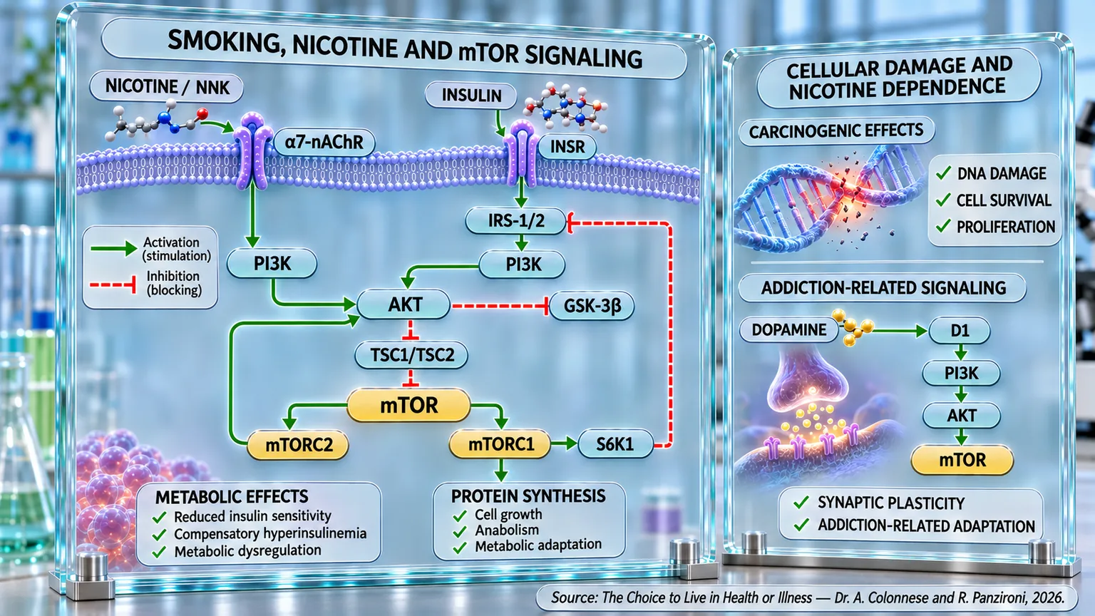
Conclusions

The scientific evidence analyzed in this chapter shows that smoking represents much more than a respiratory risk factor; it is a systemic exposure capable of interfering with metabolism, the cardiovascular system, carcinogenesis, and the brain's addiction circuits. Epidemiological studies highlight, first and foremost, the absence of a true safety threshold: even a few cigarettes a day significantly increase cardiovascular and all-cause mortality. The damage involves the endothelium, platelet aggregation, oxidative stress, inflammation, and DNA integrity, while cessation progressively alters this trajectory. Indeed, quitting reduces cardiovascular, respiratory, and oncological risk, with benefits observable even after prolonged exposure and even following a cancer diagnosis.

Electronic cigarettes do not completely eliminate this biological pressure. Although they eliminate combustion and reduce exposure to certain toxins, their aerosols contain nicotine, aldehydes, ultrafine particles, and metals. The evidence reported associates vaping with respiratory and cardiovascular alterations, while combined use with traditional cigarettes appears particularly unfavorable. Thus, a key point of the chapter emerges: changing the method of intake does not necessarily mean eliminating the biological effects of nicotine.

The discussion then shifts to metabolism. In smokers, reduced insulin sensitivity, compensatory hyperinsulinemia, and metabolic alterations correlated with the quantity and duration of exposure have been documented. Studies on nicotine reinforce this link, showing that the alteration can persist independently of combustion. At the molecular level, the PI3K/AKT/mTOR pathway emerges: nicotine can increase mTOR activity and alter IRS-1, contributing to a reduction in the insulin response. This step directly connects tobacco use to the metabolic theme of this work.

The involvement of mTOR extends to carcinogenesis. Nicotine and NNK interact with nicotinic acetylcholine receptors, especially α7-nAChR, activating PI3K/AKT/mTOR. This results in signals that favor proliferation, protein synthesis, angiogenesis, and resistance to apoptosis. NNK also adds a second level of damage: in addition to stimulating survival pathways, it has the capacity to produce mutagenic DNA alterations. The damaged cell can thus simultaneously acquire mutations and an increased capacity to survive. The studies analyzed also link this signaling to chemoresistance, showing how nicotine can support the survival of tumor cells during certain treatments.

Finally, the same pathway reaches the brain. Nicotine activates nicotinic receptors in the mesolimbic circuits, increasing dopamine in the nucleus accumbens. Dopamine does not only produce gratification: through dopaminergic receptors and mTOR, it promotes protein synthesis and synaptic plasticity, contributing to the consolidation of memory and learning associated with the substance. Evidence on addiction indeed shows that mTORC1 participates in the remodeling of neuronal circuits and the stabilization of compulsive behaviors. The chapter thus concludes its journey by connecting exposure, metabolism, carcinogenesis, and addiction: from smoking to nicotine, from nicotine to receptors, from receptors to PI3K/AKT/mTOR, and, finally, from mTOR to the cellular and neuronal modifications that can make the biological effects of tobacco persistent.

References

[1] Association of smoking status, intensity, and cessation duration with long-term incidence of nine cardiovascular outcomes and mortality: the Cross-Cohort Collaboration study

Tasdighi et al., 2025

PLoS Medicine, 2025, 22(11):e1004561.

[2] Low cigarette consumption and risk of coronary heart disease and stroke: meta-analysis of 141 cohort studies in 55 study reports

Hackshaw et al., 2018

BMJ, 2018, 360:j5855.

[3] Associations between tobacco smoking and mortality: a sex-stratified cohort analysis

Vallée, 2025

European Journal of Public Health, 2025, 35(6):1212-1218.

[4] Smoking and oral and pharyngeal cancer: a meta-analysis

Possenti et al., 2026

Oncology Reviews, 2026, 19:1672607.

[5] Tobacco smoking and cause-specific mortality: contemporary estimates from Australia

Joshy et al., 2025

BMC Medicine, 2025, 23(1):115.

[6] Light cigarette smoking increases risk of all-cause and cause-specific mortality: findings from the NHIS cohort study

Qin et al., 2020

International Journal of Environmental Research and Public Health, 2020, 17(14):5122.

[7] Smoking and cancer, cardiovascular and total mortality among older adults: the Finrisk Study

Barengo et al., 2019

Preventive Medicine Reports, 2019, 14:100875.

[8] Impact of smoking and smoking cessation on cardiovascular events and mortality among older adults: meta-analysis of individual participant data from prospective cohort studies of the CHANCES consortium

Mons et al., 2015

BMJ, 2015, 350:h1551.

[9] Smoking Cessation and Short- and Longer-Term Mortality

Cho et al., 2024

NEJM Evidence, 2024, 3(3):EVIDoa2300272.

[10] Smoking cessation and reduction in mortality risk among older adults with a long smoking history

Chen et al., 2024

BMC Public Health, 2024, 24(1):2821.

[11] Association of Smoking, Smoking Cessation, and Mortality by Race, Ethnicity, and Sex Among US Adults

Thomson et al., 2022

JAMA Network Open, 2022, 5(10):e2231480.

[12] Upgrading your best chances: postdiagnosis smoking cessation after cancer diagnosis improves patients’ life expectancy – a systematic review and meta-analysis

Caini et al., 2026

Tobacco Control, 2026, 35(2):240-249.

[13] Reduction in head and neck cancer risk following smoking cessation: a systematic review and meta-analysis

Khalifeh et al., 2024

BMJ Open, 2024, 14(8):e074723.

[14] Clinical Impact of Vaping

Petrella et al., 2025

Toxics, 2025, 13(6):470.

[15] Association Between E-Cigarettes and Asthma in Adolescents: A Systematic Review and Meta-analysis

Li et al., 2022

American Journal of Preventive Medicine, 2022, 62(6):953-960.

[16] Assessment of the Impact of Electronic Nicotine Delivery Systems on Chronic Obstructive Pulmonary Disease: A Systematic Review and Meta-analysis

Malvi et al., 2025

Respiratory Medicine, 2025;241:108059.

[17] Are E-Cigarettes Associated with Increased Risk of Myocardial Infarction and Stroke? A Systematic Review and Meta-analysis

Gupta et al., 2026

BMC Public Health, 2026, 26(1):199.

[18] Electronic Cigarettes and Cardiovascular Outcomes: A Systematic Review and Meta-analysis of Major Adverse Cardiovascular Events (MACE)

Al-Rubaye et al., 2026

BMC Public Health, 2026, 26(1):704.

[19] Electronic Cigarettes and Cardiovascular Diseases: An Updated Systematic Review and Network Meta-analysis

Tansawet et al., 2025

Tobacco Induced Diseases, 2025, 23.

[20] Insulin Resistance and Cigarette Smoking

Facchini et al., 1992

Lancet, 1992, 339(8802):1128-1130.

[21] Smoking Is Independently Associated With High Plasma Insulin Levels in Nondiabetic Men

Rönnemaa et al., 1996

Diabetes Care, 1996, 19(11):1229-1232.

[22] Insulin Resistance and Compensatory Hyperinsulinemia: The Key Player Between Cigarette Smoking and Cardiovascular Disease?

Reaven e Tsao, 2003

Journal of the American College of Cardiology, 2003, 41(6):1044-1047.

[23] Effects of Cigarette Smoking on Insulin Resistance Risk

Mouhamed et al., 2016

Annales de Cardiologie et d'Angéiologie, 2016, 65(1):21-25.

[24] Short-Term Smoking Increases the Risk of Insulin Resistance

Cho et al., 2022

Scientific Reports, 2022, 12(1):3550.

[25] Association Between E-Cigarette Smoking and Insulin Resistance Using the Triglyceride-Glucose Index in Korean Adults: Korea National Health and Nutrition Examination Survey

Lim et al., 2025

Korean Journal of Family Medicine, 2025, 46(5):327-332.

[26] Association Between Smoking Behavior and Insulin Resistance Using the Triglyceride-Glucose Index in South Korean Adults

Jeong et al., 2021

Journal of Clinical Endocrinology & Metabolism, 2021, 106(11):e4531-e4541.

[27] Long-Term Use of Nicotine Gum Is Associated With Hyperinsulinemia and Insulin Resistance

Eliasson et al., 1996

Circulation, 1996, 94(5):878-881.

[28] Weight Gain and Insulin Resistance During Nicotine Replacement Therapy

Assali et al., 1999

Clinical Cardiology, 1999, 22(5):357-360.

[29] Novel and Reversible Mechanisms of Smoking-Induced Insulin Resistance in Humans

Bergman et al., 2012

Diabetes, 2012, 61(12):3156-3166.

[30] Nicotine and Insulin Resistance: When the Smoke Clears

Bajaj, 2012

Diabetes, 2012, 61(12):3078-3080.

[31] Effect of Nicotine on Energy Metabolism of Substantia Nigra Cells in MPTP-Induced Parkinson’s Disease

Zhgenti et al., 2025

Iranian Journal of Basic Medical Sciences, 2025, 28(10):1417-1427.

[32] Nicotine Alleviates Chronic Stress-Induced Anxiety and Depressive-Like Behavior and Hippocampal Neuropathology Through Regulating Autophagy Signaling

Xiao et al., 2018

Neurochemistry International, 2018, 114:58-70.

[33] Rapid Akt Activation by Nicotine and a Tobacco Carcinogen Modulates the Phenotype of Normal Human Airway Epithelial Cells

West et al., 2003

Journal of Clinical Investigation, 2003, 111(1):81-90.

[34] The Role of α7-nAChR-Mediated PI3K/AKT Pathway in Nicotine-Induced Lung Cancer

He et al., 2024

Science of the Total Environment, 2024, 912:169604.

[35] α9- and α7-Containing Nicotinic Receptors Mediate the Proliferative Effects of Nicotine in the A549 Adenocarcinoma Cell Line

Mucchietto et al., 2018

British Journal of Pharmacology, 2018, 175(11):1957-1972.

[36] Nicotine Stimulates PPARβ/δ Expression in Human Lung Carcinoma Cells Through Activation of PI3K/mTOR and Suppression of AP-2α

Sun et al., 2009

Cancer Research, 2009, 69(16):6445-6453.

[37] The α5 Nicotinic Acetylcholine Receptor Is Involved in the Proliferation and Invasion of Human Prostate Cancer Cells

Qi et al., 2020

Oncology Reports, 2020, 43(1):159-168.

[38] The α7-Nicotinic Acetylcholine Receptor Antagonist QND7 Suppresses Non-Small Cell Lung Cancer Cell Proliferation and Migration Through Inhibition of Akt/mTOR Signaling Pathway

Witayateeraporn et al., 2020

Biochemical and Biophysical Research Communications, 2020, 521(4):977-983.

[39] Tobacco Components Stimulate Akt-Dependent Proliferation and NFκB-Dependent Survival in Lung Cancer Cells

Tsurutani et al., 2005

Carcinogenesis, 2005, 26(7):1182-1195.

[40] The Role of the Akt/mTOR Pathway in Tobacco Carcinogen-Induced Lung Tumorigenesis

Memmott e Dennis, 2010

Clinical Cancer Research, 2010, 16(1):4-10.

[41] Tobacco Carcinogen-Induced Cellular Transformation Increases Activation of the Phosphatidylinositol 3′-Kinase/Akt Pathway in Vitro and in Vivo

West et al., 2004

Cancer Research, 2004, 64(2):446-451.

[42] Survivin Expression in Normal Human Bronchial Epithelial Cells: A Critical Early Event in Tobacco Carcinogen-Induced Human Carcinogenesis

Jin et al., 2008

Carcinogenesis, 2008, 29(8):1614-1622.

[43] β-Cryptoxanthin Reduced Lung Tumor Multiplicity and Inhibited Lung Cancer Cell Motility by Down-Regulating α7-Nicotinic Acetylcholine Receptor Signaling

Iskandar et al., 2016

Cancer Prevention Research, 2016, 9(11):875-886.

[44] Nicotine Inhibits Cisplatin-Induced Apoptosis via Regulating α5-nAChR/AKT Signaling in Human Gastric Cancer Cells

Jia et al., 2016

PLoS One, 2016, 11(2):e0149120.

[45] Nicotine-Induced Cell Survival and Cisplatin Resistance Through α7-Nicotinic Acetylcholine Receptor in Oral Cancer

Hsu et al., 2020

Archives of Oral Biology, 2020, 111:104653.

[46] Nicotine Induces Tumor Growth and Chemoresistance Through Activation of the PI3K/Akt/mTOR Pathway in Bladder Cancer

Yuge et al., 2015

Molecular Cancer Therapeutics, 2015, 14(9):2112-2120.

[47] Role of Dopamine in the Behavioural Actions of Nicotine Related to Addiction

Di Chiara, 2000

European Journal of Pharmacology, 2000, 393(1-3):295-314.

[48] Nicotine-Induced Dopamine Release in the Rat Nucleus Accumbens Is Regulated by Nicotinic Receptors in the Ventral Tegmental Area

Nisell et al., 1994

Synapse, 1994, 16(1):36-44.

[49] Dopamine and Drug Addiction: The Nucleus Accumbens Shell Connection

Di Chiara et al., 2004

Neuropharmacology, 2004, 47(Suppl 1):227-241.

[50] Nicotine: From Molecular Mechanisms to Behaviour

Wonnacott et al., 2005

Current Opinion in Pharmacology, 2005, 5(1):53-59.

[51] Nicotine and Neuronal Nicotinic Acetylcholine Receptors: Unraveling the Mechanisms of Nicotine Addiction

Jiang et al., 2025

Frontiers in Neuroscience, 2025, 19:1670883.

[52] Alpha6-Containing Nicotinic Acetylcholine Receptors Dominate the Nicotine Control of Dopamine Neurotransmission in the Nucleus Accumbens

Exley et al., 2008

Neuropsychopharmacology, 2008, 33(9):2158-2166.

[53] In Vivo Activation of Midbrain Dopamine Neurons via Sensitized, High-Affinity Alpha6 Nicotinic Acetylcholine Receptors

Drenan et al., 2008

Neuron, 2008, 60(1):123-136.

[54] Pharmacological Effects of Nicotine Salts on Dopamine Release in the Nucleus Accumbens

Li et al., 2023

Frontiers in Pharmacology, 2023, 14:1279512.

[55] Combined Systemic Effects of Alcohol and Nicotine on Dopamine Release in the Nucleus Accumbens

Tizabi et al., 2007

Alcohol and Alcoholism, 2007, 42(5):413-416.

[56] Activation of D1R/PKA/mTOR Signaling Cascade in Medial Prefrontal Cortex Underlies the Antidepressant Effects of l-SPD

Zhang et al., 2017

Scientific Reports, 2017, 7(1):3809.

[57] Dopaminergic Modulation of Auditory Cortex-Dependent Memory Consolidation Through mTOR

Schicknick et al., 2008

Cerebral Cortex, 2008, 18(11):2646-2658.

[58] The First Alcohol Drink Triggers mTORC1-Dependent Synaptic Plasticity in D1 Receptor Neurons of the Nucleus Accumbens

Beckley et al., 2016

Journal of Neuroscience, 2016, 36(3):701-713.

[59] Essential Role of D1R in the Regulation of mTOR Complex 1 Signaling Induced by Cocaine

Sutton e Caron, 2015

Neuropharmacology, 2015, 99:610-619.

[60] Inhibition of mTOR Signaling in Parkinson’s Disease Prevents L-DOPA-Induced Dyskinesia

Santini et al., 2009

Science Signaling, 2009, 2(80):ra36.

[61] Repeated Stimulation of Dopamine D1-like Receptor and Hyperactivation of mTOR Signaling Lead to Generalized Seizures, Alteration of Dentate Gyrus Plasticity, and Memory Deficits

Gangarossa et al., 2014

Hippocampus, 2014, 24(12):1466-1481.

[62] Dopamine Pathway Mediated by DRD5 Facilitates Tumor Growth Through Enhancing Warburg Effect in Esophageal Cancer

Qian et al., 2021

Frontiers in Oncology, 2021, 11:655861.

[63] Signal Transduction Pathways Involved in Dopamine D2 Receptor-Induced Emesis in the Least Shrew (Cryptotis parva)

Belkacemi et al., 2021

Autonomic Neuroscience, 2021, 233:102807.

[64] mTOR Complex 1: A Key Player in Neuroadaptations Induced by Drugs of Abuse

Neasta et al., 2014

Journal of Neurochemistry, 2014, 130(2):172-184.

[65] An Emerging Role for the Mammalian Target of Rapamycin in “Pathological” Protein Translation: Relevance to Cocaine Addiction

Dayas et al., 2012

Frontiers in Pharmacology, 2012, 3:13.

[66] mTOR Signaling in the Nucleus Accumbens Mediates Behavioral Sensitization to Methamphetamine

Huang et al., 2018

Progress in Neuro-Psychopharmacology and Biological Psychiatry, 2018, 86:331-339.

[67] Inhibition of Mammalian Target of Rapamycin Signaling by Rapamycin Blocks Cocaine-Induced Locomotor Sensitization

Wu et al., 2011

Neuroscience, 2011, 172:104-109.

[68] Region-Specific Activation of AKT and mTORC1 Signaling in Response to Excessive Alcohol Drinking in Rodents

Laguesse et al., 2017

Addiction Biology, 2017, 22(6):1856-1869.

[69] AKT Signaling Pathway in the Nucleus Accumbens Mediates Excessive Alcohol Drinking Behaviors

Neasta et al., 2011

Biological Psychiatry, 2011, 70(6):575-582.