
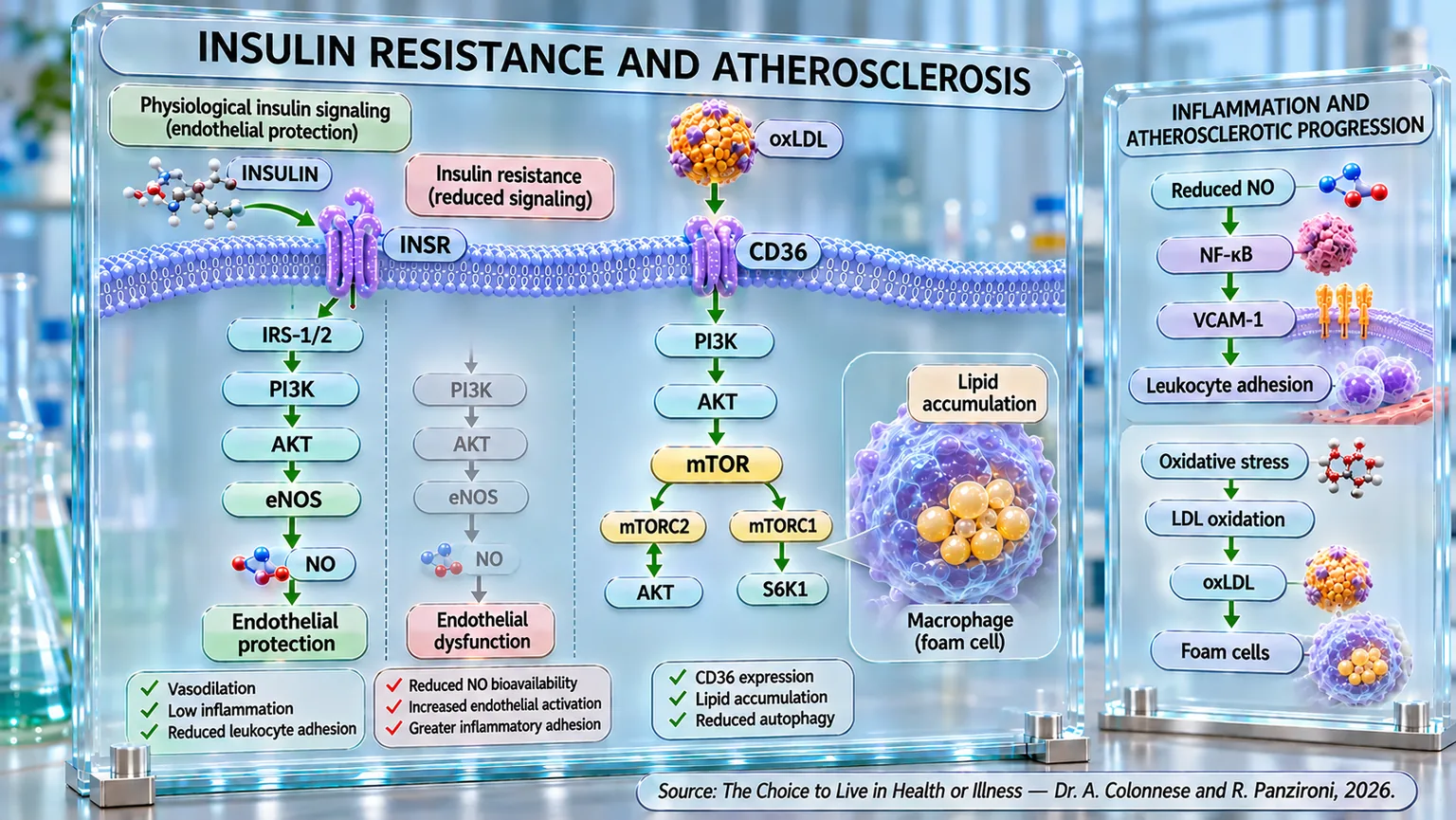
The link described in the previous paragraph between insulin resistance and endothelial dysfunction represents one of the first steps through which an initially metabolic alteration can translate, over time, into a true structural modification of the arterial wall. Atherosclerosis, in fact, does not appear suddenly with the formation of an already organized plaque, but develops through a long succession of events in which metabolic alterations, inflammation, oxidative stress, lipid accumulation, and changes in the response of vascular cells tend to progressively overlap. In this process, insulin resistance seems to play a particularly important role, so much so that its association with atherosclerosis can be detected when the disease is still subclinical, that is, before it produces evident cardiovascular manifestations.
One of the most extensive demonstrations comes from the analysis of coronary artery calcification, considered an indicator of coronary atherosclerotic burden. A meta-analysis including 15 studies and a total of 60,649 participants showed that higher HOMA-IR index values were associated with a greater prevalence of coronary calcification. Considering HOMA-IR as a continuous variable, the increase in insulin resistance was associated with an increased probability of presenting coronary calcifications, with an overall OR (a statistical indicator that measures how much more or less likely an event is) of 1.27. Even more interestingly, in the available longitudinal data, high HOMA-IR values were also associated with the progression of calcification, with an OR of 1.44. The quality of the evidence, however, was judged low for prevalence and very low for progression (Li et al., 2023) [13].
The same relationship emerges using another metabolic indicator, the triglyceride-glucose index, or TyG, obtained by combining fasting concentrations of triglycerides and glucose and frequently used as a surrogate marker of insulin resistance. A review of the available evidence compared TyG, HOMA, and triglycerides with different indicators of subclinical atherosclerosis, highlighting how TyG showed the most consistent association with coronary calcification. The relationship with arterial stiffness, assessed through pulse wave velocity (the pressure wave that propagates along the arteries every time the heart contracts), was also robust. Particularly significant is the fact that these alterations can be observed even before the onset of overt diabetes: insulin resistance therefore seems to accompany the atherosclerotic process in a phase in which traditional glycemic criteria might not yet be sufficient to identify its presence (Scott et al., 2024) [14].
The evidence becomes even more concrete by observing the carotid arteries directly. In a study conducted on 4,203 Chinese steelworkers, subjects belonging to the highest quartile of the TyG index had, compared to those in the lowest quartile, more than three times the probability of carotid atherosclerosis. The association concerned both the increase in intima-media thickness and, above all, the presence of an actual plaque: for the latter, the OR reached 3.7. The relationship was not completely linear, and the risk increased significantly beyond a TyG value of 8.7. The study also suggests that metabolic vulnerability may interact with other environmental factors, as the association appeared particularly evident in workers exposed to shift work, heat, noise, carbon monoxide, and dust (Cao et al., 2025) [15].
A consistent picture emerges in patients in whom coronary artery disease is already clinically manifest. In 1,516 subjects with symptomatic coronary artery disease, increasing TyG values were associated in parallel with a greater extent of coronary atherosclerosis and the presence of carotid plaques. Compared to the lower quartile, the one with the highest values had a 2.5 times greater probability of coronary lesions and 2.3 times greater probability of carotid plaque. It is important to emphasize that the association persisted after adjustment for various cardiovascular factors and independently of the presence of diabetes or hyperlipidemia. Furthermore, the ability of TyG to identify atherosclerotic lesions was superior to that obtained by considering glycemia or triglycerides separately (Li et al., 2023) [16].
Insulin resistance also seems to be associated not only with the presence of plaque but also with its characteristics. This aspect is particularly important because not all plaques present the same risk. A stable plaque can persist for a long time, while an unstable plaque, generally characterized by elements of greater vulnerability, can undergo complications with much more significant clinical consequences. In over 12,000 subjects who underwent carotid ultrasound, higher TyG values were significantly associated with the presence of unstable plaques. The association was present in both men and women but was more marked in men; it also became particularly evident after age 60 and in diabetic patients. The analysis showed a non-linear relationship, with a rapid increase in risk beyond a TyG of 8.63 (Huo et al., 2025) [17].
Epidemiological observations find important support in experimental studies, which allow for the isolation of the contribution of insulin signaling within the vascular wall. In mice genetically predisposed to atherosclerosis through the loss of apolipoprotein E, the selective elimination of the insulin receptor from endothelial cells produced a particularly significant result: despite systemic insulin sensitivity, glucose tolerance, insulinemia, plasma lipids, and blood pressure being substantially comparable to those of the controls, atherosclerotic lesions were more than doubled. The loss of insulin action in the endothelium was accompanied by lower activation of AKT and eNOS, impairment of vasodilation, increase in VCAM-1 (a protein present on the surface of cells lining blood vessels, which promotes the adhesion of white blood cells), and greater leukocyte adhesion to the vascular wall (Rask-Madsen et al., 2010) [18].
This experiment allows for an important conceptual step. The association between insulin resistance and atherosclerosis does not seem to depend exclusively on the systemic consequences that frequently accompany insulin resistance, such as hyperglycemia and dyslipidemia. The loss of proper insulin action directly in the endothelium can be sufficient, at least in the experimental model used, to strongly accelerate atherogenesis. In other words, physiologically functioning insulin does not simply appear as a metabolic hormone that controls glycemia, but also exerts vascular signals that contribute to maintaining the endothelium in a state less favorable to the development of atherosclerosis.
This interpretation is reinforced by an almost mirror-image experiment. If the loss of endothelial insulin signaling accelerates atherosclerosis, its enhancement can produce the opposite effect. The overexpression of IRS1 in the endothelial cells of mice predisposed to atherosclerosis has, in fact, increased the insulin response through PI3K/AKT and significantly reduced atherosclerotic lesions even in the presence of insulin resistance, hyperglycemia, and hyperinsulinemia. The improvement was accompanied by lower expression of VCAM-1 and greater production of NO. The study also identified an additional mechanism: through IRS1/PI3K/AKT, insulin increased the endothelial expression of EDNRB (endothelin receptor type B), promoting the increase of eNOS. The loss of EDNRB instead produced a reduction in NO and an acceleration of atherosclerosis (Park et al., 2016) [19].
These results also help to interpret the relationship between hyperinsulinemia and atherosclerosis with greater caution. The frequent association between high insulin concentrations, insulin resistance, and cardiovascular risk could, in fact, lead one to automatically consider insulin itself as directly atherogenic. The experimental studies provided show a more complex picture. In ApoE−/− mice made obese, insulin-resistant, hyperglycemic, and hyperlipidemic through a high-fat diet, chronic administration of insulin notably increased insulinemia but, rather than aggravating atherosclerosis, it reduced its extent and complexity, increasing the activation of AKT and eNOS and the production of NO. The reduction of glycemia through phlorizin (a natural substance present especially in the bark and roots of the apple tree), in the absence of these effects of insulin, did not determine a comparable reduction in atherosclerosis (Park et al., 2018) [20].
A fundamental pathophysiological distinction thus emerges. Insulin resistance and hyperinsulinemia cannot be considered equivalent terms, nor can their vascular effects be automatically superimposed. The available evidence indicates that the loss of proper insulin signaling, especially in the endothelium, can remove important protective vascular mechanisms and favor atherogenesis. Conversely, when the insulin signal is still able to effectively activate pathways such as IRS1/PI3K/AKT/eNOS, insulin can support the production of NO, reduce endothelial activation, and limit some processes involved in plaque progression. In the experimental models analyzed, even a marked increase in insulinemia can therefore be accompanied by a reduction in atherosclerosis when it determines an effective restoration of these actions.
Overall, epidemiological and experimental evidence therefore converge on a model in which insulin resistance represents an important metabolic terrain associated with atherosclerosis, from the subclinical phase up to the presence and, potentially, the instability of plaques. HOMA-IR and especially TyG show associations with coronary calcification, arterial stiffness, carotid and coronary atherosclerosis, and plaque instability characteristics. In parallel, experimental studies indicate that part of the link may depend directly on the loss of normal insulin action in the vascular wall. At the center of this relationship, the ability of insulin to activate the PI3K/AKT cascade and support endothelial nitric oxide production emerges once again. It is precisely this step that now allows us to shift the focus from the general association between insulin resistance and atherosclerosis to the molecular mechanism that could contribute to linking them: the relationship between PI3K/AKT and nitric oxide.