
Scientific Book

Psoriasis and signals crossing the skin

Psoriasis is an immune-mediated inflammatory disease that involves keratinocyte proliferation and the activation of specific immune responses. The IL-23/Th17, PI3K/AKT/mTOR, and HIF-1α pathways participate in the cellular processes associated with skin inflammation and alterations in epidermal growth. In the book "The Choice to Live in Health or Disease," Dr. A. Colonnese and R. Panzironi analyze the relationship between cellular metabolism, immune regulation, and the manifestations of psoriasis, offering a biological interpretation of the disease.

Cite this article
Colonnese, A. & Panzironi, R. (2026). Psoriasis and signals crossing the skin. The Choice to Live in Health or in Disease. https://lifesciencehub.info/en/la-scelta-di-vivere-in-salute-o-in-malattia/psoriasis-and-signals-crossing-the-skin
The Choice to Live in Health or in Disease · ISBN 979-12-82692-03-8

DOI lsh.en.libro.8

  • Published 05/11/2026
  • Updated 05/11/2026
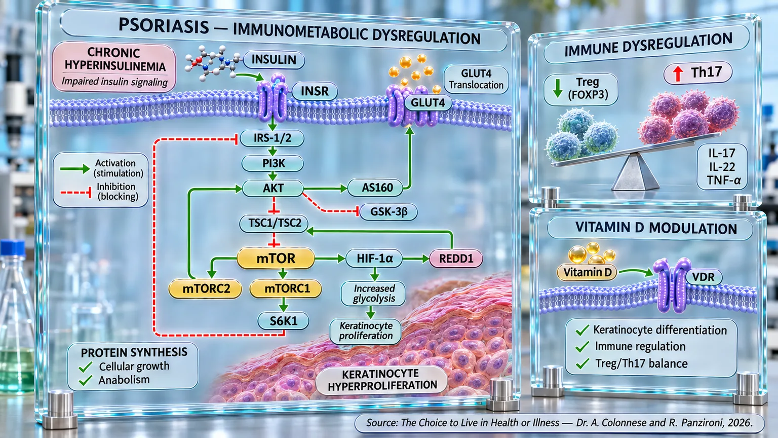
Diagram on transparent panels showing cell membrane, insulin cascade, epidermal tissue, Treg-Th17 balance, and VDR receptor.
Infographic of immunometabolic dysregulation in psoriasis: chronic hyperinsulinemia alters PI3K/AKT/mTOR insulin signaling, linked to HIF-1α, glycolysis, and keratinocyte hyperproliferation; alongside this, the imbalance between reduced Tregs and increased Th17 cells and vitamin D–VDR modulation link cellular metabolism, immunity, and epidermal growth.

ABSTRACT

Psoriasis is a chronic systemic inflammation disease in which immune and metabolic alterations accompany skin manifestations. In the book "La scelta di Vivere in Salute o in Malattia" (The Choice to Live in Health or Disease), Dr. A. Colonnese and R. Panzironi analyze the interactions between insulin metabolism, intracellular signaling, and inflammation, also considering the associations with obesity, metabolic syndrome, and cardiovascular risk.

The psoriatic immune response shows an increase in Th17 cells and IL-17, IL-22, and TNF-α mediators, associated with the impairment of regulatory T cells. Reduced FOXP3 stability may contribute to the loss of immune control. In parallel, hyperinsulinemia and insulin resistance have been observed in patients even without obesity or diabetes, sometimes with normal conventional glycemic indicators. However, it remains to be clarified to what extent hyperinsulinemia precedes and directly contributes to psoriatic pathogenesis.

A possible molecular link involves PI3K/AKT/mTOR, a pathway hyperactivated in psoriatic skin. mTORC1 supports the proliferation and glycolytic metabolism of keratinocytes, in addition to participating in the Th17 response. Through HIF-1α, metabolic signaling promotes the expression of glycolytic components and can contribute to epidermal hyperproliferation. Research on mTOR inhibition via rapamycin or sirolimus describes reductions in keratinocyte proliferation and inflammation; methotrexate can also modulate the Treg/Teff balance through AMPK and mTOR. These observations require a distinction between experimental results and clinical applicability.

Vitamin D also participates in immune regulation and keratinocyte differentiation. Lower levels are frequently associated with greater psoriasis severity, while topical analogs have documented clinical efficacy.

Overall, psoriasis emerges as a multifactorial immunometabolic condition in which insulin alterations, mTOR/HIF-1α signaling, and Th17/Treg imbalance may interact. This perspective broadens the understanding of pathological processes without attributing an exclusive causal role to a single mechanism. Integrating metabolic observations with immunological ones can guide research on the relationships between skin inflammation, alterations, and therapeutic targets, while keeping clinical associations and experimental mechanisms distinct.

Epidemiology of psoriasis

Psoriasis is one of the most widespread chronic inflammatory diseases in the world, with an impact that extends far beyond the skin. It affects approximately 2–3% of the global population, translating to over 125 million people (WHO data, 2023).

The age of onset follows a bimodal pattern, meaning there are two distinct peaks: the first between 15 and 30 years of age, and the second between 50 and 60 years of age. Early onset, often associated with the HLA-Cw6 antigen (a variant of a protein present on the cell surface that is recognized as a target by the immune system), is generally more aggressive and has a stronger genetic component. Conversely, late-onset forms tend to have a milder but more persistent course over time.

Despite the variety of clinical manifestations, the most common form is plaque psoriasis, which accounts for approximately 80–90% of cases. The typical lesions—erythematous, scaly plaques localized on the elbows, knees, scalp, and lumbar region—reflect the visible result of deep, systemic, and complex inflammation.

Beyond the dermatological aspect, psoriasis is frequently accompanied by metabolic and cardiovascular comorbidities (the presence of multiple conditions in the same individual), such as metabolic syndrome, obesity, hypertension, and insulin resistance. For this reason, the disease is now considered a systemic immuno-metabolic disorder, no longer confined to the skin but an expression of a broader inflammatory dysfunction of the body.

From a psychological perspective, the impact is equally significant: visible lesions can compromise quality of life, self-esteem, and social relationships, often leading to anxiety and depression. Psoriasis, therefore, is not just a skin disease, but a complex biological and social phenomenon in which genetics, immunity, and metabolism are intertwined in an unstable balance.

T-cell imbalance

Psoriasis is now recognized as a disease characterized by an excessive activation of the immune system, which reacts even when it is not necessary. In a healthy organism, regulatory T cells (Tregs) act as a physiological brake on immunity, maintaining the balance between activation and tolerance. When this brake loosens, as happens in psoriasis, inflammation becomes chronic and self-sustaining, leading to the uncontrolled proliferation of keratinocytes (the main cells of the epidermis that produce keratin, a protein that strengthens and protects the skin, hair, and nails) and the appearance of characteristic skin lesions. It is as if the immune system, instead of turning off the alarm once the danger has passed, continues to sound the siren, causing damage to its own tissues.

The most recent studies have shown that in psoriatic patients, Tregs are not simply reduced in number, but are primarily dysfunctional. Under normal conditions, these cells inhibit T helper 17 cells (a type of effector cell), which produce pro-inflammatory cytokines such as IL-17, IL-22, and TNF-α. In psoriasis, however, Tregs lose their suppressive capacity and in some cases transform themselves into Th17 cells, fueling the inflammatory process rather than controlling it (Kanda et al., 2021) [1]. This creates a true immune vicious cycle, in which IL-23 promotes the conversion of Tregs into Th17 cells and, simultaneously, the IL-17 produced by the latter inhibits the expression of Foxp3 (an essential gene already discussed in chapter 6 on the immune system) for the regulatory function of Tregs. The result is an immune system that, deprived of its self-regulation mechanisms, behaves like an accelerator without brakes.

Research has also shown that this loss of balance is not only quantitative but epigenetic (the set of biological mechanisms that regulate gene activity). Indeed, the Tregs of psoriatic patients show chromatin (DNA organized together with proteins inside the cell nucleus) that is more "open" and therefore more active, capable of responding abnormally to inflammatory stimuli. After treatment with anti-IL-17 biologic drugs, this hyperactivity is reduced, a sign that the inflammatory environment itself can modify the behavior of Tregs and that their function can be partially restored (Zhang et al., 2025) [2].

In parallel, experimental studies have explored new therapeutic pathways aimed not only at blocking pro-inflammatory cytokines, but at restoring immune homeostasis (the state of dynamic balance of the immune system). An innovative example is represented by the peptide dNP2-ctCTLA-4 (an experimental molecule designed to enter immune cells and attenuate their activation), which acts by promoting the expansion and stabilization of Tregs, while simultaneously reducing the production of IL-17 by Th17 and Tc17 cells (killer cells of the immune system). In murine models (experimental models that use mice or rats to study biological functions and human diseases) of psoriasis, treatment with this peptide resulted in a clear reduction of skin inflammation and clinical improvement superior to traditional biologic drugs (Lee et al., 2023) [3]. This approach suggests a change in perspective: not just putting out the fire of inflammation, but reigniting the self-regulation system that prevents the fire from restarting.

In experimental models of psoriasis, the topical application of sodium butyrate (the salt form of butyrate, a short-chain fatty acid) significantly reduced skin thickness and scaling, normalizing the production of IL-10 and FOXP3, markers of proper Treg activity, and suppressing the pro-inflammatory cytokines IL-17 and IL-6. When Tregs were artificially eliminated, the therapeutic effect of butyrate disappeared, confirming its central role in restoring immune balance (Schwarz et al., 2021) [4].

Vitamin D3 and its analogs have also demonstrated the ability to support Treg function: in a comparative study between the synthetic vitamin D analog (maxacalcitol) and the corticosteroid betamethasone, both reduced the clinical signs of psoriasiform dermatitis (skin inflammation with characteristics similar to psoriasis), but only maxacalcitol significantly increased Foxp3+ cells and IL-10 expression, strengthening the immunosuppressive environment and suggesting a deeper action on immune rebalancing (Hau et al., 2018) [5].

Taken together, these findings describe a clear picture: psoriasis is not just an excessive reaction of the immune system, but a loss of internal regulation. Tregs, normally tasked with maintaining tolerance and immunological calm, become unable to exert their function, sometimes turning into the protagonists of the inflammation they should be containing. Restoring the correct balance between Tregs and Th17 cells ultimately means bringing order back to a system that has lost its biological hierarchy.

It is as if, in an orchestra, the entire woodwind section began to play out of time, drowning out the general harmony: the music, in this case the immune response, loses coherence, turning into noise. New therapeutic strategies therefore aim not only to silence the loudest instruments, but to re-educate the conductor, restoring the immune system's ability to play in balance.

Metabolic alterations: hyperinsulinemia and insulin resistance in psoriasis

In recent years, a fundamental truth has emerged with increasing clarity: psoriasis is not just a skin disease, but a systemic disorder in which metabolism and the immune system interact in a dysfunctional way. In this sense, the skin becomes a mirror of inflammation that originates elsewhere and is fueled by altered metabolic processes, including insulin resistance.

Clinical observation of a link between psoriasis and altered normal glucose metabolism dates back several decades, but only recently have the biological connections linking inflammation and insulin resistance been quantified. Pioneering studies conducted as early as the 1990s demonstrated that, in psoriasis patients, plasma insulin levels increased disproportionately after glucose intake, while the rate of glucose clearance was reduced, a clear sign of reduced insulin sensitivity (Brenelli et al., 1995) [6]. The serum potassium response to insulin also appeared compromised, a finding suggesting that the defect did not only concern sugar metabolism, but also other cellular functions dependent on the hormone.

As knowledge has progressed, the picture has become clearer: insulin resistance in psoriasis patients also manifests in the absence of diabetes or obesity, and can be measured through specific sensitivity tests. Using the hyperinsulinemic-euglycemic clamp, the most precise method available, it has been observed that the ability of tissues to respond to insulin is significantly reduced in subjects affected by moderate or severe psoriasis, even when glucose levels and glycated hemoglobin remain within the normal range (Gyldenløve et al., 2015) [7]. This indicates that metabolic dysfunction precedes actual diabetes, representing a sort of “inflammatory prediabetes.”

In larger studies conducted on thousands of patients, the association between the HOMA-IR index (an index used to estimate insulin resistance and the functionality of pancreatic β-cells) and psoriasis has proven evident and independent of other risk factors such as age or BMI (body mass index). In a Women’s Health Initiative cohort followed for over twenty years, women with higher baseline HOMA-IR values showed a greater risk of developing psoriasis, suggesting that insulin resistance is not only a consequence but also a possible predisposing factor for the disease (Chan et al., 2022) [8].

This link has also been confirmed by cross-sectional studies based on representative populations. The METS-IR index, a new parameter that estimates insulin resistance by integrating glycemia, triglycerides, and BMI, has proven to be closely associated with psoriasis: each unit increase in this index resulted in an increase in the prevalence of the disease of approximately 1.7%, with an almost double risk for subjects in the highest quartile (Liu et al., 2024) [9]. Similar data were obtained with the TyG index, based on the ratio between triglycerides and glucose (a parameter now used to detect insulin resistance). These parameters, simple to calculate, have proven to be excellent predictors of psoriasis risk, confirming that abdominal fat distribution and lipid metabolism play a crucial role in the pathogenesis of the disease (Huang et al., 2023) [10].

At a clinical and biochemical level, numerous studies have shown that patients with psoriasis have higher levels of fasting insulin, fetuin-A (a protein produced mainly by the liver that acts as a modulator of insulin resistance), pentraxin-3 (a protein that represents an important marker of systemic and vascular inflammation), and C-reactive protein (one of the main markers of systemic inflammation) compared to healthy subjects, and that these values increase proportionally to the clinical severity of the disease, measured with the PASI index (a clinical index used to measure the severity and extent of psoriasis). The association between fetuin-A, a known inhibitor of insulin signaling, and skin severity suggests a direct link between systemic inflammation and metabolic dysfunction (Uysal et al., 2014) [11].

Other clinical analyses have highlighted that even in non-obese subjects, psoriasis is accompanied by glucose intolerance and a marked reduction in insulin sensitivity, particularly in late-onset forms (type II), which are more often associated with long-standing disease (Ucak et al., 2006) [12]. In parallel, studies conducted in India and other emerging countries have confirmed that insulin resistance can manifest in patients with psoriasis even in the absence of glycemic alterations, configuring itself as a silent prediabetic condition in which increased BMI (an index used to evaluate a person's weight in relation to their height) and waist circumference are determining factors (Dhara et al., 2015) [13].

The systemic impact of psoriasis is not limited to glucose metabolism. It has been shown that psoriasis patients exhibit early endothelial dysfunction (alteration of the normal functioning of blood vessels) and reduced release of vascular nitric oxide (a molecule that promotes the dilation of blood vessels), signs of an initial atherosclerotic alteration. In parallel, the increase in HOMA-IR and C-peptide levels (an indicator of endogenous insulin production, i.e., how much insulin the body produces on its own) suggests that chronic psoriatic inflammation can compromise both insulin sensitivity and vascular function, thus promoting progression toward a pre-atherosclerotic condition (Karadag et al., 2010) [14].

This relationship also extends to the joint forms of the disease: in psoriatic arthritis, in fact, the prevalence of metabolic syndrome and insulin resistance is significantly higher than in the general population, and their presence is independently associated with greater disease severity, a higher body mass index, and a later onset of psoriasis (Haroon et al., 2014) [15]. All these data converge toward a single interpretation: insulin resistance is already present in people affected by psoriasis, and therefore precedes the psoriasis, causing its activation.

In this light, psoriasis can be considered not only an immunological disease but also a form of chronic metabolic inflammation. Controlling the insulin axis through diet, physical activity, reduction of visceral fat, and, when necessary, the use of targeted therapies, therefore becomes an integral part of the global therapeutic strategy. It is not just about treating the skin, but about restoring a deep internal balance, bringing the entire immuno-metabolic system back toward a condition of functional harmony.

mTOR as a central node

Psoriasis represents one of the most emblematic models of a disease where inflammation and metabolism are intertwined. At the center of this dialogue lies the PI3K/AKT/mTOR signaling pathway, our insulin pathway which, as we previously discussed in chapter 4, translates nutritional and hormonal signals into responses of cell growth, proliferation, and survival.

Under physiological conditions, mTOR acts as a regulator of energy availability: when nutrients and insulin are present, the mTORC1 complex stimulates protein synthesis and cell proliferation, maintaining tissue homeostasis. However, in psoriasis, this mechanism turns into a pathological accelerator, constantly pressed due to excess insulin, inflammatory cytokines, and circulating nutrients (Mercurio et al., 2021) [16].

Numerous studies have confirmed that the PI3K/AKT/mTOR pathway is hyperactive in the skin cells of psoriatic patients. In keratinocytes, the abnormal activation of mTORC1 promotes accelerated glycolysis (a fundamental metabolic process through which glucose is broken down within cells), a behavior similar to that of tumor cells, and supports uncontrolled epidermal proliferation. In this scenario, keratinocytes behave like “nutritionally overstimulated” cells, which multiply their metabolic activity and lose the ability to differentiate correctly (Sarandi et al., 2023) [17].

It is as if, in psoriasis, the skin were convinced it is in a state of continuous growth and repair, ignoring the signals that should block the process and restore normalcy.

The role of mTOR is not limited to the skin: as explained in chapter 6, regarding the immune system, it represents a molecular bridge between metabolic alterations and the immune response. T cells activated in psoriatic lesions, particularly Th17 cells, rely heavily on mTOR to maintain their expansion and the production of pro-inflammatory cytokines such as IL-17 and TNF-α. This creates a vicious cycle in which mTOR stimulates inflammation and inflammation, in turn, fuels mTOR (Huang et al., 2014) [18].

In parallel, the insulin resistance typical of psoriatic patients further contributes to this imbalance. Persistent hyperinsulinemia stimulates the PI3K/AKT pathway, which in turn activates mTORC1 in keratinocytes and lymphocytes, reinforcing the interconnection between altered metabolism and inflammation.

Histological analyses conducted on skin biopsies have, in fact, confirmed strong hyperactivation of mTOR and ribosomal protein S6 (a protein involved in protein synthesis processes and regulated downstream of mTORC1/S6K) in psoriatic keratinocytes, especially in the basal and suprabasal layers of the epidermis. This activation pattern faithfully reflects the processes of proliferation and altered differentiation that characterize the plaques (Buerger et al., 2013) [19].

A further study has shown that mTORC1 activity in the epidermis is essential for the development of the psoriasiform phenotype and that its inhibition, both pharmacological and genetic, significantly reduces epidermal thickness and inflammatory infiltrate (Buerger, 2018) [22].

This hyperactivation does not only concern the skin: the most recent research has highlighted that mTOR is also involved in the systemic manifestations of psoriasis, including psoriatic arthritis and increased cardiovascular risk. At the joint level, the same signaling cascade promotes the proliferation of synovial cells (cells that line the synovial membrane, i.e., the thin tissue that lines the inside of the joints) and the production of inflammatory mediators; at the vascular level, it contributes to endothelial dysfunction and atherogenesis, linking the skin disease to cardiometabolic risk (Joo et al., 2024) [20].

The regulatory power of mTOR derives from its dual functional structure: the mTORC1 complex controls protein synthesis and cell growth, while mTORC2 regulates survival and cytoskeletal organization (the arrangement and regulation of the internal structure that supports the cell). In psoriasis, both complexes are altered: mTORC1 drives epidermal hyperproliferation, while mTORC2 supports the vitality and longevity of inflammatory cells (Raychaudhuri et al., 2014) [21].

These findings have stimulated interest in new therapeutic approaches targeting mTOR. Classic inhibitors like rapamycin (sirolimus) have demonstrated the ability to reduce keratinocyte proliferation and lymphocyte infiltration, but their efficacy is limited by systemic side effects. For this reason, dual mTORC1/mTORC2 inhibitors and molecules of natural origin are being studied, such as delphinidin, a plant pigment with antioxidant properties capable of selectively inhibiting PI3K and mTOR, significantly reducing inflammation and epidermal hyperplasia in murine models (Buerger, 2018) [22].

Even from a conceptual point of view, the role of mTOR in psoriasis has transformed our understanding of the disease. It is no longer just an immune dysfunction or a simple inflammatory response: psoriasis appears as an immunometabolic disease in which the skin, the immune system, and energy metabolism act in synergy.

In this perspective, psoriatic immunometabolism represents a new therapeutic frontier: intervening on mTOR means, in fact, correcting not only the proliferation of keratinocytes but also the energy balance of immune cells, restoring equilibrium in a chronically overactivated system (Karagianni et al., 2022) [23].

Ultimately, mTOR is the “power plant” of psoriasis: it receives signals from insulin, nutrients, cytokines, and growth factors, and transforms them into a single command to grow and become inflamed. Turning off or modulating this plant does not just mean reducing the plaques, but interrupting the pathogenetic cycle at its root, giving the immune system and metabolism the chance to return to balance.

mTOR inhibitors

Recently, therapeutic research on psoriasis has increasingly shifted toward the goal of modulating the intracellular mechanisms that sustain inflammation and skin hyperproliferation, rather than merely suppressing symptoms. In this context, mTOR inhibitors have gained significant attention for their ability to act at the heart of the metabolic and immune dysfunction that characterizes the disease.

The first compound in this class, rapamycin (or sirolimus), originated as a natural antibiotic with immunosuppressive properties, but its primary action consists of inhibiting the mTOR signaling pathway, reducing cell proliferation and the production of pro-inflammatory cytokines. Studies conducted on animal models of psoriasis have shown that topical treatment with rapamycin can significantly improve skin lesions induced by imiquimod (a drug used to simulate psoriasis in a mouse model), normalizing markers of epidermal differentiation and reducing the activation of the mTOR pathway, a direct indicator of pathway activity (Bürger et al., 2017) [24].

Unlike systemic administration, the topical approach allows the drug to be concentrated at the pathological site, limiting systemic side effects, which is a crucial aspect for chronic treatment.

This concept has also been confirmed by clinical studies in humans: in one of the first randomized controlled trials, topical sirolimus demonstrated a significant reduction in the clinical psoriasis score and a decrease in CD4+ cells (activated CD4+ lymphocytes that actively perform their defense function) in the epidermis, signs of effective local immune modulation, without blood abnormalities or significant systemic toxicity (Ormerod et al., 2005) [25]. These results paved the way for targeted formulations of mTOR inhibitors for cutaneous use, capable of acting selectively on lesions without compromising the patient's systemic balance.

In parallel, sirolimus has also shown promising results in systemic combination with other immunosuppressants: in a European multicenter study on patients with severe psoriasis, the combination of sirolimus with cyclosporine (an immunosuppressive drug that selectively inhibits T-lymphocyte activation), at doses lower than those required to achieve an effective therapeutic effect, resulted in a reduction of the PASI score (an index that measures the severity of psoriasis) comparable to high-dose cyclosporine, but with a significantly better safety profile and less nephrotoxicity (a condition in which a substance, drug, or toxin causes damage to the kidneys) (Reitamo et al., 2001) [26].

A further contribution to the understanding of this therapeutic strategy comes from studies on natural phytocomplexes that interfere with the PI3K/AKT/mTOR pathway. The Chinese herbal formula Liangxue Jiedu (LXJDF), for example, has been shown to simultaneously improve skin lesions and dyslipidemia in mouse models of psoriasis, reducing the PASI score, normalizing the lipid profile, and inhibiting mTOR activation in both the skin and the liver. The combined action on inflammation and metabolism confirms that the control of mTOR can represent an effective key for intervention in psoriatic comorbidities (other pathologies associated with psoriasis and present in the same patient) (Xie et al., 2021) [27].

In addition to direct mTOR inhibitors, some classic psoriasis therapies have proven capable of acting indirectly on the same pathway: methotrexate, for example, historically considered an antiproliferative, also exerts a profound immunometabolic effect, restoring the function of regulatory T cells (Treg) through the activation of AMPK and the consequent inhibition of mTOR, thereby reducing the production of IL-17 and IFN-γ (immune system molecules that promote and regulate inflammation) and restoring balance between Treg and Teff cells (Yan et al., 2018) [28].

This dual mechanism, antimitotic and immunoregulatory, explains the lasting efficacy of methotrexate and its role, which remains central today in chronic and systemic forms of psoriasis.

In summary, mTOR inhibitors and therapies that indirectly modulate this pathway represent a new frontier in the management of psoriasis. It is no longer just about blocking inflammation, but about returning the immuno-metabolic system to a state of dynamic equilibrium, correcting the overexpression of growth and inflammation signals.

From topical sirolimus to phytotherapeutic complexes, and up to methotrexate, mTOR inhibition strategies share a common goal: to turn off the biochemical engine of the disease, not just erase its visible symptoms. In this perspective, mTOR is no longer just a pharmacological target, but an interpretive key for psoriasis, a point of convergence between metabolism, immunity, and cell proliferation that allows us to understand the disease in its entirety and to imagine a treatment that is finally causal, not just palliative.

mTOR, HIF-1α and metabolic reprogramming of immune system cells

In psoriasis, the activation of mTOR can contribute to the maintenance of HIF-1α even independently of the response to hypoxia alone, placing this transcription factor within a broader program of metabolic reprogramming of keratinocytes. The hyperactivation of the PI3K/AKT/mTOR pathway, documented in psoriatic epidermis, in fact promotes cellular conditions oriented toward growth and protein synthesis; in this context, mTORC1 can support the expression of HIF-1α, which in turn promotes the expression of enzymes and transporters necessary for glycolysis. This results in a circuit in which the increased availability of metabolic intermediates supports the rapid proliferation of keratinocytes, while HIF-1α simultaneously contributes to the production of mediators involved in inflammation and angiogenesis. The mTOR–HIF-1α axis can therefore represent one of the convergence points through which inflammatory and metabolic signals participate in maintaining the psoriatic phenotype.

A particularly direct demonstration was provided by some researchers who studied the role of lysophosphatidic acid (LPA, a lipid molecule that transmits signals to cells and regulates various activities) in keratinocytes. LPA sequentially activated AKT and mTOR, simultaneously causing an increase in HIF-1α and the glycolytic enzyme PGAM1. Inhibition of LPA/LPAR1 signaling reduced the activation of mTOR, HIF-1α, PGAM1, and glycolysis. Furthermore, silencing HIF-1α prevented the increase of PGAM1 and glycolysis, experimentally identifying an AKT → mTOR → HIF-1α → PGAM1 → glycolysis → keratinocyte proliferation sequence (Kim et al., 2022) [29].

The pathological role of HIF-1α completes this picture: the increase of this factor has been documented in psoriatic lesions, where it can simultaneously coordinate metabolism, proliferation, inflammation, and angiogenesis. In keratinocytes, HIF-1α activity promotes an adaptive program that increases glycolytic capacity and supports the production of pro-angiogenic mediators, including VEGF. Its persistence can therefore contribute to the growth of the epidermis and the characteristic expansion of the vascular network present in psoriatic plaques. These data place HIF-1α as an important metabolic effector capable of transforming persistent intracellular signals into functional modifications characteristic of the disease (Torales-Cardeña et al., 2015) [30].

Further experimental evidence shows that the increase in glycolysis does not simply constitute a consequence of hyperproliferation, but can directly contribute to the maintenance of the pathological phenotype of keratinocytes. Studies on the metabolic regulation of psoriasis have identified mTOR and HIF-1α as important regulators of epidermal glycolysis; mTOR activity supports glycolytic enzymes necessary for the energy and biosynthetic availability of proliferating cells. Pharmacological interference with these metabolic programs instead reduces proliferation and inflammatory response. In this way, the hyperactivation of mTOR and the activity of HIF-1α converge on the maintenance of elevated glycolysis, a characteristic of psoriatic keratinocytes (Wang et al., 2023) [31].

Overall, the evidence allows for the proposal of a model in which mTOR represents one of the upstream regulators capable of supporting HIF-1α in psoriasis. Inflammatory cytokines and other extracellular signals activate PI3K/AKT and mTOR; mTOR, through its anabolic program, promotes the accumulation and activity of HIF-1α. The latter then increases the expression of glycolysis components, allowing keratinocytes to maintain the high energy and biosynthetic demand necessary for their proliferation. In parallel, HIF-1α supports pro-inflammatory and pro-angiogenic mediators, contributing to the maintenance of the psoriatic plaque. A coherent pathogenetic sequence is therefore outlined: inflammatory/metabolic signals → PI3K/AKT → mTOR → HIF-1α → increased glycolysis → keratinocyte hyperproliferation and maintenance of psoriatic inflammation.

Vitamin D as an immuno-metabolic modulator

In this context, it is important to note that in recent years, a well-defined scientific interest has increasingly emerged regarding a specific vitamin: vitamin D. It has moved beyond the boundaries of bone metabolism, revealing a crucial role as an immune modulator and regulator of inflammatory balance. In particular, its relationship with psoriasis has become a subject of growing attention: numerous studies have shown that psoriasis patients have significantly lower levels of 25-hydroxyvitamin D [25(OH)D] compared to the healthy population, and that this deficiency tends to correlate with the severity and duration of the disease (Lee and Song, 2018) [32].

Vitamin D acts as a true immuno-metabolic hormone: in addition to regulating keratinocyte proliferation, it guides their differentiation and reduces the production of inflammatory cytokines such as IL-17 and TNF-α. In psoriasis patients, a vitamin D deficiency weakens this control, promoting uncontrolled epidermal growth and the infiltration of activated T cells into the skin (Orgaz-Molina et al., 2012) [33].

A 2018 meta-analysis quantified this relationship in over a thousand participants, demonstrating that 25(OH)D levels were significantly lower in patients with psoriasis compared to healthy controls. Furthermore, an inverse correlation emerged between vitamin D and the PASI score, indicating that the more marked the deficiency, the more severe the skin manifestation appears. The authors emphasize that, although the correlation is moderate, it appears consistent in large samples, making a causal link between vitamin deficiency and clinical severity plausible (Lee and Song, 2018) [32].

From a clinical perspective, evidence of vitamin deficiency in psoriasis patients is often accompanied by an increase in systemic inflammatory markers such as C-reactive protein (CRP). In a Spanish study, subjects with psoriasis and low levels of 25(OH)D showed higher CRP values and a higher incidence of obesity, two conditions that contribute to the maintenance of chronic inflammation (Orgaz-Molina et al., 2012) [33].

Vitamin D, therefore, appears to be a central modulator of the “skin–immunity–metabolism” triangle, capable of influencing both the cutaneous and systemic components of the pathology.

At a therapeutic level, the use of topical vitamin D analogs has represented a turning point in the dermatological management of psoriasis. These synthetic analogs, such as calcipotriol and tacalcitol, have been shown to normalize keratinocyte growth and reduce lymphocyte activation, with clinical efficacy comparable to medium-potency corticosteroids (drugs analogous to steroid hormones). Furthermore, the combination with topical corticosteroids enhances their effects while reducing the risk of skin atrophy (thinning and weakening of the skin) (Soleymani et al., 2015) [34].

However, the scientific debate remains open regarding oral vitamin D: although its systemic administration was initially overlooked, recent studies are re-evaluating its efficacy not only in improving skin lesions but also in correcting an endemic deficiency frequently observed in psoriasis patients. According to some researchers, oral supplementation, if conducted with adequate dosages and clinical monitoring, is well-tolerated and potentially useful both in improving the dermatological condition and in reducing the cardiovascular risks associated with the disease (Kamangar et al., 2013) [35].

A particularly significant result comes from the pilot study conducted by Coimbra, in which patients suffering from psoriasis and vitiligo were treated for six months with high daily doses of vitamin D3 (35,000 IU) under strict clinical and dietary monitoring. The subjects showed a clear improvement in skin lesions, with a marked reduction in the PASI score and normalization of serum vitamin D levels, without relevant adverse events. This result suggests that high, controlled dosages could represent an effective therapeutic strategy (Finamor et al., 2013) [36].

In summary, the link between vitamin D and psoriasis can be described as a bidirectional relationship: vitamin D deficiency amplifies inflammation and skin proliferation, while psoriasis itself, by reducing the efficiency of endogenous synthesis and increasing metabolic consumption, aggravates the deficiency. Restoring optimal vitamin D levels, whether through supplementation, controlled sun exposure, or topical treatment, therefore represents a fundamental strategy for rebalancing skin metabolism and the immune system.

In this sense, vitamin D does not act as a simple nutrient, but as a signaling molecule that harmonizes the interaction between immunity and metabolism; in fact, as we saw in chapter 6 dedicated to the immune system, through the modulation of mTOR, it reduces the production of inflammatory mediators, limits the abnormal proliferation of effector T cells, and stimulates the production of regulatory T cells—processes central to the pathogenesis of the disease. This regulatory effect promotes better control of the immune response, preventing autoimmune aggression against joint tissues.

A deficiency leaves the system without a physiological brake, while its adequate presence contributes to restoring the biological calm of the skin, progressively returning it to a state of balance and health.

Conclusions

The scientific evidence analyzed in this chapter outlines psoriasis as a chronic systemic inflammatory disease in which the skin, immune system, and metabolism are closely interconnected. The disease affects approximately 2–3% of the global population and, in addition to its characteristic skin manifestations, is frequently associated with obesity, metabolic syndrome, insulin resistance, and cardiovascular complications, supporting a broader interpretation of it as an immunometabolic disorder.

The first step involves an imbalance in the immune response. Research shows an impairment of regulatory T cells, which are essential for maintaining immunological tolerance, accompanied by the expansion of Th17 cells and an increase in pro-inflammatory mediators such as IL-17, IL-22, and TNF-α. The inflammatory environment can also alter the stability of Tregs and reduce FOXP3, while interventions capable of restoring their function and number have experimentally shown a reduction in psoriatic inflammation.

This immune alteration is accompanied by significant insulin metabolism dysfunction: in patients with psoriasis, hyperinsulinemia, reduced insulin sensitivity, and insulin resistance have been documented even in the absence of diabetes or obesity, and with glycemia and glycated hemoglobin still within normal ranges. High insulin resistance indices are also associated with a higher prevalence and risk of psoriasis. In the model supported in this work, chronic hyperinsulinemia can precede and contribute to progressive insulin resistance, thus inserting itself early into the metabolic landscape upon which the disease develops.

The point of convergence between these alterations is identified as mTOR. The PI3K/AKT/mTOR pathway is hyperactivated in psoriatic skin, where mTORC1 supports the proliferation and glycolytic metabolism of keratinocytes and contributes to the activity of Th17 cells. Persistent hyperinsulinemia can represent an additional stimulus for PI3K/AKT/mTOR, thus linking insulin metabolism, epidermal proliferation, and inflammation. Consistently, pharmacological inhibition of mTOR using rapamycin/sirolimus has shown the ability to reduce keratinocyte proliferation, lymphocyte infiltration, and skin manifestations, while methotrexate can contribute to the Treg/Teff rebalance through the activation of AMPK and the inhibition of mTOR.

A further step concerns HIF-1α: experimental evidence shows a sequence in which the activation of PI3K/AKT and mTOR supports HIF-1α, which increases glycolytic enzymes and transporters and promotes the metabolism necessary for keratinocyte hyperproliferation. Thus, the PI3K/AKT → mTOR → HIF-1α → increased glycolysis → keratinocyte proliferation and maintenance of inflammation axis is outlined, directly linking metabolic signaling and the psoriatic phenotype.

Finally, vitamin D emerges as an important immunometabolic modulator. In psoriatic patients, lower levels of vitamin D are frequently associated with greater disease severity. The vitamin can regulate the proliferation and differentiation of keratinocytes, contain inflammatory mediators, and support immune balance, while topical analogues have demonstrated clinical efficacy.

Overall, a consistent common thread emerges: metabolic and insulin alteration → activation of PI3K/AKT/mTOR → HIF-1α and glycolytic reprogramming → keratinocyte hyperproliferation and immune imbalance → maintenance of psoriatic inflammation. Psoriasis thus appears as the visible expression of a more complex systemic immunometabolic alteration.

References

[1] The Defect in Regulatory T Cells in Psoriasis and Therapeutic Approaches

Kanda et al., 2021

J Clin Med. 2021 Aug 29;10(17):3880.

[2] Changes in the Immune Profile and Chromatin Accessibility of Peripheral Regulatory T Cells in Psoriasis Patients Before and After Treatment With Biologics

Zhang et al., 2025

Exp Dermatol. 2025 Apr;34(4):e70079.

[3] Alleviating psoriatic skin inflammation through augmentation of Treg cells via CTLA-4 signaling peptide

Lee et al., 2023

Front Immunol. 2023 Sep 25:14:1233514.

[4] Induction of Regulatory T Cells and Correction of Cytokine Disbalance by Short-Chain Fatty Acids: Implications for Psoriasis Therapy

Schwarz et al., 2021

J Invest Dermatol. 2021 Jan;141(1):95-104.e2.

[5] The vitamin D3 analog, maxacalcitol, reduces psoriasiform skin inflammation by inducing regulatory T cells and downregulating IL-23 and IL-17 production

Hau et al., 2018

J Dermatol Sci. 2018 Nov;92(2):117-126.

[6] Insulin resistance in psoriasis

Brenelli et al., 1995

Braz J Med Biol Res. 1995 Mar;28(3):297-301.

[7] Patients with psoriasis are insulin resistant

Gyldenløve et al., 2015

J Am Acad Dermatol. 2015 Apr;72(4):599-605.

[8] Association between baseline insulin resistance and psoriasis incidence: the Women's Health Initiative

Chan et al., 2022

Arch Dermatol Res. 2022 Nov;314(9):869-880.

[9] Association of METS-IR index with psoriasis in US adults: a cross-sectional study

Liu et al., 2024

Sci Rep. 2024 Oct 30;14(1):26123.

[10] Positive association between different triglyceride glucose index-related indicators and psoriasis: evidence from NHANES

Huang et al., 2023

Front Immunol. 2023 Dec 20:14:1325557.

[11] The levels of serum pentraxin3, CRP, fetuin-A, and insulin in patients with psoriasis

Uysal et al., 2014

Eur Rev Med Pharmacol Sci. 2014 Nov;18(22):3453-8.

[12] Comparison of various insulin sensitivity indices in psoriatic patients and their relationship with type of psoriasis

Ucak et al., 2006

J Eur Acad Dermatol Venereol. 2006 May;20(5):517-22.

[13] Clinico-Biochemical Correlation Between Psoriasis and Insulin Resistance

Dhara et al., 2015

Indian J Clin Biochem. 2015 Jan;30(1):99–103.

[14] Is psoriasis a pre-atherosclerotic disease? Increased insulin resistance and impaired endothelial function in patients with psoriasis

Karadag et al., 2010

Int J Dermatol. 2010 Jun;49(6):642-6.

[15] High prevalence of metabolic syndrome and of insulin resistance in psoriatic arthritis is associated with the severity of underlying disease

Haroon et al., 2014

J Rheumatol. 2014 Jul;41(7):1357-65.

[16] Recent Updates on the Involvement of PI3K/AKT/mTOR Molecular Cascade in the Pathogenesis of Hyperproliferative Skin Disorders

Mercurio et al., 2021

Front Med (Lausanne). 2021 Apr 30:8:665647.

[17] Psoriasis immunometabolism: progress on metabolic biomarkers and targeted therapy

Sarandi et al., 2023

Front Mol Biosci. 2023 Jun 19:10:1201912.

[18] Phosphoinositide-3 kinase/protein kinase-B/mammalian target of rapamycin pathway in psoriasis pathogenesis. A potential therapeutic target?

Huang et al., 2014

Acta Derm Venereol. 2014 Jul;94(4):371-9.

[19] Mammalian target of rapamycin and its downstream signalling components are activated in psoriatic skin

Buerger et al., 2013

Br J Dermatol. 2013 Jul;169(1):156-9.

[20] Pivotal Role of mTOR in Non-Skin Manifestations of Psoriasis

Joo et al., 2024

Int J Mol Sci. 2024 Jun 20;25(12):6778.

[21] mTOR Signaling Cascade in Psoriatic Disease: Double Kinase mTOR Inhibitor a Novel Therapeutic Target

Raychaudhuri et al., 2014

Indian J Dermatol. 2014 Jan;59(1):67-70.

[22] Epidermal mTORC1 Signaling Contributes to the Pathogenesis of Psoriasis and Could Serve as a Therapeutic Target

Buerger, 2018

Front Immunol. 2018 Nov 30:9:2786.

[23] Predominant Role of mTOR Signaling in Skin Diseases with Therapeutic Potential

Karagianni et al., 2022

Int J Mol Sci. 2022 Feb 1;23(3):1693.

[24] Blocking mTOR Signalling with Rapamycin Ameliorates Imiquimod-induced Psoriasis in Mice

Bürger et al., 2017

Acta Derm Venereol. 2017 Oct 2;97(9):1087-1094.

[25] Treatment of psoriasis with topical sirolimus: preclinical development and a randomized, double-blind trial

Ormerod et al., 2005

Br J Dermatol. 2005 Apr;152(4):758-64.

[26] Efficacy of sirolimus (rapamycin) administered concomitantly with a subtherapeutic dose of cyclosporin in the treatment of severe psoriasis: a randomized controlled trial

Reitamo et al., 2001

Br J Dermatol. 2001 Sep;145(3):438-45.

[27] Liangxue Jiedu Formula Improves Psoriasis and Dyslipidemia Comorbidity via PI3K/Akt/mTOR Pathway

Xie et al., 2021

Front Pharmacol. 2021 Mar 3:12:591608.

[28] Methotrexate restores the function of peripheral blood regulatory T cells in psoriasis vulgaris via the CD73/AMPK/mTOR pathway

Yan et al., 2018

Br J Dermatol. 2018 Oct;179(4):896-905.

[29] LPA/LPAR1 Signaling Induces PGAM1 Expression via AKT/mTOR/HIF-1α Pathway and Increases Aerobic Glycolysis, Contributing to Keratinocyte Proliferation

Kim et al., 2022

Life Sciences, vol. 311, pt. B, 2022, article 121201.

[30] Cross Talk between Proliferative, Angiogenic, and Cellular Mechanisms Orchestred by HIF-1α in Psoriasis

Torales-Cardeña et al., 2015

Mediators of Inflammation, vol. 2015, 2015, article 607363.

[31] Deciphering the Mechanism of PSORI-CM02 in Suppressing Keratinocyte Proliferation through the mTOR/HK2/Glycolysis Axis

Wang et al., 2023

Frontiers in Pharmacology, vol. 14, 2023, article 1152347.

[32] Association between circulating 25-hydroxyvitamin D levels and psoriasis, and correlation with disease severity: a meta-analysis

Lee e Song, 2018

Clin Exp Dermatol. 2018 Jul;43(5):529-535.

[33] Deficiency of serum concentration of 25-hydroxyvitamin D in psoriatic patients: a case-control study

Orgaz-Molina et al., 2012

J Am Acad Dermatol. 2012 Nov;67(5):931-8.

[34] The role of vitamin D in psoriasis: a review

Soleymani et al., 2015

Int J Dermatol. 2015 Apr;54(4):383-92.

[35] Oral vitamin D, still a viable treatment option for psoriasis

Kamangar et al., 2013

J Dermatolog Treat. 2013 Aug;24(4):261-7.

[36] A pilot study assessing the effect of prolonged administration of high daily doses of vitamin D on the clinical course of vitiligo and psoriasis

Finamor et al., 2013

Dermatoendocrinol. 2013 Jan 1;5(1):222-34.