
Scientific Book

Lupus and the complexity of immune tolerance

Systemic lupus erythematosus is a complex autoimmune disease characterized by the production of autoantibodies and inflammatory processes that can affect various organs. B-cell activity, the balance between Tfh, Tfr, and Treg populations, and metabolic regulation via mTOR are key areas of immunological research. In the book "The Choice to Live in Health or Disease," Dr. A. Colonnese and R. Panzironi explore these mechanisms and the perspectives offered by the study of immunometabolism.

Cite this article
Colonnese, A. & Panzironi, R. (2026). Lupus and the complexity of immune tolerance. The Choice to Live in Health or in Disease. https://lifesciencehub.info/en/la-scelta-di-vivere-in-salute-o-in-malattia/lupus-and-the-complexity-of-immune-tolerance
The Choice to Live in Health or in Disease · ISBN 979-12-82692-03-8

DOI lsh.en.libro.9

  • Published 05/11/2026
  • Updated 05/11/2026
  • SYSTEMIC LUPUS
  • IMMUNOMETABOLISM
  • MTOR/HIF-1Α
  • TH17/TREG LYMPHOCYTES
  • HYPERINSULINEMIA
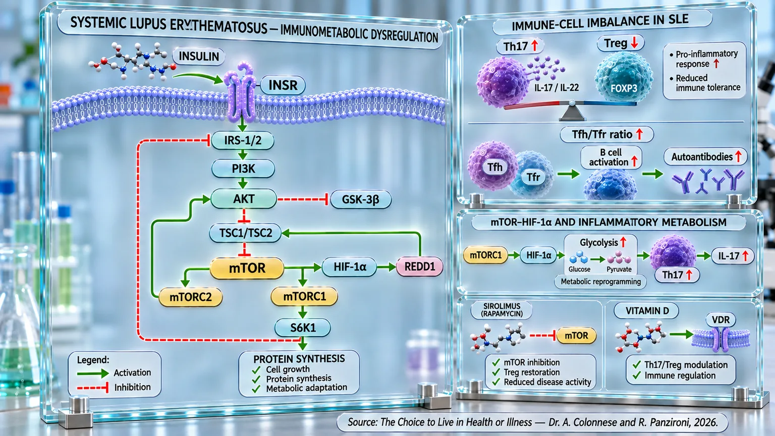
Immunometabolic scheme showing the insulin-mTOR cascade, Th17/Treg and Tfh/Tfr imbalance, HIF-1α-mediated glycolysis, and autoantibody production.
Systemic lupus erythematosus infographic: the INSR–IRS-1/2–PI3K–AKT insulin pathway converges on mTOR, mTORC1/2, HIF-1α, and protein synthesis; an increase in Th17 cells and the Tfh/Tfr ratio, along with a reduction in Tregs, accompanies B-cell activation, autoantibodies, and loss of tolerance, while sirolimus and vitamin D are indicated.

ABSTRACT

Systemic lupus erythematosus (SLE) is a complex autoimmune disease in which immune dysregulation and metabolic alterations are interconnected. In the book "The Choice to Live in Health or Disease," Dr. A. Colonnese and R. Panzironi explore the immunometabolic mechanisms involved in the loss of tolerance and the persistence of systemic inflammation.

The imbalance between Tfh and Tfr lymphocytes promotes B-cell activation and the production of autoantibodies, while the relative increase in Th17 cells compared to Tregs compromises the control of the inflammatory response. Th17 cells produce IL-17 and IL-22; Tregs, associated with FOXP3, contribute instead to immune tolerance. In terms of energy, effector populations rely more heavily on glycolysis and glutaminolysis, processes also supported by mTOR signaling.

In patients with SLE, fasting hyperinsulinemia and reduced insulin sensitivity have been observed, sometimes with normal glycemia, during clinical quiescence and in pediatric age. These alterations do not appear to be exclusively attributable to corticosteroids, without these associations demonstrating direct causality. The hyperactivation of mTOR can promote effector responses, compromise immune regulation, and be associated with reduced autophagy, mitochondrial dysfunction, and the production of reactive oxygen species. Through HIF-1α, mTORC1 also supports glycolysis and Th17 differentiation, potentially contributing to the amplification of inflammation and tissue damage.

The inhibition of mTOR with sirolimus has shown favorable results in experimental models and clinical studies, including changes in Tregs, disease activity, and autoantibodies. Lower concentrations of vitamin D are associated with higher SLE activity; some supplementation studies report modifications in lymphocyte populations and anti-DNA antibodies. Overall, these observations indicate immunometabolism as a relevant field for understanding the pathophysiology of SLE and exploring targeted therapeutic strategies, distinguishing between experimental mechanisms, clinical associations, and effectively demonstrated benefits. However, the clinical variability of the disease requires particular caution when extending results obtained in specific populations to all patients.

Epidemiology of Systemic Lupus Erythematosus

Systemic lupus erythematosus (SLE) is a chronic autoimmune disease that is relatively rare but clinically very significant, with certain epidemiological peculiarities that are important to understand from the outset. First and foremost, it predominantly affects females: many studies report that approximately 85% of diagnosed cases involve women. This strong female predilection is one of the hallmark features of the disease.

Regarding age, lupus tends to manifest primarily during childbearing years: the most frequent onset occurs between the second and fourth decades of life. However, it is important to emphasize that earlier cases (in pediatric age) or later cases (even beyond 50–60 years of age) are by no means rare.

In terms of prevalence, estimates vary considerably depending on geographical areas, ethnicities, and the diagnostic criteria used, but it is still possible to provide figures that help frame the order of magnitude: the standardized prevalence (a measure of disease spread) of SLE increased from approximately 63.5 to 70.6 cases per 100,000 inhabitants in one cohort study, while another larger analysis highlights that the incidence in the general population is around 4–5 new cases per 100,000 person-years, with a much higher ratio in women compared to men, approximately 7.6 versus 1.9 per 100,000 inhabitants.

This set of data suggests that, while it is not a "common" condition in the purely numerical sense, lupus is a condition worth focusing on for several reasons: its high predilection for women, the relatively young age of onset, and the variability in prevalence.

T-cell imbalance

In systemic lupus erythematosus, the immune system seems to progressively lose its ability to recognize what belongs to the body. It is as if the body's defenses, normally vigilant but balanced, become disoriented and begin to attack its own tissues, triggering inflammation that spreads to multiple organs. This imbalance does not arise from nothing: at its root is a complex disorder in T cells (immune cells that help recognize and combat infections), which act as the true "control centers" of immunity, normally directing the immune response like an orchestra conductor. However, when their coordination fails, the entire symphony of the immune system turns into chaotic and self-destructive noise.

One of the most critical mechanisms of this disorder concerns the balance between two fundamental populations: T helper (effector) cells and regulatory T cells. Specifically, in lupus, follicular T helper cells (a class of effector T cells), which are responsible for helping B cells (lymphocytes that contribute to the body's defense) produce antibodies, are hyperactive, while follicular regulatory T cells (Tfr), which normally control their activity, are reduced in number and function. This leads to an increase in the Tfh/Tfr ratio, which is strongly correlated with disease activity and the levels of autoantibodies such as those directed against DNA, typical of SLE. When Tfr cells decrease, the production of autoantibodies increases, and with it, systemic inflammation. Recent studies have shown that effective treatments, by restoring at least part of the Tfr count, are able to reduce this imbalance, a sign that their balance represents a sensitive indicator of the disease state (Xu et al., 2017) [1].

Alongside this Tfh/Tfr axis, another crucial balance is that between T helper 17 (Th17) cells and regulatory T (Treg) cells. Th17 cells can be considered promoters of inflammation because they are responsible for the production of pro-inflammatory cytokines such as IL-17 and IL-22, which amplify the immune response. Treg cells, on the other hand, act as regulators of the immune response, capable of containing and extinguishing inflammation. In lupus, this balance is profoundly altered: Th17 cells prevail, while Treg cells lose stability and suppressive power. This dominance pushes the body into a state of chronic inflammation that affects the kidneys, joints, skin, and nervous system, creating a vicious cycle that is difficult to break (Shan et al., 2020) [2].

Making this picture even more complex is the discovery that lymphocyte imbalance in lupus is not just a problem of "communication" between cells, but also of metabolism: to function, T cells rely on precise metabolic pathways, like an engine that chooses the right fuel depending on its needs. Pro-inflammatory Th17 and Tfh cells prefer glycolysis, that is, the rapid burning of glucose, which provides immediate energy but favors intense and often destructive activity. Treg cells, instead, prefer a "quieter" metabolism based on fatty acid oxidation, which allows them to maintain immunological calm. In lupus, however, this regulation is altered: glycolysis and glutaminolysis (a metabolic process through which the amino acid glutamine is degraded to produce energy) become hyperactive, the mTOR pathway is abnormally activated, and Treg cells lose their metabolic balance, unable to exert the necessary control. The result is an immune system in a state of "energy overconsumption," where inflammatory cells multiply unchecked (Sharabi and Tsokos, 2020) [3]; (Shan et al., 2020) [2].

Even classic regulatory T cells, identified by the markers CD4+ and CD25+ and the transcription factor FOXP3+, are deeply involved in this loss of tolerance. These cells, fundamental for maintaining immune peace, act through various mechanisms: they release calming molecules such as IL-10 and TGF-β, consume IL-2 to prevent inflammatory cells from growing, and modify the metabolism of key amino acids like tryptophan to block the proliferation of effector cells. When these Treg cells are reduced or become dysfunctional, autoimmunity runs rampant. In patients with lupus, this very condition is often observed: a numerical and functional decrease in Treg cells, which makes the immune system unable to distinguish between its own cells and foreign cells (Buckner, 2010) [4].

In summary, lupus can be imagined as the result of a breakdown in the "checks and balances" that keep the immune response in equilibrium. T helper cells push too hard on the accelerator, while regulatory cells can no longer hit the brakes. To complicate matters, the metabolic engine of the cells functions in a disordered way, fueling inflammatory activity instead of tolerance. Understanding how to rebalance these forces, bringing Tfr and Treg cells back to the center of the stage and reducing the excess energy of pro-inflammatory cells, represents one of the most promising frontiers today for restoring the immune system's natural harmony and, with it, the possibility of containing the disease.

Metabolic alterations: insulin pathway and insulin resistance

In systemic lupus erythematosus (SLE), immune dysfunction is not limited to attacks on the body's tissues: it also extends to metabolism, profoundly altering how the body manages energy, sugars, and hormones. It is as if, in addition to the immune system, the metabolic system also loses its ability to self-regulate. In recent years, various studies have highlighted how insulin resistance—a condition in which cells no longer respond effectively to the action of insulin—is surprisingly frequent in patients with lupus, even in the absence of overt diabetes.

From early clinical studies, it emerged that women with SLE show higher fasting insulin levels and reduced sensitivity to the hormone compared to healthy controls, despite maintaining normal glycemia. This means the pancreas is forced to "work harder," producing more insulin to maintain the same amount of sugar in the blood. This condition, known as compensatory hyperinsulinemia, is an early sign of insulin resistance and represents a favorable environment for the future development of metabolic syndrome and cardiovascular diseases. It is no coincidence that many patients with lupus have also been found to have higher levels of triglycerides and oxidized LDL cholesterol, factors that further increase the risk of atherosclerosis (El Magadmi et al., 2006) [5].

Similarly, a study conducted on 58 women with lupus confirmed that insulin resistance and abnormalities in insulin secretion do not necessarily depend on the severity of the disease or the presence of specific autoantibodies. Even in subjects with clinically mild or quiescent disease—that is, temporarily inactive but prone to reactivation—elevated HOMA-IR values (an index used to estimate insulin resistance and pancreatic β-cell function) and fasting insulin levels are observed, indicating reduced efficiency of the insulin-glucose system. It is interesting to note that this condition is not explained by the use of corticosteroids, which are often blamed for altering metabolism, but seems intrinsic to the pathology itself (Tso et al., 2004) [6].

Even more surprising confirmation comes from studies on children and adolescents with juvenile lupus. Even in this population, young and apparently "protected" from metabolic disorders, marked insulin resistance was found compared to healthy peers. This condition was not correlated with either the duration of the disease or the therapy, suggesting that reduced insulin sensitivity may contribute to fueling the chronic inflammation typical of lupus. It is as if insulin dysfunction generates a constant metabolic "background noise" that keeps inflammatory processes active, preventing the body from regaining physiological homeostasis (Tarçın et al., 2022) [7].

Understanding this link between lupus and insulin metabolism does not just mean explaining a biochemical alteration, but grasping a central aspect of the disease itself: the failure of regulation, primarily hormonal and then immune. It is in this dual loss of balance—of the energy system and the defense system—that lupus shows all its complexity and, perhaps, also a possible key to new therapeutic strategies capable of intervening not only in inflammation but also in the metabolism that sustains it.

mTOR as a central node

In the complex landscape of systemic lupus erythematosus, the mTOR signaling pathway emerges as a decisive hub, acting almost like a “metabolic director” that regulates the life and fate of immune cells. The activity of this protein kinase, which under physiological conditions acts as a nutrient sensor and regulator of cell growth, appears profoundly altered in lupus, where constant hyperactivation is observed: it is as if the switch controlling cell growth and energy consumption remains stuck in the “on” position, thereby fueling inflammation through effector lymphocytes and causing a loss of immunological tolerance through the reduction of regulatory T cells.

The mTOR pathway, in fact, acts as a sort of “bioenergetic control center” that decides whether a T cell should become a promoter of inflammation (Th1, Th17) or a regulatory and pacifying element (Treg). When mTOR is hyperactive, effector T cells are encouraged to proliferate and produce inflammatory cytokines, while regulatory T cells lose stability and function. This reversal of roles contributes to a state of “immune anarchy,” in which the cells that should curb immune aggression become too weak to do so (Delgoffe et al., 2009) [8]; (Zhao et al., 2022) [9].

In lupus, the dysregulation of mTOR is not limited to T cells: it affects the entire metabolic balance of the immune system, involving mitochondria—the cell's power plants—and autophagy processes, which normally eliminate damaged cellular components. When mTOR is hyperactive, autophagy is reduced, mitochondria become dysfunctional and begin to produce excessive amounts of free radicals (ROS), generating a persistent pro-inflammatory environment. This condition alters cellular metabolism and favors the survival of autoreactive cells that continue to recognize the body's own tissues as “foreign” (Robinson et al., 2022) [10]; (Saadh et al., 2023) [11].

The differentiation of T helper cells and the production of antibodies by B cells are also influenced by this imbalance. Excessive activation of mTORC1 promotes lymphocyte proliferation and the secretion of inflammatory cytokines, thus amplifying the autoimmune cascade. At the same time, the reduction in autophagy prevents the proper disposal of cellular and nuclear fragments, which become targets for the formation of autoantibodies. In other words, mTOR, which should precisely regulate energy and cell differentiation, transforms into an accelerator of immune dysfunction (He et al., 2020) [12].

The impact of this pathway is so pervasive that some authors have proposed considering lupus as a “disease of mTOR hyperactivity.” This definition, while simplified, helps to understand the profound interconnection between metabolism and immunity: chronic inflammation is not just an abnormal response of the immune system, but also a reflection of out-of-control energy metabolism, in which cells consume excessive oxygen and nutrients without being able to return to a state of rest.

Another crucial element is the ability of mTOR to integrate signals from the microenvironment: glucose, amino acids, lipids, and growth factors act as messages that guide the choice of T cells between a pro-inflammatory and a regulatory fate. When these signals are altered, as occurs in lupus, mTOR receives incorrect instructions and pushes cells toward uncontrolled proliferation. This creates a “distorted dialogue” between metabolism and immunity, where every energy error translates into an immunological error (Fernandez and Perl, 2010) [13].

The discovery of these mechanisms has also had an important therapeutic implication: drugs like rapamycin, originally developed as an immunosuppressant, have proven capable of selectively “turning off” the mTOR pathway, restoring balance to the immune system. The inhibition of mTORC1 by rapamycin, in fact, promotes the expansion of regulatory T cells and the reduction of effector cells, attenuating inflammatory activity and improving clinical parameters in patients with lupus refractory to conventional therapies. Furthermore, the antioxidant and anti-fibrotic effects of rapamycin help protect target organs, such as the kidneys and skin, which are often damaged by the disease (Zhao et al., 2022) [9]; (Fernandez and Perl, 2010) [13].

These findings suggest that mTOR is not just one player among many, but a sort of “molecular director” of lupus pathogenesis: it regulates the activity of T and B cells, modulates energy metabolism, and governs cell survival and differentiation processes; when this direction is altered, the entire immune system loses synchronization, generating the disharmony we recognize as autoimmunity. Restoring the balance of mTOR, therefore, does not simply mean suppressing inflammation, but bringing order back to the interrupted dialogue between metabolism and the immune system, restoring the body's ability to recognize and protect itself without self-destructing.

mTOR inhibitors

In recent years, the inhibition of the mTOR pathway has represented one of the most promising discoveries in the management of autoimmune diseases, particularly systemic lupus erythematosus. After decades in which treatments relied almost exclusively on corticosteroids and non-specific immunosuppressants (drugs that reduce an excessive immune response), rapamycin and its analog sirolimus have opened a new therapeutic chapter: no longer just the simple suppression of the immune system, but its reprogramming toward balance.

The principle behind these drugs is as simple as it is elegant: to modulate the mTOR signaling pathway, the “metabolic controller” of immune cells, to reduce the hyperactivity of effector cells and promote the recovery of regulatory cells. In patients with lupus, in fact, mTOR is like an accelerator stuck to the floor: inflammatory T cells proliferate unchecked, while regulatory T cells (Tregs), those responsible for maintaining immunological tolerance, are few and ineffective. By inhibiting mTOR, rapamycin turns off this excess energy and restores the biological silence necessary for tolerance (Battaglia et al., 2012) [14].

As early as the 1990s, experiments conducted on murine models (studies conducted on rodents) had shown surprising results: in mice predisposed to lupus, treatment with rapamycin not only prolonged survival but also halted pathological alterations in the kidneys, liver, and lungs, drastically reducing levels of autoantibodies and diffuse inflammatory lesions (Warner et al., 1994) [15]. In those pioneering studies, the treated animals showed a return to near-normal immunological parameters, as if the system had “remembered” its correct balance.

Years later, the same effects were also confirmed in humans: in patients suffering from refractory lupus, i.e., those resistant to conventional treatments, rapamycin led to a clear clinical improvement, with a reduction in disease activity and a significant decrease in the dose of corticosteroids required to maintain control. Even more interestingly, rapamycin normalized calcium flux in T cells, a parameter closely linked to their hyperactivation, suggesting a profound effect on cellular metabolism and not just on superficial inflammation (Fernandez et al., 2006) [16].

One of the most relevant aspects that emerged from this research is the ability of sirolimus to expand CD4+ and CD25+ regulatory T cells, and the FOXP3+ transcription factor, which are considered the cornerstone of immune tolerance. In vitro, rapamycin allows these cells to be multiplied while maintaining their phenotype—that is, the set of an individual's visible and functional characteristics—and their suppressive function, a result that has opened the door to potential applications in personalized cell therapies for autoimmune diseases (Battaglia et al., 2012) [14].

The most robust clinical evidence comes from prospective studies and meta-analyses conducted on adult and pediatric patients: in several trials, sirolimus led to a significant reduction in the SLEDAI activity index (an index created to provide an objective and reproducible assessment of SLE activity), an improvement in C3 and C4 complement levels (plasma proteins of the immune system that participate in defense against infections and the regulation of inflammation), and a decrease in anti-DNA autoantibodies, all indicators of effective immunological control (Peng et al., 2020) [17]; (Ji et al., 2020) [18]. At the same time, patients were able to consistently reduce their use of prednisone (a drug with potent anti-inflammatory action) by an average of 10-13 mg per day, thus limiting the long-term side effects of corticosteroids, one of the therapeutic priorities in the management of lupus.

Even in children with early-onset lupus, sirolimus has proven effective and well-tolerated: in a study on pediatric patients, the drug significantly reduced disease activity, improved immunological parameters, and allowed for marked steroid-sparing, without recording serious adverse events (Zhang et al., 2022) [19]. The few cases of minor infections reported resolved spontaneously, confirming a favorable safety profile.

A particularly interesting area is that of refractory cutaneous lupus, where rapamycin has shown the ability to reduce inflammatory lesions and improve skin quality even in the most resistant cases. In a patient with generalized discoid lupus erythematosus, a severe form that is poorly responsive to conventional therapies, treatment with sirolimus led to evident improvement after just a few weeks, without significant side effects (Herold et al., 2018) [20]. These results suggest that mTOR inhibitors do not act only on circulating immune system cells, but also at the tissue level, where they regulate local inflammatory activity and the skin response.

In cases of lupus nephritis, one of the most serious complications of the disease, sirolimus has shown efficacy comparable to that of mycophenolate mofetil (an immunosuppressive drug that inhibits the proliferation of T and B lymphocytes), the current standard of care. In a study of over one hundred patients, the two treatments had similar effects on the disease, but sirolimus showed some advantages and greater safety, without serious adverse effects (Bai et al., 2025) [21].

The combination of these experimental and clinical results confirms that mTOR inhibitors are not simply immunosuppressive drugs, but true immunometabolic balance modulators. They do not shut down the immune system, but “re-educate” it to recognize the boundary between defense and self-destruction. Their efficacy in lupus, and the growing interest in their use in other autoimmune pathologies, represent the transition from a medicine of containment to a medicine of restoration.

Ultimately, rapamycin and sirolimus offer a new vision for the treatment of autoimmune diseases: a therapy capable of speaking the language of cells, correcting errors in their metabolism, and restoring the immune system's ability to self-regulate. And in this perspective, mTOR inhibition appears not only as a therapeutic strategy but as an attempt to restore balance to the interrupted dialogue between immunity and biological function.

mTOR, HIF-1α and metabolic reprogramming of immune system cells

In systemic lupus erythematosus, the persistent activation of mTOR can contribute to the loss of immunological balance through the activation of HIF-1α. mTOR is, in fact, one of the cell's main metabolic sensors and, when persistently stimulated, it promotes an anabolic and glycolytic program compatible with the activation of effector lymphocytes. HIF-1α constitutes one of the central effectors of this program: it increases the expression of glycolytic genes and supports the differentiation of Th17 cells, whereas a metabolic profile less dependent on glycolysis more strongly favors the regulatory compartment. In SLE, this relationship is particularly relevant because both mTOR and HIF-1α are altered in T cells, creating the conditions for a Th17/Treg imbalance and the maintenance of autoimmune inflammation.

One of the most direct mechanistic demonstrations was provided in 2022: researchers identified SIRT2 (an enzyme that contributes to the regulation of metabolism and cellular functions) as a regulator of Th17 differentiation in CD4+ T cells. SIRT2 promoted the activation of the mTORC1–HIF-1α sequence, thereby increasing Th17 differentiation and the production of IL-17A. In T cells from patients with SLE, SIRT2 was found to be increased, and its pharmacological inhibition reduced the ability to generate Th17 cells. In mice genetically predisposed to develop a lupus-like disease, blocking SIRT2 also attenuated the autoimmune disease. This study therefore provides particularly important evidence of the functional presence of the mTORC1→HIF-1α→Th17 axis in lupus (Hisada et al., 2022) [22].

In the same year, a second study directly analyzed HIF-1α in T lymphocytes of patients with SLE: the expression of this transcription factor was found to be significantly increased in CD4+ T cells at both the mRNA and protein levels. Furthermore, HIF-1α was particularly elevated under conditions that favored Th17 differentiation; its experimental overexpression in murine T lymphocytes indeed increased the generation of Th17 cells. In patients, HIF-1α expression positively correlated with genes involved in glycolysis and with the IL-17 program. These results position HIF-1α as a metabolic effector directly involved in the expansion of the Th17 compartment in lupus (Liao et al., 2022) [23].

An important contribution to the understanding of the metabolism upstream of HIF-1α was provided by other researchers in 2019: in the same type of mice seen previously, the inhibition of glutaminase 1 (an enzyme that allows the cell to metabolically utilize glutamine) reduced glycolysis, the expression of the HIF-1α protein, and Th17 differentiation, accompanied by an improvement in the autoimmune pathology. The same treatment applied to T cells from patients with SLE decreased Th17 differentiation. Genetic silencing experiments also confirmed that the reduction of HIF-1α represented a step through which the inhibition of glutaminolysis limited glycolysis and Th17 activity. The study therefore demonstrates how closely HIF-1α is integrated with the pathological metabolism of T lymphocytes in lupus (Kono et al., 2019) [24].

The role of the upstream node had already been highlighted years earlier by other researchers, who directly analyzed T lymphocytes from patients with SLE: mTORC1 activity was found to be increased and was associated with an expansion of Th17 cells and CD4+CD8+ T cells (effector cells), while the Treg population was reduced.

The pathological importance of HIF-1α was finally confirmed at the tissue level in 2023. In a model of cutaneous lupus, CD4+ and CD8+ T lymphocytes infiltrating the skin expressed high levels of HIF-1α and showed a marked cytotoxic program (a set of mechanisms that allow immune cells to eliminate dangerous cells). Inhibition of HIF-1α reduced the cytotoxic activity of T cells and attenuated both the skin disease and systemic manifestations. In the tissues of patients with cutaneous lupus, lymphocyte infiltrates also showed high HIF-1α activity. This transcription factor therefore appears not only as a metabolic regulator but as an effector directly involved in tissue damage in lupus (Little et al., 2023) [25].

Overall, these findings allow for the reconstruction of a coherent sequence. In SLE, various metabolic and immunological stimuli favor the activation of mTOR, which downstream supports HIF-1α, increases glycolysis, and promotes the differentiation of Th17 cells. The increase in IL-17 and the simultaneous relative reduction of the Treg compartment therefore contribute to the loss of immunological tolerance. HIF-1α can also support cytotoxic programs in T cells infiltrating tissues, contributing directly to organ damage. A particularly relevant pathogenetic axis is thus outlined: mTOR activation → increase in HIF-1α → glycolysis → Th17 → IL-17 → amplification of autoimmunity and tissue damage in systemic lupus erythematosus.

Vitamin D as an immuno-metabolic modulator

The complex interplay between metabolism, the immune system, and inflammation in systemic lupus erythematosus does not end with the alteration of major metabolic hormones. Another player, often silent but crucial, enters the scene: vitamin D. In recent years, this vitamin has emerged as one of the most studied molecules in the context of autoimmune diseases, taking on a role that goes far beyond the traditional metabolism of calcium and bones. It acts as a true immunomodulatory hormone, capable of profoundly influencing the behavior of immune system cells and maintaining the balance between inflammation and tolerance. Under normal conditions, vitamin D silently but constantly regulates the activity of T and B cells, promoting a controlled response and preventing the defense system from turning against the body itself. However, when its levels drop, this balance is broken, and autoimmunity finds fertile ground to develop.

A vast meta-analysis has confirmed that patients with systemic lupus erythematosus have significantly lower levels of 25-hydroxyvitamin D, regardless of age, therapy, or season. Vitamin D deficiency was found to be not only more common among patients compared to healthy subjects but also closely correlated with disease severity: the lower the 25(OH)D values, the higher the lupus activity index (SLEDAI) (Guan et al., 2019) [26]. Another meta-analysis of over two thousand patients confirmed the same trend: vitamin D deficiency significantly increases the risk of developing lupus and worsens disease progression, while its sufficiency is associated with lower inflammatory activity and a more stable clinical course (Islam et al., 2019) [27].

European multicenter studies have provided further confirmation: by analyzing nearly four hundred patients, some researchers demonstrated an inverse correlation between serum vitamin D levels and lupus activity: every decrease in the concentration of 25-OH-vitamin D was accompanied by an increase in the disease activity score, suggesting that vitamin D acts as a sort of “biological brake” on the immune system (Amital et al., 2010) [28]. Seasonal fluctuations also influence the course of the pathology: vitamin D levels tend to decrease in winter, a period in which patients show a lower probability of reaching a state of low disease activity (LLDAS), while in summer, with greater sun exposure, the chances of clinical control improve significantly (Kim et al., 2024) [29].

On an immunological level, vitamin D acts as a fine modulator of the dialogue between regulatory T cells (Treg) and Th17 cells, two populations that represent the balance and the inflammatory drive of the immune system, respectively. In children with early-onset lupus, it has been observed that low levels of 25(OH)D are associated with a prevalence of Th17 cells and a reduced number of Tregs, with a Th17/Treg ratio skewed toward inflammation. Conversely, higher concentrations of vitamin D restore balance, increasing regulatory cells and reducing the production of pro-inflammatory cytokines such as IL-17, IL-6, and TNF-α (Jiang et al., 2023) [30]. This effect can be interpreted as a true “reset” of the immune system, which returns to distinguishing native tissue from foreign bodies.

From a clinical perspective, vitamin D supplementation has been shown to restore immunological homeostasis even in patients with documented hypovitaminosis. In a prospective study, after six months of supplementation, 25(OH)D levels increased significantly, accompanied by an increase in Treg cells and a reduction in Th1 and Th17 cells, as well as a drop in anti-DNA antibody titers, indicating better disease control. The therapy proved to be well-tolerated, with no flare-ups or significant adverse effects (Terrier et al., 2012) [31].

In summary, vitamin D acts as a universal mediator of immune and metabolic balance; in fact, as we saw in chapter 6 dedicated to the immune system, through the modulation of mTOR, it reduces the production of inflammatory mediators, limits the abnormal proliferation of effector T lymphocytes, and stimulates the production of regulatory T lymphocytes, which are central processes in the pathogenesis of the disease. This regulatory effect promotes better control of the immune response, preventing autoimmune aggression against tissues.

Restoring adequate levels of vitamin D means, in a sense, returning to the immune system its capacity for discernment, rebalancing the scales between defense and tolerance. It is like restoring light to an organism that, in the absence of this hormone-molecule, constantly operates in the shadows.

Conclusions

The scientific evidence analyzed in this chapter outlines systemic lupus erythematosus (SLE) as a complex autoimmune disease in which immunological and metabolic alterations are deeply interconnected. The disease predominantly affects females, especially during childbearing age, and presents significant epidemiological and clinical variability.

The first central element concerns the loss of balance between lymphocyte populations. In SLE, an increased Tfh/Tfr ratio promotes B-cell activation and autoantibody production, while the Th17/Treg imbalance leads to the predominance of a pro-inflammatory response. Th17 cells produce inflammatory mediators such as IL-17 and IL-22, while Tregs, which are normally responsible for maintaining tolerance through FOXP3 and regulatory mediators, are reduced or functionally impaired. This alteration also has a metabolic component: effector populations rely more heavily on glycolysis and glutaminolysis, while the abnormal activation of mTOR contributes to maintaining their inflammatory phenotype.

Associated with this picture are significant alterations in insulin metabolism: research documents higher fasting insulin levels and reduced insulin sensitivity in SLE patients, which can be observed even with normal glycemia, in clinically quiescent forms, and even in pediatric patients. These observations demonstrate that insulin dysfunction can be present independently of overt diabetes and does not appear to be explained solely by the use of corticosteroids, reinforcing the existence of an important metabolic component of the disease.

The main point of convergence between metabolic and immune regulation is represented by mTOR: its hyperactivation favors effector T cells and compromises the regulatory compartment, reduces autophagy, and is associated with mitochondrial dysfunction and ROS production. Consistently, mTOR inhibition via rapamycin/sirolimus has shown significant results in experimental models and clinical studies, promoting Treg expansion, reducing disease activity, anti-DNA autoantibodies, and the need for corticosteroids. Favorable results have also been reported in pediatric lupus, refractory cutaneous lupus, and lupus nephritis.

A further step allows for linking mTOR to metabolic reprogramming through HIF-1α: studies show that the mTORC1–HIF-1α axis supports glycolysis and promotes Th17 differentiation and IL-17 production. Conversely, inhibiting the metabolic processes that fuel HIF-1α reduces the Th17 phenotype and attenuates experimental disease. This transcription factor also participates in the cytotoxic programs of tissue-infiltrating T cells. A consistent sequence therefore emerges: mTOR → HIF-1α → glycolysis → Th17 → IL-17 → amplification of autoimmunity and tissue damage.

Finally, vitamin D emerges as an important immunometabolic modulator: in SLE patients, lower concentrations of 25(OH) D are associated with higher disease activity and a Th17/Treg imbalance. In the subjects studied, supplementation increased Tregs, reduced Th1 and Th17, and decreased anti-DNA antibodies.

Overall, SLE thus emerges as the result of an interaction between metabolic alteration, dysregulation of mTOR and HIF-1α, energy reprogramming of immune cells, and loss of tolerance, highlighting immunometabolism as a fundamental key to interpreting the pathogenesis and new therapeutic possibilities for the disease.

References

[1] The ratio of circulating follicular T helper cell to follicular T regulatory cell is correlated with disease activity in systemic lupus erythematosus

Xu et al., 2017

Clin Immunol. 2017 Oct:183:46-53.

[2] T Cell Metabolism: A New Perspective on Th17/Treg Cell Imbalance in Systemic Lupus Erythematosus

Shan et al., 2020

Front Immunol. 2020 May 22:11:1027.

[3] T cell metabolism: new insights in systemic lupus erythematosus pathogenesis and therapy

2020

Sharabi e Tsokos. Nat Rev Rheumatol. 2020 Feb;16(2):100-112.

[4] Mechanisms of impaired regulation by CD4(+) CD25(+)FOXP3(+) regulatory T cells in human autoimmune diseases

Buckner, 2010

Nat Rev Immunol. 2010 Dec;10(12):849-59.

[5] Hyperinsulinemia, insulin resistance, and circulating oxidized low density lipoprotein in women with systemic lupus erythematosus

Magadmi et al., 2006

J Rheumatol. 2006 Jan;33(1):50-6.

[6] Clinical evaluation of insulin resistance and beta-cell function by the homeostasis model assessment in patients with systemic lupus erythematosus

Tso et al., 2004

Clin Rheumatol. 2004 Oct;23(5):416-20.

[7] Insulin resistance in children with juvenile systemic lupus erythematosus and ınvestigation of the possibly responsible factors

Tarçın et al., 2022

Clin Rheumatol. 2022 Mar;41(3):795-801.

[8] The mTOR kinase differentially regulates effector and regulatory T cell lineage commitment

Delgoffe et al., 2009

Immunity. 2009 Jun 19;30(6):832-44.

[9] mTOR signaling: A pivotal player in Treg cell dysfunction in systemic lupus erythematosus

Zhao et al., 2022

Clin Immunol. 2022 Dec:245:109153.

[10] The Role of Immunometabolism in the Pathogenesis of Systemic Lupus Erythematosus

Robinson et al., 2022

Front Immunol. 2022 Jan 26:12:806560.

[11] Role of T cells in the pathogenesis of systemic lupus erythematous: Focus on immunometabolism dysfunctions

Saadh et al., 2023

Int Immunopharmacol. 2023 Jun:119:110246.

[12] Advances in systemic lupus erythematosus pathogenesis via mTOR signaling pathway

He et al., 2020

Semin Arthritis Rheum. 2020 Apr;50(2):314-320.

[13] mTOR signaling: a central pathway to pathogenesis in systemic lupus erythematosus?

Fernandez e Perl, 2010

Discov Med. 2010 Mar;9(46):173-8.

[14] Expanding human T regulatory cells with the mTOR-inhibitor rapamycin

Battaglia et al., 2012

Methods Mol Biol. 2012:821:279-93.

[15] Rapamycin prolongs survival and arrests pathophysiologic changes in murine systemic lupus erythematosus

Warner et al., 1994

Arthritis Rheum. 1994 Feb;37(2):289-97.

[16] Rapamycin reduces disease activity and normalizes T cell activation-induced calcium fluxing in patients with systemic lupus erythematosus

Fernandez et al., 2006

Arthritis Rheum. 2006 Sep;54(9):2983-8.

[17] Clinical efficacy and safety of sirolimus in systemic lupus erythematosus: a real-world study and meta-analysis

Peng et al., 2020

Ther Adv Musculoskelet Dis. 2020 Sep 14:12:1759720X20953336.

[18] Efficacy and safety of sirolimus in patients with systemic lupus erythematosus: A systematic review and meta-analysis

Ji et al., 2020

Semin Arthritis Rheum. 2020 Oct;50(5):1073-1080.

[19] Clinical efficacy and safety of sirolimus in childhood-onset systemic lupus erythematosus in real world

Zhang et al., 2022

Medicine (Baltimore). 2022 Nov 18;101(46):e31551.

[20] Rapamycin for refractory discoid lupus erythematosus

Herold et al., 2018

Dermatol Ther. 2018 Sep;31(5):e12631.

[21] Sirolimus versus mycophenolate mofetil for the treatment of lupus nephritis: Results from a real-world CSTAR cohort study

Bai et al., 2025

Rheumatol Immunol Res. 2025 Jul 1;6(2):80-89.

[22] The Deacetylase SIRT2 Contributes to Autoimmune Disease Pathogenesis by Modulating IL-17A and IL-2 Transcription.

Hisada et al. Cellular & Molecular Immunology, 2022

vol. 19, no. 6, 2022, pp. 738–750.

[23] Increased HIF-1α Expression in T Cells and Associated with Enhanced Th17 Pathway in Systemic Lupus Erythematosus.

Liao et al., 2022

Journal of the Formosan Medical Association, vol. 121, no. 12, 2022, pp. 2446–2456.

[24] Glutaminase 1 Inhibition Reduces Glycolysis and Ameliorates Lupus-like Disease in MRL/lpr Mice and Experimental Autoimmune Encephalomyelitis.

Kono et al. Arthritis & Rheumatology, 2019

vol. 71, no. 11, 2019, pp. 1869–1878.

[25] HIF-1 Regulates Pathogenic Cytotoxic T Cells in Lupus Skin Disease.

Little et al. JCI Insight, 2023

vol. 8, no. 16, 2023, e166076.

[26] Association between circulating 25-hydroxyvitamin D and systemic lupus erythematosus: A systematic review and meta-analysis

Guan et al., 2019

Int J Rheum Dis. 2019 Oct;22(10):1803-1813.

[27] Vitamin D status in patients with systemic lupus erythematosus (SLE): A systematic review and meta-analysis

Islam et al., 2019

Autoimmun Rev. 2019 Nov;18(11):102392.

[28] Serum concentrations of 25-OH vitamin D in patients with systemic lupus erythematosus (SLE) are inversely related to disease activity: is it time to routinely supplement patients with SLE with vitamin D?

Amital et al., 2010

Ann Rheum Dis. 2010 Jun;69(6):1155-7.

[29] Seasonal vitamin D levels and lupus low disease activity state in systemic lupus erythematosus

Kim et al., 2024

Eur J Clin Invest. 2024 Jan;54(1):e14092.

[30] The changes of Treg and Th17 cells relate to serum 25(OH)D in patients with initial-onset childhood systemic lupus erythematosus

Jiang et al., 2023

Front Pediatr. 2023 Aug 23:11:1228112.

[31] Restoration of regulatory and effector T cell balance and B cell homeostasis in systemic lupus erythematosus patients through vitamin D supplementation

Terrier et al., 2012

Arthritis Res Ther. 2012 Oct 17;14(5):R221.