
Scientific Book

Omega-3 and cell membrane composition

Omega-3 fatty acids, particularly EPA and DHA, play essential roles in the structure of cell membranes and the regulation of inflammation. Research is investigating their effects on lipid metabolism, cardiovascular health, immunity, and brain function, with benefits that depend on the clinical context. In the book "The Choice to Live in Health or Disease," Dr. A. Colonnese and R. Panzironi explore the biological mechanisms of omega-3 and the evidence regarding their supplementation.

Cite this article
Colonnese, A. & Panzironi, R. (2026). Omega-3 and cell membrane composition. The Choice to Live in Health or in Disease. https://lifesciencehub.info/en/la-scelta-di-vivere-in-salute-o-in-malattia/omega-3-and-cell-membrane-composition
The Choice to Live in Health or in Disease · ISBN 979-12-82692-03-8

DOI lsh.en.libro.27

  • Published 05/11/2026
  • Updated 05/11/2026
  • OMEGA-3
  • EPA
  • DHA
  • ARACHIDONIC ACID
  • PI3K/AKT/MTOR
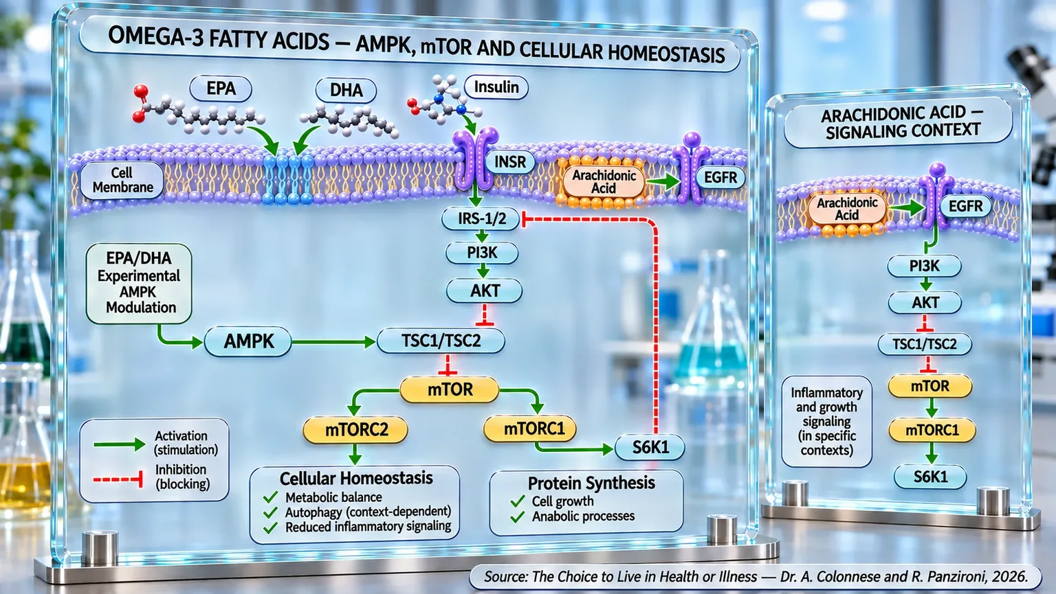
Molecular infographic showing a phospholipid bilayer, INSR and EGFR receptors, and green or red arrows pointing toward intracellular metabolic complexes.
Diagram of interactions between the cell membrane, EPA, DHA, insulin, and arachidonic acid: the INSR–IRS-1/2–PI3K–AKT and EGFR–PI3K–AKT pathways converge on TSC1/TSC2–mTOR, mTORC1, mTORC2, and S6K1. The experimental modulation of AMPK by EPA/DHA is linked to cellular homeostasis, protein synthesis, and inflammatory signals.

ABSTRACT

Omega-3 fatty acids, primarily EPA and DHA, contribute to the structure of cell membranes and the regulation of inflammatory, immune, and metabolic responses. In the book "The Choice to Live in Health or Disease," Dr. A. Colonnese and R. Panzironi examine the relationship between the availability of omega-3 and omega-6, the lipid composition of tissues, and cell signaling mechanisms, distinguishing biological adaptations from clinical benefits that have yet to be confirmed.

The intake of EPA and DHA promotes their progressive incorporation into the membranes of cardiomyocytes, erythrocytes, platelets, and immune cells, modifying their composition and reducing the proportion of arachidonic acid in certain compartments. This variation can influence the production of inflammatory mediators. In specific experimental models, arachidonic acid is involved in the activation of PI3K/AKT/mTOR, while EPA and DHA can modulate this signaling, limit mTOR activity, and promote autophagy. The activation of AMPK constitutes a further possible link to energy metabolism and the control of inflammation.

Studies report improvements in certain cardiovascular, inflammatory, and metabolic indicators. Favorable results regarding insulin sensitivity have been observed in gestational diabetes, polycystic ovary syndrome, and pediatric obesity, although the evidence is heterogeneous. Interest in Alzheimer's and Parkinson's disease primarily concerns neuroinflammation and neuronal protection; further implications are being explored in autoimmune and dermatological diseases. In oncology, the effects on cell proliferation are derived mainly from experimental observations and do not demonstrate therapeutic efficacy in humans.

Overall, omega-3s participate in interconnected cellular processes through membrane modifications and the regulation of metabolic pathways. An adequate dietary intake of EPA and DHA can be part of a balanced lifestyle, while any potential supplementation requires an individual assessment. It remains essential to distinguish the mechanisms observed in experimental models from clinical outcomes, avoiding the assumption that the modulation of AMPK and mTOR is equivalent to the proven prevention or treatment of diseases in the various populations and metabolic conditions studied to date.

Its properties

Omega-3 fatty acids belong to the family of essential polyunsaturated fatty acids—nutrients that the human body cannot synthesize on its own and must therefore be obtained through diet. Among these, eicosapentaenoic acid (EPA) and docosahexaenoic acid (DHA) are the most biologically active and widely studied forms. Although for many years Omega-3s were associated almost exclusively with cardiovascular health, modern research has progressively broadened this view, showing how these lipids participate in the regulation of inflammation, immunity, neuronal function, energy metabolism, and even the processes involved in aging and the progression of numerous chronic diseases. Today, Omega-3s are considered true biological regulators, capable of influencing cell behavior through mechanisms that go well beyond their simple structural role in cell membranes.

To understand the importance of Omega-3s, it is useful to imagine the cell membrane as a city surrounded by walls. These walls are not a passive barrier, but a dynamic system that continuously regulates the entry and exit of signals, nutrients, and information. EPA and DHA are incorporated into cell membranes, modifying their fluidity, functionality, and ability to respond to environmental stimuli. This characteristic allows Omega-3s to influence numerous biological processes, including the production of molecules involved in the inflammatory response (Lakshimi and Kavitha, 2023) [1].

One of the most relevant aspects to emerge from the literature concerns the ratio between Omega-3 and Omega-6. Both families of fatty acids are essential for health, but they must be present in an appropriate balance. Throughout human evolution, the ratio between Omega-6 and Omega-3 was likely close to 1:1. With the advent of the modern diet, characterized by a high use of refined vegetable oils and a reduction in fish consumption, this balance has progressively shifted, reaching values in many Western populations exceeding 15:1 or even 20:1. This imbalance promotes the production of pro-inflammatory lipid mediators and contributes to the development of a state of low-grade chronic inflammation, a condition now considered one of the main factors underlying modern chronic diseases (Poggioli et al., 2023) [2]; (Lakshimi and Kavitha, 2023) [1].

From a physiological perspective, EPA and DHA act as natural antagonists to numerous inflammatory mechanisms. When a cell is subjected to stress or damage, fatty acids are released from the membranes and subsequently transformed into signaling molecules. If Omega-6s prevail, the response tends to be more inflammatory; if, on the other hand, adequate amounts of Omega-3s are available, specialized mediators are produced that promote the resolution of inflammation. This does not mean suppressing the immune system, but guiding it toward an orderly and efficient conclusion of the inflammatory response (Poggioli et al., 2023) [2]; (Lakshimi and Kavitha, 2023) [1].

The immunomodulatory action of Omega-3s represents one of the most promising areas of research. EPA and DHA are able to reduce the activation of immune cells and limit the release of pro-inflammatory cytokines, molecules that, if produced in excess, can contribute to tissue damage. This effect has led many researchers to study their potential role in autoimmune diseases, conditions in which the immune system loses the ability to correctly distinguish between what belongs to the body and what is foreign. Several studies have observed a reduction in disease activity in conditions such as rheumatoid arthritis, systemic lupus erythematosus, multiple sclerosis, and type 1 diabetes in subjects who consumed adequate amounts of Omega-3s (Poggioli et al., 2023) [2].

However, the effects of Omega-3s are not limited to the peripheral immune system. A particularly high amount of DHA is present in the brain, where it contributes to the structure of neuronal membranes and the transmission of nerve signals. In recent years, the hypothesis that many neurological and psychiatric diseases share a common denominator—chronic neuroinflammation—has emerged with increasing evidence. In this condition, cells normally responsible for protecting the nervous system, such as microglia and astrocytes, remain persistently activated, producing inflammatory substances that compromise neuronal function. In major depressive disorder, for example, increases in biomarkers associated with neuronal damage and brain immune activation have been observed. Omega-3s appear to counteract these processes through the reduction of microglial activation (the response of the brain's defense cells, called microglia, to infections, damage, or other threats), the decrease in the production of inflammatory cytokines, and the modulation of specific biomarkers of neuronal damage (Malau et al., 2024) [3].

These observations help to understand why numerous studies have associated adequate levels of Omega-3s with better mental and cognitive health. The brain can be compared to an extremely complex electrical grid: for communication between nerve cells to occur correctly, neuronal membranes must maintain a flexible and functional structure. EPA and DHA help preserve this characteristic, promoting synaptic plasticity and neuronal resilience in the face of harmful stimuli (Malau et al., 2024) [3].

The importance of Omega-3s becomes particularly evident during aging. As the years pass, the risk of cognitive decline, reduced mobility, and loss of independence increases. In a population of elderly people with subjective memory complaints, a higher dietary intake of EPA and DHA was associated with better global cognitive performance, greater attention, better psychomotor speed, and better physical mobility parameters. In particular, EPA showed favorable associations with walking speed and other measures of motor function, suggesting that maintaining an adequate intake of Omega-3s can help preserve not only mental but also physical capabilities during aging (Erhardt et al., 2021) [4].

Metabolism is also strongly influenced by the availability of Omega-3s. Scientific evidence indicates that these fatty acids can improve insulin sensitivity, reduce systemic inflammation, and preserve endothelial function. Furthermore, several studies suggest a potential role in slowing the progression of diabetic complications such as retinopathy, nephropathy, and proteinuria, conditions closely associated with chronic vascular damage caused by hyperglycemia (Lakshimi and Kavitha, 2023) [1].

The cardiovascular system is probably one of the most studied areas. Omega-3s contribute to the reduction of plasma triglycerides, improve endothelial function, and favorably modulate the inflammatory processes involved in atherosclerosis. In other words, they seem to act both on the quality of the blood flowing in the vessels and on the health of the vascular walls themselves. This dual effect helps explain their established role in cardiovascular prevention (Lakshimi and Kavitha, 2023) [1]; (Poggioli et al., 2023) [2].

Interest in Omega-3s has also extended to the field of oncology. A meta-analysis that included twenty-one cohort studies observed that a high consumption of fish and marine Omega-3s was associated with better survival in cancer patients. The benefits were particularly evident in some neoplasms, including prostate cancer and ovarian cancer. Although the mechanisms are not yet fully understood, the authors hypothesize that the reduction of systemic inflammation, the modulation of the immune response, and the inhibition of processes involved in tumor growth may contribute to these effects (Wang et al., 2023) [5].

The immunomodulatory properties of Omega-3s have also attracted attention in the context of viral infections. Recent studies suggest that EPA and DHA may help modulate the systemic inflammatory response during infections, reducing the risk of the immune response itself becoming excessively aggressive. In the case of COVID-19, some papers have reported improvements in respiratory parameters and clinical outcomes in subjects taking Omega-3s, although further studies are needed to definitively confirm these results (Poggioli et al., 2023) [2]; (Lakshimi and Kavitha, 2023) [1].

Taken together, the available evidence outlines a coherent picture. Omega-3s are not simply energy nutrients or structural components of cell membranes, but true regulators of biological communication. Through the modulation of inflammation, immunity, neuronal function, and metabolism, EPA and DHA participate in processes fundamental to the maintenance of homeostasis. Furthermore, the increasing prevalence of chronic diseases associated with inflammation makes the recovery of an adequate balance between Omega-6 and Omega-3 particularly relevant—a goal that appears today not only nutritional, but profoundly physiological. The most recent research suggests that maintaining this balance may represent one of the most effective tools for supporting health throughout the lifespan, from brain function to cardiovascular health, from immune regulation to healthy aging.

Supplementation

If the first step in understanding omega-3s is knowing their physiology, the next step is understanding what happens when these nutrients are introduced in quantities higher than those usually present in the diet. Supplementation is, in fact, a particularly interesting tool because it allows us to observe, in a relatively short time, how EPA and DHA are incorporated into tissues and how this incorporation can translate into measurable biological changes.

For many years, it was hypothesized that the effects of omega-3s were the result of indirect and slow mechanisms. However, more recent research has shown that EPA and DHA can be incorporated into human tissues with surprising speed. A study conducted on patients undergoing cardiac surgery showed that supplementation with fish oil leads to a progressive accumulation of EPA and DHA in the phospholipids of myocardial tissue, with levels close to the maximum reached after only about thirty days of treatment (Metcalf et al., 2007) [6]. This finding is particularly important because it demonstrates that the heart is not simply exposed to the omega-3s circulating in the blood, but integrates them directly into its own cellular structure.

Perhaps the most interesting aspect that emerged from this research concerns the phenomenon of lipid substitution. The increase of EPA and DHA in cardiac membranes occurs, in fact, at the expense of arachidonic acid, one of the omega-6 fatty acids most involved in the synthesis of pro-inflammatory and pro-arrhythmic mediators. It is as if, within the cell membrane, new protagonists gradually take the place of the previous ones, modifying the type of messages that the cell will be able to produce in response to environmental stimuli (Metcalf et al., 2007) [6].

This dynamic does not exclusively concern the heart. Long-term supplementation studies have shown that EPA and DHA are progressively incorporated into the body's main lipid compartments, including plasma, red blood cells, platelets, and immune cells. After twelve months of supplementation, the increase in omega-3s was accompanied by a dose-dependent reduction in the omega-6/omega-3 ratio in all analyzed compartments, with reductions that reached approximately 50% in the groups with the highest intake (Walker et al., 2015) [7].

In this case, too, the change is not limited to a simple quantitative addition of new fatty acids. The increase in EPA and DHA leads to a true reorganization of cell membranes. In red blood cells and platelets, a substitution of various omega-6 PUFAs is observed, including arachidonic acid, adrenic acid, and dihomo-gamma-linolenic acid, while in immune cells, the modifications even involve saturated and monounsaturated fatty acids (Walker et al., 2015) [7]. These adaptations seem aimed at preserving the fluidity and functionality of cell membranes, influencing processes such as intracellular communication, immune system response, and the regulation of inflammation.

The clinical relevance of these modifications emerges clearly by observing the effects of supplementation in subjects at high cardiovascular risk. In a randomized study conducted on Indo-Asian individuals living in the United Kingdom, a population characterized by a particularly high risk of coronary heart disease, twelve weeks of fish oil supplementation led to a significant reduction in plasma triglycerides, a decrease in apolipoprotein B-48 (a protein produced in the intestine, essential for forming chylomicrons), an increase in HDL, and a marked growth in platelet levels of EPA and DHA. At the same time, a reduction of arachidonic acid in platelet phospholipids was observed, suggesting once again that the effects of omega-3s are closely linked to their ability to modify the lipid composition of membranes (Lovegrove et al., 2004) [8].

These results help to understand a fundamental concept: omega-3 supplementation does not act like a drug that activates a single receptor or a specific metabolic pathway. Rather, it progressively modifies the biological environment within which cells operate. It is a process that we could compare to the gradual replacement of the building materials of a house. The external structure may appear identical, but its functional characteristics change profoundly.

The effects of this lipid reorganization are particularly evident in pathologies characterized by a strong inflammatory and immune component. Multiple sclerosis is an emblematic example. This autoimmune disease of the central nervous system is characterized by chronic inflammation, demyelination, and progressive neuronal damage. A systematic review of available clinical studies highlighted that supplementation with omega-3s and fish oil is associated with a significant reduction in inflammatory markers, an improvement in the activity of glutathione reductase (an enzyme that helps protect cells from oxidative stress damage), a decrease in the rate of clinical relapses, and an improvement in the quality of life of patients (AlAmmar et al., 2021) [9].

Although the results of the studies are not completely uniform, the general picture suggests that EPA and DHA can contribute to rebalancing the immune response that characterizes the disease, reducing the production of inflammatory mediators and fostering a biological environment that is less aggressive toward nervous tissue.

Interest in omega-3 supplementation has also grown significantly in the field of neurodegenerative diseases. Alzheimer's disease represents one of the main health challenges of our time, and numerous studies have sought to evaluate whether EPA and DHA can slow the progression of cognitive decline. A recent systematic review with meta-analysis, including nearly three thousand participants, observed that more than half of the included studies reported an improvement or stabilization of cognitive functions in subjects treated with omega-3s, DHA, EPA, or enriched nutritional formulations such as Souvenaid® (a food for special medical purposes, formulated to support memory in people with early-stage Alzheimer's disease).

The analysis highlighted a significant reduction in the progression of cognitive decline evaluated through the Clinical Dementia Rating scale, suggesting that omega-3s can help preserve some cognitive functions longer in subjects affected by Alzheimer's. Although not all cognitive scales showed statistically significant differences, the data takes on particular relevance considering that the disease is characterized by progressive neuronal deterioration and that, to date, the available therapeutic options remain limited (Calderon Martinez et al., 2024) [10].

Taken together, these studies allow us to understand how omega-3 supplementation does not simply represent an increase in the intake of a nutrient. EPA and DHA are incorporated into tissues, modify the composition of cell membranes, progressively reduce the presence of pro-inflammatory fatty acids, and influence numerous biological processes ranging from cardiovascular function to immune regulation, up to neurological health. The available evidence suggests that a large part of the benefits observed in various pathologies may derive precisely from this ability to slowly remodel the lipid architecture of the body, creating a biological environment more favorable to cellular resilience and the maintenance of homeostasis.

Conditions treated

After understanding how omega-3s are incorporated into tissues and how supplementation can profoundly modify the composition of cell membranes, it is natural to wonder which major diseases these biological changes might translate into concrete clinical benefits. The answer is surprisingly broad. In recent decades, EPA and DHA have been studied in numerous, often very different, pathological contexts that share some fundamental mechanisms: chronic inflammation, oxidative stress, alterations in cell membranes, and dysfunctions in intracellular signaling systems.

One of the areas that has attracted the most interest is neurodegenerative diseases. Conditions such as Alzheimer's disease and Parkinson's disease share numerous pathological processes, including persistent neuroinflammation, accumulation of abnormal proteins, mitochondrial dysfunction, oxidative damage, and progressive neuronal loss. In this scenario, omega-3s have garnered particular attention because they possess a rare characteristic: the ability to act simultaneously on multiple mechanisms involved in the disease (Chitre et al., 2019) [11].

For many years, it was believed that the neurological benefits of omega-3s were due solely to their integration into neuronal membranes. However, more recent research has revealed a much more complex picture. EPA and DHA appear to act as true biological ligands capable of activating specific cell receptors, known as FFARs (Free Fatty Acid Receptors), which are involved in the regulation of inflammation, lipid metabolism, and neuronal signaling (Chitre et al., 2019) [11]. This discovery has opened up the possibility of considering omega-3s not just as nutrients, but as molecules with direct pharmacological potential.

In Alzheimer's disease, the leading cause of dementia in the Western world, interest in EPA and DHA stems from their ability to preserve neuronal functionality and counteract neuroinflammation. As discussed in the previous paragraph, numerous studies have observed a reduction in the progression of cognitive decline in subjects treated with omega-3s (Calderon Martinez et al., 2024) [10]. New evidence suggests that these effects may be the result of a combination of anti-inflammatory, antioxidant, and neuroprotective actions that act simultaneously on the main mechanisms involved in neurodegeneration (Chitre et al., 2019) [11].

Parkinson's disease also represents a particularly promising area. This pathology is characterized by the progressive loss of dopaminergic neurons in the substantia nigra (a region of the brain that produces dopamine, a substance essential for movement control) and the accumulation of protein aggregates consisting mainly of α-synuclein (a protein found primarily in neurons, where it contributes to communication between nerve cells). Although current pharmacological therapies can temporarily improve symptoms, they fail to block the underlying neurodegenerative process. Omega-3s have been studied precisely for their ability to intervene in the mechanisms that fuel the progression of the disease (Li and Song, 2022) [12].

Experimental evidence indicates that EPA and DHA can reduce microglial activation, decrease the release of pro-inflammatory cytokines, improve mitochondrial function, limit the production of reactive oxygen species, and promote the synthesis of neurotrophic factors essential for neuronal survival. Furthermore, some studies suggest that omega-3s may contribute to maintaining the proteostasis (the process by which cells keep proteins functioning) of α-synuclein, one of the central processes in the pathophysiology of Parkinson's (Li and Song, 2022) [12]. Although larger clinical trials are needed, current results indicate that these nutrients could represent an important support in future therapeutic strategies.

A second major chapter concerns cardiovascular diseases. In this field, omega-3s are likely one of the most studied nutritional categories in the world. Their effects are not limited to the reduction of plasma triglycerides but involve numerous levels of cardiovascular physiology; from a metabolic standpoint, EPA and DHA reduce the availability of free fatty acids for the liver, limit the production of atherogenic lipoproteins, and protect these particles from oxidation processes. On the vascular level, they directly modify the composition of endothelial cell membranes, improving their function and promoting better regulation of vascular tone (Drenjančević and Pitha, 2022) [13].

A particularly interesting aspect concerns the comparison between EPA and DHA. Several studies suggest that EPA may possess a superior ability to counteract atherothrombotic processes thanks to specific molecular properties that improve the oxidative stability of cell membranes. At the same time, omega-3s appear to exert antifibrotic and antiarrhythmic effects, and are potentially beneficial in heart failure, helping to protect both the structure and function of the myocardium (Drenjančević and Pitha, 2022) [13].

Oncology represents another sector where the role of omega-3s is receiving increasing attention. The relationship between chronic inflammation and tumor development is now widely recognized, and it is in this context that EPA and DHA seem to exert some of their most interesting actions. Epidemiological evidence indicates that a higher intake of omega-3s is associated with a lower incidence of several cancers, including breast, prostate, and colorectal cancer (Akbar et al., 2024) [14].

The observed effects appear to depend on multiple biological mechanisms. Omega-3s can alter the fluidity of tumor cell membranes, promote apoptosis of neoplastic cells, limit cell proliferation, reduce the production of inflammatory mediators, inhibit tumor angiogenesis, and modulate the expression of genes involved in carcinogenesis (Akbar et al., 2024) [14]. In other words, they do not act through a single target, but interfere simultaneously with various processes essential for tumor growth and survival.

Dermatology also offers interesting examples of the clinical application of omega-3s. Acne vulgaris, often considered exclusively an aesthetic problem, is actually a complex chronic inflammatory disease in which the activation of inflammatory pathways plays a central role. A prospective study conducted on patients suffering from acne showed that almost all participants initially had insufficient levels of EPA and DHA (Guertler et al., 2024) [15].

The combination of a Mediterranean diet and supplementation with algae-derived omega-3s led to a progressive increase in the erythrocyte omega-3 index, accompanied by a significant reduction in both inflammatory and non-inflammatory lesions. Patients who reached optimal levels of EPA and DHA generally showed the most marked clinical improvements, suggesting a close relationship between omega-3 nutritional status and the control of skin inflammation (Guertler et al., 2024) [15].

Looking at these pathologies as a whole, a common element emerges. Although Alzheimer's, Parkinson's, atherosclerosis, cancer, and acne are profoundly different conditions from a clinical perspective, they share a series of fundamental biological mechanisms: persistent inflammation, oxidative stress, alterations in cell function, and loss of homeostatic balance. Omega-3s seem to exert their effects precisely on these basic processes, acting as modulators of the biological environment rather than as simple correctors of a single symptom.

This characteristic could explain why EPA and DHA continue to arouse interest in fields of medicine that are seemingly far apart. Rather than acting as a specific treatment for a single disease, omega-3s seem to influence those fundamental mechanisms that are common to many of the major chronic diseases of the modern age. And it is probably this very ability to intervene upstream of pathological processes that makes these nutrients one of the most fascinating and promising topics in contemporary biomedical research.

But can these properties also influence the body's response to insulin? To answer this, we need to delve into an increasingly widespread metabolic condition in which chronic inflammation, fat accumulation, and alterations in insulin signaling are closely linked.

Omega-3 and insulin resistance

Among the most widespread metabolic alterations, insulin resistance represents a significant example of how inflammation and metabolism can influence each other. When cells become less sensitive to insulin, the body must adapt to keep glycemia stable. This process involves numerous tissues and various cellular mechanisms. Omega-3, thanks to their anti-inflammatory properties and their ability to modify cell membranes, could contribute to improving this balance.

At the root of this process, there is not just one mechanism, but a complex network in which low-grade chronic inflammation, visceral fat accumulation, oxidative stress, mitochondrial dysfunction, altered cell membranes, and the accumulation of lipids in the liver and muscle converge. In this context, long-chain omega-3, particularly EPA and DHA, take on a particularly interesting role because they act simultaneously on many of these alterations (Delarue, 2025) [16].

Experimental evidence shows quite consistently that EPA and DHA reduce adipose tissue inflammation, limit macrophage infiltration, decrease the production of pro-inflammatory cytokines such as TNF-α, IL-6, and MCP-1, and promote the synthesis of specialized pro-resolving mediators, including resolvins, protectins, and maresins. This step is fundamental because inflamed adipose tissue no longer represents a simple energy store, but becomes a dysfunctional endocrine organ capable of directly interfering with insulin signaling.

EPA and DHA also appear to improve insulin sensitivity through the modulation of cell membranes and intracellular pathways involved in insulin signal transmission, including the PI3K/AKT cascade. In parallel, they contribute to the improvement of mitochondrial function, the reduction of oxidative stress, and the decrease of intrahepatic triglycerides accumulation, an element particularly relevant in conditions of metabolic-associated fatty liver disease (Delarue, 2025) [16].

One of the conditions in which the effects appear most convincing is gestational diabetes. In a meta-analysis of randomized clinical trials, omega-3 supplementation resulted in a significant reduction in fasting glycemia, fasting insulin, and the HOMA-IR index, indicating an improvement in insulin sensitivity in the treated women. At the same time, various lipid parameters also improved, with a reduction in triglycerides and VLDL cholesterol and an increase in HDL cholesterol; the inflammatory data is equally relevant: in patients with gestational diabetes, omega-3 supplementation significantly reduced C-reactive protein levels, suggesting that the improvement in insulin sensitivity may be closely linked to the reduction of systemic inflammation (Liu et al., 2023) [17].

Another particularly significant area is polycystic ovary syndrome (PCOS, a hormonal and metabolic disorder that affects women of childbearing age), a condition in which insulin resistance represents one of the main pathophysiological drivers. In a meta-analysis conducted on women with this pathology, omega-3 supplementation significantly reduced serum insulin, HOMA-IR, total cholesterol, triglycerides, LDL cholesterol, VLDL, and high-sensitivity C-reactive protein, while simultaneously increasing HDL cholesterol; these results are important because they show that omega-3 do not act only on a single parameter, but simultaneously improve the insulin, lipid, and inflammatory profile (Xia et al., 2021) [18]. In a pathology like PCOS, in which hyperinsulinemia, inflammation, and dyslipidemia reinforce each other, an intervention capable of acting on multiple levels can have significant clinical value.

A review dedicated to clinical and cohort studies on PCOS confirms this direction. Omega-3 are associated with benefits on glucose metabolism, with a reduction in insulin resistance and an improvement in insulin sensitivity; they also show favorable effects on the lipid profile and, in some studies, also on the androgenic profile. This point is particularly interesting because hyperinsulinemia contributes directly to ovarian hyperandrogenism, creating a vicious cycle that sustains many clinical manifestations of the syndrome (Melo et al., 2022) [19].

The role of omega-3 in insulin resistance also emerges in childhood. In a study conducted on obese children and adolescents with insulin resistance, supplementation with 1.8 grams per day of omega-3 for twelve weeks resulted in a significant reduction in glycemia, insulinemia, and the HOMA-IR index. The same group also showed a marked reduction in triglycerides and a significant decrease in BMI (Body Mass Index, a value that relates a person's weight to their height) (Juárez-López et al., 2013) [20].

This data takes on particular value because childhood obesity represents an early window of metabolic risk. Intervening on insulin resistance as early as childhood potentially means modifying a trajectory that, in later years, could lead to type 2 diabetes, metabolic syndrome, and cardiovascular disease.

When observing diabetes as a whole, the evidence becomes more heterogeneous but remains interesting. A systematic review with meta-analysis of thirty randomized clinical trials showed that omega-3 supplementation significantly reduces fasting glycemia and insulin resistance, even without producing statistically significant effects on glycated hemoglobin (Delpino et al., 2022) [21].

Omega-3 should not be interpreted as a substitute therapy for diabetes or insulin resistance, but as a complementary nutritional intervention capable of improving the biological terrain on which these conditions develop. They can reduce inflammation, improve the lipid profile, support mitochondrial function, modulate insulin signaling, and promote a better metabolic response, especially when included in a broader context of proper nutrition, physical activity, reduction of visceral fat, and control of cardiovascular risk factors.

Taken together, the evidence suggests that omega-3 act on insulin resistance not through a single mechanism, but through a network of convergent effects. By reducing adipose tissue inflammation, improving the quality of cell membranes, modulating AMPK, containing the excessive activation of mTOR, and supporting mitochondrial function, EPA and DHA contribute to making the cell metabolically more flexible.

In this perspective, insulin resistance can be seen as a cell that has lost elasticity: it receives signals, but struggles to adapt. Omega-3 seem to help restore some of this biological elasticity. They do not impose a single direction on the cell, but help it exit an inflammatory, rigid, and dysfunctional state, fostering an environment more favorable to insulin response and metabolic homeostasis.

However, to better understand these effects, it is also necessary to consider the ratio between EPA, DHA, and arachidonic acid, an omega-6 involved in the production of inflammatory mediators. This balance could influence not only insulin sensitivity but also the activity of pathways fundamental to cell growth. Among these, the PI3K/AKT/mTOR cascade, already explored in previous chapters, takes on particular importance.

Arachidonic acid and mTOR

To understand this relationship, we must observe how arachidonic acid can directly influence the signals that regulate cell growth, inflammation, and metabolism. Although it is a physiological component of membranes, its biological activity goes well beyond the production of inflammatory mediators. Some research has, in fact, shown that it can activate the PI3K/AKT cascade and, through different mechanisms, also mTOR. These observations allow us to delve deeper into the relationship between omega-3 and omega-6, highlighting how specific fatty acids can modulate important cellular processes.

For many years, in fact, arachidonic acid was considered primarily the precursor of eicosanoids, a vast family of bioactive molecules including prostaglandins, thromboxanes, and leukotrienes. More recent research, however, has shown that its role is much broader. Arachidonic acid can actually act directly as a signaling molecule, activating specific intracellular pathways independently of the simple production of its metabolites. One of the first studies that helped clarify this concept demonstrated that arachidonic acid is capable of rapidly activating phosphoinositide 3-kinase (PI3K), one of the main regulatory nodes of metabolism and cell growth. In endothelial cells, neutrophils, and myeloid cells, exposure to arachidonic acid determines a rapid increase in PI3K activity, accompanied by the subsequent activation of AKT, one of the main effector proteins downstream of this signaling pathway (Hii et al., 2001) [22].

To understand the importance of this phenomenon, it is useful to imagine PI3K as the central electrical panel of a large company. When it is activated, numerous systems begin to function simultaneously: cell growth, survival, protein synthesis, energy metabolism, and inflammatory response. Arachidonic acid therefore seems to possess the ability to directly press one of the most important switches in entire cellular physiology.

Subsequently, it was observed that this activation does not stop at PI3K. In vascular smooth muscle cells, arachidonic acid induces sequential phosphorylation of AKT, S6K1, 4EBP1, and eIF4E, all molecules belonging to the PI3K/AKT/mTOR cascade that controls the initiation of protein synthesis (Neeli et al., 2003) [23]. In other words, the presence of arachidonic acid signals to the cell that conditions are available to increase the production of new proteins and promote cell growth.

This step is particularly important because it represents one of the main points of connection between nutrition and tissue growth. mTOR, in fact, can be considered a sort of biological sensor of abundance. When activated, it interprets the environment as favorable to growth and stimulates a series of anabolic processes. The study by Neeli and collaborators demonstrated that arachidonic acid is capable of directly activating this pathway, significantly increasing cellular protein synthesis (Neeli et al., 2003) [23].

The implications of this phenomenon become even more evident in the oncological context. In prostate carcinoma cells, it has been observed that arachidonic acid rapidly activates the PI3K/AKT cascade, subsequently favoring the nuclear translocation of the transcription factor NF-κB (Hughes-Fulford et al., 2006) [24]. NF-κB represents one of the main regulators of inflammation and cell proliferation and controls the expression of numerous genes involved in tumor growth.

Exposure of tumor cells to arachidonic acid determines, in fact, an increase in the expression of pro-inflammatory and pro-proliferative molecules such as COX-2, IL-1β, IL-6, TNF-α, GM-CSF, and ICAM-1. Even more significant is the fact that the pharmacological blockade of PI3K substantially reduces both the activation of these genes and cell proliferation itself, confirming the central role of the PI3K/AKT cascade as a mediator of the effects of arachidonic acid (Hughes-Fulford et al., 2006) [24].

A similar picture emerges in aggressive breast carcinoma. In this context, arachidonic acid has been shown to be capable of activating AKT through a mechanism dependent on EGFR (epidermal growth factor receptor) and PI3K, promoting the migration and invasion of tumor cells. These processes represent some of the fundamental characteristics of neoplastic progression, as they allow tumor cells to leave the primary tumor and colonize other tissues; the most interesting aspect of this study is that arachidonic acid does not just stimulate cell growth, but also seems to favor the acquisition of more aggressive biological behavior. Through the coordinated activation of PI3K, AKT, EGFR, and NF-κB, cells acquire a greater capacity for movement, invasion, and degradation of the extracellular matrix (Villegas-Comonfort et al., 2014) [25].

The most important discovery, however, came a few years later, when it was demonstrated that arachidonic acid can directly activate mTOR through a mechanism previously unknown. By analyzing human breast tumor samples, researchers observed a strong correlation between arachidonic acid levels and the activity of both mTORC1 and mTORC2, the two complexes that constitute the mTOR system (Wen et al., 2013) [26].

What makes these results particularly relevant is the fact that the activation of mTOR induced by arachidonic acid does not depend on the classic mechanisms normally involved in its regulation. Under physiological conditions, mTOR is generally activated by nutrients, insulin, growth factors, and amino acids. In this study, however, arachidonic acid proved capable of directly stimulating mTORC1 through a pathway independent of PI3K, amino acids, and the TSC2 complex; this means that arachidonic acid possesses the ability to bypass some of the main physiological control systems normally tasked with regulating cell growth. In animal models, this activation translated into an increase in angiogenesis, cell proliferation, and tumor incidence, effects that were completely blocked by rapamycin, a potent inhibitor of mTOR (Wen et al., 2013) [26].

These observations take on particular importance when placed in the context of the modern diet. As discussed in the previous paragraphs, the Western diet is characterized by a marked increase in the intake of omega-6 and a reduction in the consumption of omega-3. This imbalance progressively modifies the composition of cell membranes, increasing the availability of arachidonic acid in tissues. Considering the ability of arachidonic acid to activate PI3K, AKT, NF-κB, and mTOR, it becomes evident how the ratio between omega-6 and omega-3 can influence much more than just inflammation.

Taken together, the currently available evidence outlines a coherent biological model. Arachidonic acid does not represent only the precursor of inflammatory mediators, but behaves as a true regulator of cell growth. Through the coordinated activation of the PI3K/AKT/mTOR pathway, it is able to influence protein synthesis, cell proliferation, angiogenesis, and tumor progression. Understanding these mechanisms is fundamental to correctly interpreting the role of omega-3, which, by progressively replacing arachidonic acid in cell membranes, can contribute to reducing the intensity of these proliferative and inflammatory signals.

Omega-3 and arachidonic acid

If arachidonic acid is one of the main promoters of the cellular pathways involved in inflammation, proliferation, and the activation of mTOR, it is natural to wonder how omega-3s manage to exert their protective effects. The answer emerges clearly from the most recent research: EPA and DHA do not simply act by adding themselves to cell membranes, but progressively replace arachidonic acid within tissues, thus modifying the entire biochemical environment in which cells operate.

To understand this phenomenon, it is useful to imagine the cell membrane as a huge control panel composed of thousands of switches. Each fatty acid inserted into the membrane helps determine which signals will be transmitted and with what intensity. When arachidonic acid prevails, the cell has a greater reserve of substrates for the production of pro-inflammatory mediators and for the activation of pathways involved in cell growth. When EPA and DHA take its place, the very nature of cellular signaling changes.

The first evidence of this phenomenon emerged as early as the 1980s. A study conducted on healthy subjects demonstrated that a few weeks of EPA supplementation leads to a significant incorporation of this omega-3 into the phospholipids of platelet membranes, accompanied by a parallel reduction in the arachidonic acid present in the same lipid compartments (Galloway et al., 1985) [27]. This finding is particularly important because it shows that EPA does not just circulate in the blood, but physically enters cellular structures, occupying the spaces previously occupied by arachidonic acid.

Over the years, increasingly sophisticated studies have made it possible to understand that this substitution is not a phenomenon limited to platelets. In healthy young adults, the intake of fish, fish oil, or highly concentrated DHA has led to a significant increase in EPA and DHA levels in plasma lipids, erythrocyte membranes, and platelets, accompanied by a reduction in PUFAs (polyunsaturated fatty acids found in foods such as fish, nuts, and vegetable oils) belonging to the omega-6 family. A direct relationship also emerged between the amount consumed and the amount incorporated into tissues: the higher the omega-3 intake, the greater the replacement of the omega-6 fatty acids present in cell membranes (Vidgren et al., 1997) [28].

One of the most interesting aspects observed in this study concerns the behavior of DHA. In addition to incorporating directly into membranes, part of the DHA is retro-converted into EPA, further increasing the availability of biologically active omega-3 fatty acids. This mechanism further amplifies the ability of omega-3s to modify the ratio between EPA, DHA, and arachidonic acid within tissues.

Subsequent research has shown that the substitution of arachidonic acid is an extremely organized and specific phenomenon. By analyzing different biological compartments after twelve months of EPA and DHA supplementation, it was observed that the increase in omega-3s is compensated primarily by a reduction in omega-6 PUFAs, including arachidonic acid, in plasma phospholipids, red blood cells, and platelets. In some compartments, supplementation equivalent to about four weekly servings of oily fish reduced the omega-6/omega-3 ratio by about 50% (Walker et al., 2015) [7].

This finding is of enormous biological relevance. As seen in the previous paragraph, arachidonic acid is a direct activator of the PI3K/AKT/mTOR and NF-κB pathways. Progressively reducing its presence in membranes means decreasing the activation potential of these pathways. In other words, EPA and DHA do not act only by producing anti-inflammatory mediators, but also by reducing the availability of the substrate from which many pro-inflammatory and pro-proliferative signals originate.

The substitution of arachidonic acid has also been observed in pathological conditions characterized by significant alterations in lipid metabolism. In children with cystic fibrosis, supplementation with highly concentrated DHA for twelve months led to a marked increase in erythrocyte levels of DHA and EPA, accompanied by a significant reduction in arachidonic acid levels and in the ARA/DHA and ARA/EPA ratios. These changes were associated with a reduction in lipid markers of inflammation and an improvement in the overall balance between omega-6 and omega-3 PUFAs (Ayats-Vidal et al., 2023) [29].

This concept appears particularly important because it highlights that the ratio between arachidonic acid and omega-3 could represent a biologically more significant biomarker than the simple absolute quantity of each fatty acid. It is not only how much DHA or EPA is present in the body that matters, but also how much arachidonic acid is available to compete with them in cellular signaling processes.

The clinical implications of this balance also emerge clearly in critical patients admitted to intensive care. In a study conducted on severely ill elderly subjects, those who had higher concentrations of DHA and total omega-3s showed greater survival compared to patients with lower levels. Intravenous omega-3 supplementation led to a rapid increase in EPA and DHA and a concurrent reduction in arachidonic acid and the ARA/EPA ratio (Barros et al., 2013) [30].

This change was not confined to biochemical data. Patients receiving omega-3s showed a significant improvement in gas exchange and a more favorable lipid profile from an inflammatory standpoint. Once again, the same pattern observed in previous studies emerges: as EPA and DHA increase, the availability of arachidonic acid decreases, and the biological context in which inflammatory responses develop is modified.

Looking at this evidence as a whole, a very different vision emerges compared to that traditionally attributed to omega-3s. For a long time, EPA and DHA were considered simply nutrients endowed with anti-inflammatory properties. In reality, the data show that their main effect could be even more profound. Omega-3s progressively modify the composition of cell membranes, replace arachidonic acid in biological compartments, reduce the omega-6/omega-3 ratio, and alter the availability of the substrates used by cells to generate inflammatory and proliferative signals.

In this perspective, the ratio between omega-3 and arachidonic acid does not represent just a nutritional index, but a true indicator of the body's biological balance. On one hand, we find a fatty acid capable of fueling pathways such as PI3K, AKT, and mTOR; on the other, we find nutrients that progressively reduce its presence in cell membranes and limit its physiological influence. It is precisely in this delicate balance that a significant portion of the effects attributed to omega-3s on human health seems to reside.

Omega-3 and mTOR

In the previous paragraph, we saw how EPA and DHA are able to progressively replace arachidonic acid within cell membranes. This substitution produces consequences that go far beyond a simple change in lipid composition. One of the most significant effects to emerge from scientific literature concerns the regulation of mTOR, one of the main control centers for cell growth, energy metabolism, and protein synthesis.

To understand the biological significance of this relationship, it is useful to remember that mTOR acts as a sensor of abundance. When nutrients, energy, and growth factors are available in adequate quantities, this protein promotes cell growth, the synthesis of new proteins, and tissue proliferation. Conversely, when mTOR activity is reduced, the cell tends to shift toward metabolic programs more oriented toward maintenance, repair, and the recycling of damaged structures.

In recent years, a particularly interesting finding has emerged: while arachidonic acid tends to activate mTOR, omega-3s seem to exert the opposite effect. EPA and DHA act as negative regulators of this signaling pathway, helping to limit the excessive activation of proliferative and inflammatory processes.

One of the most recent demonstrations of this phenomenon comes from a study conducted on mice subjected to chronic thermal stress, a condition capable of impairing cognitive function and significantly increasing cerebral oxidative stress. In this experimental model, heat exposure led to a marked activation of the PI3K-AKT-mTOR cascade within the hippocampus, accompanied by alterations in neuronal function and a deterioration of cognitive abilities (Ren et al., 2025) [31].

Supplementation with omega-3s profoundly altered this scenario. Cerebral levels of EPA and DHA increased, while the phosphorylation of proteins belonging to the PI3K-AKT-mTOR pathway was significantly reduced. In parallel, an improvement in cognitive performance, a reduction in oxidative stress, better preservation of neuronal structure, and a decrease in mitochondrial damage were observed. What makes these results particularly interesting is the role of autophagy. As already discussed in other chapters of the book, autophagy can be compared to the cell's internal maintenance system. Through this process, damaged organelles, protein aggregates, and non-functional cellular components are eliminated. The study demonstrated that omega-3s improve autophagic flux precisely through the inhibition of the PI3K-AKT-mTOR pathway, allowing nerve cells to more effectively maintain their structural integrity (Ren et al., 2025) [31].

The inhibitory effect of omega-3s on mTOR does not, however, seem limited to the nervous system. In colorectal cancer, one of the neoplasms most frequently associated with the hyperactivation of the PI3K/AKT/mTOR cascade, EPA and DHA have shown the ability to reduce cell proliferation and increase apoptosis, i.e., the programmed death of tumor cells; in animal models of chemically induced colorectal carcinoma, omega-3s reduced the incidence and severity of tumor lesions. Cellular studies have also highlighted a significant suppression of the PI3K/AKT/Bcl-2 pathway, one of the main signaling networks that converge on mTOR and promote tumor cell survival. When researchers artificially reactivated PI3K, most of the anti-tumor effects of omega-3s were attenuated, confirming that the modulation of this pathway represents one of the central mechanisms of their biological activity (Huang et al., 2020) [32].

A similar picture also emerges in the study of liver fibrosis. This condition, characterized by a progressive replacement of normal liver tissue with fibrous tissue, represents one of the main causes of chronic liver failure. In experimental models where endogenous omega-3 production was increased through the expression of the fat-1 gene, a marked reduction in liver fibrosis and tissue inflammation was observed; molecular analyses showed that this protection was accompanied by a significant reduction in mTOR activity and a simultaneous modulation of signals that regulate cell survival, including the Bcl-2 and Bax systems. Furthermore, DHA was found to be capable of directly reducing the activation of hepatic stellate cells, which are considered the main drivers of fibrogenesis (Shan et al., 2021) [33].

Perhaps the most spectacular evidence, however, comes from oncological research. In non-small cell lung carcinoma, DHA has been shown to be capable of directly repressing mTOR through a dual biological mechanism: on one hand, it activates AMPK, the cell's main energy sensor; on the other, it inhibits the PI3K/AKT cascade. This dual intervention results in a profound reduction of mTOR activity and simultaneously induces two processes fundamental for controlling tumor growth: apoptosis and autophagy. When researchers blocked AMPK or artificially reactivated AKT, mTOR activity increased again and much of the cell death induced by DHA disappeared (Kim et al., 2015) [34]. This result suggests that the inhibition of mTOR does not represent a secondary effect of omega-3s, but one of the central mechanisms through which they exert their biological activity.

A further element of confirmation comes from studies on the relationship between omega-3s and omega-6s. Using mice genetically modified to convert omega-6s into omega-3s through the expression of the fat-1 enzyme, researchers observed that increasing the n-3/n-6 PUFA ratio results in a significant suppression of mTORC1, one of the two main functional complexes of mTOR. In mice carrying APC mutations, an experimental model that faithfully reproduces human colorectal carcinogenesis, increasing the omega-3/omega-6 ratio drastically reduced the number and size of intestinal tumors. Even more interesting is the direct comparison between DHA, EPA, and arachidonic acid: while EPA and DHA inhibited cell proliferation and promoted apoptosis, arachidonic acid showed opposite effects (Liu et al., 2016) [35].

This finding creates a direct link with the previous paragraph. On one side, we find arachidonic acid, capable of activating PI3K, AKT, and mTOR; on the other, EPA and DHA, capable of suppressing these same signaling pathways. These are not simply nutrients with different effects, but molecules that seem to exert opposite biological actions on the same regulatory nodes of cellular metabolism.

Taken together, this evidence suggests that a significant portion of the benefits attributed to omega-3s may derive precisely from their ability to negatively modulate mTOR. Through the inhibition of the PI3K/AKT/mTOR cascade, the activation of AMPK, the promotion of autophagy, and the reduction of proliferative signals, EPA and DHA contribute to shifting the cellular balance from uncontrolled growth toward the maintenance of homeostasis. In this perspective, the relationship between omega-3s and mTOR represents one of the most important points of intersection between nutrition, metabolism, and the prevention of chronic diseases.

Omega-3 and AMPK

If mTOR can be considered the cellular sensor of abundance, AMPK represents its physiological counterweight. The two proteins function like the pans of a metabolic scale. When mTOR prevails, the cell interprets the environment as favorable for growth, protein synthesis, and proliferation. When AMPK is activated, however, the biological message changes radically: the priority is no longer to grow, but to conserve energy, repair damage, and maintain homeostasis.

AMPK, whose full name is AMP-activated protein kinase, is often defined as the cell's primary energy sensor. When energy reserves decrease or when the cell is exposed to stress conditions, this protein is activated and coordinates a vast adaptive response aimed at restoring metabolic balance. Among its most important effects are the stimulation of autophagy, the inhibition of lipid synthesis, the reduction of inflammation, and the suppression of mTOR activity.

In recent years, it has become increasingly clear that EPA and DHA possess a remarkable ability to activate AMPK in numerous different tissues and pathological conditions. This observation is particularly relevant because it suggests that many of the effects attributed to omega-3s could be mediated precisely by this central energy sensor.

One of the most comprehensive demonstrations comes from studies on sepsis (a severe reaction of the body to an infection, which causes widespread inflammation and can damage vital organs). During sepsis, the immune system loses its physiological control and generates widespread damage that simultaneously involves the liver, kidneys, heart, and other vital organs. In an experimental model of sepsis induced by cecal ligation and puncture (the first part of the large intestine), omega-3s significantly reduced the production of inflammatory cytokines and attenuated multiorgan damage; molecular analysis showed that these effects were closely associated with a potent activation of AMPK and a concomitant inhibition of mTOR. Even more interesting is the fact that the pharmacological blockade of AMPK or the artificial reactivation of mTOR significantly reduced the protection induced by omega-3s (Liu et al., 2023) [36].

This result suggests that AMPK does not simply represent a biological marker associated with the effects of omega-3s, but is one of the main mediators responsible for their therapeutic activity.

An equally important role emerges in the regulation of fibrotic processes. Fibrosis, in fact, represents a pathological response in which normal repair mechanisms are replaced by an excessive production of scar tissue. In peritoneal fibrosis associated with peritoneal dialysis, EPA and DHA have shown the ability to activate a sophisticated signaling axis involving AMPK; in this model, omega-3s reduced so-called hyperglycolysis, a metabolic reprogramming that closely resembles the Warburg effect observed in tumor cells. Through the activation of AMPK, EPA and DHA inhibited mTOR, reduced lactate production, and limited the transformation of mesothelial cells into fibrotic cells (Zhang et al., 2024) [37]. In other words, omega-3s did not merely counteract inflammation, but directly corrected the metabolic alteration that fueled the pathological process.

These results are particularly interesting because they introduce a concept that is assuming increasing importance in modern medicine: many chronic diseases seem to share profound alterations in cellular metabolism. In this context, AMPK emerges as a sort of metabolic supervisor capable of restoring order within dysfunctional cellular systems. The activation of AMPK by omega-3s has also been observed in the oncological context. In non-small cell lung carcinoma, DHA induces a marked activation of AMPK accompanied by a simultaneous inhibition of the PI3K/AKT/mTOR cascade. This dual action causes a reduction in cell proliferation and promotes both autophagy and apoptosis.

A particularly significant aspect that emerged from this study is that the experimental blockade of AMPK drastically reduces the autophagy and cell death induced by DHA. This demonstrates that the activation of this protein represents one of the central events through which omega-3s exert their antitumor effects. In animal models with high endogenous production of omega-3s, tumor growth was in fact significantly reduced, accompanied by an increase in AMPK phosphorylation and a decrease in AKT activity (Kim et al., 2015) [34].

A similar mechanism has been observed in breast cancer. In MCF-7 cells, DHA induces a particularly complex autophagic response that involves the OSGIN1 protein, a gene associated with the cellular response to oxidative stress. Following exposure to DHA, cells show increased levels of OSGIN1, greater production of reactive oxygen species at the mitochondrial level, and marked activation of AMPK. This activation is accompanied by the phosphorylation of Raptor, one of the main regulators of mTORC1, and the consequent suppression of mTOR activity. The final effect is the formation of autophagosomes (small cellular vesicles that collect damaged or useless components) and the activation of cellular programs incompatible with the survival of the tumor cell. When OSGIN1 is silenced, most of the effects of DHA disappear, indicating that the relationship between controlled oxidative stress, AMPK activation, and mTOR inhibition represents one of the fundamental mechanisms of the biological action of omega-3s (Tsai et al., 2021) [38].

The importance of AMPK, however, is not limited to tumors or metabolic diseases. The nervous system and sensory organs also seem to depend heavily on this protein to maintain their functional integrity. In ischemic retina, a condition that characterizes numerous eye diseases responsible for vision loss, DHA was found to be capable of reactivating AMPK through the GPR120 receptor; the activation of this cascade resulted in a reduction in the expression of HIF-1α and VEGF, two of the main promoters of pathological retinal angiogenesis. Furthermore, DHA improved the integrity of the endothelial barrier and reduced the formation of new abnormal vessels. When AMPK was genetically eliminated, omega-3s completely lost their protective capacity (Dátilo et al., 2023) [39]. This result represents one of the most convincing proofs of the central role of AMPK as a mediator of the biological effects of omega-3s.

Looking at all this evidence together, a surprisingly coherent picture emerges. Although the pathologies studied are profoundly different from one another—sepsis, fibrosis, cancer, neurodegeneration, and ischemic retinal diseases—the molecular mechanism that unites the effects of omega-3s tends to converge always on the same biological node: the activation of AMPK.

This protein appears, in fact, as one of the main intracellular interpreters of the signal transmitted by EPA and DHA. Through its activation, excessive proliferative processes are inhibited, mTOR activity is reduced, autophagy is promoted, inflammation is limited, and the cell's ability to adapt to stress conditions is improved. In this perspective, AMPK represents not only a molecular target of omega-3s, but one of the main meeting points between nutrition, energy metabolism, cellular longevity, and the prevention of chronic diseases.

Conclusions

The journey taken in this chapter has allowed us to understand how important omega-3s are for the body's balance. EPA and DHA are not just components of cell membranes; they also participate in the regulation of inflammation, immunity, and metabolism. Their presence influences communication between cells and contributes to the maintenance of biological functions. In particular, the ratio with omega-6s plays a significant role, as the availability of different fatty acids conditions the production of mediators involved in the inflammatory response.

These properties explain the growing interest in supplementation. Research has shown that EPA and DHA are progressively incorporated into the membranes of the heart, red blood cells, platelets, and immune cells. This process modifies the lipid composition of tissues and reduces the presence of arachidonic acid in various compartments. It is, therefore, not just a simple increase in available nutrients, but a structural transformation that can influence cellular behavior.

From these modifications arise potential implications for numerous pathologies. Studies have highlighted favorable effects on certain cardiovascular, inflammatory, and metabolic parameters. Further research has explored the potential of omega-3s in neurodegenerative, autoimmune, oncological, and dermatological diseases. In Alzheimer's and Parkinson's, the focus is primarily on neuroinflammation and neuronal protection. In oncology, meanwhile, experimental results suggest possible effects on cell proliferation. However, many benefits still require clinical confirmation and cannot be considered equivalent to established treatments.

A particularly important connection concerns insulin resistance. EPA and DHA can help reduce adipose tissue inflammation and improve certain metabolic indicators. Favorable results have been observed in gestational diabetes, polycystic ovary syndrome, and pediatric obesity. These effects suggest possible support for insulin sensitivity, although the evidence remains heterogeneous and depends on the conditions studied.

To better understand these results, we have delved into the role of arachidonic acid. In specific experimental models, this omega-6 can activate an important PI3K/AKT/mTOR signaling pathway. Its progressive replacement in membranes by EPA and DHA could therefore modify the availability of signals involved in inflammation and cell growth.

From this emerges the relationship with mTOR, a protein fundamental for the regulation of protein synthesis and proliferation. Several experimental studies have shown that omega-3s can limit its activation, favoring autophagy and the maintenance of cellular balance under certain conditions.

This mechanism ultimately leads to AMPK, the cell's main energy sensor. Research has highlighted that EPA and DHA can activate it, contributing to the regulation of mTOR, inflammation, and metabolic processes.

Overall, omega-3s emerge as important biological regulators capable of acting on mechanisms shared by numerous pathological conditions. Their value lies primarily in their ability to support cellular balance. Ensuring an adequate dietary intake of EPA and DHA is therefore a concrete nutritional choice to be included in a balanced lifestyle. Supplementation, when necessary, must instead be evaluated by considering individual conditions and available evidence.

References

[1] New Insights into the Health Potential of ω-3 Polyunsaturated Fatty Acids

Lakshimi e Kavitha, 2023

Current Nutrition Reports, 2023;12(4):813-829.

[2] Modulation of Inflammation and Immunity by Omega-3 Fatty Acids: A Possible Role in the Prevention and Arresting of Autoimmune, Viral and Age-Related Diseases

Poggioli et al., 2023

European Review for Medical and Pharmacological Sciences, 2023;27(15):7380-7400.

[3] Omega-3 Fatty Acids and Neuroinflammation in Depression: Targeting Damage-Associated Molecular Patterns and Neural Biomarkers

Malau et al., 2024

Cells, 2024;13(21):1791.

[4] Long Chain Omega-3 Polyunsaturated Fatty Acids: Are They Beneficial for Physical and Cognitive Function in the Elderly?

Erhardt et al., 2021

Journal of Nutrition Health and Aging, 2021;25(4):454-461.

[5] Dietary Fish Intake and Omega-3 Polyunsaturated Fatty Acids and Cancer Survival: A Systematic Review and Meta-Analysis

Wang et al., 2023

Critical Reviews in Food Science and Nutrition, 2023;63(23):6235-6251.

[6] Effects of Fish-Oil Supplementation on Myocardial Fatty Acids in Humans

Metcalf et al., 2007

American Journal of Clinical Nutrition, 2007;85(5):1222-1228.

[7] The Pattern of Fatty Acids Displaced by EPA and DHA Following 12 Months Supplementation Varies Between Blood Cell and Plasma Fractions

Walker et al., 2015

Nutrients, 2015;7(8):6281-6293.

[8] Moderate Fish-Oil Supplementation Reverses Low Platelet Long-Chain n-3 Polyunsaturated Fatty Acid Status and Reduces Plasma Triacylglycerol Concentrations in British Indo-Asians

Lovegrove et al., 2004

American Journal of Clinical Nutrition, 2004;79(6):974-982.

[9] Effect of Omega-3 Fatty Acids and Fish Oil Supplementation on Multiple Sclerosis: A Systematic Review

AlAmmar et al., 2021

Nutritional Neuroscience, 2021;24(7):569-579.

[10] The Effects of Omega-3, DHA, EPA, Souvenaid® in Alzheimer's Disease: A Systematic Review and Meta-Analysis

Calderon Martinez et al., 2024

Neuropsychopharmacology Reports, 2024; 44(3):545-556.

[11] Omega-3 Fatty Acids as Druggable Therapeutics for Neurodegenerative Disorders

Chitre et al., 2019

CNS & Neurological Disorders - Drug Targets, 2019;18(10):735-749.

[12] Potential Treatment of Parkinson's Disease with Omega-3 Polyunsaturated Fatty Acids

Li e Song, 2022

Nutritional Neuroscience, 2022;25(1):180-191.

[13] Omega-3 Polyunsaturated Fatty Acids: Vascular and Cardiac Effects on the Cellular and Molecular Level (Narrative Review)

Drenjančević e Pitha, 2022

International Journal of Molecular Sciences, 2022;23(4):2104.

[14] Understanding the Role of Polyunsaturated Fatty Acids in Cancer Development and Prevention

Akbar et al., 2024

Cancer Treatment and Research, 2024;191:57-93.

[15] Exploring the Potential of Omega-3 Fatty Acids in Patients with Acne: A Prospective Intervention Study

Guertler et al., 2024

Journal of Cosmetic Dermatology, 2024;23(10):3295-3304.

[16] Beneficial Effects of Long-Chain n-3 Fatty Acids on Insulin Resistance: Basic and Clinical Aspects

Delarue, 2025

Biochimie, 2025;239(Pt A):30-40.

[17] Effects of Omega-3 Supplementation on Glucose and Lipid Metabolism in Patients with Gestational Diabetes: A Meta-Analysis of Randomized Controlled Trials

Liu et al., 2023

Journal of Diabetes and its Complications, 2023;37(4):108451.

[18] Efficacy of Omega-3 Fatty Acid Supplementation on Cardiovascular Risk Factors in Patients with Polycystic Ovary Syndrome: A Systematic Review and Meta-Analysis

Xia et al., 2021

Annals of Palliative Medicine, 2021;10(6):6425-6437.

[19] Omega-3 supplementation in the treatment of polycystic ovary syndrome (PCOS) - a review of clinical trials and cohort

Melo et al., 2022

Endocrine Regulations, 2022;56(1):66-79.

[20] Omega-3 Polyunsaturated Fatty Acids Reduce Insulin Resistance and Triglycerides in Obese Children and Adolescents

Juárez-López et al., 2013

Pediatric Diabetes, 2013;14(5):377-383.

[21] Omega-3 Supplementation and Diabetes: A Systematic Review and Meta-Analysis

Delpino et al., 2022

Critical Reviews in Food Science and Nutrition, 2022;62(16):4435-4448.

[22] Activation of the Phosphatidylinositol 3-Kinase-AKT/Protein Kinase B Signalling Pathway by Arachidonic Acid in Human Myeloid and Endothelial Cells: Involvement of the ErbB Receptor Family

Hii et al., 2001

Journal of Biological Chemistry, 2001;276(29):27246-27255.

[23] Arachidonic Acid Activation of Translation Initiation Signalling in Vascular Smooth Muscle Cells

Neeli et al., 2003

Biochemical and Biophysical Research Communications, 2003;309(4):755-761.

[24] Arachidonic Acid Activates Phosphatidylinositol 3-Kinase Signaling and Induces Gene Expression in Prostate Cancer

Hughes-Fulford et al., 2006

Cancer Research, 2006;66(3):1427-1433.

[25] Arachidonic Acid Promotes Migration and Invasion Through a PI3K/AKT-Dependent Pathway in MDA-MB-231 Breast Cancer Cells

Villegas-Comonfort et al., 2014

Prostaglandins, Leukotrienes and Essential Fatty Acids, 2014;90(5):169-177.

[26] Critical Role of Arachidonic Acid-Activated mTOR Signaling in Breast Carcinogenesis and Angiogenesis

Wen et al., 2013

Oncogene, 2013;32(2):160-170.

[27] Effects of Dietary Fish Oil Supplementation on the Fatty Acid Composition of Human Platelet Membrane Phospholipids: Demonstration of Selective Incorporation of Eicosapentaenoic Acid into Membrane Phospholipid Pools

Galloway et al., 1985

Clinical Science, 1985;68(4):449-454.

[28] Incorporation of n-3 Fatty Acids into Plasma Lipid Fractions, and Erythrocyte membranes and platelets During Dietary Supplementation with Fish, Fish Oil and Docosahexaenoic Acid-Rich Oil Among Healthy Young Men

Vidgren et al., 1997

Lipids, 1997;32:697-705.

[29] Changes of Erythrocyte Fatty Acids after Supplementation with Highly Concentrated Docosahexaenoic Acid (DHA) in Pediatric Cystic Fibrosis: A Randomized Double-Blind Controlled Trial

Ayats-Vidal et al., 2023

Journal of Clinical Medicine, 2023;12(11):3704.

[30] Intravenous n-3 Fatty Acid Supplementation and n-3 Fatty Acid Status and Outcome in Critically Ill Elderly Patients Receiving Enteral Nutrition

Barros et al., 2013

Clinical Nutrition, 2013;32(4):599-605.

[31] Omega-3 Polyunsaturated Fatty Acids Improve Cognitive Function in Heat-Stressed Mice by Enhancing Autophagy Through Inhibition of PI3K-AKT-mTOR Pathway Phosphorylation

Ren et al., 2025

Food & Function, 2025;16(5):1931-1946.

[32] Omega-3 Polyunsaturated Fatty Acids Attenuate MNU-Induced Colorectal Cancer in Rats by Blocking PI3K/AKT/Bcl-2 Signaling

Huang et al., 2020

OncoTargets and Therapy, 2020;13:1953-1965.

[33] Endogenous Production of n-3 Polyunsaturated Fatty Acids Protects Mice from Carbon Tetrachloride-Induced Liver Fibrosis by Regulating mTOR and Bcl-2/Bax Signaling Pathways

Shan et al., 2021

Experimental Physiology, 2021;106(4):983-993.

[34] Docosahexaenoic Acid Induces Cell Death in Human Non-Small Cell Lung Cancer Cells by Repressing mTOR via AMPK Activation and PI3K/AKT Inhibition

Kim et al., 2015

BioMed Research International, 2015;2015:239764.

[35] Elevation of n-3/n-6 PUFAs ratio Suppresses mTORC1 and Prevents Colorectal Carcinogenesis Associated with APC Mutation

Liu et al., 2016

Oncotarget, 2016;7(47):76944-76954.

[36] Protective Effect of Omega-3 Polyunsaturated Fatty Acids on Sepsis via the AMPK/mTOR Pathway

Liu et al., 2023

Pharmaceutical Biology, 2023;61(1):306-315.

[37] Omega-3 Polyunsaturated Fatty Acids Protect Peritoneal Mesothelial Cells from Hyperglycolysis and Mesothelial-to-Mesenchymal Transition Through the FFAR4/CaMKKβ/AMPK/mTOR Signaling Pathway

Zhang et al., 2024

International Immunopharmacology, 2024;128:111561.

[38] Docosahexaenoic acid promotes the formation of autophagosomes in MCF-7 breast cancer cells through oxidative stress-induced growth inhibitor 1 mediated activation of AMPK/mTOR pathway

Tsai et al., 2021

Food and Chemical Toxicology, 2021;154:112318.

[39] Omega-3 Polyunsaturated Fatty Acids Activate AMP Kinase and Protect the Retina from Ischemic Injury: An In Vitro and In Vivo Study

Dátilo et al., 2023

Experimental Eye Research, 2023;226:109345.