Scientific Book

Die Entscheidung, gesund oder krank zu leben

„Die Entscheidung, gesund oder krank zu leben“ ist das Buch von A. Colonnese und R. Panzironi über die Zusammenhänge zwischen Ernährung, Stoffwechsel, Insulin, Entzündung und Immunsystem. Auf Grundlage wissenschaftlicher Evidenz untersucht es die biologischen Mechanismen, die an der Entstehung chronischer Krankheiten beteiligt sind, sowie die Rolle von Ernährung, körperlicher Aktivität, Stress und Mikronährstoffen. Ein Weg der Wissenschaftsvermittlung, um die Funktionsweise des Körpers zu verstehen, Risikofaktoren zu erkennen und im Alltag bewusste Entscheidungen für Prävention, Gesundheit und Wohlbefinden zu treffen.

Diesen Artikel zitieren
Colonnese, A. & Panzironi, R. (2026). Die Entscheidung, gesund oder krank zu leben. Die Entscheidung, gesund oder krank zu leben. https://lifesciencehub.info/de/la-scelta-di-vivere-in-salute-o-in-malattia
Die Entscheidung, gesund oder krank zu leben · ISBN 979-12-82692-06-9

DOI lsh.de.libro

  • Veröffentlichung 05/11/2026
  • Aktualisierung 05/11/2026
  • LIFE PATHWAY
  • INSULINSIGNALWEG
  • INSULINRESISTENZ
  • PI3K/AKT/MTOR
  • AMPK
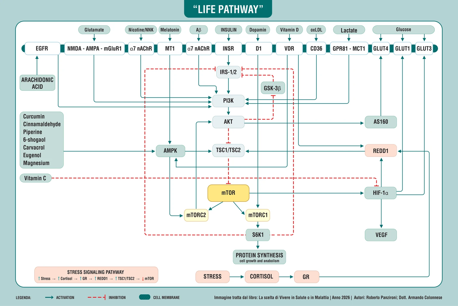
Life Pathway: Schema der Membranrezeptoren sowie der Signalwege IRS, PI3K, AKT, AMPK, TSC1/TSC2, mTOR, REDD1 und HIF-1α, die Ernährung, Stress, Nährstoffe und Zellstoffwechsel miteinander verbinden
Der Life Pathway integriert Membranrezeptoren und Signale von Glutamat, Insulin, Dopamin, Vitamin D, Laktat und Glukose mit den Signalwegen IRS-1/2–PI3K–AKT, AMPK–TSC1/TSC2–mTOR und REDD1–HIF-1α. Er verknüpft Stress, Nährstoffe und Stoffwechsel mit der Proteinsynthese und dem zellulären Gleichgewicht, dem Dreh- und Angelpunkt für die Entscheidung zwischen Gesundheit und Krankheit.

ABSTRACT

Das Buch „Die Entscheidung, gesund oder krank zu leben“ von Dr. A. Colonnese und R. Panzironi untersucht die Zusammenhänge zwischen Ernährung, Stoffwechselregulation, Immunität und chronischen Erkrankungen. Gesundheit erscheint dabei als dynamisches Ergebnis des Zusammenspiels von zellulären Prozessen, Umweltbedingungen und Verhaltensweisen, ohne dass individuelle Entscheidungen das Krankheitsrisiko vollständig bestimmen könnten.

An der Reaktion auf eine Mahlzeit sind glykämische Variabilität, Pankreassekretion, hepatische Clearance und Insulinsensitivität beteiligt. Vom Insulinrezeptor aus durchläuft das Signal IRS-1/2, PI3K und AKT und wird mit mTORC1, mTORC2, AMPK, HIF-1α, REDD1 und TSC1/TSC2 verknüpft. Diese Regulatoren koordinieren Energieverfügbarkeit, Proteinsynthese, Autophagie und Stressanpassung. Die Verbindungen zu Lipidstoffwechsel, Prostaglandinen und Immunmetabolismus helfen, Entzündung und Immundifferenzierung zu deuten, einschließlich des Th17/Treg-Gleichgewichts. Die Funktion jedes Signalwegs hängt vom Kontext ab: Signale, die für die Reparatur notwendig sind, können unter anderen Bedingungen krankhafte Prozesse unterstützen.

Die betrachteten Belege betreffen Diabetes, Autoimmunität, Neurodegeneration, Tumoren und Atherosklerose und zeigen, wie gemeinsame Regelkreise je nach Gewebe und Bedingungen unterschiedliche Rollen übernehmen. Epidemiologische Zusammenhänge, klinische Studien und experimentelle Modelle erfordern jeweils eine eigene Deutung. Ernährung, Bewegung, Rauchen, soziale Beziehungen, Stress und Schlafrhythmus wirken mit diesen Prozessen zusammen. Vitamine, Magnesium, Omega-3-Fettsäuren, Melatonin und bioaktive Verbindungen aus Gewürzen liefern weitere Beispiele zellulärer Regulation.

Das Konzept des Life Pathway schlägt eine integrierte Betrachtung der stoffwechselbezogenen, energetischen und umweltbedingten Netzwerke vor, ohne einen neuen molekularen Signalweg zu postulieren. Dieser Deutungsrahmen verbindet biologische Mechanismen mit dem alltäglichen Verhalten und unterstützt realistische, individuelle Präventionsentscheidungen, die mit einer angemessenen Behandlung vereinbar sind. Es bleibt notwendig, die klinische Relevanz experimenteller Mechanismen zu klären, monokausale Erklärungen multifaktorieller Erkrankungen zu vermeiden, biologische Plausibilität von Wirksamkeitsnachweisen zu unterscheiden und auch den Beitrag genetischer, umweltbedingter und sozialer Faktoren anzuerkennen.

Wir sind am Ende dieses Buches angelangt. Bevor wir uns verabschieden, möchten wir gemeinsam den Weg zurückverfolgen, der uns von Ernährungsgewohnheiten zu zellulären Signalen, von chronischen Krankheiten zu den Möglichkeiten der Prävention geführt hat. Diese Schlussfolgerungen fassen die Bedeutung der einzelnen Etappen zusammen und stellen die Verbindungen zwischen ihnen wieder her, damit das, was wir kennengelernt haben, zu einem Wissen wird, das im Alltag nutzbar ist.

Unsere Absicht als Autoren war es, den Leser in die biologischen Mechanismen einzuführen, die zur Gesundheit beitragen. Zu wissen, warum ein bestimmtes Verhalten wichtig ist, kann den Willen, es zu ändern, konkreter machen. Eine Mahlzeit, ein Spaziergang, Ruhe und eine bedeutsame zwischenmenschliche Beziehung erhalten einen anderen Stellenwert, wenn wir verstehen, dass sie beeinflussen können, wie der Organismus Energie nutzt, mit Stress umgeht und seine Funktionen aufrechterhält.

Der Titel „Die Entscheidung, gesund oder krank zu leben“ drückt diese Möglichkeit der Teilhabe aus. Gesundheit hängt auch von genetischen, umweltbedingten und sozialen Faktoren ab, die wir uns nicht aussuchen, und niemand entscheidet sich freiwillig dafür, krank zu werden. Es gibt jedoch Gewohnheiten, auf die wir Einfluss nehmen können, wobei die Möglichkeiten für jeden Einzelnen unterschiedlich sind. Das Bewusstsein, das wir fördern möchten, betrifft genau diesen Spielraum: zu erkennen, was wir verbessern können, ohne Krankheit zu einer Schuld oder Prävention zu einem Versprechen auf Unverwundbarkeit zu machen.

Indem wir der Forschung gefolgt sind, sind wir auf klinische Ergebnisse, epidemiologische Zusammenhänge und im Labor untersuchte Mechanismen gestoßen. Jede Ebene trägt zum Verständnis bei, beantwortet jedoch unterschiedliche Fragen. Diese Unterscheidung beizubehalten, ermöglicht es uns, Chancen zu erkennen, ohne Hypothesen den Wert von Gewissheiten beizumessen. Dies ist auch die Voraussetzung dafür, einen bewussten Lebensstil mit angemessenen medizinischen Behandlungen zu verbinden und dabei die Komplexität des Menschen zu respektieren.

Wir werden den Weg daher in der Reihenfolge wieder aufnehmen, in der er sich entwickelt hat, und dabei das hervortreten lassen, was scheinbar weit voneinander entfernte Themen verbindet. Am Ende werden wir der Perspektive, die sie alle durchzieht, einen Namen geben: Life Pathway, das redaktionelle Konzept, mit dem wir die Kommunikation zwischen Stoffwechsel, Umwelt und zellulärer Anpassung zusammenführen. Um die Bedeutung zu verstehen, kehren wir zu dem Punkt zurück, an dem alles begann: unsere Beziehung zum Essen.

Ernährung und Veränderungen unserer Gewohnheiten

Das erste Kapitel hat uns dazu eingeladen, die Ernährung als Teil unserer biologischen und täglichen Geschichte zu betrachten. Essen ist eine gewohnheitsmäßige Handlung, die oft vollzogen wird, ohne sich zu fragen, was danach geschieht. Doch die Auswahl der Lebensmittel, ihre Verarbeitung und die Art und Weise, wie sie kombiniert werden, tragen dazu bei, die Reaktionen des Organismus zu bestimmen. Aus dieser Erkenntnis entstand die Frage, die dieses Buch geleitet hat: Wie sehr sind die Stoffwechselzustände, denen wir heute begegnen, mit über die Zeit wiederholten Gewohnheiten verknüpft?

Der Bezug zur mediterranen Ernährungstradition hat uns auf die Qualität der Mahlzeit und ihre Gesamtzusammensetzung zurückgeführt. Ihr Wert liegt in dieser Perspektive in der Aufmerksamkeit für die Lebensmittel und die Vielfalt der Nährstoffquellen. Um diese Bedeutung wiederzuentdecken, muss man über den Namen einer Diät hinausblicken und berücksichtigen, was tatsächlich konsumiert wird – in den Mengen und Kombinationen, die das reale Leben prägen.

Wir haben also begonnen, den Fokus von reinen Kalorien auf die Stoffwechselreaktion zu verlagern. Zwei Mahlzeiten können zwar Energie liefern, aber unterschiedliche Auswirkungen auf die Geschwindigkeit der Nährstoffaufnahme, den Blutzuckerspiegel und die Insulinausschüttung haben. Dieser Unterschied erschöpft zwar nicht den gesamten Nährwert von Lebensmitteln, hilft aber zu verstehen, warum das Verhältnis zum Essen eine umfassendere Bewertung verdient als das bloße Zählen von Kalorien.

Das erste Ergebnis dieser Überlegung ist eine größere Aufmerksamkeit für die Beständigkeit der Gewohnheiten. Stoffwechselgesundheit baut sich durch Verhaltensweisen auf, die sich wiederholen, eingebettet in persönliche und umweltbedingte Rahmenbedingungen, die diese erleichtern oder erschweren können. Deshalb muss eine sinnvolle Ernährungsentscheidung in den Alltag integrierbar sein und dabei sowohl die Nährstoffversorgung als auch die Nachhaltigkeit gewährleisten. Wissen dient dazu, solche Entscheidungen verständlich zu machen, damit sie nicht allein von Verboten oder kurzzeitigem Enthusiasmus abhängen.

Mit dieser Perspektive hat unser Weg seinen konkreten Ausgangspunkt gefunden. Nahrung führt dem Körper Substanzen zu, die er erkennen, verteilen und verwerten muss; die Reaktion auf diese Zufuhr verrät etwas über seine Anpassungsfähigkeit. Den Weg der Glukose zu verfolgen, hat es uns ermöglicht, in diesen Prozess einzutauchen und zu verstehen, warum ein im Blut gemessener Wert nur einen Teil der Geschichte erzählt.

Blutzuckerspiegel und Insulin bei der Reaktion auf eine Mahlzeit

Vom Essen sind wir zur Reaktion übergegangen, die eine Mahlzeit im Organismus hervorruft. Die aufgenommene Glukose gelangt in das Blut, während die Bauchspeicheldrüse und das Gewebe ihre Verwertung koordinieren. Der Blutzuckerspiegel beschreibt ein Ergebnis dieser Arbeit: Um ihn zu interpretieren, müssen die Prozesse berücksichtigt werden, die ihn bestimmt haben. Die Menge der Kohlenhydrate, die Struktur der Lebensmittel, Ballaststoffe, die Verarbeitung und die Zusammensetzung der Mahlzeit tragen gemeinsam dazu bei, wie schnell die Glukose verfügbar wird.

In diesem Zusammenhang sind wir auf den glykämischen Index und die glykämische Last gestoßen. Ersterer hilft dabei, die Reaktion auf verschiedene Kohlenhydratquellen zu vergleichen; Letztere bezieht auch die verzehrte Menge mit ein. Es handelt sich um Analyseinstrumente, die im Kontext der Mahlzeit und der individuellen Merkmale betrachtet werden müssen. Proteine, Fette und Ballaststoffe verändern nämlich die Verdauung, die Aufnahme und die hormonelle Reaktion, weshalb die Bewertung eines Lebensmittels anhand einer einzigen Zahl nicht ausreicht.

Auch das Insulin erfordert die gleiche Aufmerksamkeit. Seine Ausschüttung reagiert auf Glukose, integriert aber weitere Signale, darunter Aminosäuren und Darmhormone. Der Food Insulin Index hat uns geholfen zu erkennen, dass eine Insulinreaktion nicht automatisch mit der glykämischen Reaktion übereinstimmt. Zudem durchläuft das Insulin, bevor es vollständig den peripheren Kreislauf erreicht, die Leber, die einen Teil davon entfernt. Die gemessene Konzentration hängt daher sowohl von der Produktion in der Bauchspeicheldrüse als auch von der Clearance ab, also der Entfernung des Hormons.

Diese Unterscheidung hat auch die Bedeutung der Untersuchungen erweitert. Der Nüchternblutzucker bildet einen Basiszustand ab; das glykierte Hämoglobin liefert Informationen über die durchschnittliche Glukoseexposition; der Glukosetoleranztest beobachtet die Reaktion auf einen definierten Reiz. In diesem Kapitel hat uns die Betrachtung des Blutzuckerspiegels nach einer Stunde gezeigt, wie bestimmte Anzeichen von Schwierigkeiten auftreten können, bevor offensichtlichere Veränderungen sichtbar werden. Keiner dieser Werte erzählt jedoch isoliert betrachtet die gesamte metabolische Funktionsweise.

Das Fazit ist eine Einladung, den Stoffwechsel im Zeitverlauf zu betrachten. Ein scheinbar normaler Blutzuckerspiegel kann durch eine zunehmende Beanspruchung der Regulationssysteme aufrechterhalten werden. Das Verständnis dieser Möglichkeit ermöglicht es, der Prävention und der umfassenden Bewertung der Person einen höheren Stellenwert beizumessen. Der nächste Schritt bestand daher darin, zu beobachten, was passiert, wenn die Schwankungen zunehmen und die Zusammenarbeit zwischen Bauchspeicheldrüse, Leber und Gewebe an Effizienz verliert.

Glykämische Variabilität und Veränderungen im Vorfeld eines Diabetes

Die Beobachtung des Glukosespiegels im Zeitverlauf hat uns eine Dimension erkennen lassen, die bei einer Einzelmessung verborgen bleibt: die glykämische Variabilität. Glukose schwankt physiologisch, doch das Ausmaß und die Häufigkeit dieser Schwankungen können sich auf dem Weg zu Prädiabetes und Diabetes verändern. Kontinuierliche Glukosemessungen und deren Indikatoren haben diese Verläufe sichtbar gemacht und zeigen, dass ein ähnlicher Durchschnittswert mit völlig unterschiedlichen Tagesprofilen einhergehen kann.

Die untersuchten Studien bringen eine stärkere glykämische Instabilität mit oxidativem Stress, Entzündungen und Gefäßveränderungen in Verbindung. Experimentelle Modelle zeigen zudem, dass eine intermittierende Exposition gegenüber hohen Glukosekonzentrationen die β-Zellen der Bauchspeicheldrüse schädigen kann. Diese Beobachtungen haben uns geholfen, einen möglichen Kreislauf zu verstehen: Die Schwierigkeit, den Glukosespiegel zu regulieren, kann die Belastung für genau die Zellen erhöhen, die an dieser Regulation beteiligt sind.

Die Funktion der Bauchspeicheldrüse muss daher im Verhältnis zu den Bedürfnissen des Gewebes interpretiert werden. Eine hohe Insulinmenge garantiert keine angemessene Reaktion, wenn die Empfindlichkeit gegenüber dem Hormon reduziert ist. Aus demselben Grund beschreibt die zirkulierende Konzentration allein nicht, wie viel die Bauchspeicheldrüse tatsächlich produziert. Die Leber verändert durch die Insulinclearance die Exposition des Organismus gegenüber dem Hormon; eine Verringerung dieser Clearance kann auch ohne einen entsprechenden Anstieg der Sekretion zu Hyperinsulinämie beitragen.

An dieser Stelle haben wir eines der zentralen Themen dieses Werks vertieft: die bidirektionale Beziehung zwischen Hyperinsulinämie und Insulinresistenz. Die Resistenz kann eine kompensatorische Reaktion der Bauchspeicheldrüse auslösen; in manchen Kontexten kann eine hohe und anhaltende Insulinexposition ihrerseits zur Desensibilisierung des Gewebes beitragen. Sekretion, Abbau und zelluläre Reaktion beeinflussen sich gegenseitig, wobei das Gewicht dieser Faktoren je nach Person und Stadium der Stoffwechselstörung variiert.

Diese Betrachtungsweise macht die schrittweise Verschlechterung verständlicher. Dem Auftreten einer manifesten Hyperglykämie können jahrelange Anpassungsprozesse vorausgehen, während derer das System bestimmte Ergebnisse auf Kosten eines zunehmenden Ungleichgewichts aufrechterhält. Das Interesse an diesen Übergängen gilt der Möglichkeit, eine ungünstige Entwicklung durch eine angemessene Beurteilung frühzeitig zu erkennen. Um zu verstehen, wie ein Signal schrittweise an Wirksamkeit verlieren kann, haben wir uns daraufhin auf zellulärer Ebene mit den Proteinen befasst, die die Insulinbotschaft in eine Aktion übersetzen.

Das Insulinsignal und die Anpassungsfähigkeit der Zelle

Der Einstieg in den Insulin-Signalweg bedeutete zu beobachten, was nach der Bindung des Hormons an seinen Rezeptor geschieht. IRS-1 und IRS-2 sind an der Übertragung der Nachricht beteiligt, PI3K unterstützt deren Weiterleitung und AKT trägt dazu bei, diese in Stoffwechsel- und Überlebensreaktionen umzuwandeln. Die Verfügbarkeit von Nährstoffen wird so zu einer Information, die die Glukoseverwertung, die Synthese und die Speicherung von Ressourcen steuern kann.

Der Signalweg erreicht dann das TSC1/TSC2-System und mTOR, einen der wichtigsten Integrationspunkte für Energie, Nährstoffe und Wachstumssignale. Wir haben zwischen mTORC1 und mTORC2 unterschieden, Komplexe mit unterschiedlichen, aber miteinander verbundenen Funktionen. Ersterer ist an der Steuerung der Synthese und der Autophagie beteiligt; letzterer trägt unter anderem zur Regulierung von AKT bei. Diese Unterscheidung ist notwendig, um zu verstehen, warum eine Veränderung von mTOR je nach Kontext unterschiedliche Folgen haben kann.

Wenn mTORC1 dauerhaft stimuliert wird, kann der Schaltkreis, an dem S6K1 beteiligt ist, das vorgeschaltete Signal abschwächen und so mit IRS interferieren. Ein physiologisches Kontrollsystem kann auf diese Weise an der Verringerung der Insulinantwort mitwirken. Andererseits kann auch die Beeinträchtigung notwendiger mTOR-Funktionen den Stoffwechsel stören. Die daraus resultierende Erkenntnis ist daher die Notwendigkeit, eine angemessene Regulation zu bewahren: Wachstum, Reparatur und Erneuerung erfordern unterschiedliche Zeitpunkte und Bedingungen.

In diesem Netzwerk sind wir auf HIF-1α und REDD1 gestoßen. Ersterer verbindet Stoffwechselsignale mit der zellulären Anpassung, insbesondere in Bezug auf die Sauerstoffversorgung; letzterer ist an der Stressantwort beteiligt und kann mTORC1 über das TSC-System hemmen. Diese Regulatoren zeigen, wie die Zelle Informationen integriert, die aus verschiedenen Richtungen kommen. Nahrung ist eine davon, zusammen mit dem Energiezustand und dem Zustand des Gewebes.

Der Wert dieses Abschnitts besteht nicht darin, alle Abkürzungen auswendig zu lernen. Er besteht darin zu erkennen, dass die Insulinantwort von einer verteilten und regulierten Kommunikation abhängt, deren Wirksamkeit sich im Laufe der Zeit ändern kann. Insulin spielt weiterhin eine unverzichtbare Rolle; seine Exzesse und Veränderungen zu verstehen bedeutet, die Qualität seines Signals zu schützen. Von hier aus sind wir zu einer weiterreichenden Konsequenz übergegangen: Dieselben Schaltkreise, die den Stoffwechsel steuern, können mit jenen interagieren, die Entzündungsprozesse unterstützen.

Der Dialog zwischen Stoffwechsel und Entzündung

Die Verbindung zur Entzündung hat uns über die reine Energieverteilung hinausgeführt. Wir haben beobachtet, wie eine anhaltende metabolische Stimulation die Verfügbarkeit von Substanzen beeinflussen kann, die zur Produktion von Entzündungsmediatoren verwendet werden. In dieser Perspektive stellt die Hyperinsulinämie eines der möglichen Elemente eines veränderten biologischen Milieus dar, das in der Lage ist, sich mit den Prozessen zu verflechten, über die Zellen einen Schaden oder einen Aktivierungszustand kommunizieren.

Der beschriebene Weg beginnt beim Lipidstoffwechsel. Über SREBP-1c und Desaturase-Enzyme kann das Insulinsignal dazu beitragen, die Produktion von Arachidonsäure zu verändern. Diese Fettsäure gehört zur normalen Physiologie der Membranen und stellt den Vorläufer verschiedener Mediatoren dar. Ihre Präsenz wird für die Entzündungskaskade dann relevant, wenn andere Signale ihre Freisetzung und Nutzung ermöglichen.

Wir haben diesen zweiten Schritt über MAPK/ERK, intrazelluläres Kalzium und cPLA2α verfolgt, das Enzym, das Arachidonsäure aus Membranphospholipiden verfügbar machen kann. Anschließend beteiligt sich COX-2 an der Produktion von Prostaglandin E2. In den beschriebenen Kontexten kann das Prostaglandinsignal, vermittelt durch den EP4-Rezeptor, mit EGFR kommunizieren und erneut PI3K, AKT und mTOR einbeziehen. Lipidstoffwechsel und Wachstumssignalisierung können sich somit in Kreisläufe gegenseitiger Beeinflussung einfügen.

Diese Sequenz hat es uns ermöglicht, dem Ausdruck metabolische Entzündung eine konkrete Bedeutung beizumessen. Eine Veränderung in der Nutzung von Nährstoffen kann dazu beitragen, die Botschaften zu verändern, die von benachbarten Zellen empfangen werden, mit Auswirkungen auf das Gewebemilieu. Die Rekonstruktion eines Mechanismus zeigt eine biologische Möglichkeit auf; seine tatsächliche Relevanz hängt von der Intensität des Reizes, der Dauer und den anderen im Organismus vorhandenen Bedingungen ab.

Die Aufmerksamkeit verlagerte sich daraufhin von den Mediatoren auf die Zellen, die diese produzieren und interpretieren. Der Makrophage kann weitere Abwehrmechanismen aktivieren, Zytokine freisetzen und die Reparatur beeinflussen. Ein intrazelluläres Phänomen nimmt so eine kollektive Dimension an, die Gewebe und Organe einbezieht. Um zu verstehen, wie eine Antwort eingeleitet, verstärkt und schließlich eingedämmt wird, ist es notwendig geworden, das Immunsystem und die energetischen Mechanismen zu kennen, die seine verschiedenen Funktionen unterstützen.

Die Immunabwehr und ihr metabolisches Gleichgewicht

Mit dem Immunsystem haben wir ein Netzwerk kennengelernt, das in der Lage ist, Bedrohungen zu erkennen, Reaktionen zu organisieren und Gewebeschäden zu begrenzen. Die angeborene Immunität bietet eine erste Eingriffsmöglichkeit, während die adaptive Immunität durch T- und B-Lymphozyten spezifischere Antworten entwickelt. Die Zusammenarbeit zwischen diesen Systemen erfordert Kommunikation, Energie und Kontrolle: Eine Aktivierung ist notwendig, aber ebenso die Beendigung einer Reaktion, sobald sie ihre Funktion erfüllt hat.

Immunzellen passen ihren Stoffwechsel an die jeweilige Aufgabe an. Entzündungsprozesse gehen häufig mit einem verstärkten Rückgriff auf die Glykolyse einher, während bestimmte regulatorische und reparative Funktionen stärker vom oxidativen Stoffwechsel abhängen. Die Beschreibung von Makrophagen als M1 und M2 hat uns geholfen, diese Unterscheidung einzuordnen, auch wenn sie eine Vereinfachung komplexerer und variablerer Zellverhaltensweisen darstellt. Aus dieser Beziehung zwischen Funktion und Energie ist das Immunmetabolismus-Konzept entstanden.

mTOR und AMPK sind an der Koordination dieser Programme beteiligt. mTORC1 unterstützt Prozesse, die für die Aktivierung und das Wachstum von Effektorzellen erforderlich sind; AMPK trägt zur energetischen Anpassung bei und kann aufwendige anabole Aktivitäten begrenzen. HIF-1α verknüpft einen Teil dieser Signalübertragung mit der Glykolyse und der Immunzell-Differenzierung. Dieselben Proteine, denen wir bereits im Stoffwechsel begegnet sind, erhalten dadurch eine zusätzliche Bedeutung: Sie tragen dazu bei, die Art und Weise zu steuern, wie die Abwehrkräfte ihre Ressourcen nutzen.

Das Verhältnis zwischen Th17-Zellen und Treg-Zellen hat diesen Zusammenhang besonders deutlich gemacht. Erstere sind an Entzündungsreaktionen beteiligt; letztere helfen dabei, die Toleranz gegenüber dem eigenen Organismus aufrechtzuerhalten. FOXP3 ist essenziell für die Identität und die regulatorische Funktion der Treg-Zellen. Forschungen zu Rapamycin, Glukokortikoiden und Vitamin D haben gezeigt, dass ein Eingreifen in metabolische Signale diese Gleichgewichte verändern kann, wobei die Effekte von den jeweiligen Zellen und den untersuchten Bedingungen abhängen.

Wir haben somit verstanden, dass eine wirksame Abwehr Maß, Dauer und Regulationsfähigkeit erfordert. Eine undifferenzierte Stärkung der Immunität ist kein ausreichendes biologisches Ziel; vielmehr muss die kontextgerechte Reaktion bewahrt werden. Dieses Bewusstsein hat uns darauf vorbereitet, Autoimmunerkrankungen zu verstehen, bei denen die Toleranz nachlassen kann und die Immunaktivierung zum Schaden beiträgt. Die erste Etappe dieses Übergangs führte uns zu den Gelenken, wo die rheumatoide Arthritis den Dialog zwischen Entzündung und Stoffwechsel greifbar macht.

Rheumatoide Arthritis und der Stoffwechsel der Gelenkreaktion

Bei rheumatoider Arthritis trifft der molekulare Signalweg auf eine Erkrankung, die tiefgreifende Auswirkungen auf die Beweglichkeit, das Schmerzempfinden und den Alltag haben kann. Die Gelenkentzündung betrifft Immunzellen und Synovialgewebe im Rahmen eines komplexen Autoimmunprozesses. Die Betrachtung durch die Linse des Immunmetabolismus hat es uns ermöglicht, unser Verständnis dafür zu erweitern, was die Entzündungsreaktion aufrechterhält, wobei der Mensch und die konkreten Folgen der Krankheit stets im Mittelpunkt stehen.

Der erste Aspekt, der sich herauskristallisierte, betrifft das Gleichgewicht zwischen den Lymphozytenpopulationen. Th17-Zellen und ihre Mediatoren können zur Entzündung beitragen, während Treg-Zellen an deren Eindämmung beteiligt sind. Auch die Wechselwirkungen zwischen Tfh-, Tfr-Zellen und B-Lymphozyten helfen dabei, die Produktion von Autoantikörpern zu verstehen. Wenn diese Systeme nicht angemessen koordiniert sind, kann die Reaktion über längere Zeit bestehen bleiben und das Gelenkumfeld zunehmend in Mitleidenschaft ziehen.

Die untersuchten Forschungsarbeiten haben zudem Veränderungen der Insulinsensitivität dokumentiert, die bei einigen Patienten bereits in frühen Stadien auftreten und nicht allein durch die Behandlung mit Glukokortikoiden erklärt werden können. Dieser Befund deutet auf eine kontinuierliche Wechselwirkung zwischen entzündlicher Aktivität und Stoffwechsel hin.

mTOR stellt einen der Punkte dar, an denen diese Aspekte zusammenlaufen können. Seine Aktivität ist an den Programmen der Effektorzellen, der synovialen Proliferation und der Angiogenese beteiligt. Über HIF-1α und die Glykolyse können Wachstumssignale den Energiebedarf der betroffenen Zellen decken. Studien zu mTOR-Inhibitoren zeigen das Potenzial dieser Verbindung als Forschungsziel. Auch Vitamin D wurde hinsichtlich seines Zusammenhangs mit der Immuntoleranz und der Krankheitsaktivität untersucht.

Die Erkenntnis, die wir daraus mitnehmen, betrifft die Weite unseres Blickwinkels. Die Krankheitskontrolle erfordert spezifische Therapien, während die metabolische Gesundheit eine wesentliche Dimension der umfassenden Patientenversorgung darstellt. Die Kenntnis dieser Zusammenhänge hilft zu verstehen, warum sich scheinbar weit entfernte Organe und Systeme gegenseitig beeinflussen können. Vom Gelenk haben wir uns dann der Haut zugewandt: Bei der Psoriasis hat uns eine unmittelbar sichtbare Manifestation erneut Prozesse vor Augen geführt, die über das Gewebe hinausgehen, in dem sie auftreten.

Psoriasis und Signale, die die Haut durchdringen

Die Psoriasis hat uns gelehrt, über die Oberfläche hinauszublicken. Die Hauterscheinungen machen einen Zustand sichtbar, in dem sich die Proliferation der Keratinozyten, die Immunaktivität und Entzündungsmediatoren gegenseitig beeinflussen. Die Haut wird so zu einem Ort, an dem wir die Auswirkungen einer gestörten biologischen Kommunikation beobachten können, ohne dass die Größe einer Läsion allein die Komplexität der betroffenen Person beschreibt.

Die Aufmerksamkeit für Th17-Zellen, IL-17 und andere Mediatoren hat uns auf das Verhältnis zwischen Effektorantwort und regulatorischer Kontrolle zurückgeführt. Treg-Zellen können in der entzündlichen Umgebung weniger effektiv sein oder an Stabilität verlieren. Währenddessen beteiligen sich die Keratinozyten aktiv am Prozess, indem sie ihr Wachstum und die Kommunikation mit den Abwehrmechanismen verändern. Die Persistenz der Entzündung hängt daher von mehreren Akteuren ab, die dazu beitragen, das veränderte Haut-Mikromilieu aufrechtzuerhalten.

Neben dieser Dimension tritt die metabolische Ebene. In den untersuchten Studien wurden Hyperinsulinämie und eine verringerte Insulinsensitivität auch bei Personen ohne manifesten Diabetes und in einigen Fällen ohne Adipositas beobachtet. Diese Daten legen nahe, Bedingungen zu berücksichtigen, die bei bloßer Betrachtung des Blutzuckerspiegels oder des Gewichts möglicherweise unbemerkt bleiben. Die kardiovaskulären und metabolischen Zusammenhänge unterstreichen die Bedeutung einer umfassenden Beurteilung, die über die Behandlung der Hauterscheinungen hinausgeht.

Im Psoriasis-Gewebe haben wir PI3K/AKT/mTOR und HIF-1α wiedergefunden. Diese Signale können den glykolytischen Stoffwechsel unterstützen, der für die epidermale Proliferation und die Aktivität bestimmter Immunzellen erforderlich ist. Die Forschung zu deren Modulation zeigt, dass die Kontrolle von Wachstum und Entzündung gemeinsame Zielstrukturen haben kann. Vitamin D fügt durch die Regulierung der Hautdifferenzierung und der Immunität eine weitere Verbindung hinzu.

Diese Etappe hat uns zu einer konkreten Erkenntnis geführt: Sich um die Gesundheit zu kümmern bedeutet, auch das zu berücksichtigen, was nicht unmittelbar sichtbar ist. Die Krankheit definiert den Menschen nicht und rechtfertigt keine Urteile über seine Lebensgewohnheiten. Sie bietet stattdessen einen Anlass, Erkenntnisse zu integrieren und unterschiedliche Bedürfnisse zu identifizieren. Derselbe Blickwinkel wird umso notwendiger, wenn Autoimmunität verschiedene Organe betreffen kann, wie wir es beim systemischen Lupus erythematodes gesehen haben.

Lupus und die Komplexität der Immuntoleranz

Beim systemischen Lupus erythematodes haben wir einen Verlust der Toleranz festgestellt, der verschiedene Organe betreffen und sich in sehr unterschiedlichen klinischen Bildern äußern kann. Diese Vielfalt hat uns daran erinnert, wie wichtig es ist, den Überblick zu behalten. Autoantikörper und die Aktivierung von B-Lymphozyten stellen einen Teil des Prozesses dar, der in ein Netzwerk aus Beziehungen zwischen Abwehrmechanismen, Geweben und metabolischen Signalen eingebettet ist.

Die Gleichgewichte zwischen Tfh- und Tfr-Zellen sowie zwischen Th17- und Treg-Zellen haben die Bedeutung der Systeme verdeutlicht, die die Immunantwort koordinieren und begrenzen. Wenn bestimmte Effektorprogramme überwiegen und die Regulation unzureichend wird, kann die Fähigkeit zur Aufrechterhaltung der Entzündung zunehmen. Der Stoffwechsel begleitet diese Veränderungen: Glykolyse, Glutaminverwertung und mitochondriale Aktivität tragen dazu bei, die für die verschiedenen Zellpopulationen notwendigen Ressourcen bereitzustellen.

Die in Studien beschriebenen Insulinveränderungen haben das Bild weiter vervollständigt. Eine verringerte Insulinsensitivität kann auch bei normalem Blutzuckerspiegel und in klinisch wenig aktiven Phasen vorliegen. Ihre Beobachtung in verschiedenen Populationen legt nahe, dass die metabolische Interaktion Aufmerksamkeit verdient, ohne sie automatisch auf ein einziges Medikament oder ein einzelnes Krankheitsmerkmal zurückzuführen. Auch hier erfordert die Interpretation der Zusammenhänge eine vorsichtige und kontextbezogene Betrachtung.

mTOR und HIF-1α haben uns eine Verbindung zwischen diesen Ebenen aufgezeigt. Ihre Aktivität kann glykolytische Programme und Effektorantworten unterstützen, während Veränderungen der Autophagie und der Mitochondrien zum zellulären Bild beitragen. Studien zu Sirolimus haben gezeigt, wie einige dieser Verbindungen Gegenstand therapeutischer Forschung werden können. Vitamin D wurde aufgrund seines Zusammenhangs mit dem Immunstatus und dem Gleichgewicht zwischen regulatorischen und Effektorpopulationen in Betracht gezogen.

Der Wert dieser Etappe besteht darin, dass sie uns einer komplexen Krankheit nähergebracht hat, wobei die Unterscheidung zwischen gemeinsamen Mechanismen und individuellen Merkmalen gewahrt blieb. Ein gemeinsames Netzwerk hilft dabei, Fragen zu formulieren und Beobachtungen zu interpretieren; die Wahl der Behandlung erfordert spezifische Nachweise und Bewertungen. Mit dieser Aufmerksamkeit sind wir zu einem anderen Bereich übergegangen, in dem Toleranz jeden Tag unerlässlich ist: dem Darm, der den Kontakt mit der Außenwelt, die Aufnahme von Nährstoffen und den Schutz des Organismus in Einklang bringen muss.

Darm und Aufrechterhaltung der Immunbarriere

Bei chronisch entzündlichen Darmerkrankungen gewinnt das Verhältnis zwischen Abwehr und Toleranz eine unmittelbare Bedeutung. Die Darmschleimhaut ist an der Nährstoffaufnahme beteiligt und bildet gleichzeitig eine Schnittstelle zur Außenwelt. Morbus Crohn und Colitis ulcerosa weisen zwar wichtige Unterschiede auf, teilen jedoch eine anhaltende oder wiederkehrende Entzündungsreaktion, die die Lebensqualität und den Ernährungszustand tiefgreifend beeinträchtigen kann.

Der bisherige Weg hat die Rolle der Th17- und Treg-Populationen sowie die der Makrophagen und Epithelzellen verdeutlicht. Das Problem liegt in der Fähigkeit, eine angemessene Reaktion aufrechtzuerhalten, um das Gewebe zu schützen, während man sich mit Reizen auseinandersetzt. Die Immunaktivität erfordert Energie, und metabolische Programme tragen dazu bei, diese zu steuern. Deshalb können die Barriere und die Abwehrmechanismen nicht verstanden werden, wenn man sie von den energetischen Bedingungen der Zellen trennt.

Die Zusammenhänge zwischen metabolischen Indikatoren, wie dem TyG-Index, und dem Krankheitsrisiko haben eine weitere Perspektive eröffnet. Diese Daten deuten auf mögliche Verbindungen zwischen metabolischer Dysfunktion und Darmentzündungen hin und helfen uns zu erkennen, dass die Darmgesundheit in den Rahmen des gesamten Organismus eingebettet ist, wobei Einflüsse in beide Richtungen wirken können.

Wir haben mTOR und HIF-1α in den Programmen wiedergefunden, die die Glykolyse und die Entzündungsaktivität unterstützen. In den untersuchten Modellen kann die Modulation dieser Signalwege Immunpopulationen, die Autophagie und die Integrität der Barriere verändern. Studien mit mTOR-Inhibitoren und natürlichen Verbindungen haben Forschungsansätze geliefert, während Beobachtungen zu Vitamin D die Aufmerksamkeit auf den Ernährungszustand und die Immunregulation gelenkt haben. Die Funktion jedes Signals bleibt an den Zelltyp und den Kontext gebunden.

Aus diesem Abschnitt ergibt sich der Wert einer Behandlung, die Entzündung, Ernährung und den allgemeinen Gesundheitszustand gemeinsam betrachtet. Das biologische Verständnis macht deutlicher, warum ein Darmproblem Folgen haben kann, die über den Verdauungstrakt hinausgehen. Derselbe Dialog zwischen Immunität und Stoffwechsel findet sich in Geweben mit sehr unterschiedlichen Anforderungen wieder. Im nächsten Kapitel sind wir diesem bis zum Nervensystem gefolgt, wo die Multiple Sklerose den Schutz und die Reparatur der Myelinscheiden in den Vordergrund rückt.

Multiple Sklerose und Schutz des Nervensystems

Bei der Multiplen Sklerose hat sich der Fokus auf ein Gewebe verlagert, in dem die Kommunikation eine besonders feine Organisation erfordert. Die autoimmune Beteiligung des Myelins geht mit Entzündungsprozessen einher, die die neurologische Funktion beeinträchtigen können. Das Verständnis der metabolischen Komponente hat es uns ermöglicht, die Beschreibung des Schadens um eine weitere Frage zu ergänzen: Welche Ressourcen und Signale unterstützen die Zellen, die diesen Prozess begünstigen, und welche jene, die versuchen, ihn zu reparieren?

Forschungen zu Treg-Zellen haben die Rolle der Toleranz hervorgehoben, während Th1, Th17 sowie die Wechselwirkungen zwischen T- und B-Lymphozyten unterschiedliche Beiträge zur Autoimmunantwort aufgezeigt haben. Auch die Mikroglia, ein Teil der Abwehrmechanismen des Nervensystems, trägt zur Entstehung des entzündlichen Milieus bei. Der Stoffwechsel der einzelnen Zellpopulationen wird somit relevant, um zu verstehen, wie sie aktiviert werden und wie sie ihre Funktion im Gewebe aufrechterhalten.

In den untersuchten Studien wurden Veränderungen der Insulinsensitivität und verstärkte Insulinreaktionen bereits in den frühen Stadien der Erkrankung beobachtet. Die Verbindung zu Entzündungsmarkern und dem Grad der Behinderung deutet auf ein komplexes Zusammenspiel hin, das Aufmerksamkeit verdient. Das Vorhandensein dieser Zusammenhänge erweitert die Beurteilung des Patienten, erlaubt es jedoch nicht, die Multiple Sklerose auf eine einzige Stoffwechselstörung zurückzuführen oder daraus automatisch eine heilende Intervention abzuleiten.

Die wichtigste Erkenntnis betrifft mTOR. In einigen Immunkompartimenten kann seine Aktivität pathogene Programme unterstützen; in Oligodendrozyten trägt es hingegen zu Funktionen bei, die für die Myelinisierung notwendig sind. HIF-1α und die Glykolyse vervollständigen die Verbindung zu den Effektorantworten. Experimente zur Modulation dieses Signalwegs müssen daher unter Berücksichtigung der verschiedenen Zelltypen, Zeitpunkte und Ziele interpretiert werden. Auch Vitamin D fügt sich in dieses Netzwerk ein, wobei Assoziationen und Ergebnisse stets im Kontext der Evidenzlage zu bewerten sind.

Dieser Abschnitt verdeutlicht ein zentrales Prinzip des gesamten Buches: Ein und dasselbe Protein kann je nach Kontext sowohl an der Fehlfunktion als auch an der Erholung beteiligt sein. Gesundheit erfordert ein Gleichgewicht zwischen Abwehr und Reparatur. Nachdem wir dieses Erfordernis im Nervensystem betrachtet haben, sind wir dem Zusammenhang zwischen Autoimmunität und Stoffwechsel in einer Drüse gefolgt, die zur Regulierung der Körperaktivität beiträgt: der Schilddrüse, dem Gegenstand der nachfolgenden Betrachtung zur Hashimoto-Thyreoiditis.

Hashimoto und die Verflechtung von Immunität und Schilddrüsenfunktion

Die Hashimoto-Thyreoiditis hat uns ermöglicht, das Zusammenspiel zwischen dem Immunsystem und der endokrinen Regulation zu beobachten. Der Verlust der Toleranz gegenüber Schilddrüsenbestandteilen kann mit einer fortschreitenden Schädigung der Drüse einhergehen, deren Folgen von der verbleibenden Funktion und dem individuellen Krankheitsverlauf abhängen. Die Verfolgung dieses Prozesses hat uns geholfen zu verstehen, warum die Beurteilung einer endokrinen Autoimmunerkrankung mehrere Ebenen gleichzeitig berücksichtigen muss.

Wir haben das Ungleichgewicht zwischen Th17- und Treg-Zellen sowie die Rolle der regulatorischen Populationen bei der Eindämmung der Immunantwort festgestellt. Innerhalb des Gewebes können oxidativer Stress, Autophagie und Wachstumssignale die Fähigkeit der Zellen beeinflussen, ihre Funktion aufrechtzuerhalten. Autoimmunität beinhaltet daher einen Dialog zwischen den Abwehrmechanismen und dem Zustand der Zielzellen, die ihrerseits an der Umgebung mitwirken, in der sich die Immunantwort entwickelt.

Die Beobachtungen zur Insulinresistenz haben eine metabolische Dimension hinzugefügt. In den betrachteten Studien wurde eine verringerte Insulinsensitivität auch bei Personen mit erhaltener Schilddrüsenfunktion dokumentiert. Dieser Zusammenhang legt nahe, dass Autoimmunität und Stoffwechsel über die Auswirkungen hormoneller Schwankungen hinaus interagieren können. Das Gewicht der verschiedenen Faktoren bleibt individuell und erfordert eine ganzheitliche Betrachtung, in der auch viszerale Adipositas und andere Begleiterkrankungen ihren Platz finden.

Auf molekularer Ebene verknüpfen PI3K/AKT/mTOR und HIF-1α Wachstum, Autophagie und Energieprogramme der Immunzellen. Experimentelle Ergebnisse mit Rapamycin und anderen Verbindungen zeigen, wie die Modulation dieser Signale einige Aspekte der Schädigung in den untersuchten Modellen reduzieren kann. Vitamin D wurde hinsichtlich seiner Auswirkungen auf Autoantikörper und die Immunregulation analysiert. Solche Beobachtungen bereichern das biologische Verständnis, wobei präklinische Ergebnisse strikt von etablierten Therapien unterschieden werden müssen.

Die zentrale Botschaft bleibt die Möglichkeit, komplementäre Bedürfnisse zu erkennen: die endokrine Funktion zu bewahren, den Autoimmunprozess zu verfolgen und den metabolischen sowie ernährungsphysiologischen Status zu berücksichtigen. Das Wissen um diese Zusammenhänge hilft, eine fragmentierte Interpretation von Untersuchungsergebnissen und Symptomen zu vermeiden. Von dieser Drüse aus sind wir zur Bauchspeicheldrüse übergegangen, wo der Verlust der Toleranz genau jene Zellen betrifft, die das Hormon produzieren, das uns von Anfang an begleitet hat: Insulin, das für das Verständnis von Typ-1-Diabetes unerlässlich ist.

Typ-1-Diabetes und der Verlust von Bauchspeicheldrüsenzellen

Bei Typ-1-Diabetes wird der Zusammenhang zwischen Immunsystem und Stoffwechsel besonders deutlich. Die Autoimmunreaktion schädigt die pankreatischen β-Zellen und verringert schrittweise die Fähigkeit zur Insulinproduktion. Das Hormon, das wir als Stoffwechselsignal betrachtet haben, erhält hier die konkrete Bedeutung einer Funktion, die bei ihrem Ausfall ersetzt werden muss. Diese Eigenschaft unterscheidet die Erkrankung und macht es unerlässlich, ihre Spezifität während der gesamten Betrachtung zu wahren.

Der Verlauf hat gezeigt, dass die Toleranz nicht nur von der Anzahl, sondern auch von der Funktion der Treg-Zellen abhängt. Eine unzureichende Regulation kann autoreaktiven Populationen Raum geben, einschließlich jener, die an der Reaktion gegen die Bauchspeicheldrüsenzellen beteiligt sind. Forschungen zur Expansion oder zum Transfer von Treg-Zellen haben das Interesse an Strategien verdeutlicht, die in der Lage sind, eine selektivere Kontrolle der Immunantwort wiederherzustellen.

Auch die Insulinsensitivität kann das Bild verändern. Bei einigen gefährdeten Personen sowie bei Menschen mit manifester Erkrankung wurde eine verringerte Wirksamkeit des verfügbaren Insulins beobachtet. Dieser Faktor kann die metabolische Komplexität erhöhen, ohne die zentrale Rolle des autoimmunbedingten Verlusts der β-Zellen zu ersetzen. Die Verfügbarkeit des Hormons und die Reaktion der Gewebe sind unterschiedliche Dimensionen, die sich gegenseitig bei der Bestimmung der glykämischen Kontrolle beeinflussen können.

mTOR, HIF-1α und Glykolyse sind an den Energieprogrammen der Effektorzellen beteiligt, während AMPK und Autophagie auch im Zusammenhang mit der Widerstandsfähigkeit der Bauchspeicheldrüsenzellen untersucht wurden. Studien zu Rapamycin haben die Möglichkeiten und Grenzen einer Modulation aufgezeigt, die unterschiedliche Funktionen berücksichtigen muss. Vitamin D wurde aufgrund möglicher Zusammenhänge mit Toleranz und Stoffwechsel in Betracht gezogen, ohne dass diese Ergebnisse die Notwendigkeit einer Insulintherapie in Frage stellen, wenn diese indiziert ist.

Die Lehre aus diesem Kapitel ist daher zweifach: gemeinsame Mechanismen erkennen und klinische Unterschiede wahren. Die metabolische Versorgung begleitet eine Erkrankung, deren autoimmuner Ursprung auch auf eine Ernährungsweise zurückgeführt werden kann. Das Verständnis dieses Schrittes ermöglicht es, den nächsten mit größerer Präzision anzugehen. Beim Typ-2-Diabetes fügt sich der Verlust der Pankreasfunktion in eine andere Geschichte ein, in der Insulinsensitivität der Gewebe, Sekretion und Insulin-Clearance charakteristische Verhältnisse annehmen.

Typ-2-Diabetes und fortschreitende Stoffwechselverschlechterung

Typ-2-Diabetes hat uns zu den frühen Signalen zurückgeführt, denen wir zu Beginn begegnet sind, und zeigt, wie diese Teil eines langen und komplexen Prozesses sein können. Der Anstieg des Blutzuckerspiegels ist ein zentrales Element der Diagnose, doch die Erkrankung umfasst das Zusammenspiel von Bauchspeicheldrüse, Leber, Muskeln und Fettgewebe. Im Laufe der Zeit kann sie mit Komplikationen einhergehen, die zahlreiche Organe betreffen, was das systemische Ausmaß des Stoffwechselungleichgewichts verdeutlicht.

Postprandiale Schwankungen und die glykämische Variabilität können bereits vor einer stabileren Hyperglykämie zunehmen. Die Zusammensetzung der Mahlzeiten beeinflusst diese Reaktionen und bietet einen der Ansatzpunkte für Interventionen. Studien zu kohlenhydratreduzierten Ernährungsstrategien haben mögliche Vorteile für die Blutzuckerkontrolle gezeigt, die jedoch im Zusammenhang mit der Qualität der Lebensmittel, der Therapietreue und den individuellen Bedürfnissen zu bewerten sind. Der Wert einer Strategie misst sich auch daran, ob sie langfristig angemessen beibehalten werden kann.

Die Verschlechterung der β-Zellen umfasst Veränderungen der frühen Insulinantwort sowie der Fähigkeit, den Bedarf des Organismus auszugleichen. Parallel dazu kann eine verringerte Clearance die Insulinexposition erhöhen. Hyperinsulinämie und Insulinresistenz können so zu sich gegenseitig verstärkenden Kreisläufen beitragen. Es besteht daher eine dynamische Beziehung zwischen Produktion, Abbau und Wirksamkeit des Hormons.

Innerhalb der Zelle hilft das Feedback von mTORC1 und S6K1 auf IRS dabei, einen der Mechanismen zu verstehen, die die Signalqualität verringern können. AMPK hingegen verweist auf die Fähigkeit, sich an den Energiebedarf anzupassen und die Ressourcennutzung zu unterstützen. Wenn das Ungleichgewicht anhält, können oxidativer Stress, Glukotoxizität, Lipotoxizität und mitochondriale Störungen auf die Bauchspeicheldrüse einwirken und deren funktionelle Kapazität verringern.

Die präventive Bedeutung dieser Rekonstruktion liegt in der Erkenntnis, dass sich der metabolische Verlauf über die Zeit entwickelt und durch geeignete ernährungsphysiologische Interventionen in den verschiedenen Phasen angegangen werden kann. Eine Diagnose erfordert eine Behandlung; die ihr vorausgehenden Signale verdienen Aufmerksamkeit und Bewertung. Diese Sichtweise hat uns dann über den peripheren Stoffwechsel hinaus zu einem Organ geführt, das kontinuierlich von der Energieverfügbarkeit abhängt: dem Gehirn. Bei Alzheimer haben wir beobachtet, wie Insulinsignalisierung, Glukoseverwertung und zelluläre Instandhaltung mit dem neurologischen Verfall verknüpft sein können.

Alzheimer und die Fragilität des zerebralen Stoffwechsels

Im Kapitel über Alzheimer haben wir die Perspektive auf die Neurodegeneration erweitert und neben der Ansammlung von Beta-Amyloid und Tau auch das Problem des Energiegleichgewichts im Gehirn beleuchtet. Neuronen müssen ihre Kommunikation, Integrität und den Austausch ihrer Bestandteile aufrechterhalten. Wenn diese Funktionen ihre Koordination verlieren, können sich die Veränderungen gegenseitig beeinflussen und zur zunehmenden Anfälligkeit der Netzwerke beitragen, die das Gedächtnis und die kognitiven Fähigkeiten stützen.

Die verminderte Reaktion des Gehirns auf Insulin wurde als Teil dieses Bildes untersucht und kann auch bei Abwesenheit von Diabetes beobachtet werden. Die beschriebenen Modelle verknüpfen das Insulinsignal mit dem Management von Beta-Amyloid, der Proteinverarbeitung und der Autophagie. Das Enzym IDE, das sowohl am Abbau von Insulin als auch von Beta-Amyloid beteiligt ist, stellt einen weiteren möglichen Interaktionspunkt dar, dessen Relevanz vom biologischen Kontext abhängt.

mTOR ist an Funktionen beteiligt, die für die synaptische Plastizität und das Gedächtnis notwendig sind. Eine veränderte Regulation kann jedoch mit einer geringeren Austauschkapazität und der Ansammlung schädlicher Komponenten einhergehen. Auch das Feedback auf IRS sowie die Regulation von AKT und GSK3β helfen dabei, mögliche Verbindungen zu den Tau-Veränderungen nachzuvollziehen. Das Verständnis dieser Mechanismen erfordert, die physiologische Rolle genau der Proteine zu bewahren, die an der Fehlfunktion beteiligt sind.

Wir haben zudem das Glutamat verfolgt, dessen Kommunikation kontrolliert werden muss, um eine übermäßige Erregung zu vermeiden. Veränderungen bei der Freisetzung und der Entfernung können den neuronalen Stress verschlimmern. Gleichzeitig können mitochondriale Schwierigkeiten und Veränderungen der Transporter GLUT1 und GLUT3 die tatsächliche Energieverfügbarkeit verringern. HIF-1α tritt in diesem Netzwerk als Regulator metabolischer Antworten auf und zeigt erneut gewebespezifische Effekte, die von den jeweiligen Bedingungen abhängen.

Das Kapitel hat uns somit eine Lesart vermittelt, in der Proteine, Signale und Stoffwechsel an demselben biologischen Problem beteiligt sind. Die metabolischen Zusammenhänge entsprechen keiner einfachen Erklärung für Alzheimer, tragen aber dazu bei, dessen Komplexität zu verstehen. Von einem Gewebe, das Schwierigkeiten hat, seine Funktionen aufrechtzuerhalten, sind wir zu Zellen übergegangen, die in der Lage sind, ihre Ressourcen neu zu organisieren, um ein pathologisches Wachstum zu unterstützen. Dieser Perspektivwechsel hat uns in den Stoffwechsel von Tumoren eingeführt.

Tumoren und die Organisation des metabolischen Mikromilieus

Bei Tumoren haben wir beobachtet, wie eine Zelle die Nutzung von Ressourcen und die Kommunikation mit ihrer Umgebung verändern kann. Genetische Veränderungen spielen weiterhin eine wesentliche Rolle, während Stoffwechsel, Wachstumssignale und Interaktionen mit anderen Zellen zur Komplexität des Prozesses beitragen. Die Perspektive dieses Werks hat uns dazu veranlasst, diesen Zusammenhängen zu folgen und dabei zwischen dem, was eine biologische Möglichkeit stützt, und dem, was einen therapeutischen Nutzen belegt, zu unterscheiden.

Insulin überträgt auch Wachstums- und Überlebenssignale. Epidemiologische Zusammenhänge zwischen Hyperinsulinämie, Insulinresistenz und bestimmten onkologischen Erkrankungen haben die Frage nach dem metabolischen Kontext aufgeworfen, in dem sich Neoplasien entwickeln. Über PI3K, AKT und mTOR können diese Signale mit Synthese- und Proliferationsprogrammen interagieren. Ihre Bedeutung hängt jedoch von den Eigenschaften des Tumors und den in den Zellen vorhandenen Veränderungen ab.

Wachstum erfordert neben Energie auch Materialien. Der Warburg-Effekt beschreibt den verstärkten Rückgriff auf die Glykolyse, selbst wenn Sauerstoff verfügbar ist, was bei vielen neoplastischen Zellen beobachtet wird. HIF-1α trägt zur Anpassung an die Sauerstoffverhältnisse sowie zur Regulierung von Transportern und Enzymen bei. Glukose gelangt so in Stoffwechselprogramme, die den Aufbau neuer Zellkomponenten und die Produktion von Laktat unterstützen können.

Gerade das Laktat hat unser Verständnis erweitert. Über MCT1 und MCT4 kann es zwischen Zellen ausgetauscht werden und wird so in einigen Tumorarealen zu einer Ressource. Seine Signalwirkung und die Prozesse der Laktylierung verbinden Stoffwechsel und Zellregulation. Tumorassoziierte Fibroblasten beteiligen sich an diesem Austausch: Beim inversen Warburg-Effekt stellen bestimmte Stromazellen Metaboliten bereit, die von den neoplastischen Zellen genutzt werden können. Das Tumorgewebe erscheint somit als eine kooperative und heterogene Umgebung.

Diese Organisation interagiert mit den Systemen, die Schäden und Proliferation kontrollieren. p53 trägt zur zellulären Überwachung bei; das Verhältnis zwischen AKT, MDM2 und p53 zeigt, wie Überlebenssignale diesen Schutz beeinträchtigen können. AMPK und TSC1/TSC2 fügen Verbindungen zur Kontrolle von mTOR hinzu. Die Tumorentwicklung hängt daher auch von der Fähigkeit der veränderten Zellen ab, die normalerweise dem Wachstum auferlegten Grenzen zu überwinden.

Die Mikroumgebung umfasst zudem die Immunabwehr. Hypoxie, Laktat und Adenosin können Bedingungen begünstigen, die die Wirksamkeit der Antitumor-Antwort verringern. CD39 und CD73 sind an der Produktion von Adenosin beteiligt, während dessen Rezeptoren die Wirkungen übertragen. Makrophagen und Treg-Zellen können Funktionen übernehmen, die in diesem Kontext die Toleranz gegenüber dem Tumor fördern. VEGF verbindet schließlich die metabolische Anpassung mit der Bildung neuer Gefäße.

Dieser Abschnitt hat uns geholfen zu verstehen, wie ein und derselbe Mechanismus je nach Kontext unterschiedliche Auswirkungen haben kann: Die Hemmung der Immunantwort kann vor Schäden durch Autoimmunität schützen, aber auch die Abwehr gegen einen Tumor schwächen. Die Untersuchung des Stoffwechsels ermöglicht es uns daher, diese Gleichgewichte besser zu verstehen und eröffnet neue Forschungsperspektiven. Indem wir dem Faden folgten, der Stoffwechsel, Lipide und Entzündungen verbindet, haben wir unsere Aufmerksamkeit anschließend von den Blutgefäßen, die den Tumor versorgen, auf die Arterienwand gelenkt, um zu verstehen, wie diese Faktoren zur Entstehung von Arteriosklerose beitragen.

Atherosklerose und Schutz der Gefäßwand

Bei der Atherosklerose haben wir einen Prozess kennengelernt, der sich schleichend entwickeln kann, bevor er durch ein kardiovaskuläres Ereignis offensichtlich wird. Die Arterienwand ist aktiv an der Regulierung des Blutflusses, der Immunkommunikation und dem Lipidstoffwechsel beteiligt. Das Endothel, die innere Auskleidung der Gefäße, bietet einen privilegierten Beobachtungspunkt, um zu verstehen, wie die Qualität metabolischer Signale die Gefäßgesundheit beeinflussen kann.

Unter physiologischen Bedingungen trägt der IRS/PI3K/AKT-Signalweg zur Aktivierung von eNOS und zur Produktion von Stickstoffmonoxid bei. Dieses Molekül fördert die Vasodilatation und unterstützt die Schutzfunktionen des Endothels. Wenn das Insulinsignal an Wirksamkeit verliert, kann dieser Beitrag abnehmen. Das Kapitel hat uns somit daran erinnert, dass Insulin wichtige Funktionen für die Gefäßphysiologie beibehält und dass seine Konzentration stets im Zusammenhang mit der Reaktion des Gewebes interpretiert werden muss.

Die Verringerung des endothelialen Schutzes geht mit Entzündungsprozessen und der Adhäsion von Immunzellen einher. Signale wie NF-κB und VCAM-1 sind an der Rekrutierung von Leukozyten beteiligt, während Lipoproteine in der Gefäßwand oxidativen Veränderungen unterliegen können. Oxidierte LDL werden von Rezeptoren wie CD36 erkannt, die deren Aufnahme in Makrophagen fördern. Die Ansammlung von Lipiden trägt zur Bildung von Schaumzellen bei, die charakteristische Bestandteile atherosklerotischer Läsionen sind.

CD36 hat uns gezeigt, wie Aufnahme und Signalübertragung Hand in Hand gehen können. In Makrophagen trägt es zu Entzündungsreaktionen und Lipidansammlungen bei; auf Blutplättchen kann es an der Erhöhung der Reaktivität gegenüber Reizen beteiligt sein. PI3K/AKT/mTOR und die Autophagie ergänzen die Zusammenhänge mit der Fähigkeit der Zellen, Lipide zu verarbeiten. Diese Prozesse gehören zu einem Netzwerk, in dem sich Insulinmetabolismus, oxidativer Stress und das vaskuläre Milieu gegenseitig beeinflussen können.

Diese Rekonstruktion hat das Konzept der Prävention im Zeitverlauf greifbarer gemacht. Die Gefäßgesundheit hängt vom Zusammenspiel der Risikofaktoren und einer angemessenen Behandlung ab, während die metabolische Dimension hilft, einige weniger sichtbare Zusammenhänge zu verstehen. Nachdem wir diese Beziehungen bei verschiedenen Krankheitsbildern verfolgt haben, war es notwendig, zur täglichen Lebensweise zurückzukehren. Die Ernährung, die zu Beginn als Ausgangspunkt eingeführt wurde, ist mit einer umfassenderen Bedeutung wieder aufgetaucht: als eine der wiederholten Einwirkungen, durch die wir dazu beitragen können, den biologischen Kontext des Organismus zu verändern.

Ernährungsqualität in der täglichen Prävention

Die Rückkehr zum Thema Ernährung nach der Betrachtung verschiedener Krankheitsbilder hat es uns ermöglicht, deren Rolle als Risikofaktor besser zu verstehen. Jede Mahlzeit löst glykämische und insulinbezogene Reaktionen aus, die bei wiederholtem Auftreten das metabolische Gleichgewicht verändern können. Der Fokus lag dabei vor allem auf der Menge und Qualität der Kohlenhydrate: Schnell verdauliche Lebensmittel und Portionen mit einer hohen glykämischen Last können zu einem stärkeren Anstieg des Glukosespiegels und einem erhöhten Insulinbedarf führen.

Die untersuchten Studien haben einen hohen glykämischen Index, eine hohe glykämische Last und ein höheres insulinämisches Potenzial der Ernährung mit einem erhöhten Risiko für Typ-2-Diabetes sowie in verschiedenen Bevölkerungsgruppen mit einer höheren allgemeinen, kardiovaskulären und onkologischen Sterblichkeit in Verbindung gebracht. Diese Ergebnisse legen nahe, dass die Qualität der Ernährung auch jenseits der Gewichtskontrolle Aufmerksamkeit verdient. Insbesondere bei Prädiabetes wurde eine hohe glykämische Last mit einer höheren Wahrscheinlichkeit für das Fortschreiten zur Erkrankung assoziiert.

Die kardiovaskuläre Beteiligung hat uns gezeigt, wie sich die Folgen auf die Blutgefäße ausweiten können. Wiederholte Blutzuckerschwankungen können oxidativen Stress, Entzündungen und endotheliale Dysfunktionen fördern – Prozesse, die zur Anfälligkeit der Arterienwand beitragen. Die Zusammenhänge mit koronarer Herzkrankheit, Arteriosklerose und Schlaganfall fügen sich in dieses Bild ein. Auch Studien zu Demenz und Alzheimer haben die Aufmerksamkeit auf die glykämische Belastung durch die Ernährung gelenkt, ohne jedoch zu beweisen, dass diese direkt eine Neurodegeneration verursacht.

Im onkologischen Bereich bieten Hyperinsulinämie und die Signale der PI3K/AKT/mTOR-Achse mögliche Verbindungen zwischen Ernährung, Zellwachstum und Zellüberleben. Bei Autoimmunerkrankungen deuten Veränderungen der Insulinsensitivität zwar auf eine Wechselwirkung mit Entzündungsprozessen hin, es ist jedoch nicht erwiesen, dass eine Reduzierung der glykämischen Last diesen Krankheiten vorbeugt.

Interventionsstudien haben daher einige konkrete Möglichkeiten aufgezeigt. Ernährungsweisen mit niedrigerem glykämischen Index und geringerer glykämischer Last können die Insulinämie, den HOMA-IR-Wert und bestimmte Entzündungsmarker verbessern; Low-Carb-Strategien können die glykämische Kontrolle unterstützen sowie Triglyzeridwerte und eine Fettleber verbessern. Die Auswirkungen hängen jedoch von der Gesamtqualität der Ernährung, den Ersatzlebensmitteln und der Kontinuität der Intervention ab.

Die abschließende Botschaft betrifft daher die Fähigkeit, ungünstige metabolische Belastungen durch angemessene Portionsgrößen, hochwertige Nahrungsquellen und auf den individuellen Bedarf abgestimmte Mahlzeiten zu reduzieren. Das Verständnis dieser Zusammenhänge macht die täglichen Entscheidungen bewusster. Deren Wirkung ist zudem eng mit der Verwertung der Nährstoffe verknüpft: Dies führt zum Thema Bewegung, welche den Energiebedarf der Muskeln erhöht und zur Regulierung des Glukosespiegels beiträgt.

Bewegung sowie der Wechsel zwischen Arbeit und Erholung

Nachdem wir beobachtet haben, wie die Ernährung die Verfügbarkeit von Nährstoffen beeinflusst, haben wir untersucht, inwieweit Bewegung deren Verwertung bestimmt. Langanhaltende körperliche Inaktivität reduziert den Energiebedarf der Muskeln und kann Veränderungen der Insulinsensitivität begünstigen. Die untersuchten Studien bringen das Ansammeln von Stunden im Sitzen mit einer erhöhten Mortalität sowie einem gesteigerten kardiovaskulären und metabolischen Risiko in Verbindung und lenken die Aufmerksamkeit auch auf die Dauer der aufeinanderfolgenden Phasen der Immobilität.

Dieser Aspekt hat es uns ermöglicht, zwischen gezieltem Training und über den Tag verteilter Bewegung zu unterscheiden. Körperliche Aktivität ist wichtig, doch viele Stunden ununterbrochenes Sitzen können auch bei Menschen, die trainieren, ein gewisses Risiko aufrechterhalten. Die Unterbrechung der Immobilität und deren teilweise Ersetzung durch leichte Aktivitäten stellt daher eine konkrete Chance dar. Untersuchungen zur täglichen Schrittzahl zeigen bereits beim Übergang von Inaktivität zu relativ moderaten Bewegungsmengen Vorteile, die besonders für ältere und gebrechliche Menschen relevant sind.

Der Zusammenhang mit der Insulinresistenz verdeutlicht die biologische Bedeutung dieser Beobachtungen. Selbst kurze Phasen stark reduzierter Aktivität können die Reaktion des Gewebes auf Insulin verschlechtern. Im Gegensatz dazu können aerobes Training, Krafttraining und kombinierte Programme die Glukoseaufnahme, die mitochondriale Funktion und die Indikatoren der Insulinsensitivität verbessern. Diese Effekte können auch ohne signifikanten Gewichtsverlust auftreten: Der Nutzen betrifft direkt die Funktionsweise des Muskels.

Während der Kontraktion steigt der Verbrauch von ATP, der wichtigsten zellulären Energieressource. AMPK trägt dazu bei, diesen erhöhten Bedarf zu erkennen, und fördert die notwendigen Prozesse, um ihn zu decken, indem es die Nutzung von Fettsäuren und die Glukoseaufnahme über GLUT4 unterstützt. Bewegung verändert somit die Fähigkeit der Zellen, die verfügbaren Nährstoffe zu nutzen.

Diese Reaktion ist mit mTOR koordiniert, das an der Proteinsynthese, der Reparatur und der muskulären Anpassung beteiligt ist. Das Verhältnis zwischen den beiden Signalwegen variiert je nach Art der Übung, ihrer Intensität und der Erholungsphase. Auch die Autophagie und der Austausch von Mitochondrien tragen zu den Anpassungen bei und unterstützen die Aufrechterhaltung der zellulären Effizienz.

Die Schlussfolgerung ist daher konkret: Die Reduzierung der sitzenden Zeit, die Unterbrechung langer Sitzphasen und die Ausübung einer an die eigenen Bedingungen angepassten körperlichen Aktivität sind komplementäre Verhaltensweisen. Ihre Kontinuität unterstützt die Glukoseregulation und die Muskelfunktion und bietet Vorteile, die über den Kalorienverbrauch hinausgehen. Neben diesen förderlichen Gewohnheiten haben wir zudem eine Exposition untersucht, die den Stoffwechsel und die Gefäßgesundheit beeinträchtigen kann: das Rauchen, dessen Auswirkungen auch die Schaltkreise betreffen, die es schwierig machen, den Konsum zu beenden.

Rauchen und die Schaltkreise, die die Abhängigkeit aufrechterhalten

Nachdem wir untersucht haben, wie Bewegung die Stoffwechselgesundheit unterstützt, haben wir uns einer Belastung zugewandt, die diese beeinträchtigen kann: das Rauchen. Seine Auswirkungen erreichen Lunge, Blutgefäße und Gewebe und beziehen auch die Suchtzentren des Gehirns mit ein. Die analysierten Studien zeigen, dass selbst wenige Zigaretten täglich das kardiovaskuläre Risiko und die Sterblichkeit erhöhen. Eine Reduzierung des Konsums eliminiert daher das mit der Exposition verbundene Risiko nicht.

Der Schaden entsteht durch Prozesse, die sich gegenseitig verstärken können. Oxidativer Stress und Entzündungen beeinträchtigen die Endothelfunktion, während eine erhöhte Thrombozytenreaktivität die Bildung von Thromben begünstigt. Parallel dazu können die krebserregenden Substanzen des Tabaks die DNA schädigen. Ein Rauchstopp verändert diese Entwicklung schrittweise und bietet kardiovaskuläre, respiratorische und onkologische Vorteile, selbst nach Jahren des Konsums.

Die metabolische Beteiligung hat uns zurück zum Insulinsignal geführt. Bei Rauchern wurden eine geringere Insulinsensitivität sowie Veränderungen dokumentiert, die mit der Menge und Dauer der Exposition zusammenhängen. Nikotin kann auch unabhängig von der Verbrennung zu diesen Effekten beitragen. In experimentellen Modellen helfen die Modulation von mTOR und Veränderungen von IRS-1 zu erklären, wie die Wirksamkeit der Insulinantwort abnehmen kann, was den Kompensationsbedarf des Organismus erhöht.

Dieselben Signale gewinnen bei Tumorprozessen eine zusätzliche Bedeutung. Nikotin und NNK, ein krebserregendes Tabak-Nitrosamin, können nikotinische Rezeptoren einbeziehen und PI3K/AKT/mTOR aktivieren. In den untersuchten Modellen unterstützt dieser Signalweg die Proliferation, Angiogenese und Resistenz gegen den Zelltod. NNK kann zudem DNA-Veränderungen hervorrufen: Genetische Schäden und Überlebenssignale können so konvergieren und das Fortbestehen veränderter Zellen begünstigen.

Auch elektronische Zigaretten erfordern Aufmerksamkeit. Das Fehlen von Verbrennung reduziert die Exposition gegenüber einigen Giftstoffen, macht die Aerosole jedoch nicht harmlos, da sie Nikotin, Aldehyde, ultrafeine Partikel und Metalle enthalten können. Die verschiedenen Produkte weisen unterschiedliche Profile auf, und der kombinierte Gebrauch mit herkömmlichen Zigaretten hält die Belastung durch die Schäden des Rauchens aufrecht.

Die Schwierigkeit, den Konsum zu beenden, findet schließlich eine Erklärung in den Suchtkreisläufen. Nikotin erhöht die Dopaminausschüttung in den Belohnungssystemen; Prozesse der synaptischen Plastizität, an denen auch mTOR beteiligt ist, tragen dazu bei, die Verbindungen zwischen Substanz, Belohnung und Gewohnheiten zu festigen.

Diese Mechanismen zu verstehen bedeutet, sowohl den Wert der Raucherentwöhnung als auch die Notwendigkeit zu erkennen, diejenigen zu unterstützen, die diesen Weg gehen. Das Wissen um die Risiken kann motivieren, während ein angemessener Begleitprozess hilft, die Veränderung beizubehalten. Von hier aus sind wir zum Stellenwert sozialer Beziehungen übergegangen, die eine konkrete Unterstützung bei der Gesundheitsvorsorge und der Bewältigung von Schwierigkeiten bieten können.

Soziale Beziehungen und biologische Reaktionen auf Einsamkeit

Nachdem wir das Rauchen und die mit der Abhängigkeit verbundenen Schwierigkeiten untersucht haben, sind wir auf einen weiteren Faktor eingegangen, der die Gesundheit beeinflussen kann: den Mangel an bedeutsamen Beziehungen. Die analysierten Studien bringen soziale Isolation und anhaltende Einsamkeit mit einer erhöhten Sterblichkeit und einem höheren kardiovaskulären Risiko in Verbindung. Die Qualität der Bindungen hat daher einen konkreten Wert, da sie sowohl das tägliche Verhalten als auch die biologischen Systeme beeinflussen kann, mit denen wir Stress bewältigen.

Die evolutionäre Perspektive hat uns geholfen, diesen Zusammenhang zu verstehen. Kooperation, das Teilen von Ressourcen und soziales Lernen haben das Überleben des Menschen gesichert. Auch heute noch erleichtert es die Bewältigung von Schwierigkeiten und die Selbstfürsorge, wenn man auf andere zählen kann. Beobachtungen zur Isolation während der Kindheit und Jugend deuten zudem darauf hin, dass die Anfälligkeit bereits früh beginnen kann und in den Folgejahren mit stärkeren Entzündungsprozessen und kardiometabolischen Veränderungen einhergeht.

Ein Teil des Risikos ergibt sich aus den Lebensgewohnheiten. Isolation kann mit einer weniger ausgewogenen Ernährung, Nährstoffmangel, höherem Tabakkonsum, geringerer körperlicher Aktivität und Schwierigkeiten bei der Einhaltung von Therapien einhergehen. Diese Bedingungen können sich anhäufen und gegenseitig verstärken. Soziale Unterstützung hingegen kann Veränderungen praktikabler machen und deren Beständigkeit fördern. Vor allem die Qualität der Beziehungen zählt: Zahlreiche Kontakte zu haben, garantiert noch nicht, dass man sich gehört, verstanden oder unterstützt fühlt.

Die Verbindung erstreckt sich zudem auf den Stoffwechsel. Die untersuchten Forschungsergebnisse bringen Einsamkeit und mangelnde soziale Unterstützung mit Insulinresistenz und Störungen der glykämischen Regulation in Verbindung. Eine mögliche Erklärung betrifft die Hypothalamus-Hypophysen-Nebennieren-Achse: Anhaltende Einsamkeit kann mit Veränderungen der Stressreaktion und der Cortisolrhythmen einhergehen, was die Verfügbarkeit und Nutzung von Energie beeinflusst.

Auf zellulärer Ebene können Glucocorticoide tatsächlich REDD1 induzieren, das über TSC1/TSC2 dazu beiträgt, die Aktivität von mTORC1 zu begrenzen. Diese Reaktion schränkt energieaufwendige Prozesse wie die Proteinsynthese vorübergehend ein, doch eine anhaltende Aktivierung von Stresssignalen kann Katabolismus und Stoffwechselstörungen begünstigen. Weitere experimentelle Belege zeigen, dass eine Beeinträchtigung von mTORC2 mit AKT, AS160 und GLUT4 interferieren kann, wodurch die Glukoseaufnahme verringert wird; diese Mechanismen helfen dabei, den Zusammenhang zwischen Stress und Stoffwechsel zu interpretieren.

Die Schlussfolgerung lautet, dass Gesundheit auch Aufmerksamkeit für die zwischenmenschlichen Bedingungen erfordert. Das Erkennen von Einsamkeit und die Förderung verlässlicher Bindungen können die tägliche Gesundheitsvorsorge unterstützen und die Last von Schwierigkeiten verringern. Um besser zu verstehen, wie diese Erfahrungen die Gewebe erreichen, wurde der Weg daher mit dem Thema Stress und den hormonellen Signalen fortgesetzt, die die persönliche Erfahrung mit der Stoffwechselregulation verbinden.

Chronischer Stress und Stoffwechselregulation

Nachdem wir die Rolle sozialer Beziehungen vertieft haben, sind wir der wichtigsten Verbindung zwischen Alltagserfahrungen und Stoffwechselregulation gefolgt: der Stressreaktion. Angesichts einer realen oder wahrgenommenen Bedrohung aktiviert der Organismus Systeme, die die notwendigen Ressourcen zur Bewältigung bereitstellen. Das Risiko entsteht, wenn diese Belastung anhält und die Erholung unzureichend wird, was Störungen der hormonellen Regulation und der Energienutzung begünstigt.

Im Zentrum dieser Reaktion steht die Hypothalamus-Hypophysen-Nebennieren-Achse und das Cortisol. Die untersuchten Forschungsergebnisse beschreiben Veränderungen seines Tagesrhythmus und der Aufwachreaktion sowie Zusammenhänge mit Bluthochdruck und Stoffwechselstörungen. Chronischer Stress kann sich daher durch eine Fehlregulation der neuroendokrinen Antwort auf die Gesundheit auswirken, was nicht zwangsläufig bei jedem Menschen mit einem konstant erhöhten Cortisolspiegel einhergeht.

Auf metabolischer Ebene erhöht Cortisol die Glukoseproduktion in der Leber und kann deren Nutzung im peripheren Gewebe verringern. Die Interferenz mit IRS-1, PI3K/AKT und den Mechanismen, die GLUT4 mobilisieren, kann die Insulinwirkung weniger effektiv machen. Anfangs kann die Bauchspeicheldrüse dies kompensieren, indem sie mehr Insulin produziert und den Blutzuckerspiegel scheinbar normal hält. Diese Kompensation hilft zu verstehen, warum sich eine veränderte Insulinsensitivität bereits vor einem offensichtlichen Anstieg des Blutzuckerspiegels entwickeln kann, auch unabhängig von Fettleibigkeit.

Die Verbindung zur zellulären Regulation betrifft auch REDD1, ein Protein, dessen Expression durch Glukokortikoide induziert werden kann. Über TSC1/TSC2 trägt REDD1 dazu bei, die Aktivität von mTORC1 und die Proteinsynthese zu reduzieren. Eine vorübergehende Reaktion kann die Anpassung unterstützen; hält sie jedoch an, kann dies zu Katabolismus und Muskelabbau beitragen. Die Erkenntnisse zur Beeinträchtigung von mTORC2 zeigen zudem mögliche Folgen für die Aktivierung von AKT auf, wobei dieser Mechanismus jedoch von der bloßen Reduktion von mTORC1 unterschieden werden muss.

Der Signalweg erreicht schließlich das System AKT–AS160–GLUT4. Die Phosphorylierung von AS160 durch AKT begünstigt den Transport von GLUT4-haltigen Vesikeln zur Membran, wodurch die Glukoseaufnahme insbesondere im Muskel- und Fettgewebe erhöht wird. Störungen der Signalübertragung oder des Vesikeltransports können diesen Prozess behindern und zur Insulinresistenz beitragen.

Wir haben somit verstanden, warum anhaltender Stress einen Risikofaktor darstellt, der neben Ernährung, Bewegung und anderen persönlichen Bedingungen berücksichtigt werden muss. Das Erkennen von Überlastungsquellen und das Ermöglichen von Erholung bedeutet, auch auf die metabolische Gesundheit zu achten. Um dieses Verständnis zu vervollständigen, sind wir daher zum Ursprung der Reaktion zurückgekehrt: den Gehirnschaltkreisen, die die Wahrnehmung einer Bedrohung in hormonelle Signale umwandeln, die den gesamten Organismus erreichen können.

Vom Nervensignal zur hormonellen Antwort

Nachdem wir die Auswirkungen von Cortisol auf den Stoffwechsel beobachtet haben, sind wir den Pfad zurückgegangen, um zu verstehen, wie die Stressreaktion entsteht. Bevor die Hormone das Gewebe erreichen, muss das Gehirn eine Bedrohung – ob real oder wahrgenommen – erkennen und die notwendigen Signale koordinieren, um ihr zu begegnen. Das Risiko entsteht, wenn diese Aktivierung anhält und die Systeme, die sie eigentlich eindämmen sollten, keine angemessene Erholung ermöglichen.

Glutamat, der wichtigste erregende Neurotransmitter, ist an der Kommunikation zwischen den beteiligten Schaltkreisen beteiligt. Seine Aktivität ist eng mit mTOR verknüpft, das zur Funktion und Plastizität der Synapsen beiträgt. Astrozyten bewahren das Gleichgewicht dieser Kommunikation, indem sie Glutamat aus dem extrazellulären Raum entfernen und an dessen Recycling mitwirken. In den beschriebenen experimentellen Modellen kann eine gestörte Regulation von mTOR diese Funktionen beeinträchtigen, was zu einer Glutamatansammlung, erhöhter Erregbarkeit und neuroinflammatorischen Signalen führt.

Dieser Zusammenhang hat uns gezeigt, wie der Stoffwechselzustand der Zellen die Aktivität neuronaler Netzwerke beeinflussen kann. Glutamaterge Signale erreichen auch die Neuronen, die Orexin produzieren und sich im lateralen Hypothalamus befinden. Orexin fördert das Wachsein und trägt zur Aktivierung der Alarmsysteme bei, wodurch Verbindungen geschaffen werden, über die die Reaktion aufrechterhalten und verstärkt werden kann.

Zu den beteiligten Strukturen gehört der Locus coeruleus, ein Hirnkern, der Noradrenalin produziert. Das Zusammenlaufen orexinerger und glutamaterger Signale begünstigt dessen Aktivierung und bereitet den Organismus auf Aufmerksamkeit und Handeln vor. Diese Reaktion ist sinnvoll, wenn sie einen vorübergehenden Bedarf begleitet; ihre Persistenz kann jedoch die Rückkehr zu Zuständen behindern, die mit Ruhe und Erholung vereinbar sind.

Der Übergang zur endokrinen Reaktion involviert den Nucleus paraventricularis des Hypothalamus. Noradrenerge Signale tragen zur Regulierung der Neuronen bei, die CRH produzieren – das Hormon, das den Hypophysenvorderlappen zur Freisetzung von ACTH anregt. Letzteres erreicht die Nebennierenrinde und induziert die Produktion von Cortisol. Über dieses Netzwerk nimmt die cerebrale Aktivierung eine systemische Dimension an, die die Energieverfügbarkeit, die Herz-Kreislauf-Funktion und die Immunaktivität verändert.

Zirkadiane Rhythmen und negative Rückkopplungsmechanismen helfen normalerweise bei der Regulierung der Reaktion. Wenn Stress anhält, können sich die hormonelle Rhythmik, die Empfindlichkeit gegenüber Glukokortikoiden und die Entzündungskontrolle verändern. Die beschriebenen Schaltkreise sind daher wichtige Verbindungsglieder zwischen der Wahrnehmung einer Bedrohung und den biologischen Konsequenzen innerhalb einer umfassenderen und komplexeren Regulation.

Die Schlussfolgerung betrifft die Fähigkeit zur Erholung nach einer Aktivierung: Gesundheit zu schützen bedeutet auch, die Bedingungen zu erkennen, die den Organismus in Alarmbereitschaft halten. Das Funktionieren dieser Netzwerke hängt zudem von den Ressourcen ab, die den Zellen zur Verfügung stehen. Von hier aus hat uns der Weg zurück zur Ernährung geführt, wobei wir die Rolle von Vitaminen und Mineralstoffen bei der Unterstützung des metabolischen Gleichgewichts vertieft haben.

Mikronährstoffe und angemessene Ernährung

Nachdem wir die Stressreaktion vertieft haben, untersuchten wir einen weiteren Zustand, der das Gleichgewicht des Organismus beeinträchtigen kann: den Mangel an Mikronährstoffen. Vitamine, Mineralstoffe und Spurenelemente sind notwendig, um Energie zu produzieren, die Immunabwehr zu unterstützen, die DNA zu reparieren und den antioxidativen Schutz aufrechtzuerhalten. Eine unzureichende Verfügbarkeit kann daher mehrere Funktionen gleichzeitig schwächen, selbst wenn die Ernährung ausreichend oder übermäßig viele Kalorien liefert.

Dies ist die Bedeutung des sogenannten verborgenen Hungers: Genug zu essen garantiert nicht, dass man alles aufnimmt, was die Zellen benötigen. Übergewicht und Adipositas können mit Nährstoffmängeln einhergehen, wodurch eine Bewertung, die allein auf dem Gewicht basiert, unzureichend wird. Die untersuchten Forschungsergebnisse bringen eine bessere mikronährstoffbezogene Qualität der Ernährung mit einer geringeren allgemeinen Sterblichkeit sowie in verschiedenen Bevölkerungsgruppen mit einer geringeren kardiovaskulären und onkologischen Sterblichkeit in Verbindung. Diese Beobachtungen unterstreichen die Bedeutung der Zusammensetzung von Lebensmitteln zusätzlich zu ihrem Energiegehalt.

Der Zusammenhang mit der Insulinresistenz macht dieses Risiko konkreter. Magnesium, Zink, Calcium, Kalium und Vitamin D sind mit unterschiedlichen Rollen an der Insulinausschüttung, der ATP-Produktion, der mitochondrialen Funktion und der Übertragung der Signale beteiligt, die die Nutzung von Glukose ermöglichen. Eine unzureichende Verfügbarkeit kann diese Prozesse stören und mit oxidativem Stress und Entzündungen einhergehen. Bei Profilen, die durch Mehrfachmängel gekennzeichnet sind, wurden höhere HOMA-IR-Werte und eine schlechtere glykämische Kontrolle beobachtet.

Die Beziehung kann jedoch in beide Richtungen verlaufen. Die bei Menschen mit Typ-2-Diabetes festgestellten Mängel, einschließlich derer an Vitamin D, Magnesium und Vitamin B12, stellen nicht notwendigerweise die ursprüngliche Ursache der Stoffwechselstörung dar, können diese jedoch begleiten oder begünstigen. Das Verständnis dieser Dynamik hilft dabei, individuelle Bedürfnisse zu bewerten, ohne jede Veränderung einem einzelnen Nährstoff zuzuschreiben.

Die Anfälligkeit kann zudem bereits vor der Geburt beginnen. Das mütterliche Ernährungsumfeld trägt zur Entwicklung der Bauchspeicheldrüse und zur Stoffwechselregulation der Nachkommen bei. Experimentelle Studien zeigen, dass Mängel während der Schwangerschaft und Stillzeit energetische und epigenetische Programme verändern können, was Auswirkungen hat, die erst später zutage treten. Diese Ergebnisse unterstreichen den Wert einer angemessenen Ernährung in den frühen Lebensphasen.

Prävention erfordert daher Aufmerksamkeit für die Qualität und Vielfalt der Ernährung sowie das Erkennen tatsächlicher Mängel. Eine Supplementierung kann nützlich sein, wenn sie einen Mangel korrigiert oder auf spezifische Bedürfnisse eingeht; bei Menschen, die bereits ausreichend ernährt sind, garantiert eine wahllose Erhöhung der Zufuhr von Vitaminen und Mineralstoffen keine zusätzlichen Vorteile. Von dieser Unterscheidung ausgehend haben wir die einzelnen Nährstoffe vertieft, beginnend mit Vitamin D und seinem Beitrag zur Immun- und Stoffwechselregulation.

Vitamin D und die Regulierung zellulärer Funktionen

Nachdem wir die Folgen von Mikronährstoffmängeln erkannt haben, haben wir uns eingehender mit Vitamin D befasst und Funktionen entdeckt, die weit über die Erhaltung der Knochengesundheit hinausgehen. Über den VDR-Rezeptor, der in zahlreichen Geweben vorhanden ist, trägt seine aktive Form dazu bei, zelluläre Programme zu regulieren, die an der Immunität, dem Stoffwechsel und der Stressreaktion beteiligt sind. Diese Verteilung hilft zu verstehen, warum seine Rolle in verschiedenen Organen und bei unterschiedlichen Erkrankungen untersucht wurde.

Auf immunologischer Ebene ist Vitamin D an der Koordination zwischen Abwehr und Toleranz beteiligt. Es kann antigenpräsentierende Zellen beeinflussen, bestimmte entzündungsfördernde Reaktionen eindämmen und die Aktivität regulatorischer T-Zellen fördern. Zudem trägt es zur Aufrechterhaltung der Darm- und Atemwegsbarrieren bei, die eine grundlegende Schnittstelle zwischen Organismus und Umwelt darstellen. Diese Funktionen bieten eine biologische Grundlage, um die Zusammenhänge zwischen Vitaminmangel, Entzündungen und Autoimmunität zu interpretieren.

Die Verbindung zum Stoffwechsel betrifft vor allem die Pankreasfunktion und die Insulinantwort. Die untersuchten Forschungsergebnisse deuten darauf hin, dass Vitamin D auf die β-Zellen, das entzündliche Milieu und die Empfindlichkeit des peripheren Gewebes einwirken kann. Einige Supplementierungsstudien haben Verbesserungen der glykämischen und insulinbezogenen Parameter dokumentiert, insbesondere bei Personen mit anfänglich unzureichenden Spiegeln. Das Ausmaß des Nutzens bleibt jedoch variabel und hängt von den Merkmalen der Teilnehmer und der Intervention ab.

Um diese Effekte zu verstehen, haben wir uns mit den intrazellulären Mechanismen befasst. In experimentellen Modellen kann die Aktivierung des VDR die Expression von REDD1 erhöhen, was über TSC1/TSC2 dazu beiträgt, mTORC1 zu hemmen. Diese Regulation kann proliferative Signale begrenzen und die Autophagie fördern, den Prozess, bei dem Zellen Teile ihrer eigenen Komponenten abbauen und recyceln. Die Bedeutung dieses Effekts hängt vom untersuchten Gewebe und den jeweiligen Bedingungen ab.

Neben diesem Signalweg sind wir auf AMPK gestoßen, das an der energetischen Anpassung beteiligt ist. Forschungen zur Fettleber, zum Darm und zur Osteoarthrose deuten auf Zusammenhänge mit der Glukoseverwertung, der Fettoxidation und der Bewältigung von Zellstress hin. Andere Studien haben mögliche Auswirkungen bei diabetischer Nephropathie und neurologischen Erkrankungen untersucht, während viele Anwendungen klinisch noch bestätigt werden müssen.

Auch in der Onkologie ist es notwendig, zwischen den Ergebnissen zu unterscheiden: Einige Meta-Analysen zur Supplementierung haben eine Verringerung der Krebssterblichkeit festgestellt, ohne jedoch einen signifikanten Rückgang der Neuerkrankungen zu belegen. Molekulare und klinische Beobachtungen beantworten daher unterschiedliche Fragen.

Der Wert von Vitamin D ergibt sich aus seiner Beteiligung an essenziellen Funktionen und der Möglichkeit, einen Mangel bei Bedarf zu korrigieren. Eine ausreichende Versorgung sicherzustellen bedeutet, zum normalen Funktionieren des Organismus im Rahmen einer umfassenden Gesundheitsfürsorge beizutragen. Von dieser Regulation zellulärer Programme sind wir zu Vitamin C übergegangen und haben dessen Beitrag zum oxidativen Schutz, zur Gewebeintegrität und zur Kontrolle der Reaktionen auf die Sauerstoffversorgung vertieft.

Vitamin C zwischen Gewebeintegrität und zellulärer Antwort

Nachdem wir uns eingehend mit Vitamin D befasst haben, sind wir mit Vitamin C auf ein weiteres essenzielles Element für die Erhaltung der Gesundheit gestoßen. Der Mensch kann es nicht selbst synthetisieren und muss es über die Nahrung aufnehmen. Seine Verfügbarkeit unterstützt den antioxidativen Schutz, die Gewebebildung, die Eisenaufnahme und verschiedene Immunfunktionen. Zudem trägt es zur Synthese von Molekülen bei, die an der neuronalen Kommunikation beteiligt sind, sowie zur Aktivität von Enzymen, die bei der Regulierung der Genexpression mitwirken.

Eine seiner grundlegenden Funktionen betrifft das Kollagen, ein Strukturprotein, das in Haut, Sehnen, Bändern, Knochen und Gefäßwänden vorkommt. Vitamin C fungiert als Cofaktor für Enzyme, die für dessen korrekte Bildung notwendig sind, und begünstigt Modifikationen, die seine Struktur stabilisieren. Dieser Beitrag hilft, seine Rolle bei der Gewebeintegrität und bei Reparaturprozessen zu verstehen, ebenso wie die Folgen eines anhaltenden Mangels.

Der strukturelle Schutz steht in direktem Zusammenhang mit der Immunabwehr. Haut und Schleimhäute bilden die ersten Barrieren gegen äußere Einflüsse, während Vitamin C zudem die Aktivität der Zellen unterstützt, die eingreifen, wenn diese Barrieren überwunden werden. Es fördert die Migration von Neutrophilen und die Phagozytose, also die Fähigkeit, Mikroorganismen aufzunehmen und zu eliminieren, und beteiligt sich an den Prozessen, die mit der Auflösung von Entzündungen einhergehen. Die untersuchten Forschungsergebnisse beschreiben zudem eine Beteiligung an den Funktionen der Natural-Killer-Zellen und an der Reifung der T-Lymphozyten.

Während Infektionen kann der Verbrauch von Vitamin C ansteigen, doch seine physiologischen Funktionen sind von den Ergebnissen einer Supplementierung zu unterscheiden. Studien zur Erkältung zeigen begrenzte Vorteile, vor allem hinsichtlich der Dauer der Symptome unter bestimmten Bedingungen, ohne jedoch eine allgemeine Prävention der Infektion zu belegen.

Ein weiterer Zusammenhang betrifft HIF-1α, ein Protein, das einen Teil der zellulären Reaktion auf Sauerstoffmangel koordiniert. Vitamin C unterstützt die Aktivität der Hydroxylasen, die – wenn Sauerstoff und andere Cofaktoren verfügbar sind – dazu beitragen, dessen Stabilität und Aktivität zu begrenzen. Diese Regulierung betrifft Programme, die mit der Glukosenutzung, der Laktatproduktion und der Bildung neuer Blutgefäße verbunden sind.

In onkologischen Modellen hat die Beziehung zu HIF-1α Forschungsperspektiven zum Stoffwechsel und zur Tumormikroumgebung eröffnet. Auch die hohen Konzentrationen, die intravenös erreicht werden können, wurden hinsichtlich ihrer Wirkung auf das oxidative Gleichgewicht neoplastischer Zellen untersucht. Diese Ergebnisse beziehen sich jedoch auf Bedingungen, die sich von der Nahrungsaufnahme unterscheiden, und belegen keine antitumorale Wirksamkeit einer gewöhnlichen Supplementierung.

Vitamin C erweist sich somit als ein Nährstoff, der notwendig ist, um komplementäre Funktionen zu unterstützen: schützen, aufbauen, reparieren und regulieren. Eine ausreichende Zufuhr trägt zum normalen Funktionieren des Organismus bei. Von der Integrität der Gewebe sind wir dann zur Zusammensetzung der Zellmembranen übergegangen und haben mit Omega-3-Fettsäuren die Beziehung zwischen Struktur, Kommunikation und Entzündungskontrolle vertieft.

Omega-3 und die Zusammensetzung der Zellmembranen

Nachdem wir den Beitrag von Vitamin C zur Integrität des Gewebes untersucht haben, haben wir uns den Zellmembranen zugewandt. Hier sind EPA und DHA, zwei wichtige Omega-3-Fettsäuren, an der Struktur beteiligt, über die Zellen Signale empfangen und weiterleiten. Ihr Einbau verändert die Lipidzusammensetzung der Membranen und kann die Funktionsweise der Systeme beeinflussen, die an Kommunikation, Immunität und Stoffwechsel beteiligt sind.

Die untersuchten Studien haben diesen Prozess im Herzen, in den roten Blutkörperchen, in den Blutplättchen und in den Immunzellen dokumentiert. Der Einbau erfolgt schrittweise und geht in verschiedenen Kompartimenten mit einer Verringerung des Anteils an Arachidonsäure einher. Auf diese Weise verändert sich die Verfügbarkeit der Vorstufen, die zur Produktion von Lipidmediatoren verwendet werden, was dazu beiträgt, die Entzündungsreaktion zu modulieren. Dieses Verhältnis muss unter Berücksichtigung der physiologischen Funktionen beider Fettsäurefamilien interpretiert werden, ohne den Omega-6-Fettsäuren eine ausschließlich ungünstige Rolle zuzuschreiben.

Aus diesen Veränderungen ergeben sich Zusammenhänge mit verschiedenen Gesundheitsbereichen. Die Forschung hat günstige Auswirkungen auf bestimmte kardiovaskuläre, entzündliche und metabolische Parameter aufgezeigt, während Studien zu Autoimmunerkrankungen die Regulierung der Immunantworten vertieft haben. Im Nervensystem gilt das Interesse vor allem der Neuroinflammation und der Aufrechterhaltung der neuronalen Funktion. Die bei Alzheimer, Parkinson und in der Onkologie untersuchten Anwendungen weisen jedoch unterschiedliche Evidenzgrade auf und belegen keine verallgemeinerbare therapeutische Wirksamkeit.

Ein zentraler Punkt betrifft die Insulinsensitivität. EPA und DHA können dazu beitragen, die Entzündung des Fettgewebes zu modulieren, wodurch potenziell günstigere Bedingungen für die Reaktion auf Insulin geschaffen werden. Einige Studien zu Schwangerschaftsdiabetes, dem polyzystischen Ovarialsyndrom und kindlicher Adipositas haben Verbesserungen bei Stoffwechselindikatoren beobachtet. Die Ergebnisse bleiben heterogen und hängen von den Merkmalen der Personen und den Interventionen ab.

Um diese Effekte zu verstehen, haben wir auch die intrazelluläre Signalübertragung verfolgt. In spezifischen experimentellen Modellen kann Arachidonsäure PI3K/AKT/mTOR einbeziehen; eine Veränderung ihrer Verfügbarkeit in den Membranen könnte daher einen Teil dieser Signale beeinflussen. Andere Untersuchungen zeigen, dass EPA und DHA die Aktivität von mTOR eindämmen und unter bestimmten Bedingungen die Autophagie fördern können, was zum Austausch zellulärer Komponenten beiträgt.

AMPK vervollständigt diese Verbindung zur Energieregulierung. Ihre Aktivierung, die in verschiedenen Modellen beobachtet wurde, kann zu den Auswirkungen von Omega-3-Fettsäuren auf den Stoffwechsel, die Entzündung und die Kontrolle von mTOR beitragen. Diese Mechanismen helfen dabei, die Beobachtungen zu interpretieren, wobei zwischen biologischer Plausibilität und nachgewiesenem klinischem Nutzen unterschieden werden muss.

Die ernährungsphysiologische Bedeutung von Omega-3-Fettsäuren liegt daher in ihrem Beitrag zur Struktur und Regulierung der Zellen. Eine angemessene Zufuhr ist Teil einer qualitativ hochwertigen Ernährung, während eine Supplementierung eine Bewertung der individuellen Bedürfnisse erfordert. Von der Zusammensetzung der Membranen sind wir dann zum zeitlichen Ablauf biologischer Reaktionen übergegangen: Mit Melatonin haben wir untersucht, wie tägliche Rhythmen, Ruhe und zelluläre Instandhaltung an demselben Gleichgewicht teilhaben.

Melatonin und die Koordination biologischer Rhythmen

Nachdem wir die Rolle der Omega-3-Fettsäuren in den Zellmembranen vertieft haben, haben wir beobachtet, wie die Funktionsweise des Organismus auch von der Regelmäßigkeit seiner Rhythmen abhängt. Melatonin, das von der Zirbeldrüse sowie in verschiedenen peripheren Geweben produziert wird, ist an der circadianen Signalübertragung und der Koordination zwischen Aktivität und Ruhe beteiligt. Die untersuchten Forschungsarbeiten beschreiben zudem antioxidative, entzündungshemmende und immunmodulierende Wirkungen mit möglichen Effekten auf die Funktion der Mitochondrien.

Der bekannteste Zusammenhang betrifft den Schlaf. Melatonin trägt dazu bei, die Prozesse zu synchronisieren, die den Übergang vom Wachzustand zur Ruhe begleiten. Klinische Studien haben unter spezifischen Bedingungen eine Verkürzung der Einschlafzeit sowie Verbesserungen einiger Schlafparameter dokumentiert. Das Ausmaß der Wirkungen variiert jedoch je nach Störung, individuellen Merkmalen und Art der Verabreichung.

Um die Wirkungsweise zu verstehen, haben wir den MT1-Rezeptor verfolgt, über den bestimmte Signale an die Nervenzellen übertragen werden. In den beschriebenen experimentellen Modellen ist dieser Rezeptor an der Regulierung der Schaltkreise beteiligt, die die Schlafphasen organisieren. Im Locus coeruleus geht die Verringerung der Aktivität noradrenerger Neuronen mit dem Übergang in einen Zustand geringerer Wachsamkeit einher und ist während des REM-Schlafs besonders ausgeprägt.

Diese Schaltkreise stehen in Wechselwirkung mit den orexinergen Neuronen des lateralen Hypothalamus, die den Wachzustand aufrechterhalten und die Wachsysteme stimulieren. Experimentelle Untersuchungen legen nahe, dass Melatonin auch deren Aktivität über MT1 modulieren und so zum Übergang in den Ruhezustand beitragen kann. Wir sind somit auf Strukturen gestoßen, die uns bereits aus dem Bereich Stress bekannt waren, und haben verstanden, wie sehr Aktivierung und Erholung von einer koordinierten Regulierung abhängen.

Das Vorhandensein von Rezeptoren in der Bauchspeicheldrüse, im Darm, in den Gefäßen und in den Immunzellen erweitert diese Kommunikation über das Gehirn hinaus. Melatonin kann die Insulinausschüttung, den Glukosestoffwechsel, den Gefäßtonus und Entzündungsreaktionen beeinflussen. Diese Verbindungen zeigen, wie das circadiane Signal an der Organisation der Funktionen des gesamten Organismus beteiligt ist.

Ein weiterer Aspekt betrifft die zelluläre Instandhaltung. In den untersuchten Modellen moduliert Melatonin die Autophagie, durch die zelluläre Bestandteile abgebaut und recycelt werden, sowie die Mitophagie, die dem Abbau geschädigter Mitochondrien dient. AMPK und mTORC1 sind an dieser Regulierung beteiligt und verknüpfen Energieverfügbarkeit, Synthese und Erneuerung. Die Wirkungen hängen vom Gewebe und den Bedingungen ab: Die Aufrechterhaltung dieser effizienten Prozesse erfordert eine angemessene Modulation.

Melatonin hat uns somit geholfen, den Zusammenhang zwischen biologischen Rhythmen, Ruhe und zellulärem Gleichgewicht zu verstehen. Seine physiologischen Funktionen bieten eine breite Perspektive, während therapeutische Anwendungen spezifische Nachweise erfordern. Von dieser zeitlichen Organisation sind wir nun zum Magnesium übergegangen, das für zahlreiche Reaktionen notwendig ist, die die Energieproduktion und die Signalübertragung unterstützen.

Magnesium und die Funktionsweise metabolischer Reaktionen

Nachdem wir den Zusammenhang zwischen Melatonin, Ruhe und zellulärer Instandhaltung vertieft haben, sind wir mit Magnesium auf ein Element gestoßen, das für das Funktionieren zahlreicher biologischer Prozesse unerlässlich ist. Dieser Mineralstoff ist an hunderten enzymatischen Reaktionen beteiligt, die bei der Energieproduktion und -nutzung, der Nervenübertragung und der Muskelaktivität eine Rolle spielen. Zudem trägt er zum Stoffwechsel von Vitamin D bei, was verdeutlicht, dass die Wirksamkeit von Nährstoffen auch von der Verfügbarkeit der für ihre Nutzung notwendigen Cofaktoren abhängt.

Seine Rolle erstreckt sich auch auf die kardiovaskuläre Regulation. Die untersuchten klinischen Studien dokumentierten moderate Senkungen des Blutdrucks durch eine Supplementierung, insbesondere bei Personen mit Bluthochdruck oder einer unzureichenden Versorgung mit dem Mineralstoff. Epidemiologische Forschungen bringen zudem eine höhere Magnesiumaufnahme über die Nahrung mit einem geringeren Risiko für Schlaganfälle, Typ-2-Diabetes und andere kardiometabolische Erkrankungen in Verbindung. Diese Beobachtungen müssen im Kontext der gesamten Ernährung interpretiert werden, da magnesiumreiche Lebensmittel auch andere nützliche Substanzen liefern.

Die Beteiligung an der Nerven- und Muskelfunktion hat zu weiteren Forschungsbereichen geführt. Einige Studien berichten von Vorteilen bei der Erholung nach dem Sport, bei bestimmten Schlafparametern sowie bei Angst- oder Depressionssymptomen. Ein besonders wichtiger Zusammenhang betrifft die Insulinsensitivität. Magnesium trägt zu den Reaktionen bei, die die Aktivität des Insulinrezeptors und die Übertragung der für die Glukosenutzung notwendigen Signale unterstützen. Eine unzureichende Verfügbarkeit kann diese Prozesse stören und mit Entzündungen sowie oxidativem Stress einhergehen. Einige Meta-Analysen und klinische Studien haben Verbesserungen des HOMA-IR und glykämischer Parameter beobachtet, vor allem bei Menschen mit Stoffwechselstörungen oder einem Mangel an diesem Mineralstoff.

Das Verhältnis kann wechselseitig sein: Stoffwechselstörungen können den Verlust von Magnesium begünstigen, während ein Mangel die Insulinantwort weniger effizient machen kann. Die Korrektur eines Mangels hat daher eine konkrete Bedeutung für die Unterstützung normaler Zellfunktionen.

In experimentellen Modellen haben wir auch die Verbindungen zu AMPK, mTOR und Autophagie verfolgt. Unter bestimmten Bedingungen kann Magnesium diese Systeme beeinflussen und so zur Energieregulation und zum Recycling zellulärer Komponenten beitragen; die im Knochengewebe, in der Leber und in Tumormodellen beschriebenen Effekte sind dabei kontextabhängig.

Die abschließende Botschaft betrifft daher die Angemessenheit: Eine ausreichende Magnesiumversorgung bedeutet, für die Gesundheit unverzichtbare Reaktionen zu unterstützen, während eine Supplementierung auf individuellen Bedürfnissen basieren sollte. Von dieser ernährungsphysiologischen Grundlage aus sind wir zu Gewürzen übergegangen und haben untersucht, wie deren bioaktive Verbindungen mit einigen derselben Stoffwechselsignale interagieren können.

Gewürze und ihre metabolischen Zusammenhänge

Die letzten sechs Kapitel haben unsere Aufmerksamkeit wieder auf vertraute Zutaten gelenkt und die biologische Komplexität aufgezeigt, die sich hinter einem Aroma verbergen kann. Zimt, Kurkuma, Gewürznelken, Oregano, schwarzer Pfeffer und Ingwer enthalten Verbindungen, die mit Entzündungs- und Stoffwechselsignalen interagieren können. Wenn man sie gemeinsam betrachtet, erkennt man den roten Faden, der sie verbindet, sowie gleichzeitig die Unterschiede in der Beweislage für jedes einzelne Gewürz.

Zimt hat uns mit Zimtaldehyd, Zimtsäure und Polyphenolen vertraut gemacht. Klinische Studien haben Ergebnisse zu einigen glykämischen und lipidbezogenen Parametern geliefert, wobei eine Variabilität auch auf die verschiedenen Formulierungen zurückzuführen ist. In experimentellen Modellen bietet die Beziehung zu GLUT4 und AMPK mögliche Erklärungen für die metabolischen Effekte. Die Beobachtungen zu PI3K/AKT/mTOR, HIF-1α und Autophagie gehören hingegen zu spezifischen Kontexten, darunter auch tumorösen, und erfordern eine andere Betrachtungsweise als die Verwendung des Gewürzes in einer Mahlzeit.

Kurkuma hat dieses Thema durch Curcumin und andere Curcuminoide erweitert. Die Modulation von NF-κB, oxidativem Stress und Zytokinen bildete eine Verbindung zur Entzündung. Die klinischen Ergebnisse zu Gelenkschmerzen und Stoffwechselindikatoren waren interessant, zeigten jedoch auch Unterschiede zwischen den Präparaten. Die Bioverfügbarkeit ist zu einem wesentlichen Teil der Interpretation geworden: Die aufgenommene Menge entspricht nicht unbedingt der Menge, die das Gewebe erreicht oder die im Labor beobachteten Effekte hervorruft.

In Tumormodellen kann Curcumin die Proliferation, Apoptose, Autophagie und Ferroptose beeinflussen. In anderen Geweben sind AMPK und mTOR an Schutz- und Anpassungsreaktionen beteiligt. Diese Vielfalt bestätigt, dass ein Molekül keine einzige, vom Kontext unabhängige Wirkung besitzt. Das Ziel der Forschung ist es zu verstehen, wo, wie und unter welchen Bedingungen sich eine Eigenschaft in ein nützliches Ergebnis übersetzen lässt.

Bei Gewürznelken sind wir vor allem auf Eugenol gestoßen. Die experimentell beschriebenen antioxidativen, entzündungshemmenden und antimikrobiellen Eigenschaften haben Perspektiven für verschiedene Gewebe eröffnet. Die ersten klinischen Beobachtungen zu Extrakten und glykämischen Parametern stellen ein interessantes, aber im Vergleich zur Breite der untersuchten Mechanismen noch begrenztes Evidenzniveau dar. Die Beteiligung von AMPK, GLUT4 und Autophagie macht das metabolische Interesse verständlich, ohne dass man die Effekte jeder untersuchten Zubereitung direkt auf die kulinarische Verwendung übertragen kann.

Oregano hat dieses Netzwerk um Carvacrol und Thymol ergänzt. In den untersuchten Modellen geht die Modulation von oxidativem Stress und Entzündungen mit Auswirkungen auf die Insulinsignalisierung und den Leberstoffwechsel einher. IRS, AKT, AS160 und GLUT4 tauchen in dem Pfad wieder auf, der die Glukoseaufnahme erleichtert. In anderen Kontexten ist die Reduzierung proliferativer Signale an PI3K/AKT/mTOR beteiligt. Das Vorhandensein gemeinsamer Zielstrukturen hilft bei der Hypothesenbildung, macht eine Verbindung jedoch nicht gleichwertig mit einem Medikament, das auf einen Teil desselben Netzwerks wirkt.

Schwarzer Pfeffer hat uns zu Piperin und einem weiteren Aspekt geführt: der Fähigkeit, die Bioverfügbarkeit anderer Substanzen zu verändern. Die Kombinationen mit Curcumin wurden unter verschiedenen Bedingungen untersucht, was es notwendig macht, die Effekte der Kombination von denen der einzelnen Komponenten zu unterscheiden. In präklinischen Modellen sind Piperin, AMPK und GLUT4 mit der Glukoseaufnahme verknüpft, während Autophagie und Wachstumssignale in neurologischen und onkologischen Studien auftreten. Die Veränderung der Aufnahme und des Stoffwechsels erklärt auch die Möglichkeit von Wechselwirkungen mit Medikamenten und Nahrungsergänzungsmitteln.

Schließlich hat Ingwer viele dieser Verbindungen durch Gingerole und Shogaole zusammengeführt. Die klinischen Ergebnisse zu Blutzucker, glykiertem Hämoglobin und anderen Stoffwechselindikatoren sind ermutigend, auch wenn die Ergebnisse nicht immer einheitlich sind. In Muskelmodellen kann 6-Gingerol AMPK, AS160 und GLUT4 einbeziehen; 6-Shogaol wurde auch hinsichtlich der Modulation proliferativer Signale und der Autophagie untersucht. Die Beobachtungen zur Darmbarriere haben den Pfad zurück zur Kommunikation zwischen Entzündung und Gewebeintegrität geführt.

Zusammengenommen zeigen die sechs Gewürze einen gemeinsamen Punkt: Die Ernährung führt Substanzen ein, die über ihren energetischen Beitrag hinaus eine biologische Aktivität haben können. Die Stärke der Schlussfolgerungen variiert jedoch je nachdem, was untersucht wurde. Ein Zellexperiment, ein standardisierter Extrakt und eine kulinarische Menge stellen nicht die gleiche Exposition dar. Dosierung, Aufnahme und die Bedingungen der Person bestimmen den Abstand zwischen einer molekularen Eigenschaft und einem möglichen Nutzen.

Ihre Verwendung in der Ernährung kann zur Vielfalt und zum Genuss der Mahlzeit beitragen, während Extrakte eine eigene Bewertung erfordern. Kein Gewürz allein fasst den Wert einer angemessenen Ernährung zusammen oder ersetzt medizinische Behandlungen. Der Sinn dieser Kapitel bestand darin, anhand verschiedener Beispiele die Rückkehr derselben biologischen Knotenpunkte aufzuzeigen: Insulin, AMPK, mTOR, Entzündung und Zellerneuerung. Von dieser Konvergenz aus können wir nun den letzten Schritt machen und der Perspektive, die das gesamte Werk verbunden hat, einen Namen geben.

Life Pathway als einheitliches redaktionelles Angebot

An dieser Stelle können wir den roten Faden, der sich durch das Buch zieht, klarer erkennen. Wir haben den Weg der Glukose von der Mahlzeit bis in die Gewebe verfolgt, Insulin als Botenstoff betrachtet und die Proteine vertieft, die diese Signale übersetzen. Wir sind dann denselben Netzwerken in der Immunologie, bei Krankheiten und als Reaktion auf alltägliche Verhaltensweisen begegnet. Das Konzept des „Life Pathway“ entspringt dem Wunsch, diese Kontinuität lesbar zu machen, und bildet den redaktionellen Vorschlag, mit dem wir das Werk abschließen möchten.

Mit diesem Namen definieren wir das integrierte funktionelle Netzwerk, über das metabolische, energetische und umweltbedingte Signale in zellulären Antworten zusammenlaufen. Im Zentrum unserer Betrachtung steht der Signalweg, der vom Insulinrezeptor über IRS-1/2, PI3K und AKT verläuft, zusammen mit den Regulatoren und Effektoren, die dessen Bedeutung erweitern. mTOR stellt einen der wichtigsten Knotenpunkte dieser Integration dar; AMPK, HIF-1α, REDD1 und TSC1/TSC2 tragen dazu bei, Energie, Sauerstoffversorgung, Nährstoffverfügbarkeit und Stressbedingungen miteinander zu verknüpfen.

Die von uns vorgeschlagene Neuerung ist redaktioneller und interpretativer Natur: diese Verbindungen in einer erkennbaren Perspektive zusammenzuführen, die den Leser durch Themen begleiten kann, die oft getrennt voneinander behandelt werden. Der „Life Pathway“ bezeichnet nicht die Entdeckung eines neuen molekularen Weges, der sich von den bereits bekannten unterscheidet. Es ist der Name, der in diesem Werk einer integrierten Lesart ihrer Beziehungen gegeben wurde. Sein Wert bemisst sich an der Fähigkeit, die Zusammenhänge zu verdeutlichen, während die Komplexität und die Grenzen des verfügbaren Wissens gewahrt bleiben.

Diese Präzisierung ermöglicht es auch, die Rolle des Insulins zu verstehen. Das Hormon signalisiert die Verfügbarkeit von Ressourcen und trägt dazu bei, diese zu nutzen, zu speichern und für die Synthese bereitzustellen. Die Qualität der Antwort hängt von der Sekretion, der Clearance und der Empfindlichkeit der Gewebe ab. Eine anhaltende Hyperinsulinämie kann Teil von Funktionsstörungen sein, aber das Netzwerk empfängt viele andere Signale, und nicht alle Krankheiten gehen auf eine einzige Insulinveränderung zurück. Der „Life Pathway“ bewahrt diese Zusammenhänge, ohne sie auf eine universelle Sequenz zu reduzieren.

Das Prinzip, das sich daraus ergibt, betrifft die Fähigkeit zur Modulation. Eine Zelle muss in der Lage sein, bei Bedarf zu wachsen, Energie bei Anforderung zu nutzen, Strukturen zu reparieren und beschädigte Komponenten zu recyceln. mTOR und AMPK beteiligen sich an dieser Koordination, wobei ihre Funktionen je nach Gewebe und Zeitpunkt variieren. HIF-1α trägt zur metabolischen Anpassung bei; REDD1 und das TSC-System führen Kontrollsignale ein. Gesundheit hängt auch von der Möglichkeit ab, zwischen geeigneten Programmen zu wechseln und die Reaktion proportional zu den Bedingungen zu halten.

Deshalb sind wir auf unterschiedliche Wirkungen desselben Signalwegs gestoßen. Im Muskel unterstützen die Synthesesignale die Erholung; in einigen Tumorzellen können sie ein krankhaftes Wachstum fördern. Treg-Zellen helfen, Autoimmunität einzudämmen, können aber in der Tumormikroumgebung die Antitumor-Antwort behindern. Die Autophagie trägt zum Austausch bei, während ihre Rolle je nach Zustand variiert. Die einheitliche Perspektive gewinnt gerade dann an Wert, wenn sie uns hilft, diese Unterschiede zu interpretieren, anstatt absolute Etiketten für einzelne Moleküle zu verwenden.

Der „Life Pathway“ verbindet diese Komplexität mit dem täglichen Leben. Die Ernährung verändert die Nährstoffverfügbarkeit und die hormonellen Reaktionen; Bewegung erhöht den Energiebedarf und unterstützt Anpassungen; Stress, Schlaf und soziale Beziehungen interagieren mit den neuroendokrinen Systemen. Rauchen führt schädliche Stoffe ein und involviert die Schaltkreise der Abhängigkeit. Mikronährstoffe und Lipide tragen zur Funktion und Struktur der Zellen bei. Diese Faktoren treffen im Organismus aufeinander und tragen mit unterschiedlichem Gewicht zu dem Kontext bei, in dem er effizient bleiben muss.

Das Bewusstsein, das wir dem Leser vermitteln möchten, entspringt dieser Vision. Eine Gewohnheit gewinnt an Bedeutung, wenn wir verstehen, welche Funktionen sie unterstützen kann und warum ihre Beständigkeit zählt. Veränderung kann bedeuten, die Ernährung anzupassen, Gelegenheiten für Bewegung zu finden, die Erholung zu schützen oder das Rauchen mit angemessener Unterstützung anzugehen. Jeder Weg muss die Gesundheit, die Möglichkeiten und die Lebensumstände der Person berücksichtigen, um Wissen in realistische Handlungen zu verwandeln.

Die Wahl, auf die der Buchtitel anspielt, findet hier ihre verantwortungsvollste Bedeutung. Wir können an der Pflege unserer Gesundheit teilhaben, auch ohne jeden Faktor zu kontrollieren, der sie bestimmt. Krank zu werden beweist nicht, dass man eine falsche Wahl getroffen hat, genauso wenig wie ein förderliches Verhalten die Abwesenheit von Krankheit garantiert. Die Mechanismen zu kennen, ermöglicht es jedoch, einen Handlungsspielraum zu erkennen und den Veränderungen, die wir unterstützen können, Wert beizumessen, zusammen mit den notwendigen Behandlungen und Untersuchungen.

Mit dem „Life Pathway“ bieten wir dem Leser daher ein Werkzeug an, um das Gelernte zusammenzuhalten. Der Weg endet auf der Ebene der Erzählung, aber die Fragen, die ihn geleitet haben, können die täglichen Entscheidungen begleiten: Welche Bedingungen biete ich meinem Organismus, welche Bedürfnisse vernachlässige ich, welche Veränderung kann ich dauerhaft machen? Das Ziel des Werkes ist diese stärkere bewusste Teilhabe. Zu verstehen, was in uns vorgeht, kann uns helfen, uns mit mehr Kontinuität, Kompetenz und Respekt für die biologische Komplexität des Lebens darum zu kümmern.

Quellen

Kapitel 1 · Ernährung und die Veränderung unserer Gewohnheiten (29)

[1] Stone agers in the fast lane: chronic degenerative diseases in evolutionary perspective

Eaton et al., 1988

Am J Med. 1988 Apr;84(4):739-49.

[2] Plant-animal subsistence ratios and macronutrient energy estimations in worldwide hunter-gatherer diets

Cordain et al., 2000

Am J Clin Nutr. 2000 Mar;71(3):682-92.

[3] Evolution of the human diet: linking our ancestral diet to modern functional foods as a means of chronic disease prevention

Jew et al., 2009

J Med Food. 2009 Oct;12(5):925-34.

[4] Paleolithic nutrition: twenty-five years later

Konner et al., 2010

Nutr Clin Pract. 2010 Dec;25(6):594-602.

[5] Por que comemos o que comemos?

Zucoloto, 2008

Editora Mauad. Rio de Janeiro. FS (2008)

[6] Australian aboriginal plant foods: a consideration of their nutritional composition and health implications

Brand-Miller et al., 1998

Nutr Res Rev. 1998 Jun;11(1):5-23.

[7] Origins and evolution of the Western diet: health implications for the 21st century

Cordain et al., 2005

Am J Clin Nutr. 2005 Feb;81(2):341-54.

[8] Evolution of the human feeding behavior

Zucoloto, 2011

Psychology & Neuroscience, 2011 4(1), 131–141.

[9] Starvation in humans: evolutionary background and contemporary implications

Prentice, 2005

Mech Ageing Dev. 2005 Sep;126(9):976-81.

[10] Animal source foods and human health during evolution

Larsen, 2003

J Nutr. 2003 Nov;133(11 Suppl 2):3893S-3897S.

[11] The paleo diet: lose weight and get healthy by eating the food you were designed to eat.

Cordain, 2002

New York: Wiley. 2002.

[12] Sedentary lifestyle a disease from XXI century

Rodulfo, 2019

Clin Investig Arterioscler. 2019 Sep-Oct;31(5):233-240.

[13] Amitriptyline at an Environmentally Relevant Concentration Alters the Profile of Metabolites Beyond Monoamines in Gilt-Head Bream

Ziarrusta et al., 2019

Environ Toxicol Chem. 2019 May;38(5):965-977. Epub 2019 Apr 2.

[14] Climate change and overfishing increase neurotoxicant in marine predators

Schartup et al., 2019

Nature. 2019 Aug;572(7771):648-650. Epub 2019 Aug 7.

[15] International table of glycemic index and glycemic load values: 2002

Foster-Powell et al., 2002

Am J Clin Nutr. 2002 Jul;76(1):5-56.

[16] Assessing the evolution of wheat grain traits during the last 166 years using archived samples

Ben Mariem et al., 2020

Sci Rep. 2020 Dec 11;10(1):21828.

[17] Domestication and the storage starch biosynthesis pathway: signatures of selection from a whole sorghum genome sequencing strategy

Campbell et al., 2016

Plant Biotechnol J. 2016 Dec;14(12):2240-2253.

[18] Kernel Carbohydrates Concentration in Sugary-1, Sugary Enhanced and Shrunken Sweet Corn Kernels

Szymanek et al., 2015

Agriculture and Agricultural Science Procedia Volume 7, 2015, Pages 260-264.

[19] CChanges in USDA food composition data for 43 garden crops, 1950 to 1999

Davis et al., 2004

J Am Coll Nutr. 2004 Dec;23(6):669-82.

[20] Colorless tetrapyrrolic chlorophyll catabolites found in ripening fruit are effective antioxidants

Müller et al., 2007

Angew Chem Int Ed Engl. 2007;46(45):8699-702.

[21] Assessment of Morphological Traits and Fruit Metabolites in Eleven Fig Varieties (Ficus Carica L.)

Hssaini et al., 2020

International Journal of Fruit Science Volume 20, 2020 - Issue sup2.

[22] Gains and Losses of Agricultural Food Production: Implications for the Twenty-First Century

Komarnytsky et al., 2022

Annu Rev Food Sci Technol. 2022 Mar 25:13:239-261.

[23] Nitrates/Nitrites in Food-Risk for Nitrosative Stress and Benefits

Karwowska et al., 2020

Antioxidants (Basel). 2020 Mar 16;9(3):241.

[24] A Tonoplast Sugar Transporter Underlies a Sugar Accumulation QTL in Watermelon

Ren et al., 2018

Plant Physiol. 2018 Jan;176(1):836-850.

[25] Serge Renaud: from French paradox to Cretan miracle

Simini, 2000

Lancet. 2000 Jan 1;355(9197):48.

[26] Origin, components and mechanisms of action of the Mediterranean diet

Urquiaga et al., 2017

Rev Med Chil. 2017 Jan;145(1):85-95.

[27] Dietary carbohydrate restriction as the first approach in diabetes management: critical review and evidence base

Feinman et al., 2015

Nutrition. 2015 Jan;31(1):1-13.

[28] The loss of postprandial glycemic control precedes stepwise deterioration of fasting with worsening diabetes

Monnier et al., 2007

Diabetes Care. 2007 Feb;30(2):263-9.

[29] European guidelines on cardiovascular disease prevention in clinical practice: executive summary: Fourth Joint Task Force of the European Society of Cardiology and Other Societies on Cardiovascular Disease Prevention in Clinical Practice (Constituted by representatives of nine societies and by invited experts)

Graham et al., 2007

Eur Heart J. 2007 Oct;28(19):2375-414.

Kapitel 2 · Blutzucker und Insulin als Reaktion auf die Mahlzeit (56)

[1] Glycemic Index: Physiological Significance

Esfahani et al., 2009

Journal of the American College of Nutrition, vol. 28, suppl. 4, 2009, pp. 439S–445S.

[2] Glycemic Index, Glycemic Load and Glycemic Response: An International Scientific Consensus Summit from the International Carbohydrate Quality Consortium (ICQC).

Augustin et al., 2015

Nutrition, Metabolism and Cardiovascular Diseases, vol. 25, no. 9, 2015, pp. 795–815.

[3] Acute Effect of Meal Glycemic Index and Glycemic Load on Blood Glucose and Insulin Responses in Humans.

Galgani et al., 2006

Nutrition Journal, vol. 5, 2006, article 22.

[4] Prediction of Postprandial Glycemia and Insulinemia in Lean, Young, Healthy Adults: Glycemic Load Compared with Carbohydrate Content Alone.

Bao et al., 2011

Am J Clin Nutr. 2011 May;93(5):984-96.

[5] Glycemic Impact, Glycemic Glucose Equivalents, Glycemic Index, and Glycemic Load: Definitions, Distinctions, and Implications.

Monro et al., 2008

The American Journal of Clinical Nutrition, vol. 87, no. 1, 2008, pp. 237S–243S.

[6] Effects of Diet, Lifestyle, Chrononutrition and Alternative Dietary Interventions on Postprandial Glycemia and Insulin Resistance

Papakonstantinou et al., 2022

Nutrients. 2022 Feb 16;14(4):823.

[7] Glycemic Index, Postprandial Glycemia, and the Shape of the Curve in Healthy Subjects: Analysis of a Database of More than 1000 Foods.

Brand-Miller et al., 2009

The American Journal of Clinical Nutrition, vol. 89, no. 1, 2009, pp. 97–105.

[8] Glycemic Index (GI) or Glycemic Load (GL) and Dietary Interventions for Optimizing Postprandial Hyperglycemia in Patients with Type 2 Diabetes: A Review

Vlachos et al., 2020

Nutrients, vol. 12, no. 6, 2020, article 1561.

[9] Evaluation of Glycemic Response to Model Breakfasts with Different Glycemic Index (GI) in 5–7-Year-Old School Children.

Sünram-Lea et al., 2021

Nutrients, vol. 13, no. 12, 2021, article 4246.

[10] Consumption of a Low Glycemic Index Asian Diet over 24 Hours Improves Glycemic Response and Promotes Fat Oxidation: A Randomized Crossover Trial.

Camps et al., 2017

Nutrition Journal, vol. 16, no. 1, 2017, article 43.

[11] Nutrient and non-nutrient regulators of insulin secretion.

Kolic et al., 2026

Nature Reviews Endocrinology, 2026.

[12] Algorithms to Improve the Prediction of Postprandial Insulinaemia in Response to Common Foods.

Bell et al., 2016

Nutrients, vol. 8, no. 4, 2016, article 210.

[13] Impact of Overweight and Glucose Tolerance on Postprandial Responses to High- and Low-Glycaemic Index Meals.

Perälä et al., 2011

British Journal of Nutrition, vol. 105, no. 11, 2011, pp. 1627–1634.

[14] High-Glycemic Index, Low-Fiber Breakfast Affects Postprandial Glucose, Insulin and Ghrelin Responses in Patients with Type 2 Diabetes

Silva et al., 2015

The Journal of Nutrition, vol. 145, no. 4, 2015, pp. 736–741.

[15] A Low-Glycemic Index Diet Does Not Affect Postprandial Energy Metabolism but Decreases Postprandial Insulinaemia and Increases Satiety in Healthy Women.

Krog-Mikkelsen et al., 2011

The Journal of Nutrition, vol. 141, no. 9, 2011, pp. 1679–1684.

[16] Reducing the Glycemic Index or Carbohydrate Content of Mixed Meals Reduces Postprandial Glycemia and Insulinemia throughout the Day but Does Not Affect Satiety.

Liu et al., 2012

Diabetes Care, vol. 35, no. 8, 2012, pp. 1633–1637.

[17] Cellular Biology of Systemic Insulin Function.

Tokarz et al., 2018

Journal of Cell Biology, vol. 217, no. 7, 2018, pp. 2273–2289.

[18] Hepatic Insulin Clearance: Mechanism and Physiology.

Najjar et al., 2019

Physiology, vol. 34, no. 3, 2019, pp.198–215.

[19] Adaptation to Insulin Resistance in Obesity: Role of Insulin Secretion and Clearance.

Jung et al., 2018

Diabetologia, vol. 61, no. 3, 2018, pp.681–687.

[20] Hepatic insulin clearance is the primary determinant of insulin sensitivity in the normal dog

Ader et al., 2014

Obesity, vol. 22, no. 5, 2014, pp.1238–1245.

[21] Adaptation of Insulin Clearance to Metabolic Demand Is a Determinant of Glucose Tolerance.

Gastaldelli et al., 2021

Diabetes, vol. 70, no. 2, 2021, pp. 377–385.

[22] Glucose Homeostatic Law: Insulin Clearance Predicts the Progression of Glucose Intolerance in Humans.

Ohashi et al., 2015

PLoS ONE, vol. 10, no. 12, 2015, article e0143880.

[23] Prediction of glucose and insulin responses of normal subjects after consuming mixed meals varying in energy, protein, fat, carbohydrate and glycemic index

Wolever e Bolognesi, 1996

The Journal of Nutrition, vol. 126, no. 11, 1996, pp. 2807–2812.

[24] Exponential Increase in Postprandial Blood Glucose Exposure with Increasing Dietary Carbohydrate Load Using a Linear Carbohydrate-to-Insulin Ratio.

Marran et al., 2013

South African Medical Journal, vol. 103, no. 7, 2013, pp. 461–463.

[25] The Effects of Fat and Protein on Glycemic Responses in Nondiabetic Humans Vary with Waist Circumference, Fasting Plasma Insulin, and Dietary Fiber Intake.

Moghaddam et al., 2006

The Journal of Nutrition, vol. 136, no. 10, 2006, pp. 2506–2511.

[26] Effect of Chicken, Fat and Vegetable on Glycaemia and Insulinaemia to a White Rice-Based Meal in Healthy Adults.

Sun et al., 2014

European Journal of Nutrition, vol. 53, no. 8, 2014, pp. 1719–1726.

[27] Ingestion of a Mixed Meal Decreases Glucose Excursions in Comparison with Glucose Ingestion via Several Adaptive Mechanisms in People with and without Type 2 Diabetes.

Alsalim et al., 2016

Diabetes, Obesity and Metabolism, vol. 18, no. 1, 2016, pp. 24–33.

[28] Acute Effects of Adding Intact and Homogenised Vegetables to Rice Meals on Postprandial Glycaemia and in Vitro Carbohydrate Digestion.

Zhu et al., 2018

British Journal of Nutrition, vol. 120, no. 9, 2018, pp. 1023–1033.

[29] Glycaemic and Insulinaemic Responses to Mashed Potato Alone or with Broccoli, Broccoli Fibre or Cellulose in Healthy Adults.

Ballance et al., 2018

European Journal of Nutrition, vol. 57, no. 1, 2018, pp. 199–207.

[30] Effects of Carbohydrate Restriction on Postprandial Glucose Metabolism, β-Cell Function, Gut Hormone Secretion and Satiety in Patients with Type 2 Diabetes.

Skytte et al., 2021

American Journal of Physiology-Endocrinology and Metabolism, vol. 320, no. 1, 2021, pp. E7–E18.

[31] A Modified Mediterranean Diet Improves Fasting and Postprandial Glycaemia in Overweight and Obese Adults: A Pilot Study.

Gioxari et al., 2022

International Journal of Environmental Research and Public Health, vol. 19, no. 22, 2022, article 15347.

[32] Carbohydrate Restriction for Diabetes: Rediscovering Centuries-Old Wisdom.

Lennerz et al., 2021

The Journal of Clinical Investigation, vol. 131, no. 1, 2021, article e142246.

[33] Diagnosis and Classification of Diabetes: Standards of Care in Diabetes—2026

American Diabetes Association, 2026

Diabetes Care. 2026;49(Suppl. 1):S27–S49.

[34] Fasting Blood Glucose Trajectories and Risk of Atherosclerosis: A Longitudinal Cohort Study with Threshold Analysis in Chongqing, China

Li et al., 2026

Journal of Health, Population and Nutrition. 2026;45(1):103.

[35] The Effect of Changes in Fasting Glucose on the Risk of Myocardial Infarction, Stroke, and All-Cause Mortality: A Nationwide Cohort Study

Lee et al., 2018

Cardiovascular Diabetology. 2018;17(1):51.

[36] Performance of Fasting Plasma Glucose for Community Screening of Undiagnosed Diabetes and Prediabetes in Sub-Saharan Africa

Baye et al., 2025

Frontiers in Endocrinology. 2025;16:1501383.

[37] Evaluation of Glycated Hemoglobin (HbA1c) for Diagnosing Type 2 Diabetes and Prediabetes among Palestinian Arab Population

Kharroubi et al., 2014

PLoS ONE. 2014;9(2):e88123.

[38] Diagnostic Accuracy of Tests for Type 2 Diabetes and Prediabetes: A Systematic Review and Meta-Analysis

Kaur et al., 2020

PLoS ONE. 2020;15(11):e0242415.

[39] Prevalence of Prediabetes and Undiagnosed Diabetes in Canada (2007–2011) According to Fasting Plasma Glucose and HbA1c Screening Criteria

Rosella et al., 2015

Diabetes Care. 2015;38(7):1299–1305.

[40] Risk of Progression to Diabetes from Prediabetes Defined by HbA1c or Fasting Plasma Glucose Criteria in Koreans

Kim et al., 2016

Diabetes Research and Clinical Practice. 2016;118:105–111.

[41] Predictive Performance of Glycated Hemoglobin for Incident Diabetes Compared with Glucose Tolerance Test According to Central Obesity

Yoo et al., 2020

Endocrinology and Metabolism. 2020;35(4):873–881.

[42] Associations between Three Diagnostic Glycemic Measures with All-Cause and Cardiovascular Mortality among Individuals without Antidiabetic Medication: A Prospective Cohort Study

Zhang et al., 2025

Journal of Diabetes and Its Complications. 2025;39(3):108970.

[43] Hemoglobin A1c versus Oral Glucose Tolerance Test in Identifying Prediabetes among the Qatari Population

Iskandar et al., 2019

BMC Endocrine Disorders. 2019;19(1):87.

[44] Performance of HbA1c Compared with Oral Glucose Tolerance Test as a Screening Tool to Diagnose Dysglycemia in High-Risk Thai Patients

Thewjitcharoen et al., 2019

BMC Endocrine Disorders. 2019;19(1):23.

[45] Screening for Diabetes with HbA1c: Test Performance of HbA1c Compared to Fasting Plasma Glucose among Chinese, Malay and Indian Community Residents in Singapore

Lim et al., 2018

Scientific Reports. 2018;8:12419.

[46] International Diabetes Federation Position Statement on the 1-Hour Plasma Glucose for the Diagnosis of Intermediate Hyperglycaemia and Type 2 Diabetes

Bergman et al., 2024

Diabetes Research and Clinical Practice. 2024;209:111589.

[47] Accuracy of 1-Hour Plasma Glucose during the Oral Glucose Tolerance Test in Diagnosing Type 2 Diabetes in Adults: A Meta-Analysis

Ahuja et al., 2021

Diabetes Care. 2021;44(4):1062–1069.

[48] One-Hour Plasma Glucose Is the Best Predictor of Future Dysglycemia among Individuals with Normal Glucose Tolerance

Sai Prasanna et al., 2017

Journal of Diabetes and Its Complications. 2017;31(11):1592–1596.

[49] The 1-hour post-load glucose level is more effective than HbA1c for screening dysglycemia

Jagannathan et al., 2016

Acta Diabetologica. 2016;53(4):543–550.

[50] Diagnostic Performance of One-Hour Plasma Glucose and Glycemic Curve Shape during Oral Glucose Tolerance Testing: A Cross-Sectional Study in a Brazilian Cohort

Castro et al., 2025

Diabetology & Metabolic Syndrome. 2025;17(1):428.

[51] Normal Fasting Plasma Glucose and Risk of Type 2 Diabetes Diagnosis

Nichols et al., 2008

The American Journal of Medicine. 2008;121(6):519–524.

[52] Normal Fasting Plasma Glucose and Risk of Type 2 Diabetes

Brambilla et al., 2011

Diabetes Care. 2011;34(6):1372– 1374.

[53] Fasting Plasma Glucose Levels Between 90 and 99 mg/dL and the Risk of Type 2 Diabetes: The Panasonic Cohort Study Based on the General Population

Munekawa et al., 2022

Journal of Diabetes Investigation. 2022;13(3):453–459.

[54] J-Shaped Relationship Between Normal Fasting Plasma Glucose and Risk of Type 2 Diabetes in the General Population: Results from Two Cohort Studies

He et al., 2023

Journal of Translational Medicine. 2023;21(1):175.

[55] Glucose Levels Are Associated with Cardiovascular Disease and Mortality in a Prospective Study of Men and Women from 21 Countries: The EpiDREAM Cohort Study

Anand et al., 2012

European Journal of Preventive Cardiology. 2012;19(4):755–764.

[56] Is the Current Definition for Diabetes Relevant to Mortality Risk from All Causes and Cardiovascular and Noncardiovascular Diseases?

DECODE Study Group, 2003

Diabetes Care. 2003;26(3):688–696.

Kapitel 3 · Glykämische Variabilität und Veränderungen im Vorfeld eines Diabetes (54)

[1] Contributions of Preprandial and Postprandial Glucose Exposures, Glycemic Variability, and Non-Glycemic Factors to HbA1c in People with and without Diabetes

Færch et al., 2018

Nutrition & Diabetes, 2018; 8:38.

[2] Glucose Fluctuations in Subjects with Normal Glucose Tolerance, Impaired Glucose Regulation and Newly Diagnosed Type 2 Diabetes Mellitus

Wang et al., 2012

Clinical Endocrinology, 2012; 76(6):810–815.

[3] The Rationale for Pre- and Postprandial Blood Glucose Monitoring: The Role of Glycemic Variability in Micro- and Macrovascular Risk

Gerich, 2007

Current Medical Research and Opinion, 2007; 23(8):1791–1798.

[4] Dietary Modification with Food Order and Split Carbohydrate Intake Improves Glycemic Excursions in Healthy Young Women

Higuchi et al., 2025

Nutrients, 2025; 17(20):3194.

[5] Optimizing Glycemic Variability in Type 2 Diabetes through Simple Dietary and Culinary Recommendations to Modulate Starch Digestibility: A Randomized Controlled Trial

Chisbert et al., 2025

The American Journal of Clinical Nutrition, 2025; 122(6):1591–1601.

[6] Continuous Glucose Monitoring Glycemic Variability Parameters as Predictors of Diabetes: A Prospective Evaluation in a General Non-Diabetic Population

Rodríguez García et al., 2025

Advances in Laboratory Medicine, 2025; 6(1):46–51.

[7] Glycemic Variability: How Do We Measure It and Why Is It Important?

Suh e Kim, 2015

Diabetes & Metabolism Journal, 2015; 39(4):273–282.

[8] Glycemic Variability: Adverse Clinical Outcomes and How to Improve It?

Zhou et al., 2020

Cardiovascular Diabetology, 2020;19(1):102.

[9] Glycemic Variability in Relation to Oral Disposition Index in Subjects with Different Stages of Glucose Tolerance

Chen et al., 2013

Diabetology & Metabolic Syndrome, 2013; 5:38.

[10] Glucose Variability: A Review of Clinical Applications and Research Developments

Rodbard, 2018

Diabetes Technology & Therapeutics, 2018; 20(Suppl. 2):S25–S215.

[11] One-Hour Plasma Glucose Concentration During the OGTT: What Does It Tell About β-Cell Function Relative to Insulin Sensitivity in Overweight/Obese Children?

Tfayli et al., 2011

Pediatric Diabetes, 2011; 12(6):572–579.

[12] Normal Glucose Tolerance with a High 1-Hour Postload Plasma Glucose Level Exhibits Decreased β-Cell Function Similar to Impaired Glucose Tolerance

Oh et al., 2015

Diabetes & Metabolism Journal, 2015; 39(2):147–153.

[13] Elevated 1-Hour Plasma Glucose Levels Are Associated with Dysglycemia, Impaired β-Cell Function and Insulin Sensitivity: Pilot Study from a Real-World Health Care Setting

Jagannathan et al., 2016

Endocrine, 2016; 52(1):172–175.

[14] Relationship Between Glycemic Variability, Hemoglobin A1c, and Mean Glucose in Phase 3 Clinical Trials Among Patients with Type 1 and Type 2 Diabetes

Luo et al., 2018

Journal of Diabetes Science and Technology, 2018; 12(2):325–332.

[15] Insulin Resistance and β-Cell Dysfunction in Individuals with Normal Glucose Tolerance but Elevated 1-Hour Plasma Glucose After Oral Glucose Load

Nie et al., 2025

Frontiers in Endocrinology, 2025; 16:1507107.

[16] Postprandial Glycemic Profiles Can Reflect Heterogeneity in Insulin Secretion and Insulin Sensitivity in Type 2 Diabetes

Giosuè et al., 2026

Metabolism, 2026; 181:156626.

[17] Beta-Cell Dysfunction and Glucose Intolerance: Results from the San Antonio Metabolism (SAM) Study

Gastaldelli et al., 2004

Diabetologia, 2004; 47(1):31–39.

[18] The Normal Glucose Tolerance Continuum in Obese Youth: Evidence for Impairment in β-Cell Function Independent of Insulin Resistance

Yeckel et al., 2005

The Journal of Clinical Endocrinology & Metabolism, 2005; 90(2):747–754.

[19] The Relationship Between Fasting Hyperglycemia and Insulin Secretion in Subjects with Normal or Impaired Glucose Tolerance

Abdul-Ghani et al., 2008

American Journal of Physiology-Endocrinology and Metabolism, 2008; 295(2)–E406.

[20] Pancreatic β-Cell Dysfunction in Normoglycemic Patients and Risk Factors

Vilchis-Flores et al., 2019

Acta Diabetologica, 2019; 56(12):1305–1314.

[21] Evidence of Reduced β-Cell Function in Asian Indians with Mild Dysglycemia

Staimez et al., 2013

Diabetes Care, 2013; 36(9):2772–2778.

[22] Associations of β-Cell Function and Insulin Resistance with Youth-Onset Type 2 Diabetes and Prediabetes Among Asian Indians

Mohan et al., 2013

Diabetes Technology & Therapeutics, 2013; 15(4):315–322.

[23] Deterioration of Beta Cell Function Is the Dominant Determinant of Progression from Normal Glucose Tolerance to Prediabetes/Diabetes in Young Women After Pregnancy

Retnakaran et al., 2023

Diabetologia, 2023; 66(11):2154–2163.

[24] Early Beta Cell Dysfunction Versus Insulin Hypersecretion as the Primary Event in the Pathogenesis of Dysglycaemia

Esser et al., 2020

Diabetologia, 2020; 63(10):2007–2021.

[25] Association Between Glucose Fluctuation During 2-h Oral Glucose Tolerance Test, Inflammatory Markers, Oxidative Stress and β-Cell Function in a Chinese Population with Normal Glucose Tolerance

Ma et al., 2021

Annals of Translational Medicine, 2021; 9(4):327.

[26] Effect of Glucose Fluctuation on Pancreatic Beta-Cell Apoptosis and Function

Kim et al., 2010

Korean Diabetes Journal, 2010; 34(1):47–54.

[27] Intermittent High Glucose Enhances Apoptosis in INS-1 Cells

Shi et al., 2011

Experimental Diabetes Research, 2011; 2011:754673.

[28] Enhanced Apoptosis of INS-1 Cells Under Glucose Fluctuation and the Involvement of FOXO-SIRT Pathway

Kim et al., 2012

Diabetes Research and Clinical Practice, 2012; 98(1):132–139.

[29] Systematic Investigation of the Role of Intermittent Hyperglycemia in Pancreatic Beta Cells

Shao et al., 2015

International Journal of Clinical and Experimental Medicine, 2015; 8(4):5462–5469. PMID: 26131124.

[30] Cytotoxic Effect of Intermittent High Glucose on Apoptosis and Cell Viability in Pancreatic Beta Cells

Zhang et al., 2014

Journal of Diabetes Research, 2014; 2014:712781.

[31] Reduction of Insulin Clearance During Hyperglycemia

Tillil et al., 1988

Diabetes, 1988; 37(10):1351–1357.

[32] Pulsatile Insulin Secretion Dictates Systemic Insulin Delivery by Regulating Hepatic Insulin Extraction in Humans

Meier et al., 2005

Diabetes, 2005; 54(6):1649–1656.

[33] Impaired Insulin Clearance as a Cause Rather than a Consequence of Insulin Resistance

Watada et al., 2017

Journal of Diabetes Investigation, 2017; 8(6):723–725.

[34] Hypothesis: Role of Reduced Hepatic Insulin Clearance in the Pathogenesis of Type 2 Diabetes

Bergman et al., 2019

Diabetes. 2019 Sep;68(9):1709-1716.

[35] Hepatic Insulin Clearance Is the Primary Determinant of Insulin Sensitivity in the Normal Dog

Ader et al., 2014

Obesity (Silver Spring), 2014; 22(5):1238–1245.

[36] Reduced Insulin Clearance Is Associated with Increased Risk of Type 2 Diabetes in Native Americans

Shah et al., 2021

Diabetologia, 2021; 64(4):914–922.

[37] A Systematic Review of Beta Cell Function in Adults of Black African Ethnicity

Ladwa et al., 2019

Journal of Diabetes Research, 2019; 2019:7891359.

[38] Pathophysiology of type 2 diabetes in sub-Saharan Africans

Goedecke e Mendham, 2022

Diabetologia. 2022 Dec;65(12):1967-1980.

[39] Hyperinsulinism and polycystic ovary syndrome (PCOS): role of insulin clearance

Amato et al., 2015

J Endocrinol Invest. 2015 Dec;38(12):1319-26.

[40] Fasting Insulin Reflects Heterogeneity in Predominant Pathophysiological Processes Underlying Glucose Intolerance: The Insulin Resistance Atherosclerosis Study (IRAS)

Goodarzi et al., 2011

Diabetes Care, 2011; 34(8):1824–1829.

[41] Variability in Directly Measured First-Pass Hepatic Insulin Extraction and Its Association with Insulin Sensitivity and Plasma Insulin

Asare-Bediako et al., 2018

Diabetes, 2018; 67(8):1495– 1503.

[42] Development of hyperinsulinemia and insulin resistance during the early stage of weight gain

Erdmann et al., 2008

Am J Physiol Endocrinol Metab. 2008 Mar;294(3):E568-75.

[43] Weight-dependent differential contribution of insulin secretion and clearance to hyperinsulinemia of obesity

Erdmann et al., 2009

Regul Pept. 2009 Jan 8;152(1-3):1-7.

[44] Impaired Insulin Clearance as the Initial Regulator of Obesity-Associated Hyperinsulinemia: Novel Insight Into the Underlying Mechanism Based on Serum Bile Acid Profiles

Fu et al., 2022

Diabetes Care. 2022 Feb 1;45(2):425-435.

[45] Disturbances of basal and postprandial insulin secretion and clearance in obese patients with type 2 diabetes mellitus

Erdmann et al., 2012

Horm Metab Res. 2012 Jan;44(1):60-9.

[46] Relationship between disease severity, hyperinsulinemia, and impaired insulin clearance in patients with nonalcoholic steatohepatitis

Bril et al., 2014

Hepatology. 2014 Jun;59(6):2178-87.

[47] Insights into circulating CEACAM1 in insulin clearance and disease progression: Evidence from the Portuguese PREVADIAB2 study

Patarrão et al., 2024

Eur J Clin Invest. 2024 Dec 14;54(Suppl 2):e14344.

[48] Hyperinsulinemia drives hepatic insulin resistance in male mice with liver-specific Ceacam1 deletion independently of lipolysis

Ghadieh et al., 2019

Metabolism. 2019 Apr:93:33-43.

[49] Effect of sustained physiologic hyperinsulinaemia and hyperglycaemia on insulin secretion and insulin sensitivity in man

Del Prato et al., 1994

Diabetologia. 1994 Oct;37(10):1025-35.

[50] Characterising hyperinsulinemia-induced insulin resistance in human skeletal muscle cells

Turner et al., 2020

J Mol Endocrinol. 2020 Apr;64(3):125-132.

[51] Insulin resistance induced by hyperinsulinemia coincides with a persistent alteration at the insulin receptor tyrosine kinase domain

Catalano et al., 2014

PLoS One. 2014 Sep 26;9(9):e108693.

[52] Human and mouse muscle transcriptomic analyses identify insulin receptor mRNA downregulation in hyperinsulinemia-associated insulin resistance

Cen et al., 2022

FASEB J. 2022 Jan;36(1):e22088.

[53] Chronic hyperinsulinism induced down-regulation of insulin post-receptor signaling transduction in Hep G2 cells

Yuan et al., 2002

J Huazhong Univ Sci Technolog Med Sci. 2002;22(4):313-6.

[54] Hyperinsulinemia alters insulin receptor presentation and internalization in brain microvascular endothelial cells

Watson et al., 2022

Diab Vasc Dis Res. 2022 Jul-Aug; 19 (4):14791641221118626.

Kapitel 4 · Das Insulinsignal und die Anpassungsfähigkeit der Zelle (91)

[1] Human insulin receptor and its relationship to the tyrosine kinase family of oncogenes.

Ullrich et al., 1985

Nature. 1985;313(6005):756–761.

[2] An in vivo and in vitro study on the mechanism of prednisone-induced insulin resistance in healthy individuals.

Pagano et al., 1983

Institute of Internal Medicine, University of Turin, 10126 Torino-Corso Polonia 14, Italy; Lepetit Group spA, Milan, Italy Test clinico J Clin Investire.1983 novembre;72(5):1814-20.

[3] Mechanisms of stress, energy homeostasis and insulin resistance in European adolescents--the HELENA study

Huybrechts et al., 2014

Nutr Metab Cardiovasc Dis. 2014 Oct;24(10):1082-9.

[4] Reduced Tyrosine and Serine-632 Phosphorylation of Insulin Receptor Substrate-1 in the Gastrocnemius Muscle of Obese Zucker Rat

Ola, 2022

Curr Issues Mol Biol 2022 Nov 29;44(12):6015-6027.

[5] Distinct Signalling Properties of Insulin Receptor Substrate (IRS)-1 and IRS-2 in Mediating Insulin/IGF-1 Action

Rabiee et al., 2018

Cell Signal. 2018 Mar 14;47:1–15.

[6] PI3K/PTEN Signaling in Angiogenesis and Tumorigenesis

Jiang & Liu, 2009

Adv Cancer Res. 2009:102:19-65.

[7] The PTEN/PI3K pathway governs normal vascular development and tumor angiogenesis

Hamada et al., 2005

Genes Dev. 2005 Sep 1;19(17):2054–2065.

[8] AKT ISOFORMS-AS160-GLUT4: The defining axis of insulin resistance

Sharma e Dey, 2021

Rev Endocr Metab Disord. 2021 Dec;22(4):973-986.

[9] Preferential effect of montelukast on dapagliflozin: Modulation of IRS-1/AKT/GLUT4 signaling and endoplasmic reticulum stress response elements improves insulin sensitivity in soleus muscle of type 2 diabetic rat model

Fleifel et al., 2022

Life Sciences. 2022;307:120865.

[10] Luteolin extracted from Lonicera japonica Thunb. restores GLUT4-mediated glucose uptake through the PI3K/Akt signaling pathway in insulin resistance

Park et al., 2026

Biomedicine & Pharmacotherapy. 2026;201:119616.

[11] Neuregulin-1β increases glucose uptake and promotes GLUT4 translocation in palmitate-treated C2C12 myotubes by activating the PI3K/AKT signaling pathway

Yu et al., 2023

Frontiers in Pharmacology. 2023;13:1066279.

[12] Trilobatin ameliorates insulin resistance through the IRS-AKT-GLUT4 signaling pathway in C2C12 myotubes and ob/ob mice

Liu et al., 2020

Chinese Medicine. 2020;15:110.

[13] Stevioside attenuates insulin resistance in skeletal muscle by facilitating IR/IRS-1/Akt/GLUT4 signaling pathways: An in vivo and in silico approach

Deenadayalan et al., 2021

Molecules. 2021;26(24):7689.

[14] Insulin signaling pathway activation and GLUT4 membrane translocation in human induced pluripotent stem cell-derived cardiomyocytes

Querio et al., 2024

International Journal of Molecular Sciences. 2024;25(15):8197.

[15] Rheb GTPase is a direct target of TSC2 GAP activity and regulates mTOR signaling

Inoki et al., 2003

Genes Dev. 2003 Aug 1;17(15):1829-34.

[16] Mammalian Target of Rapamycin (mTOR) Inhibition as a Potential Antiepileptogenic Therapy: From Tuberous Sclerosis to Common Acquired Epilepsies

Wong, 2009

Epilepsia. 2009 Oct 8;51(1):27–36.

[17] Regulation of mTOR function in response to hypoxia by REDD1 and the TSC1/TSC2 tumor suppressor complex

Brugarolas et al., 2004

Genes Dev. 2004 Dec 1;18(23):2893-904.

[18] The hypoxia-induced paralogs Scylla and Charybdis inhibit growth by down-regulating S6K activity upstream of TSC in Drosophila

Reiling & Hafen, 2004

Genes Dev. 2004 Dec 1;18(23):2879-92.

[19] Role of mTOR in Glucose and Lipid Metabolism

Mao & Zhang, 2018

Int. J. Mol. Sci. 2018, 19, 2043.

[20] Role of mTORC1-S6K1 Signaling in Regulation of Hematopoietic Stem Cells and Acute Myeloid Leukemia

Ghosh & Kapur, 2017

Experimental Hematology. 2017;50:13–21.

[21] Nuclear S6K1 Regulates cAMP Response Element-Dependent Gene Transcription through Activation of the mTOR Signaling Pathway

Jeon et al., 2022

Biochemical and Biophysical Research Communications. 2022;594:101–108.

[22] Direct Visualization of S6K1 Recruitment and Phosphorylation in the mTORC1 Signaling Pathway in Living Cells

Ahmed et al., 2019

Scientific Reports. 2019;9:3408.

[23] Hyperbaric oxygen treatment increases intestinal stem cell proliferation through the mTORC1/S6K1 signaling pathway in Mus musculus

Casanova-Maldonado et al., 2023

Biol Res. 2023 Jul 13;56(1):41.

[24] Dysregulation of m6A RNA Methylation Impairs Adaptive Hypertrophic Response and Induces Maladaptation through Hyperactivation of mTORC1-S6K1 and Impaired Autophagy

Annamalai et al., 2025

Cell Communication and Signaling. 2025;23(1):522.

[25] Arginine Deficiency Induced by Acute Liver Failure Impairs the Blood-Brain Barrier by Inhibiting the mTORC1-S6K1/4EBP1 Pathway and Inducing Autophagy

Yang et al., 2025

Cell Death & Disease. 2025;16(1):842.

[26] Effect of the lysosomotropic agent chloroquine on mTORC1 activation and protein synthesis in human skeletal muscle

Borack et al., 2021

Nutr Metab (Lond). 2021 Jun 12;18(1):61.

[27] Nutrient Overload, Insulin Resistance, and Ribosomal Protein S6 Kinase 1, S6K1

Um et al., 2006

Cell Metabolism. 2006;3(6):393–402.

[28] Nutrients suppress phosphatidylinositol 3-kinase/Akt signaling via raptor-dependent mTOR-mediated insulin receptor substrate 1 phosphorylation

Tzatsos & Kandror, 2006

Molecular and Cellular Biology. 2006;26(1):63–76.

[29] The TSC1-2 tumor suppressor controls insulin-PI3K signaling via regulation of IRS proteins

Harrington et al., 2004

J Cell Biol. 2004 Jul 19;166(2):213-23.

[30] Inappropriate Activation of the TSC/Rheb/mTOR/S6K Cascade Induces IRS1/2 Depletion, Insulin Resistance, and Cell Survival Deficiencies

Shah et al., 2004

Current Biology. 2004;14(18):1650–1656.

[31] Saturated fatty acids induce insulin resistance in podocytes through inhibition of IRS1 via activation of both IKKβ and mTORC1

Denhez et al., 2020

Sci Rep. 2020 Dec 10;10(1):21628.

[32] Chronic Increase in Physical Activity Inhibits mTOR/S6K1 Signaling and Reduces IRS-1 Serine Phosphorylation in Rat Skeletal Muscle in the Fed State

Glynn et al., 2008

Applied Physiology, Nutrition, and Metabolism. 2008;33(1):93–101.

[33] Targeting HIF-1 for cancer therapy

Semenza, 2003

Nat Rev Cancer. 2003 Oct;3(10):721-32.

[34] Insulin activates hypoxia-inducible factor-1alpha in human and rat vascular smooth muscle cells via phosphatidylinositol-3 kinase and mitogen-activated protein kinase pathways: impairment in insulin resistance owing to defects in insulin signalling

Doronzo et al., 2006

Diabetologia. 2006 May;49(5):1049-63.

[35] Insulin stimulates hypoxia-inducible factor 1 through a phosphatidylinositol 3-kinase/target of rapamycin-dependent signaling pathway

Treins et al., 2002

J Biol Chem. 2002 Aug 2;277(31):27975-81.

[36] Insulin induces REDD1 expression through hypoxia-inducible factor 1 activation in adipocytes

Regazzetti et al., 2010

J Biol Chem. 2010 Feb 19;285(8):5157-64.

[37] REDD1 enhances protein phosphatase 2A-mediated dephosphorylation of Akt to repress mTORC1 signaling

Dennis et al., 2014

Sci Signal. 2014 Jul 22;7(335):ra68.

[38] Hypoxia regulates TSC1/2-mTOR signaling and tumor suppression through REDD1-mediated 14-3-3 shuttling

DeYoung et al., 2008

Genes Dev. 2008 Jan 15;22(2):239-51.

[39] Aberrant REDD1-mTORC1 responses to insulin in skeletal muscle from Type2 diabetics

Williamson et al., 2015

Am J Physiol Regul Integr Comp Physiol. 2015 Oct 15;309(8):R855-63. Epub 2015 Aug 12.

[40] mTOR complex 2 signaling and functions

Oh & Jacinto, 2011

Cell Cycle. 2011;10(14):2305–2316.

[41] Association of mSin1 with mTORC2, Ras, and Akt Reveals a Crucial Domain on mSin1 Involved in Akt Phosphorylation

Yao et al., 2017

Oncotarget. 2017;8(38):63392–63404.

[42] mTORC2 Associates with Ribosomes to Promote Cotranslational Phosphorylation and Stability of Nascent Akt Polypeptide

Oh et al., 2010

EMBO Journal. 2010;29(23):3939–3951.

[43] mTORC2 Controls Folding and Stability of Akt and Protein Kinase C

Facchinetti et al., 2008

EMBO Journal. 2008;27(14):1932–1943.

[44] Autoregulation of the Mechanistic Target of Rapamycin (mTOR) Complex 2 Integrity Is Controlled by an ATP-Dependent Mechanism

Chen et al., 2013

Journal of Biological Chemistry. 2013;288(38):27019–27030.

[45] Hepatic mTORC2 Activates Glycolysis and Lipogenesis Through Akt, Glucokinase, and SREBP1c

Hagiwara et al., 2012

Cell Metabolism. 2012;15(5):725–738.

[46] Prolonged Rapamycin Treatment Inhibits mTORC2 Assembly and Akt/PKB

Sarbassov et al., 2006

Molecular Cell. 2006;22(2):159–168.

[47] Inhibition of histone deacetylase overcomes rapamycin-mediated resistance in diffuse large B-cell lymphoma by inhibiting Akt signaling through mTORC2

Gupta et al., 2009

Blood. 2009;114(14):2926–2935.

[48] Role of Glycogen Synthase Kinase-3 in the Phosphatidylinositol 3-Kinase/Akt Cell Survival Pathway

Pap & Cooper, 1998

Journal of Biological Chemistry. 1998;273(32):19929–19932.

[49] Essential Role for Protein Kinase B (PKB) in Insulin-induced Glycogen Synthase Kinase-3 Inactivation. Characterization of Dominant-negative Mutant of PKB

Van Weeren et al., 1998

Journal of Biological Chemistry. 1998;273(21):13150–13156.

[50] Akt Signalling Through GSK-3β, mTOR and Foxo1 Is Involved in Human Skeletal Muscle Hypertrophy and Atrophy

Léger et al., 2006

The Journal of Physiology. 2006;576(Pt 3):923–933.

[51] Insulin Regulates AKT/GSK-3β Signaling, Tau Phosphorylation and Redox Homeostasis in SH-SY5Y Neuroblastoma Cells

Jorda et al., 2026

International Journal of Molecular Sciences. 2026;27(12):5565.

[52] Targeting of the PI3 K/AKT/GSK3β Pathway in Parkinson's Disease: A Therapeutic Blueprint

AlRuwaili et al., 2025

Molecular Neurobiology. 2025;62(11):15108–15131.

[53] Nup358 restricts ER-mitochondria connectivity by modulating mTORC2/Akt/GSK3β signalling

Kalarikkal et al., 2024

EMBO Reports. 2024;25(10):4226–4251.

[54] AKT/GSK3β Signaling in Glioblastoma

Majewska & Szeliga, 2017

Neurochemical Research. 2017;42(3):918–924.

[55] Salidroside Inhibits Platelet Function and Thrombus Formation Through AKT/GSK3β Signaling Pathway

Wei et al., 2020

Aging (Albany NY). 2020;12(9):8151–8166.

[56] Phosphorylation of IRS-1 by Glycogen Synthase Kinase 3 Impairs Insulin Action

Eldar-Finkelman & Krebs, 1997

Proceedings of the National Academy of Sciences of the United States of America. 1997;94(18):9660–9664.

[57] Constitutive activity of glycogen synthase kinase-3β: Positive regulation of steady-state levels of insulin receptor substrates-1 and -2 in adrenal chromaffin cells

Nemoto et al., 2006

Brain Res. 2006 Sep 19;1110(1):1-12.

[58] Modulation of Muscle Insulin Resistance by Selective Inhibition of Glycogen Synthase Kinase-3 in Zucker Diabetic Fatty Rats

Henriksen et al., 2003

American Journal of Physiology – Endocrinology and Metabolism. 2003;284(5):E892– E900.

[59] Short-term in vitro inhibition of glycogen synthase kinase 3 potentiates insulin signaling in type I skeletal muscle of Zucker Diabetic Fatty rats

Henriksen & Teachey, 2007

Metabolism, Volume 56, Issue 7, July 2007, Pages 931-938.

[60] Acute selective glycogen synthase kinase-3 inhibition enhances insulin signaling in prediabetic insulin-resistant rat skeletal muscle

Dokken et al., 2005

American Journal of Physiology – Endocrinology and Metabolism. 2005;288(6):E1188–E1194.

[61] Role of Glycogen Synthase Kinase-3 in Insulin Resistance and Type 2 Diabetes

Henriksen & Dokken, 2006

Current Drug Targets. 2006;7(11):1435–1441.

[62] Hyperinsulinemia drives glomerular podocyte injury and albuminuria via a self-perpetuating GSK3β-IRS1 insulin desensitization circuit

Chen et al., 2026

Metabolism. 2026 Jun:179:156571.

[63] Dexamethasone impairs insulin signalling and glucose transport by depletion of insulin receptor substrate-1, phosphatidylinositol 3-kinase and protein kinase B in primary cultured rat adipocytes

Buren et al., 2002

Eur J Endocrinol. 2002 Mar;146(3):419-29.

[64] REDD1 deletion prevents dexamethasone-induced skeletal muscle atrophy

Britto et al., 2014

Am J Physiol Endocrinol Metab. 2014 Dec 1;307(11):E983-93.

[65] Glucocorticoid-Mediated Skeletal Muscle Atrophy: Molecular Mechanisms and Potential Therapeutic Targets

Permpoon et al., 2025

Int. J. Mol. Sci. 2025, 26(15), 7616.

[66] Effect of dexamethasone on insulin binding, glucose transport, and glucose oxidation of isolated rat adipocytes

Olefsky, 1975

J Clin Invest. 1975 Dec;56(6):1499-1508.

[67] Glucocorticoid-dependent REDD1 expression reduces muscle metabolism to enable adaptation under energetic stress

Britto et al., 2018

BMC Biology (2018) 16:65.

[68] Investigation of the Relationship Between Chronic Stress and Insulin Resistance in a Chinese Population

Yan et al., 2016

J Epidemiol. 2016 Jul 5;26(7):355-60.

[69] Molecular Mechanisms of Glucocorticoid-Induced Insulin Resistance

Beaupere et al., 2021

Int. Journal of Molecular. Science. 2021, 22, 623.

[70] Association of hair glucocorticoid levels with sleep quality indicators: a pilot study in apparently healthy perimenopausal and menopausal women

Mazgelytė et al., 2023

Front Endocrinol (Lausanne). 2023 Jul 17:14:1186014.

[71] A single night of partial sleep deprivation induces insulin resistance in multiple metabolic pathways in healthy subjects

Donga et al., 2010

J Clin Endocrinol Metab. 2010 Jun;95(6):2963-8.

[72] Effects of Sleep Fragmentation on Glucose Metabolism in Normal Subjects

Stamatakis & Punjabi, 2010

Chest. 2010 Jan;137(1):95-101.

[73] Sleep loss results in an elevation of cortisol levels the next evening

Leproult et al., 1997

Sleep, Volume 20, Issue 10, October 1997, Pages 865–870.

[74] Impact of Sleep and Its Disturbances on Hypothalamo-Pituitary-Adrenal Axis Activity

Balbo et al., 2010

Int J Endocrinol. 2010:2010:759234.

[75] Skeletal muscle insulin signaling and whole-body glucose metabolism following acute sleep restriction in healthy males

Sweeney et al., 2017

Physiol Rep. 2017; Dec 12;5(23):e13498.

[76] Sleep loss: a novel risk factor for insulin resistance and Type 2 diabetes

Spiegel et al., 2008

J Appl Physiol 99: 2008 –2019, 2005.

[77] Associations between sleep loss and increased risk of obesity and diabetes

Knutson & Eve Van Cauter, 2008

Ann N Y Acad Sci. 2008; 1129: 287–304.

[78] Chronic Insufficient Sleep in Women Impairs Insulin Sensitivity Independent of Adiposity Changes: Results of a Randomized Trial

Zuraikat et al., 2024

Diabetes Care. 2024 Jan 1;47(1):117-125.

[79] Sleep influences on obesity, insulin resistance, and risk of type 2 diabetes

Reutrakul & Eve Van Cauter, 2018

Metabolism. 2018 Jul:84:56-66.

[80] Caffeine Stimulation of Cortisol Secretion Across the Waking Hours in Relation to Caffeine Intake Levels

Lovallo et al., 2005

Psychosom Med. 2005 Sep-Oct;67(5):734-9.

[81] Caffeinated coffee consumption impairs blood glucose homeostasis in response to high and low glycemic index meals in healthy men

Moisey et al., 2008

Am J Clin Nutr. 2008 May;87(5):1254-61.

[82] Effects of intravenous caffeine administered to healthy males during sleep

Lin et al., 1997

Depress Anxiety. 1997;5(1):21-8. <21::AID-DA4>3.0.CO;2-8.

[83] Inhibitory mechanism of caffeine on insulin-stimulated glucose uptake in adipose cells

Akiba et al., 2004

Biochem Pharmacol. 2004 Nov 15;68(10):1929-37.

[84] Caffeine Can Decrease Insulin Sensitivity in Humans

Keijzers et al., 2002

Diabetes Care 25:364–369, 2002.

[85] Consumption of caffeinated coffee and a high carbohydrate meal affects postprandial metabolism of a subsequent oral glucose tolerance test in young, healthy males

Moisey et al., 2010

Br J Nutr. 2010 Mar;103(6):833-41.

[86] Acute caffeine ingestion reduces insulin sensitivity in healthy subjects: a systematic review and meta-analysis

Shi et al., 2016

Nutr J. 2016 Dec 28;15(1):103.

[87] Caffeine Ingestion Is Associated With Reductions in Glucose Uptake Independent of Obesity and Type 2 Diabetes Before and After Exercise Training

Lee et al., 2005

Diabetes Care. 2005 Mar;28(3):566-72.

[88] Simultaneous coffee caffeine intake and sleep deprivation alter glucose homeostasis in Iranian men: a randomized crossover trial

Rasaei et al., 2016

Asia Pac J Clin Nutr. 2016 Dec;25(4):729-739.

[89] Dexamethasone represses signaling through the mammalian target of rapamycin in muscle cells by enhancing expression of REDD1

Wang et al., 2006

J Biol Chem. 2006 Dec 22;281(51):39128-34.

[90] Glucocorticoid elevation of dexamethasone-induced gene 2 (Dig2/RTP801/REDD1) protein mediates autophagy in lymphocytes

Molitoris et al., 2011

J Biol Chem. 2011 Aug 26;286(34):30181-9.

[91] REDD1 (regulated in development and DNA damage response 1) expression in skeletal muscle as a surrogate biomarker of the efficiency of glucocorticoid receptor blockade

Kumari et al., 2011

Biochem Biophys Res Commun. 2011 Sep 9;412(4):644-7.

Kapitel 5 · Der Dialog zwischen Stoffwechsel und Entzündung (48)

[1] Hyperinsulinemia in Obesity, Inflammation, and Cancer

Zhang et al., 2021

Diabetes Metab J. 2021 May;45(3):285-311.

[2] Chronic hyperinsulinemia promotes meta-inflammation and extracellular matrix deposition in adipose tissue: Implications of nitric oxide

Kumar et al., 2018

Mol Cell Endocrinol. 2018 Dec 5:477:15-28.

[3] Hyperinsulinemia-induced microglial mitochondrial dynamic and metabolic alterations lead to neuroinflammation in vivo and in vitro

Yang et al., 2022

Front Neurosci. 2022 Nov 16:16:1036872.

[4] Macrophages, Low-Grade Inflammation, Insulin Resistance and Hyperinsulinemia: A Mutual Ambiguous Relationship in the Development of Metabolic Diseases

Püschel et al., 2022

J Clin Med. 2022 Jul 27;11(15):4358.

[5] Dual regulation of mouse Delta(5)- and Delta(6)-desaturase gene expression by SREBP-1 and PPARalpha

Matsuzaka et al., 2002

J Lipid Res. 2002 Jan;43(1):107-14.

[6] Cytosolic phospholipase A2 is required for macrophage arachidonic acid release by agonists that Do and Do not mobilize calcium. Novel role of mitogen-activated protein kinase pathways in cytosolic phospholipase A2 regulation

Gijón et al., 2000

J Biol Chem. 2000 Jun30;275(26):20146-56.

[7] Activation of epidermal growth factor receptor signaling by the prostaglandin E(2) receptor EP4 pathway during gastric tumorigenesis

Oshima et al., 2011

Cancer Sci. 2011 Apr;102(4):713-9.

[8] Bifurcation of insulin signaling pathway in rat liver: mTORC1 required for stimulation of lipogenesis, but not inhibition of gluconeogenesis

Li et al., 2010

Proc Natl Acad Sci U S A. 2010 Feb 23;107(8):3441-6.

[9] Insulin stimulation of SREBP-1c processing in transgenic rat hepatocytes requires p70 S6-kinase

Owen et al., 2012

Proc Natl Acad Sci U S A. 2012 Oct 2;109(40):16184-9.

[10] SREBP activity is regulated by mTORC1 and contributes to Akt-dependent cell growth

Porstmann et al., 2008

Cell Metab. 2008 Sep;8(3):224-36.

[11] Insulin induces fatty acid desaturase expression in human monocytes

Arbo et al., 2011

Scand J Clin Lab Invest. 2011 Jul;71(4):330-9.

[12] Essential fatty acid synthesis and its regulation in mammals

Nakamura e Nara, 2003

Prostaglandins Leukot Essent Fatty Acids. 2003 Feb;68(2):145-50.

[13] Delta5 desaturase mRNA levels are increased by simvastatin via SREBP-1 at early stages, not via PPARalpha, in THP-1 cells

Patrizia Risé, 2007

Silvia Ghezzi, Romina Carissimi, Francesca Mastromauro, Anna Petroni, Claudio Galli Eur J Pharmacol. 2007 Oct 1;571(2-3):97-105.

[14] Gene regulation of mammalian desaturases

Nakamura e Nara, 2002

Biochem Soc Trans. 2002 Nov;30(Pt 6):1076-9.

[15] Direct in vivo characterization of delta 5 desaturase activity in humans by deuterium labeling: effect of insulin

El Boustani et al., 1989

Metabolism. 1989 Apr;38(4):315-21.

[16] A Novel Orally Available Delta-5 Desaturase Inhibitor Prevents Atherosclerotic Lesions Accompanied by Changes in Fatty Acid Composition and Eicosanoid Production in ApoE Knockout Mice

Takagahara et al., 2019

J Pharmacol Exp Ther. 2019 Nov;371(2):290-298.

[17] A Novel Selective Inhibitor of Delta-5 Desaturase Lowers Insulin Resistance and Reduces Body Weight in Diet-Induced Obese C57BL/6J Mice

Yashiro et al., 2016

PLoS One. 2016 Nov 10;11(11):e0166198.

[18] Dynamic Modeling and Analysis of the Cross-Talk between Insulin/AKT and MAPK/ERK Signaling Pathways

Yaman Arkun, 2016

PLoS One. 2016 Mar 1;11(3):e0149684.

[19] Mitotic regulators and the SHP2-MAPK pathway promote IR endocytosis and feedback regulation of insulin signaling

Choi et al., 2019

Nat Commun. 2019 Apr 1;10(1):1473.

[20] MAPK signalling in cellular metabolism: stress or wellness?

Gehart et al., 2010

EMBO Rep. 2010 Nov;11(11):834-40.

[21] MAPK/ERK signaling regulates insulin sensitivity to control glucose metabolism in Drosophila

Zhang et al., 2011

PLoS Genet. 2011 Dec;7(12):e1002429.

[22] cPLA2 is phosphorylated and activated by MAP kinase

Lin et al., 1993

Cell. 1993 Jan 29;72(2):269-78.

[23] Regulation of arachidonic acid release and cytosolic phospholipase A2 activation

Gijón e Leslie, 1999

J Leukoc Biol. 1999 Mar;65(3):330-6.

[24] Calcium influx: a possible role for insulin modulation of intracellular distribution and activity of 6-phosphofructo-1-kinase in human erythrocytes

Zancan e Sola-Penna, 2005

Mol Genet Metab. 2005 Nov;86(3):392-400.

[25] IGF-I and insulin induce different intracellular calcium signals in skeletal muscle cells

Espinosa et al., 2004

J Endocrinol. 2004 Aug;182(2):339-52.

[26] Calcium signaling in insulin action on striated muscle

Contreras-Ferrat et al., 2014

Cell Calcium. 2014 Nov;56(5):390-6.

[27] Insulin elicits a ROS-activated and an IP2-dependent Ca2+ release, which both impinge on GLUT4 translocation

Contreras-Ferrat et al., 2014

J Cell Sci. 2014 May 1;127(Pt 9):1911-23.

[28] Properties and regulation of cytosolic phospholipase A2

Leslie, 1997

J Biol Chem. 1997 Jul 4;272(27):16709-12.

[29] Localization and regulation of cytosolic phospholipase A(2)

Hirabayashi e Shimizu, 2000

Biochim Biophys Acta. 2000 Oct 31;1488(1-2):124-38.

[30] Induction of cyclooxygenase-2 by the activated MEKK1 --> SEK1/MKK4 --> p38 mitogen-activated protein kinase pathway

Guan et al., 1998

J Biol Chem. 1998 May 22;273(21):12901-8.

[31] Rottlerin enhances IL-1β-induced COX-2 expression through sustained p38 MAPK activation in MDA-MB-231 human breast cancer cells

Park e Kwon, 2011

Exp Mol Med. 2011 Dec 31;43(12):669-75.

[32] Insulin activates microglia and increases COX-2/IL-1β expression in young but not in aged hippocampus

Haas et al., 2020

Brain Res. 2020 Aug 15:1741:146884.

[33] Signal transduction pathways (MAPKs, NF-κB, and C/EBP) regulating COX-2 expression in nasal fibroblasts from asthma patients with aspirin intolerance

Garcia-Garcia et al., 2012

PLoS One. 2012;7(12):e51281.

[34] Induction of prostaglandin endoperoxide synthase 2 by mitogen-activated protein kinase cascades

McGinty et al., 2000

Biochem J. 2000 Dec 1;352 Pt 2(Pt 2):419-24.

[35] TGF-beta1 induces COX-2 expression and PGE2 synthesis through MAPK and PI3K pathways in human mesangial cells

Rodríguez-Barbero et al., 2006

Kidney Int. 2006 Sep;70(5):901-9.

[36] Effect of selective cyclooxygenase-2 (COX-2) inhibitor treatment on glucose-stimulated insulin secretion in C57BL/6 mice

Fujita et al., 2007

Biochem Biophys Res Commun. 2007 Nov 9;363(1):37-43.

[37] Induction of COX-2-PGE2 synthesis by activation of the MAPK/ERK pathway contributes to neuronal death triggered by TDP-43-depleted microglia

Xia et al., 2015

Cell Death Dis. 2015 Mar 26;6(3):e1702.

[38] Prostaglandin E2 regulates cell migration via the intracellular activation of the epidermal growth factor receptor

Buchanan et al., 2003

J Biol Chem. 2003 Sep 12;278(37):35451-7.

[39] PGE2-induced hypertrophy of cardiac myocytes involves EP4 receptor-dependent activation of p42/44 MAPK and EGFR transactivation

Mendez e LaPointe, 2005

Am J Physiol Heart Circ Physiol. 2005 May;288(5):H2111-7.

[40] Role of beta-arrestin 1 in the metastatic progression of colorectal cancer

Buchanan et al., 2006

Proc Natl Acad Sci U S A. 2006 Jan 31;103(5):1492-7. Epub 2006 Jan 23.

[41] Prostaglandin E2 induced functional expression of early growth response factor-1 by EP4, but not EP2, prostanoid receptors via the phosphatidylinositol 3-kinase and extracellular signal-regulated kinases

Fujino et al., 2003

J Biol Chem. 2003 Apr 4;278(14):12151-6.

[42] Colon carcinoma cell growth is associated with prostaglandin E2/EP4 receptor-evoked ERK activation

Pozzi et al., 2004

J Biol Chem. 2004 Jul 9;279(28):29797-804.

[43] Prostaglandin E receptor 4 (EP4) promotes colonic tumorigenesis

Chang et al., 2015

Oncotarget. 2015 Oct 20;6(32):33500-11.

[44] Eicosanoids in Cancer: Prostaglandin E2 Receptor 4 in Cancer Therapeutics and Immunotherapy

Ching et al., 2020

Front Pharmacol. 2020 May 29:11:819.

[45] Regulation of innate immune cell function by mTOR

Weichhart et al., 2015

Review Nat Rev Immunol. 2015 Oct;15(10):599-614.

[46] Regulation of endothelial cell adhesion molecule expression by mast cells, macrophages, and neutrophils

Zhang et al., 2011

PLoS One. 2011 Jan 14;6(1):e14525.

[47] Macrophage cytokines: involvement in immunity and infectious diseases

Duque e Descoteaux, 2014

Front Immunol. 2014 Oct 7:5:491.

[48] mTOR-Mediated Regulation of Dendritic Cell Differentiation and Function

Sukhbaatar et al., 2016

Trends Immunol. 2016 Nov;37(11):778-789.

Kapitel 6 · Die Immunabwehr und ihr metabolisches Gleichgewicht (40)

[1] Alternative activation of macrophages: mechanism and functions

Gordon e Martinez, 2010

Immunity. 2010 May 28;32(5):593-604.

[2] Janeway’s Immunobiology

Murphy e Weaver, 2016

Garland Science. (2016)

[3] A guide to immunometabolism for immunologists

O'Neill et al., 2016

Nat Rev Immunol. 2016 Sep;16(9):553-65.

[4] Inflammation, metaflammation and immunometabolic disorders

Hotamisligil, 2017

Nature. 2017 Feb 8;542(7640):177-185.

[5] HIF-1αlpha-dependent glycolytic pathway orchestrates a metabolic checkpoint for the differentiation of TH17 and Treg cells

Shi et al., 2011

J Exp Med. 2011 Jul 4;208(7):1367-76.

[6] Hypoxia-inducible factor-1α: a promising therapeutic target for autoimmune diseases

Guan et al., 2017

Expert Opin Ther Targets. 2017 Jul;21(7):715-723.

[7] Control of T(H)17/T(reg) balance by hypoxia-inducible factor 1

Dang et al., 2011

Cell. 2011 Sep 2;146(5):772-84.

[8] mTOR promotes an inflammatory response through the HIF1 signaling pathway in ulcerative colitis

Wang et al., 2024

Int Immunopharmacol. 2024 Jun 15:134:112217.

[9] Anti-arthritis effect of berberine associated with regulating energy metabolism of macrophages through AMPK/ HIF-1α pathway

Yu et al., 2020

Int Immunopharmacol. 2020 Oct:87:106830.

[10] Costunolide ameliorates colitis via specific inhibition of HIF1α/glycolysis-mediated Th17 differentiation

Lv et al., 2021

Int Immunopharmacol. 2021 Aug:97:107688.

[11] RNAi Silencing of HIF-1α Ameliorates Lupus Development in MRL/lpr Mice

Zhao et al., 2018

Inflammation. 2018 Oct;41(5):1717-1730.

[12] Cassiaside C Inhibits M1 Polarization of Macrophages by Downregulating Glycolysis

Kim et al., 2022

Int J Mol Sci. 2022 Feb 1;23(3):1696.

[13] Dexmedetomidine inhibits LPS-induced proinflammatory responses via suppressing HIF1α-dependent glycolysis in macrophages

Meng et al., 2020

Aging (Albany NY). 2020 May 20;12(10):9534-9548.

[14] The mammalian target of rapamycin: linking T cell differentiation, function, and metabolism

Powell e Delgoffe, 2010

Immunity. 2010 Sep 24;33(3):301-11.

[15] Metabolism Controls the Balance of Th17/T-Regulatory Cells

Sun et al., 2017

Front Immunol. 2017 Nov 27:8:1632.

[16] Cutting edge: distinct glycolytic and lipid oxidative metabolic programs are essential for effector and regulatory CD4+ T cell subsets

Michalek et al., 2011

J Immunol. 2011 Mar 15;186(6):3299-303.

[17] Regulation and function of mTOR signalling in T cell fate decisions

Chi, 2012

Nat Rev Immunol. 2012 Apr 20;12(5):325-38.

[18] mTOR, metabolism, and the regulation of T-cell differentiation and function

Waickman e Powell, 2012

Immunol Rev. 2012 Sep;249(1):43-58.

[19] Hyperglycemia exacerbates acetaminophen-induced acute liver injury by promoting liver-resident macrophage proinflammatory response via AMPK/PI3K/AKT-mediated oxidative stress

Wang et al., 2019

Cell Death Discov. 2019 Jul 19:5:119.

[20] Metabolic pathways in T cell fate and function Gerriets e Rathmell, Trends Immunol.

2012

2012 Apr;33(4):168-73.

[21] Cryptotanshinone activates AMPK-TSC2 axis leading to inhibition of mTORC1 signaling in cancer cells

Chen et al., 2017

BMC Cancer. 2017 Jan 7;17(1):34.

[22] Adiponectin attenuates the osteoblastic differentiation of vascular smooth muscle cells through the AMPK/mTOR pathway

Zhan et al., 2014

Exp Cell Res. 2014 May 1;323(2):352-8.

[23] Role of AMP-activated protein kinase in cancer therapy

Rehman et al., 2014

Arch Pharm (Weinheim). 2014 Jul;347(7):457-68.

[24] Rapamycin selectively expands CD4+CD25+FoxP3+ regulatory T cells

Battaglia et al., 2005

Blood. 2005 Jun 15;105(12):4743-8.

[25] Rapamycin inhibits relapsing experimental autoimmune encephalomyelitis by both effector and regulatory T cells modulation

Esposito et al., 2010

J Neuroimmunol. 2010 Mar 30;220(1-2):52-63.

[26] Arctigenin ameliorates inflammation in vitro and in vivo by inhibiting the PI3K/AKT pathway and polarizing M1 macrophages to M2-like macrophages

Hyam et al., 2013

Eur J Pharmacol. 2013 May 15;708(1-3):21-9.

[27] LZ205, a newly synthesized flavonoid compound, exerts anti-inflammatory effect by inhibiting M1 macrophage polarization through regulating PI3K/AKT/mTOR signaling pathway

Bai et al., 2018

Exp Cell Res. 2018 Mar 1;364(1):84-94.

[28] Role of regulatory T cells and FOXP3 in human diseases

Bacchetta et al., 2007

J Allergy Clin Immunol. 2007 Aug;120(2):227-35; quiz 236-7.

[29] Foxp3+ CD25+ CD4+ natural regulatory T cells in dominant self-tolerance and autoimmune disease

Sakaguchi et al., 2006

Immunol Rev. 2006 Aug:212:8-27.

[30] T cell receptor signaling controls Foxp3 expression via PI3K, Akt, and mTOR

Sauer et al., 2008

Proc Natl Acad Sci U S A. 2008 Jun 3;105(22):7797-802.

[31] The AKT-mTOR axis regulates de novo differentiation of CD4+Foxp3+ cells

Haxhinasto et al., 2008

J Exp Med. 2008 Mar 17;205(3):565-74.

[32] Glucocorticoid Hormone Treatment Enhances the Cytokine Production of Regulatory T Cells by Upregulation of Foxp3 Expression

Ugor et al. Immunobiology, 2018

vol. 223, nos. 4–5, 2018, pp. 422–431.

[33] Anti-inflammatory Roles of Glucocorticoids Are Mediated by Foxp3+ Regulatory T Cells via a miR-342-Dependent Mechanism

Kim et al., 2020

Immunity, vol. 53, no. 3, 2020, pp. 581– 596.e5.

[34] Comparative Study of Immunomodulatory Agents to Induce Human T Regulatory (Treg) Cells: Preferential Treg-Stimulatory Effect of Prednisolone and Rapamycin

Janyst et al., 2020

Archivum Immunologiae et Therapiae Experimentalis, vol. 68, 2020, article 20.

[35] Cutting Edge: Steroid Responsiveness in Foxp3+ Regulatory T Cells Determines Steroid Sensitivity during Allergic Airway Inflammation in Mice

Nguyen et al., 2021

The Journal of Immunology, vol. 207, no. 3, 2021, pp. 765–770.

[36] Restoration of regulatory and effector T cell balance and B cell homeostasis in systemic lupus erythematosus patients through vitamin D supplementation

Terrier et al., 2012

Arthritis Res Ther. 2012 Oct 17;14(5):R221.

[37] Vitamin D3 Priming of Dendritic Cells Shifts Human Neutrophil-Dependent Th17 Cell Development to Regulatory T Cells

Hafkamp et al., 2022

Front Immunol. 2022 Jul 7:13:872665.

[38] Vitamin D Supplementation and T Cell Regulation in Preterm Infants: A Randomized Controlled Trial

Aly et al., 2019

J Pediatr Gastroenterol Nutr. 2019 Nov;69(5):607-610.

[39] Vitamin D: a new player in the world of mTOR signaling

Lisse e Hewison, 2011

Cell Cycle. 2011 Jun 15;10(12):1888-9.

[40] Exploring vitamin D signalling within skin cancer

Vishlaghi e Lisse, 2020

Clin Endocrinol (Oxf). 2020 Apr;92(4):273-281.

Kapitel 7 · Rheumatoide Arthritis und der Stoffwechsel der Gelenkreaktion (36)

[1] The Th17/Treg imbalance and cytokine environment in peripheral blood of patients with rheumatoid arthritis

Wang et al., 2012

Rheumatol Int. 2012 Apr;32(4):887-93.

[2] Mechanisms of impaired regulation by CD4+CD25+FOXP3+ regulatory T cells in human autoimmune diseases

Buckner, 2011

Nature Rev Immunol. 2011 Jul 1.2010 Dec; 10(12): 849–859.

[3] Augmenting regulatory T cells: new therapeutic strategy for rheumatoid arthritis

Zhang et al., 2024

Front Immunol. 2024 Jan 23:15:1312919. eCollection 2024.

[4] Regulatory T cells in patients with early untreated rheumatoid arthritis: Phenotypic changes in the course of methotrexate treatment

Avdeeva et al., 2020

Biochimie. 2020 Jul:174:9-17.

[5] Increased Circulating Follicular Treg Cells Are Associated With Lower Levels of Autoantibodies in Patients With Rheumatoid Arthritis in Stable Remission

Liu et al., 2018

Arthritis Rheumatol. 2018 May; 70(5):711-721.

[6] Imbalance of circulating Tfr/Tfh ratio in patients with rheumatoid arthritis

Wang et al., 2019

Clin Exp Med. 2019 Feb;19(1):55-64.

[7] An imbalance between blood CD4+CXCR5+Foxp3+ Tfr cells and CD4+CXCR5+Tfh cells may contribute to the immunopathogenesis of rheumatoid arthritis

Cao et al., 2020

Mol Immunol. 2020 Sep:125:1-8.

[8] Insulin Resistance in Early Rheumatoid Arthritis Is Associated with Low Appendicular Lean Mass

Müller et al., 2017

Biomed Res Int. 2017 Aug 28;2017:9584720.

[9] Insulin resistance and adverse lipid profile in untreated very early rheumatoid arthritis patients: A single-center, cross-sectional study in China

Ye et al., 2022

Arch Rheumatol. 2022 Jun 6;37(4):593-602.

[10] Relationship Between Insulin Sensitivity and β-Cell Secretion in Nondiabetic Subjects with Rheumatoid Arthritis

Tejera-Segura et al., 2019

J Rheumatol. 2019 Mar;46(3):229-236.

[11] Glucose tolerance, insulin sensitivity and β-cell function in patients with rheumatoid arthritis treated with or without low-to-medium dose glucocorticoids

Hoes et al., 2011

Ann Rheum Dis. 2011 Nov;70(11):1887-94.

[12] Activation of mTOR (mechanistic target of rapamycin) in rheumatic diseases

Perl, 2016

Nat Rev Rheumatol. 2016 Mar;12(3):169-82.

[13] Mechanistic target of rapamycin (mTOR): a potential new therapeutic target for rheumatoid arthritis

Zhang et al., 2023

Arthritis Res Ther. 2023 Oct 2;25(1):187.

[14] Sirolimus selectively increases circulating Treg cell numbers and restores the Th17/Treg balance in rheumatoid arthritis patients with low disease activity or in DAS28 remission who previously received conventional disease-modifying anti-rheumatic drugs

Niu et al., 2020

Clin Exp Rheumatol. 2020 Jan-Feb;38(1):58-66. Epub 2019 Apr 29.

[15] Expanding human T regulatory cells with the mTOR-inhibitor rapamycin

Battaglia et al., 2012

Methods Mol Biol. 2012:821:279-93.

[16] The mTOR kinase differentially regulates effector and regulatory T cell lineage commitment

Delgoffe et al., 2009

Immunity. 2009 Jun 19;30(6):832-44.

[17] WTD Attenuating Rheumatoid Arthritis via Suppressing Angiogenesis and Modulating the PI3K/AKT/mTOR/HIF-1α Pathway

Ba et al., 2021

Front Pharmacol. 2021 Sep 27:12:696802.

[18] Low-Dose Sirolimus Immunoregulation Therapy in Patients with Active Rheumatoid Arthritis: A 24-Week Follow-Up of the Randomized, Open-Label, Parallel-Controlled Trial

Wen et al. Journal of Immunology Research, 2019

vol. 2019, 2019, article 7684352.

[19] Sustained codelivery of heat shock protein peptide and rapamycin via nano-in-hydrogel induces immune tolerance in rheumatoid arthritis

Cao et al., 2025

J Control Release. 2025 Jul 10:383:113842.

[20] Mesoporous Silica-Based Nanomotors Loaded with Rapamycin for Synergistic Treatment of Rheumatoid Arthritis

Ma et al., 2025

ACS Nano. 2025 Jul 1;19(25):22914-22930.

[21] Modulation of the immune response in rheumatoid arthritis with strategically released rapamycin

Shao et al., 2017

Mol Med Rep. 2017 Oct;16(4):5257-5262.

[22] Iguratimod Restrains Circulating Follicular Helper T Cell Function by Inhibiting Glucose Metabolism via Hif1α-HK2 Axis in Rheumatoid Arthritis

Bai et al., 2022

Front Immunol. 2022 Jun 1:13:757616.

[23] Berberine Ameliorates Collagen-Induced Arthritis in Mice by Restoring Macrophage Polarization via AMPK/mTORC1 Pathway Switching Glycolytic Reprogramming.

Cheng et al. International Immunopharmacology, 2023

vol. 124, part B, 2023, article 111024.

[24] Ermiao San Attenuating Rheumatoid Arthritis via PI3K/AKT/mTOR Signaling Activate HIF-1α Induced Glycolysis.

Zhang et al. Journal of Ethnopharmacology, 2025

vol. 345, 2025, article 119615.

[25] The Glycolysis-HIF-1α Axis Induces IL-1β of Macrophages in Rheumatoid Arthritis.

Jia et al. Arthritis Research & Therapy, 2025

vol. 27, 2025, article 180.

[26] Triptolide Alleviates Rheumatoid Arthritis by Inhibiting Lactate-Mediated Activation of mTORC1/HIF-1α to Restrain Th17 Cell Differentiation.

Zhang et al. International Immunopharmacology, 2026

vol. 180, 2026, article 116676.

[27] Rheumatoid Arthritis Synovial Fibroblasts Promote the Glycolysis of Myeloid-Derived Suppressor Cells via TREM1/mTOR Axis.

Deng et al. International Immunopharmacology, 2026

vol. 184, 2026, article 116961.

[28] Association Between Rheumatoid Arthritis and Serum Vitamin D Levels

Sukharani et al., 2021

Cureus. 2021 Sep 24;13(9):e18255.

[29] Vitamin D level in rheumatoid arthritis and its correlation with the disease activity: a meta-analysis

Lee e Bae, 2016

Clin Exp Rheumatol. 2016 Sep-Oct;34(5):827-833. Epub 2016 Apr 6.

[30] Serum Vitamin D Level and Rheumatoid Arthritis Disease Activity: Review and Meta-Analysis

Lin et al., 2016

PLoS One. 2016 Jan 11;11(1):e0146351.

[31] Vitamin D deficiency in rheumatoid arthritis patients of India - a single-arm meta-analysis

Bose et al., 2023

Afr Health Sci. 2023 Mar;23(1):795-806.

[32] Correlation of Rheumatoid arthritis disease severity with serum vitamin D levels

Tv et al., 2023

Clin Nutr ESPEN. 2023 Oct:57:697-702.

[33] 1,25-Dihydroxyvitamin D3 Ameliorates Collagen-Induced Arthritis via Suppression of Th17 Cells Through miR-124 Mediated Inhibition of IL-6 Signaling

Zhou et al., 2019

Front Immunol. 2019 Feb 7:10:178.

[34] Role of vitamin D supplementation in improving disease activity in rheumatoid arthritis: An exploratory study

Chandrashekara e Patted, 2017

Int J Rheum Dis. 2017 Jul;20(7):825-831.

[35] Vitamin D: does it help Tregs in active rheumatoid arthritis patients

El-Banna e Gado, 2020

Expert Rev Clin Immunol. 2020 Aug;16(8):847-853.

[36] Vitamin D and Muscle Status in Inflammatory and Autoimmune Rheumatic Diseases: An Update

Hysa et al., 2024

Nutrients. 2024 Jul 19;16(14):2329.

Kapitel 8 · Psoriasis und Signale, die die Haut durchdringen (36)

[1] The Defect in Regulatory T Cells in Psoriasis and Therapeutic Approaches

Kanda et al., 2021

J Clin Med. 2021 Aug 29;10(17):3880.

[2] Changes in the Immune Profile and Chromatin Accessibility of Peripheral Regulatory T Cells in Psoriasis Patients Before and After Treatment With Biologics

Zhang et al., 2025

Exp Dermatol. 2025 Apr;34(4):e70079.

[3] Alleviating psoriatic skin inflammation through augmentation of Treg cells via CTLA-4 signaling peptide

Lee et al., 2023

Front Immunol. 2023 Sep 25:14:1233514.

[4] Induction of Regulatory T Cells and Correction of Cytokine Disbalance by Short-Chain Fatty Acids: Implications for Psoriasis Therapy

Schwarz et al., 2021

J Invest Dermatol. 2021 Jan;141(1):95-104.e2.

[5] The vitamin D3 analog, maxacalcitol, reduces psoriasiform skin inflammation by inducing regulatory T cells and downregulating IL-23 and IL-17 production

Hau et al., 2018

J Dermatol Sci. 2018 Nov;92(2):117-126.

[6] Insulin resistance in psoriasis

Brenelli et al., 1995

Braz J Med Biol Res. 1995 Mar;28(3):297-301.

[7] Patients with psoriasis are insulin resistant

Gyldenløve et al., 2015

J Am Acad Dermatol. 2015 Apr;72(4):599-605.

[8] Association between baseline insulin resistance and psoriasis incidence: the Women's Health Initiative

Chan et al., 2022

Arch Dermatol Res. 2022 Nov;314(9):869-880.

[9] Association of METS-IR index with psoriasis in US adults: a cross-sectional study

Liu et al., 2024

Sci Rep. 2024 Oct 30;14(1):26123.

[10] Positive association between different triglyceride glucose index-related indicators and psoriasis: evidence from NHANES

Huang et al., 2023

Front Immunol. 2023 Dec 20:14:1325557.

[11] The levels of serum pentraxin3, CRP, fetuin-A, and insulin in patients with psoriasis

Uysal et al., 2014

Eur Rev Med Pharmacol Sci. 2014 Nov;18(22):3453-8.

[12] Comparison of various insulin sensitivity indices in psoriatic patients and their relationship with type of psoriasis

Ucak et al., 2006

J Eur Acad Dermatol Venereol. 2006 May;20(5):517-22.

[13] Clinico-Biochemical Correlation Between Psoriasis and Insulin Resistance

Dhara et al., 2015

Indian J Clin Biochem. 2015 Jan;30(1):99–103.

[14] Is psoriasis a pre-atherosclerotic disease? Increased insulin resistance and impaired endothelial function in patients with psoriasis

Karadag et al., 2010

Int J Dermatol. 2010 Jun;49(6):642-6.

[15] High prevalence of metabolic syndrome and of insulin resistance in psoriatic arthritis is associated with the severity of underlying disease

Haroon et al., 2014

J Rheumatol. 2014 Jul;41(7):1357-65.

[16] Recent Updates on the Involvement of PI3K/AKT/mTOR Molecular Cascade in the Pathogenesis of Hyperproliferative Skin Disorders

Mercurio et al., 2021

Front Med (Lausanne). 2021 Apr 30:8:665647.

[17] Psoriasis immunometabolism: progress on metabolic biomarkers and targeted therapy

Sarandi et al., 2023

Front Mol Biosci. 2023 Jun 19:10:1201912.

[18] Phosphoinositide-3 kinase/protein kinase-B/mammalian target of rapamycin pathway in psoriasis pathogenesis. A potential therapeutic target?

Huang et al., 2014

Acta Derm Venereol. 2014 Jul;94(4):371-9.

[19] Mammalian target of rapamycin and its downstream signalling components are activated in psoriatic skin

Buerger et al., 2013

Br J Dermatol. 2013 Jul;169(1):156-9.

[20] Pivotal Role of mTOR in Non-Skin Manifestations of Psoriasis

Joo et al., 2024

Int J Mol Sci. 2024 Jun 20;25(12):6778.

[21] mTOR Signaling Cascade in Psoriatic Disease: Double Kinase mTOR Inhibitor a Novel Therapeutic Target

Raychaudhuri et al., 2014

Indian J Dermatol. 2014 Jan;59(1):67-70.

[22] Epidermal mTORC1 Signaling Contributes to the Pathogenesis of Psoriasis and Could Serve as a Therapeutic Target

Buerger, 2018

Front Immunol. 2018 Nov 30:9:2786.

[23] Predominant Role of mTOR Signaling in Skin Diseases with Therapeutic Potential

Karagianni et al., 2022

Int J Mol Sci. 2022 Feb 1;23(3):1693.

[24] Blocking mTOR Signalling with Rapamycin Ameliorates Imiquimod-induced Psoriasis in Mice

Bürger et al., 2017

Acta Derm Venereol. 2017 Oct 2;97(9):1087-1094.

[25] Treatment of psoriasis with topical sirolimus: preclinical development and a randomized, double-blind trial

Ormerod et al., 2005

Br J Dermatol. 2005 Apr;152(4):758-64.

[26] Efficacy of sirolimus (rapamycin) administered concomitantly with a subtherapeutic dose of cyclosporin in the treatment of severe psoriasis: a randomized controlled trial

Reitamo et al., 2001

Br J Dermatol. 2001 Sep;145(3):438-45.

[27] Liangxue Jiedu Formula Improves Psoriasis and Dyslipidemia Comorbidity via PI3K/Akt/mTOR Pathway

Xie et al., 2021

Front Pharmacol. 2021 Mar 3:12:591608.

[28] Methotrexate restores the function of peripheral blood regulatory T cells in psoriasis vulgaris via the CD73/AMPK/mTOR pathway

Yan et al., 2018

Br J Dermatol. 2018 Oct;179(4):896-905.

[29] LPA/LPAR1 Signaling Induces PGAM1 Expression via AKT/mTOR/HIF-1α Pathway and Increases Aerobic Glycolysis, Contributing to Keratinocyte Proliferation

Kim et al., 2022

Life Sciences, vol. 311, pt. B, 2022, article 121201.

[30] Cross Talk between Proliferative, Angiogenic, and Cellular Mechanisms Orchestred by HIF-1α in Psoriasis

Torales-Cardeña et al., 2015

Mediators of Inflammation, vol. 2015, 2015, article 607363.

[31] Deciphering the Mechanism of PSORI-CM02 in Suppressing Keratinocyte Proliferation through the mTOR/HK2/Glycolysis Axis

Wang et al., 2023

Frontiers in Pharmacology, vol. 14, 2023, article 1152347.

[32] Association between circulating 25-hydroxyvitamin D levels and psoriasis, and correlation with disease severity: a meta-analysis

Lee e Song, 2018

Clin Exp Dermatol. 2018 Jul;43(5):529-535.

[33] Deficiency of serum concentration of 25-hydroxyvitamin D in psoriatic patients: a case-control study

Orgaz-Molina et al., 2012

J Am Acad Dermatol. 2012 Nov;67(5):931-8.

[34] The role of vitamin D in psoriasis: a review

Soleymani et al., 2015

Int J Dermatol. 2015 Apr;54(4):383-92.

[35] Oral vitamin D, still a viable treatment option for psoriasis

Kamangar et al., 2013

J Dermatolog Treat. 2013 Aug;24(4):261-7.

[36] A pilot study assessing the effect of prolonged administration of high daily doses of vitamin D on the clinical course of vitiligo and psoriasis

Finamor et al., 2013

Dermatoendocrinol. 2013 Jan 1;5(1):222-34.

Kapitel 9 · Lupus und die Komplexität der Immuntoleranz (31)

[1] The ratio of circulating follicular T helper cell to follicular T regulatory cell is correlated with disease activity in systemic lupus erythematosus

Xu et al., 2017

Clin Immunol. 2017 Oct:183:46-53.

[2] T Cell Metabolism: A New Perspective on Th17/Treg Cell Imbalance in Systemic Lupus Erythematosus

Shan et al., 2020

Front Immunol. 2020 May 22:11:1027.

[3] T cell metabolism: new insights in systemic lupus erythematosus pathogenesis and therapy

2020

Sharabi e Tsokos. Nat Rev Rheumatol. 2020 Feb;16(2):100-112.

[4] Mechanisms of impaired regulation by CD4(+) CD25(+)FOXP3(+) regulatory T cells in human autoimmune diseases

Buckner, 2010

Nat Rev Immunol. 2010 Dec;10(12):849-59.

[5] Hyperinsulinemia, insulin resistance, and circulating oxidized low density lipoprotein in women with systemic lupus erythematosus

Magadmi et al., 2006

J Rheumatol. 2006 Jan;33(1):50-6.

[6] Clinical evaluation of insulin resistance and beta-cell function by the homeostasis model assessment in patients with systemic lupus erythematosus

Tso et al., 2004

Clin Rheumatol. 2004 Oct;23(5):416-20.

[7] Insulin resistance in children with juvenile systemic lupus erythematosus and ınvestigation of the possibly responsible factors

Tarçın et al., 2022

Clin Rheumatol. 2022 Mar;41(3):795-801.

[8] The mTOR kinase differentially regulates effector and regulatory T cell lineage commitment

Delgoffe et al., 2009

Immunity. 2009 Jun 19;30(6):832-44.

[9] mTOR signaling: A pivotal player in Treg cell dysfunction in systemic lupus erythematosus

Zhao et al., 2022

Clin Immunol. 2022 Dec:245:109153.

[10] The Role of Immunometabolism in the Pathogenesis of Systemic Lupus Erythematosus

Robinson et al., 2022

Front Immunol. 2022 Jan 26:12:806560.

[11] Role of T cells in the pathogenesis of systemic lupus erythematous: Focus on immunometabolism dysfunctions

Saadh et al., 2023

Int Immunopharmacol. 2023 Jun:119:110246.

[12] Advances in systemic lupus erythematosus pathogenesis via mTOR signaling pathway

He et al., 2020

Semin Arthritis Rheum. 2020 Apr;50(2):314-320.

[13] mTOR signaling: a central pathway to pathogenesis in systemic lupus erythematosus?

Fernandez e Perl, 2010

Discov Med. 2010 Mar;9(46):173-8.

[14] Expanding human T regulatory cells with the mTOR-inhibitor rapamycin

Battaglia et al., 2012

Methods Mol Biol. 2012:821:279-93.

[15] Rapamycin prolongs survival and arrests pathophysiologic changes in murine systemic lupus erythematosus

Warner et al., 1994

Arthritis Rheum. 1994 Feb;37(2):289-97.

[16] Rapamycin reduces disease activity and normalizes T cell activation-induced calcium fluxing in patients with systemic lupus erythematosus

Fernandez et al., 2006

Arthritis Rheum. 2006 Sep;54(9):2983-8.

[17] Clinical efficacy and safety of sirolimus in systemic lupus erythematosus: a real-world study and meta-analysis

Peng et al., 2020

Ther Adv Musculoskelet Dis. 2020 Sep 14:12:1759720X20953336.

[18] Efficacy and safety of sirolimus in patients with systemic lupus erythematosus: A systematic review and meta-analysis

Ji et al., 2020

Semin Arthritis Rheum. 2020 Oct;50(5):1073-1080.

[19] Clinical efficacy and safety of sirolimus in childhood-onset systemic lupus erythematosus in real world

Zhang et al., 2022

Medicine (Baltimore). 2022 Nov 18;101(46):e31551.

[20] Rapamycin for refractory discoid lupus erythematosus

Herold et al., 2018

Dermatol Ther. 2018 Sep;31(5):e12631.

[21] Sirolimus versus mycophenolate mofetil for the treatment of lupus nephritis: Results from a real-world CSTAR cohort study

Bai et al., 2025

Rheumatol Immunol Res. 2025 Jul 1;6(2):80-89.

[22] The Deacetylase SIRT2 Contributes to Autoimmune Disease Pathogenesis by Modulating IL-17A and IL-2 Transcription.

Hisada et al. Cellular & Molecular Immunology, 2022

vol. 19, no. 6, 2022, pp. 738–750.

[23] Increased HIF-1α Expression in T Cells and Associated with Enhanced Th17 Pathway in Systemic Lupus Erythematosus.

Liao et al., 2022

Journal of the Formosan Medical Association, vol. 121, no. 12, 2022, pp. 2446–2456.

[24] Glutaminase 1 Inhibition Reduces Glycolysis and Ameliorates Lupus-like Disease in MRL/lpr Mice and Experimental Autoimmune Encephalomyelitis.

Kono et al. Arthritis & Rheumatology, 2019

vol. 71, no. 11, 2019, pp. 1869–1878.

[25] HIF-1 Regulates Pathogenic Cytotoxic T Cells in Lupus Skin Disease.

Little et al. JCI Insight, 2023

vol. 8, no. 16, 2023, e166076.

[26] Association between circulating 25-hydroxyvitamin D and systemic lupus erythematosus: A systematic review and meta-analysis

Guan et al., 2019

Int J Rheum Dis. 2019 Oct;22(10):1803-1813.

[27] Vitamin D status in patients with systemic lupus erythematosus (SLE): A systematic review and meta-analysis

Islam et al., 2019

Autoimmun Rev. 2019 Nov;18(11):102392.

[28] Serum concentrations of 25-OH vitamin D in patients with systemic lupus erythematosus (SLE) are inversely related to disease activity: is it time to routinely supplement patients with SLE with vitamin D?

Amital et al., 2010

Ann Rheum Dis. 2010 Jun;69(6):1155-7.

[29] Seasonal vitamin D levels and lupus low disease activity state in systemic lupus erythematosus

Kim et al., 2024

Eur J Clin Invest. 2024 Jan;54(1):e14092.

[30] The changes of Treg and Th17 cells relate to serum 25(OH)D in patients with initial-onset childhood systemic lupus erythematosus

Jiang et al., 2023

Front Pediatr. 2023 Aug 23:11:1228112.

[31] Restoration of regulatory and effector T cell balance and B cell homeostasis in systemic lupus erythematosus patients through vitamin D supplementation

Terrier et al., 2012

Arthritis Res Ther. 2012 Oct 17;14(5):R221.

Kapitel 10 · Darm und Aufrechterhaltung der Immunbarriere (36)

[1] The Function and Role of the Th17/Treg Cell Balance in Inflammatory Bowel Disease

Yan et al., 2020

J Immunol Res. 2020 Dec 15:2020:8813558.

[2] Peripheral and intestinal regulatory CD4+ CD25(high) T cells in inflammatory bowel disease

Maul et al., 2005

Gastroenterology. 2005 Jun;128(7):1868-78.

[3] Metabolic regulation of the Th17/Treg balance in inflammatory bowel disease

Zhang et al., 2024

Review Pharmacol Res. 2024 May:203:107184. Epub 2024 Apr 12.

[4] mTOR inhibitor INK128 attenuates dextran sodium sulfate-induced colitis by promotion of MDSCs on Treg cell expansion

Shi et al., 2019

J Cell Physiol. 2019 Feb;234(2):1618-1629.

[5] mTOR Inhibition Attenuates Dextran Sulfate Sodium-Induced Colitis by Suppressing T Cell Proliferation and Balancing TH1/TH17/Treg Profile

Hu et al., 2016

PLoS One. 2016 Apr 29;11(4):e0154564.

[6] Increased insulin resistance and beta cell activity in patients with Crohn's disease

Bregenzer et al., 2006

Inflamm Bowel Dis. 2006 Jan;12(1):53-6.

[7] Increased pulse wave velocity and relationship with inflammation, insulin, and insulin resistance in inflammatory bowel disease

Korkmaz et al., 2014

Eur J Gastroenterol Hepatol. 2014 Jul;26(7):725-32.

[8] Prolonged QT dispersion in inflammatory bowel disease

Yorulmaz et al., 2013

World J Gastroenterol. 2013 Jan 7;19(1):65-71.

[9] Evaluating the triglyceride-glucose index as a biomarker for inflammatory bowel disease risk: insights from a UK Biobank prospective study

Zhang et al., 2025

Front Immunol. 2025 Aug 27:16:1548444.

[10] Activation of PI3K/Akt/mTOR signaling pathway triggered by PTEN downregulation in the pathogenesis of Crohn's disease

Long et al., 2013

J Dig Dis. 2013 Dec;14(12):662-9.

[11] The mTOR kinase differentially regulates effector and regulatory T cell lineage commitment

Delgoffe et al., 2009

Immunity. 2009 Jun 19;30(6):832-44.

[12] Expanding human T regulatory cells with the mTOR-inhibitor rapamycin

Battaglia et al., 2012

Methods Mol Biol. 2012:821:279-93.

[13] The colonic epithelium plays an active role in promoting colitis by shaping the tissue cytokine profile

Lyons et al., 2018

PLoS Biol. 2018 Mar 29;16(3):e2002417.

[14] Targeting Mammalian Target of Rapamycin: Prospects for the Treatment of Inflammatory Bowel Diseases

Lashgari et al., 2021

Curr Med Chem. 2021;28(8):1605-1624.

[15] mTOR as a Potential Target for the Treatment of Microbial Infections, Inflammatory Bowel Diseases, and Colorectal Cancer

Afzal et al., 2022

Int J Mol Sci. 2022 Oct 18;23(20):12470.

[16] Therapeutic effect of a new immunosuppressive agent, everolimus, on interleukin-10 gene-deficient mice with colitis

Matsuda et al., 2007

Clin Exp Immunol. 2007 May;148(2):348-59.

[17] Sirolimus ameliorates inflammatory responses by switching the regulatory T/T helper type 17 profile in murine colitis

Yin et al., 2013

Immunology. 2013 Aug;139(4):494-502.

[18] The use of sirolimus (rapamycin) in the management of refractory inflammatory bowel disease in children

Mutalib et al., 2014

J Crohns Colitis. 2014 Dec;8(12):1730-4.

[19] Anti-inflammatory effect of Rhein on ulcerative colitis via inhibiting PI3K/Akt/mTOR signaling pathway and regulating gut microbiota

Dong et al., 2022

Phytother Res. 2022 May;36(5):2081-2094.

[20] Glaucocalyxin A alleviates ulcerative colitis by inhibiting PI3K/AKT/mTOR signaling

Zhou et al., 2025

Sci Rep. 2025 Feb 24;15(1):6556.

[21] Moringin alleviates DSS-induced ulcerative colitis in mice by regulating Nrf2/NF-κB pathway and PI3K/AKT/mTOR pathway

Zhang et al., 2024

Int Immunopharmacol. 2024 Jun 15:134:112241.

[22] Triptolide attenuates LPS-induced activation of RAW 264.7 macrophages by inducing M1-to-M2 repolarization via the mTOR/STAT3 signaling

Zhu et al., 2022

Immunopharmacol Immunotoxicol. 2022 Dec;44(6):894-901.

[23] Targeting Immune Cell Metabolism in the Treatment of Inflammatory Bowel Disease

Bittencourt et al., 2021

Inflamm Bowel Dis. 2021 Oct 18;27(10):1684-1693.

[24] mTOR Promotes an Inflammatory Response through the HIF1 Signaling Pathway in Ulcerative Colitis.

Wang et al. International Immunopharmacology, 2024

vol. 134, 2024, article 112217.

[25] Dioscin Ameliorates Murine Ulcerative Colitis by Regulating Macrophage Polarization.

Wu et al. Pharmacological Research, 2021

vol. 172, 2021, article 105796.

[26] Mechanisms of Astragalus Polysaccharide Alleviated Experimental Colitis Involved mTreg Cells and the mTOR/HIF-1α Pathway.

Liu et al. The Journal of Nutritional Biochemistry, 2025

vol. 145, 2025, article 110010.

[27] Artesunate Alleviated DSS Induced Colitis via Inhibiting M1 Polarization through Directly Targeting INSR/mTORC1/HIF-1α Pathway.

Huang et al. Phytomedicine, 2026

vol. 150, 2026, article 157752.

[28] Wenyang Decoction Ameliorates DSS-Induced Ulcerative Colitis by Regulating Macrophage Polarization and the PI3K/AKT/mTOR/HIF-1α Signaling Pathway.

Li et al. Mediators of Inflammation, 2026

vol. 2026, 2026, article 7737168.

[29] Systematic review with meta-analysis: association of vitamin D status with clinical outcomes in adult patients with inflammatory bowel disease

Gubatan et al., 2019

Aliment Pharmacol Ther. 2019 Dec;50(11-12):1146-1158.

[30] Vitamin D Deficiency is Associated with Increased Disease Activity in Patients with Inflammatory Bowel Disease

Hausmann et al., 2019

J Clin Med. 2019 Aug 27;8(9):1319.

[31] The Association of Vitamin D Status with Disease Activity in a Cohort of Crohn's Disease Patients in Canada

Alrefai et al., 2017

Nutrients. 2017 Oct 12;9(10):1112.

[32] Vitamin D status in relation to Crohn's disease: Meta-analysis of observational studies

Sadeghian et al., 2016

Nutrition. 2016 May;32(5):505-14.

[33] Efficacy of vitamin D in treatment of inflammatory bowel disease: A meta-analysis

Li et al., 2018

Medicine (Baltimore). 2018 Nov;97(46):e12662.

[34] The Role of Vitamin D in Immune System and Inflammatory Bowel Disease

Wu et al., 2022

J Inflamm Res. 2022 May 28;15:3167–3185.

[35] Therapeutic effect of vitamin d supplementation in a pilot study of Crohn's patients

Yang et al., 2013

Clin Transl Gastroenterol. 2013 Apr 18;4(4):e33.

[36] The association between serum vitamin D and inflammatory bowel disease

Zhao et al., 2019

Medicine (Baltimore). 2019 May;98(18):e15233.

Kapitel 11 · Multiple Sklerose und Schutz des Nervensystems (34)

[1] Loss of functional suppression by CD4+CD25+ regulatory T cells in patients with multiple sclerosis

Viglietta et al., 2004

J Exp Med. 2004 Apr 5;199(7):971-9.

[2] Abnormal effector and regulatory T cell subsets in paediatric-onset multiple sclerosis

Mexhitaj et al., 2019

Brain. 2019 Mar 1;142(3):617-632.

[3] Alterations of Thymus-Derived Tregs in Multiple Sclerosis

Lorenzini et al., 2024

Neurol Neuroimmunol Neuroinflamm. 2024 Jul;11(4):e200251.

[4] Peripheral imbalanced TFH/TFR ratio correlates with intrathecal IgG synthesis in multiple sclerosis at clinical onset

Puthenparampil et al., 2019

Mult Scler. 2019 Jun;25(7):918-926.

[5] Sorafenib inhibits multiple sclerosis by regulating T cell differentiation

Wang et al., 2025

Cell Signal. 2025 Sep:133:111872.

[6] Adoptive Transfer of Regulatory T Cells as a Promising Immunotherapy for the Treatment of Multiple Sclerosis

Duffy et al., 2019

Front Neurosci. 2019 Oct 15:13:1107.

[7] Mechanisms of impaired regulation by CD4(+) CD25(+)FOXP3(+) regulatory T cells in human autoimmune diseases

Buckner, 2010

Nat Rev Immunol. 2010 Dec;10(12):849-59.

[8] Regulatory T cells in autoimmune neuroinflammation

Kleinewietfeld e Hafler, 2014

Immunol Rev. 2014 May;259(1):231-44.

[9] Hyperinsulinemia in newly diagnosed patients with multiple sclerosis

Penesova et al., 2015

Metab Brain Dis. 2015 Aug;30(4):895-901.

[10] Association between insulin resistance and multiple sclerosis: a systematic review and meta-analysis

Sepidarkish et al., 2024

Metab Brain Dis. 2024 Jun;39(5):1015-1026.

[11] Disability in patients with multiple sclerosis: influence of insulin resistance, adiposity, and oxidative stress

Oliveira et al., 2014

Nutrition. 2014 Mar;30(3):268-73.

[12] Metabolomic profile of insulin resistance in patients with multiple sclerosis is associated to the severity of the disease

Ruiz-Argüelles et al., 2018

Mult Scler Relat Disord. 2018 Oct:25:316-321.

[13] Could the performance of oral glucose tolerance test contribute to the brain health-focused care in multiple sclerosis?

Maric et al., 2020

Mult Scler Relat Disord. 2020 Nov:46:102536.

[14] Insulin resistance, atherogenicity, and iron metabolism in multiple sclerosis with and without depression: Associations with inflammatory and oxidative stress biomarkers and uric acid

Oliveira et al., 2017

Psychiatry Res. 2017 Apr:250:113-120.

[15] The mTOR Signaling Pathway in Multiple Sclerosis; from Animal Models to Human Data

Vakrakou et al., 2022

Int J Mol Sci. 2022 Jul 22;23(15):8077.

[16] Preclinical evaluation of the PI3K/Akt/mTOR pathway in animal models of multiple sclerosis

Mammana et al., 2018

Oncotarget. 2018 Jan 3;9(9):8263-8277.

[17] mTOR kinase, a key player in the regulation of glial functions: relevance for the therapy of multiple sclerosis

Dello Russo et al., 2013

Glia. 2013 Mar;61(3):301-11.

[18] The mitochondriogenic but not the immunosuppressant effects of mTOR inhibitors prompt neuroprotection and delay disease evolution in a mouse model of progressive multiple sclerosis

Buonvicino et al., 2024

Neurobiol Dis. 2024 Feb:191:106387.

[19] Rapamycin Ameliorates Experimental Autoimmune Encephalomyelitis by Suppressing the mTOR-STAT3 Pathway

Hou et al., 2017

Neurochem Res. 2017 Oct;42(10):2831-2840.

[20] Rapamycin rescues loss of function in blood-brain barrier-interacting Tregs

Baeten et al., 2024

JCI Insight. 2024 Apr 8;9(7):e167457.

[21] Tip-concentrated microneedle patch delivering everolimus for therapy of multiple sclerosis

Yang et al., 2022

Biomater Adv. 2022 Apr:135:212729.

[22] Fingolimod ameliorates the development of experimental autoimmune encephalomyelitis by inhibiting Akt-mTOR axis in mice

Hou et al., 2016

Int Immunopharmacol. 2016 Jan:30:171-178.

[23] Discovery of Novel Azaindoles as Potent and Selective PI3Kδ Inhibitors for Treatment of Multiple Sclerosis

Yu et al., 2024

J Med Chem. 2024 Jun 13;67(11):9628-9644.

[24] HIF-1αlpha-Dependent Glycolytic Pathway Orchestrates a Metabolic Checkpoint for the Differentiation of TH17 and Treg Cells.

Shi et al. Journal of Experimental Medicine, 2011

vol. 208, no. 7, 2011, pp. 1367–1376.

[25] Dynamic Regulation of Th17 Differentiation by Oxygen Concentrations.

Ikejiri et al. International Immunology, 2012

vol. 24, no. 3, 2012, pp. 137–146.

[26] Metformin Ameliorates the Development of Experimental Autoimmune Encephalomyelitis by Regulating T Helper 17 and Regulatory T Cells in Mice.

Sun et al. Journal of Neuroimmunology, 2016

vol. 292, 2016, pp. 58–67.

[27] Intranuclear Delivery of HIF-1α-TMD Alleviates EAE via Functional Conversion of TH17 Cells.

Shin et al. Frontiers in Immunology, 2021

vol. 12, 2021, article 741938.

[28] Functional genomics analysis of vitamin D effects on CD4+ T cells in vivo in experimental autoimmune encephalomyelitis ‬

Zeitelhofer et al., 2017

Proc Natl Acad Sci U S A. 2017 Feb 28;114(9):E1678-E1687.

[29] Association of seasonal serum 25-hydroxyvitamin D levels with disability and relapses in relapsing-remitting multiple sclerosis

Brola et al., 2016

Eur J Clin Nutr. 2016 Sep;70(9):995-9.

[30] Vitamin D intake and incidence of multiple sclerosis

Munger et al., 2004

Neurology. 2004 Jan 13;62(1):60-5.

[31] The association between vitamin D deficiency and multiple sclerosis: an updated systematic review and meta-analysis

Balasooriya et al., 2024

Mult Scler Relat Disord. 2024 Oct:90:105804.

[32] Vitamin D status and disability among patients with multiple sclerosis: a systematic review and meta-analysis

Moosazadeh et al., 2021

AIMS Neurosci. 2021 Feb 5;8(2):239-253.

[33] Radiological Benefits of Vitamin D Status and Supplementation in Patients with MS-A Two-Year Prospective Observational Cohort Study

Galus et al., 2023

Observational Study Nutrients. 2023 Mar 17;15(6):1465.

[34] Vitamin D increases glucocorticoid efficacy via inhibition of mTORC1 in experimental models of multiple sclerosis

Hoepner et al., 2019

Acta Neuropathol. 2019 Sep;138(3):443-456.

Kapitel 12 · Hashimoto und die Verflechtung von Immunität und Schilddrüsenfunktion (28)

[1] The possible role of CD4+CD25(high)Foxp3+/CD4+IL-17A+ cell imbalance in the autoimmunity of patients with Hashimoto thyroiditis

Xue et al., 2015

Endocrine. 2015 Dec;50(3):665-73.

[2] Imbalances in T Cell-Related Transcription Factors Among Patients with Hashimoto's Thyroiditis

Safdari et al., 2017

Sultan Qaboos Univ Med J. 2017 May;17(2):e174-e180.

[3] The changes in peripheral blood Th17 and Treg ratios in Hashimoto's thyroiditis are accompanied by differential PD-1/PD-L1 expression

Fang et al., 2022

Front Endocrinol (Lausanne). 2022 Aug 25:13:959477.

[4] T regulatory (Treg) and T helper 17 (Th17) lymphocytes in thyroid autoimmunity

González-Amaro e Marazuela, 2016

Review Endocrine. 2016 Apr;52(1):30-8.

[5] Selenium regulates T cell differentiation in experimental autoimmune thyroiditis in mice

Wang et al., 2023

Int Immunopharmacol. 2023 Nov;124(Pt B):110993.

[6] Protective effects of Jiayan Kangtai granules on autoimmune thyroiditis in a rat model by modulating Th17/Treg cell balance

Hou et al., 2018

J Tradit Chin Med. 2018 Jun;38(3):380-390.

[7] Treg cells as a protective factor for Hashimoto`s thyroiditis: a Mendelian randomization study

Guo et al., 2024

Front Endocrinol (Lausanne). 2024 Mar 8:15:1347695.

[8] The association of subclinical insulin resistance with thyroid autoimmunity in euthyroid individuals

Blaslov et al., 2020

Acta Clin Croat. 2020 Dec;59(4):696-702.

[9] Insulin resistance attenuates the impact of levothyroxine on thyroid autoimmunity and hypothalamic-pituitary-thyroid axis activity in women with autoimmune subclinical hypothyroidism

Krysiak et al., 2021

Clin Exp Pharmacol Physiol. 2021 Sep;48(9):1215-1223. Epub 2021 Jun 21.

[10] Endoplasmic reticulum stress contributes to insulin resistance in Hashimoto's thyroiditis

Zhou et al., 2025

Endocr Connect. 2025 Aug 11;14(8):e250158.

[11] Glutamic Acid Decarboxylase Antibody Positivity Is Associated with an Impaired Insulin Response to Glucose and Arginine in Nondiabetic Patients with Autoimmune Thyroiditis

Lethagen et al., 2002

J Clinic Endocr & Metab, Volume 87, Issue 3, 1 March 2002, Pages 1177–1183.

[12] Mediating role of triglyceride-glucose index and its derivatives in the relationship between central obesity and Hashimoto thyroiditis in type 2 diabetes

Zhai et al., 2024

Lipids Health Dis. 2024 Dec 19;23(1):411.

[13] A Comparative Study on Insulin Secretion, Insulin Resistance and Thyroid Function in Patients with Polycystic Ovary Syndrome with and without Hashimoto's Thyroiditis

Zhao et al., 2021

Diabetes Metab Syndr Obes. 2021 Apr 28:14:1817-1821.

[14] DINP aggravates autoimmune thyroid disease through activation of the Akt/mTOR pathway and suppression of autophagy in Wistar rats

Duan et al., 2019

Environ Pollut. 2019 Feb:245:316-324.

[15] Increased Interleukin-23 in Hashimoto's Thyroiditis Disease Induces Autophagy Suppression and Reactive Oxygen Species Accumulation

Zheng et al., 2018

Front Immunol. 2018 Jan 29:9:96.

[16] The immune mechanism of the mTOR/ACC1/CPT1A fatty acid oxidation signaling pathway in Hashimoto's thyroiditis

Zhang et al., 2025

J Endocrinol Invest. 2025 Apr;48(4):845-859.

[17] Reduced serum sestrin 2 levels in Hashimoto's disease: a cross-sectional study on a potential pathophysiological and diagnostic role

Ates et al., 2025

Endocr Connect. 2025 Sep 29;14(9):e250546.

[18] Dysregulated tryptophan metabolism: driving T cell subsets and PI3K-Akt pathway alterations in Hashimoto's thyroiditis

Zhang et al., 2025

Front Immunol. 2025 Sep 2:16:1605739.

[19] Reversal of Abnormal CD4+ T Cell Metabolism Alleviates Thyroiditis by Deactivating the mTOR/HIF1a/Glycolysis Pathway

Zhao et al., 2021

Front Endocrinol (Lausanne). 2021 Jun 4:12:659738.

[20] Dioscin Ameliorates Experimental Autoimmune Thyroiditis via the mTOR and TLR4/NF-κB Signaling.

Zhang et al. Drug Design, 2023

Development and Therapy, vol. 17, 2023, pp. 2273–2285.

[21] Notch Signaling Regulates Th17 Cells Differentiation through PI3K/AKT/mTORC1 Pathway and Involves in the Thyroid Injury of Autoimmune Thyroiditis.

He et al. Journal of Endocrinological Investigation, 2024

vol. 47, no. 8, 2024, pp. 1971–1986.

[22] Is vitamin D related to pathogenesis and treatment of Hashimoto's thyroiditis?

Mazokopakis et al., 2015

Clinical Trial Hell J Nucl Med. 2015 Sep-Dec;18(3):222-7.

[23] Physiological serum 25-hydroxyvitamin D concentrations are associated with improved thyroid function-observations from a community-based program

Mirhosseini et al., 2017

Endocrine. 2017 Dec;58(3):563-573.

[24] Impact of Vitamin D on Immunopathology of Hashimoto's Thyroiditis: From Theory to Practice

Lebiedziński e Lisowska, 2023

Review Nutrients. 2023 Jul 17;15(14):3174.

[25] Vitamin D supplementation reduces thyroid peroxidase antibody levels in patients with autoimmune thyroid disease: An open-labeled randomized controlled trial

Chaudhary et al., 2016

Indian J Endocrinol Metab. 2016 May-Jun;20(3):391-8.

[26] Effects of vitamin D supplements on frequency of CD4+ T-cell subsets in women with Hashimoto's thyroiditis: a double-blind placebo-controlled study

Nodehi et al., 2019

Eur J Clin Nutr. 2019 Sep;73(9):1236-1243.

[27] Effects of vitamin D supplementation on autoantibodies and thyroid function in patients with Hashimoto's thyroiditis: A systematic review and meta-analysis

Tang et al., 2023

Medicine (Baltimore). 2023 Dec 29;102(52):e36759.

[28] Effects of vitamin D on thyroid autoimmunity markers in Hashimoto's thyroiditis: systematic review and meta-analysis

Zhang et al., 2021

J Int Med Res. 2021 Dec;49(12):3000605211060675.

Kapitel 13 · Typ-1-Diabetes und Verlust von Pankreaszellen (30)

[1] CD4+ CD45RA+ FoxP3high activated regulatory T cells are functionally impaired and related to residual insulin-secreting capacity in patients with type 1 diabetes

Haseda et al., 2013

Clin Exp Immunol. 2013 Aug;173(2):207-16.

[2] Expansion of Th17 cells and functional defects in T regulatory cells are key features of the pancreatic lymph nodes in patients with type 1 diabetes

Ferraro et al., 2011

Diabetes. 2011 Nov;60(11):2903-13.

[3] Type 1 diabetes in BioBreeding rats is critically linked to an imbalance between Th17 and regulatory T cells and an altered TCR repertoire

Van den Brandt et al., 2010

J Immunol. 2010 Aug 15;185(4):2285-94.

[4] Rapamycin selectively expands CD4+CD25+FoxP3+ regulatory T cells

Battaglia et al., 2005

Blood. 2005 Jun 15;105(12):4743-8.

[5] Administration of CD4+CD25highCD127- regulatory T cells preserves β-cell function in type 1 diabetes in children

Marek-Trzonkowska et al., 2012

Clinical Trial Diabetes Care. 2012 Sep;35(9):1817-20.

[6] Clinical application of regulatory T cells in type 1 diabetes

Marek-Trzonkowska et al., 2013

Pediatr Diabetes. 2013 Aug;14(5):322-32.

[7] Therapy of type 1 diabetes with CD4(+)CD25(high) CD127-regulatory T cells prolongs survival of pancreatic islets - results of one year follow-up

Marek-Trzonkowska et al., 2014

Clin Immunol. 2014 Jul;153(1):23-30.

[8] Mechanisms of impaired regulation by CD4+CD25+FOXP3+ regulatory T cells in human autoimmune diseases

Buckner, 2010

Nat Rev Immunol. 2010 Dec;10(12):849–859.

[9] Role of insulin resistance in predicting progression to type 1 diabetes

Xu et al., 2007

Diabetes Care. 2007 Sep;30(9):2314-20.

[10] Insulin resistance is a risk factor for progression to type 1 diabetes

Fourlanos et al., 2004

Diabetologia. 2004 Oct;47(10):1661-7.

[11] HOMA-IR and the Matsuda Index as predictors of progression to type 1 diabetes in autoantibody-positive relatives

Petrelli et al., 2024

Diabetologia. 2024 Feb;67(2):290-300.

[12] Insulin resistance in patients with type 1 diabetes assessed by glucose clamp studies: systematic review and meta-analysis

Donga et al., 2015

Eur J Endocrinol. 2015 Jul;173(1):101-9.

[13] Inverse association of insulin antibody levels with insulin sensitivity in adults with Type 1 diabetes

Burkart et al., 2018

Diabet Med. 2018 May;35(5):595-601.

[14] The mTOR kinase differentially regulates effector and regulatory T cell lineage commitment

Delgoffe et al., 2009

Immunity. 2009 Jun 19;30(6):832-44.

[15] Metformin prevents diabetes development in type 1 diabetes models via suppression of mTOR and STAT3 signaling in immune cells

Suzuki et al., 2025

Scientific Reports volume 15, Article number: 10641 (2025)

[16] Cai's herbal tea enhances mitochondrial autophagy of type 1 diabetic mellitus β cells through the AMPK/mTOR pathway and alleviates inflammatory response

Li et al., 2024

Acta Diabetol. 2024 Dec;61(12):1553-1567.

[17] Rapamycin Prevents the Onset of Insulin-Dependent Diabetes Mellitus (IDDM) in NOD Mice.

Baeder et al., 1992

Clinical and Experimental Immunology, vol. 89, no. 2, 1992, pp. 174–178.

[18] Modification of Adverse Inflammation Is Required to Cure New-Onset Type 1 Diabetic Hosts.

Koulmanda et al., 2007

Proc. Natl. Acad. Sci. U.S.A., vol. 104, no. 32, 2007, pp. 13074–13079.

[19] Mitochondrial-Encoded MOTS-c Prevents Pancreatic Islet Destruction in Autoimmune Diabetes.

Kong et al., 2021

Cell Reports, vol. 36, no. 4, 2021, article 109447.

[20] Rapamycin Nanoparticles Increase the Therapeutic Window of Engineered Interleukin-2 and Drive Expansion of Antigen-Specific Regulatory T Cells for Protection against Autoimmune Disease.

Kishimoto et al., 2023

Journal of Autoimmunity, vol. 140, 2023, article 103125.

[21] Metformin Mitigates Autoimmune Insulitis by Inhibiting Th1 and Th17 Responses while Promoting Treg Production.

Duan et al., 2019

American Journal of Translational Research, vol. 11, no. 4, 2019, pp. 2393–2402. PMID: 31105845; PMCID: PMC6511786.

[22] Activation of the HIF1α/PFKFB3 Stress Response Pathway in Beta Cells in Type 1 Diabetes.

Nomoto et al., 2020

Diabetologia, vol. 63, no. 1, 2020, pp. 149–161.

[23] Intake of vitamin D and risk of type 1 diabetes: a birth-cohort study

Hyppönen et al., 2001

Lancet. 2001 Nov 3;358(9292):1500-3.

[24] Lower serum 25 (OH) D concentrations in type 1 diabetes: A meta-analysis

Feng et al., 2015

Diabetes Res Clin Pract. 2015 Jun;108(3):e71-5.

[25] Vitamin D Deficiency as a Possible Cause of Type 1 Diabetes in Children and Adolescents up to 15 Years Old: A Systematic Review

Daskalopoulou et al., 2022

Rev Diabet Stud. 2022 Jun 30;18(2):58-67.

[26] Immunomodulatory Effect of Vitamin D and Its Potential Role in the Prevention and Treatment of Type 1 Diabetes Mellitus-A Narrative Review

Rak e Bronkowska, 2018

Molecules. 2018 Dec 24;24(1):53.

[27] Dietary supplementation with high doses of regular vitamin D3 safely reduces diabetes incidence in NOD mice when given early and long term

Takiishi et al., 2014

Diabetes. 2014 Jun;63(6):2026-36.

[28] Effect of vitamin D on HbA1c levels of children and adolescents with diabetes mellitus type 1

Ordooei et al., 2017

Minerva Pediatr. 2017 Oct;69(5):391-395.

[29] T-lymphocyte and glycemic status after vitamin D treatment in type 1 diabetes: A randomized controlled trial with sequential crossover

Bogdanou et al., 2017

Diabetes Metab Res Rev. 2017 Mar;33(3)

[30] Treating vitamin D deficiency in children with type I diabetes could improve their glycaemic control

Giri et al., 2017

BMC Res Notes. 2017 Sep 7;10:465.

Kapitel 14 · Typ-2-Diabetes und fortschreitende Stoffwechselverschlechterung (96)

[1] Life expectancy associated with different ages at diagnosis of type 2 diabetes in high-income countries: 23 million person-years of observation

Emerging Risk Factors Collaboration, 2023

Lancet Diabetes & Endocrinology. 2023;11(10):731-742.

[2] Editorial: New insights into comorbidities and mortality in patients with diabetes

Li et al., 2024

Frontiers in Endocrinology. 2024;15:1406131.

[3] The burden of multiple long-term conditions associated with diabetes on years of life lived and lost

Gregg et al., 2024

Nature Medicine. 2024;30:2830-2837.

[4] Global prevalence of multimorbidity among people with type 2 diabetes: a systematic review and meta-analysis

Ferede et al., 2025

BMC Public Health. 2025;26(1):193.

[5] Variation in comorbidity burden in people with type 2 diabetes over the disease duration: a population-based real-world data analysis

Pearson-Stuttard et al., 2022

eClinicalMedicine. 2022;52.

[6] The cardio-renal-metabolic connection: a review of the evidence

Marassi e Fadini, 2023

Cardiovascular Diabetology. 2023;22:195.

[7] Prevalence of cardiovascular and chronic kidney diseases in individuals with type 2 diabetes mellitus: a global umbrella review

Hassen et al., 2026

Diabetes & Metabolic Syndrome. 2026;20(2):103381.

[8] Emerging trends and disparities in cardiovascular, renal and diabetes-related mortality: a retrospective analysis of a large online epidemiological database

Goyal et al., 2025

PLoS One. 2025;20(5):e0320670.

[9] The burden and risks of emerging complications of diabetes mellitus

Tomic et al., 2022

Nature Reviews Endocrinology. 2022;18(9):525-539.

[10] Evaluation of the impact of type 2 diabetes on mortality and life expectancy according to the number of risk factor targets achieved: an observational study

Wang et al., 2024

BMC Medicine. 2024;22:114.

[11] Classifying diabetes and prediabetes through continuous glucose monitoring glycemic variability indices

Acciaroli et al., 2018

Journal of Diabetes Science and Technology. 2018;12(1):105-113.

[12] From Stability to Variability: Classification of Healthy Individuals, Prediabetes, and Type 2 Diabetes Using Glycemic Variability Indices Derived from Continuous Glucose Monitoring Data

Cichosz et al., 2025

Diabetes Technology & Therapeutics. 2025;27(1):34-44.

[13] Differences in Continuous Glucose Monitoring Profiles in Prediabetes Compared with Normoglycemic Individuals: A Matched-Pair Analysis

Rizos et al., 2024

Journal of Diabetes Science and Technology. 2024;18(2):414-422.

[14] Continuous glucose monitoring reveals high intraindividual variability in fasting glucose

Shilo et al., 2024

Nature Medicine. 2024;30:1424-1431.

[15] Continuous Glucose Monitoring for Prediabetes: What Are the Best Metrics?

Zahalka et al., 2024

Journal of Diabetes Science and Technology. 2024;18(4):835-846.

[16] Glycemic Variability Parameters in Continuous Glucose Monitoring as Predictors of Diabetes: A Prospective Assessment in a General Non-Diabetic Population

Rodríguez García et al., 2025

Advances in Laboratory Medicine. 2025;6(1):46-51.

[17] A Mixed Meal Tolerance Test Predicts Type 2 Diabetes in Southwest Native American Adults

Mitchell et al., 2024

Nutrition & Diabetes. 2024;14(1):50.

[18] Individual Variations in Glycemic Responses to Carbohydrates and the Underlying Metabolic Physiology

Wu et al., 2025

Nature Medicine. 2025;31:2232-2243.

[19] Continuous Glucose Monitoring for Prediabetes: Roles, Evidence, and Gaps

Zahalka et al., 2025

Endocrine Practice. 2025;31(8):1054-1060.

[20] Glycemic Index, Glycemic Load and Glycemic Response: An International Scientific Consensus Summit from the International Carbohydrate Quality Consortium (ICQC)

Augustin et al., 2015

Nutrition, Metabolism and Cardiovascular Diseases. 2015;25(9):795-815.

[21] Associations of Diet Composition and Quality With Continuous Glucose Monitoring-Derived Glycemic Metrics in a Community-Based Cohort

Bakhshi et al., 2025

American Journal of Clinical Nutrition. 2025;122(4):942-953.

[22] Effect of Macronutrients and Fiber on Postprandial Glycemic Responses and Meal Glycemic Index and Glycemic Load Value Determinations

Meng et al., 2017

American Journal of Clinical Nutrition. 2017;105(4):842-853.

[23] Age, Sex, BMI, Meal Timing, and Meal Glycemic Load Shape Postprandial Glycemic Responses

Calvo-Malvar et al., 2025

JAMA Network Open. 2025;8(9):e2533193.

[24] Reducing Breakfast Glycemic Index and Glycemic Load Attenuates Postprandial Glycemic Response: A Systematic Review and Meta-analysis of Randomized Controlled Trials

Toh et al., 2020

Nutrition. 2020;71:110634.

[25] Effects of Different Proportions of Carbohydrates in Breakfast on Postprandial Glucose Excursions in Individuals With Normal Glucose Tolerance and Impaired Glucose Regulation

Kang et al., 2013

Diabetes Technology & Therapeutics. 2013;15(7):569-574.

[26] Restricting Carbohydrates at Breakfast Is Sufficient to Reduce 24-Hour Exposure to Postprandial Hyperglycemia and Improve Glycemic Variability

Chang et al., 2019

American Journal of Clinical Nutrition. 2019;109(5):1302-1309.

[27] Individual Postprandial Glycemic Responses to Different Types of Meals According to Carbohydrate Levels and Their Associations With Glycemic Variability Measured by Continuous Glucose Monitoring

Song et al., 2023

Nutrients. 2023;15(16):3571.

[28] Glycemic Variability and Control Assessed by Continuous Glucose Monitoring in Healthy Young and Older Adults and Their Relationship With Diet

Köhlmoos e Dittmar, 2025

Journal of the Endocrine Society. 2025;9(7):bvaf081.

[29] The Beneficial Impact of a Low-Carbohydrate Diet on Glycemic Variability in Insulin-Deficient Diabetes

Song et al., 2026

Frontiers in Nutrition. 2026;12:1733037.

[30] Effects of Carbohydrate-Restricted Diets on 24-Hour Mean Blood Glucose Levels Measured by Continuous Glucose Monitoring in Type 2 Diabetes: An Exploratory Meta-Analysis

Wang e Pei, 2025

Frontiers in Nutrition. 2025;12:1670022.

[31] The Clinical Effects of a Carbohydrate-Reduced High-Protein Diet on Glycaemic Variability in Patients with Type 2 Diabetes Mellitus Treated with Metformin: A Randomised Controlled Trial

Thomsen et al., 2020

Clinical Nutrition ESPEN. 2020;39:46–52.

[32] Effects of Carbohydrate Restriction on Postprandial Glucose Metabolism, β-Cell Function, Gut Hormone Secretion, and Satiety in Patients with Type 2 Diabetes

Skytte et al., 2021

American Journal of Physiology-Endocrinology and Metabolism. 2021;320(1):E7-E18.

[33] Association Between Low-Carbohydrate Diets and Time in Range Assessed by Continuous Glucose Monitoring in Patients with Type 2 Diabetes

Osugi et al., 2023

Journal of Diabetes Investigation. 2023;14(5):659-668.

[34] Long-Term Effects of a Novel Continuous Remote Care Intervention Including Nutritional Ketosis for the Management of Type 2 Diabetes: A 2-Year Non-randomized Clinical Trial

Athinarayanan et al., 2019

Frontiers in Endocrinology. 2019;10:348.

[35] Effectiveness of Real-Time Continuous Glucose Monitoring to Improve Effects of a Lifestyle Intervention in Type 2 Diabetes: A Pilot Study

Taylor et al., 2019

Diabetes Therapy. 2019;10(2):509-522.

[36] The Effect of Carbohydrate Intake on Glycemic Control in Individuals with Type 1 Diabetes: An Open-Label Randomized Crossover Trial

Sterner Isaksson et al., 2023

The Lancet Regional Health – Europe. 2023;37:100806.

[37] Phasic Insulin Release and Metabolic Regulation in Type 2 Diabetes

Del Prato et al., 2002

Diabetes. 2002;51(Suppl 1):S109-S116.

[38] Loss of Early Insulin Secretion Leads to Postprandial Hyperglycaemia

Del Prato, 2003

Diabetologia. 2003;46(Suppl 1):M2-M8.

[39] Oral disposition index in obese youth from normal to prediabetes to diabetes: relationship to clamp disposition index

Sjaarda et al., 2012

J Pediatr. 2012 Jul;161(1):51-7.

[40] Insulin Resistance and β-Cell Dysfunction in Individuals With Normal Glucose Tolerance but Elevated 1-Hour Plasma Glucose After Oral Glucose Load

Nie et al., 2025

Frontiers in Endocrinology. 2025;16:1507107.

[41] Glycemic Variability Is Associated With Beta-Cell Function in Patients With Different Glucose Tolerance Statuses

Chen et al., 2013

Diabetology & Metabolic Syndrome. 2013;5:38.

[42] Glycemic variability correlates strongly with postprandial beta-cell dysfunction in a segment of type 2 diabetic patients using oral hypoglycemic agents

Kohnert et al., 2009

Diabetes Care. 2009;32(6):1058-1062.

[43] Daily Glycemic Variability Is Associated With Intrahepatic Fat Content, β-Cell Sensitivity, and Glycolytic Biomarkers in Youth With Obesity

Barbieri et al., 2025

Obesity (Silver Spring). 2025;33(1):116-124.

[44] Glycemic Variability in Gestational Diabetes Mellitus and Its Association With β-Cell Function

Su et al., 2013

Endocrine. 2013;43(2):370-375.

[45] Glycemic Variability Assessed by Continuous Glucose Monitoring in Non-Diabetic Individuals and Associations With Cardiometabolic Risk Markers: A Systematic Review and Meta-analysis

Hjort et al., 2024

Clinical Nutrition. 2024;43(4):915-925.

[46] Insulin Clearance in Obesity and Type 2 Diabetes

Koh et al., 2022

Int J Mol Sci. 2022 Jan 6;23(2):596.

[47] The Physiology of Insulin Clearance

Bergman et al., 2022

Int J Mol Sci. 2022 Feb 5;23(3):1826.

[48] Hypothesis: Role of Reduced Hepatic Insulin Clearance in the Pathogenesis of Type 2 Diabetes

Bergman et al., 2019

Diabetes. 2019;68(9):1709-1716.

[49] Longitudinal Compensation for Fat-Induced Insulin Resistance Includes Reduced Insulin Clearance and Enhanced β-Cell Response

Mittelman et al., 2000

Diabetes. 2000;49(12):2116-2125.

[50] Beta-cell "rest" accompanies reduced first-pass hepatic insulin extraction in the insulin-resistant, fat-fed canine model

Kim et al., 2007

Am J Physiol Endocrinol Metab. 2007 Jun;292(6):E1581-9.

[51] Reduced insulin clearance in paediatric metabolic (dysfunction)-associated fatty liver disease and its dual role in beta-cell offload and diabetes risk

Jiang et al., 2024

Diabetes, Obesity and Metabolism. 2024;26(11):5390–5398.

[52] Postprandial Insulin Response and Clearance Among Black and White Women: The Federal Women's Study

Chung et al., 2019

Journal of Clinical Endocrinology & Metabolism. 2019;104(1):181-192.

[53] Three Days of a Eucaloric Low-Carbohydrate High-Fat Diet Increases Insulin Clearance in Healthy Non-Obese Japanese Men

Suzuki et al., 2019

Scientific Reports. 2019;9(1):3857.

[54] Reduced Insulin Clearance Is Associated With Increased Risk of Type 2 Diabetes in American Indians

Shah et al., 2021

Diabetologia. 2021;64(4):914-922.

[55] Reduced Insulin Clearance and Insulin-Degrading Enzyme Activity Contribute to Hyperinsulinemia in African Americans

Fosam et al., 2020

J Clin Endocrinol Metab. 2020 Apr 1;105(4):e1835-e1846.

[56] Fasting insulin reflects heterogeneous physiological processes: role of insulin clearance

Goodarzi et al., 2011

Am J Physiol Endocrinol Metab. 2011 Aug;301(2):E402-8.

[57] β Cell function and plasma insulin clearance in people with obesity and different glycemic status

Mittendorfer et al., 2022

J Clin Invest. 2022 Feb 1;132(3):e154068.

[58] Development of Hyperinsulinemia and Insulin Resistance During the Early Stage of Weight Gain

Erdmann et al., 2008

American Journal of Physiology-Endocrinology and Metabolism. 2008;294(3):E568-E575.

[59] Three Days of Mild Hyperglycemia Impairs β-Cell Function, Insulin Sensitivity, and Insulin Clearance in Healthy Individuals

Merovci et al., 2021

Diabetes. 2021;70(1):204–213.

[60] Insulin Resistance and Associated Compensatory Responses in African-American and Hispanic Children

Goran et al., 2002

Diabetes Care. 2002;25(12):2184-2190.

[61] Lower Insulin Clearance Parallels a Reduced Insulin Sensitivity and Is Associated With a Decline in β-Cell Function Over Time in Obese Youth

Galderisi et al., 2019

Diabetes. 2019;68(11):2074-2084.

[62] Metabolic Clearance Rate of Insulin Across the Glucose Tolerance Spectrum by Race and Ethnicity in Youth With Obesity

Cho et al., 2025

Obesity (Silver Spring). 2025;33(7):1365-1374.

[63] A Low n-6:n-3 Polyunsaturated Fatty Acid Ratio Diet Improves Hyperinsulinemia by Restoring Insulin Clearance in Obese Youth

Tricò et al., 2022

Diabetes, Obesity and Metabolism. 2022;24(7):1267-1276.

[64] Overnutrition, Hyperinsulinemia and Ectopic Fat Accumulation: Time for a Paradigm Shift in the Management of Type 2 Diabetes

Janssen, 2024

International Journal of Molecular Sciences. 2024;25(10):5488.

[65] Pathophysiology of Prediabetes Hyperinsulinemia and Insulin Resistance in the Cardiovascular System

2025

Bkaily et al. Biomedicines. 2025;13(8):1842.

[66] Banting Lecture 2011: Hyperinsulinemia: Cause or Consequence?

Corkey, 2012

Diabetes. 2012;61(1):4-13.

[67] Hyperinsulinemia: An Early Indicator of Metabolic Dysfunction

Thomas et al., 2019

J Endocr Soc. 2019 Jul 24;3(9):1727-1747.

[68] Pancreatic beta-cell specific BAG3 knockout results in chronic hyperinsulinemia inducing insulin resistance

Damiani et al., 2023

Mol Metab. 2023 Jun 10;74:101752.

[69] Diabetes: Have We Got It All Wrong? Hyperinsulinism as the Culprit: Surgery Provides the Evidence

Pories e Dohm, 2012

Diabetes Care. 2012;35(12):2438-2442.

[70] Hyperinsulinemia in Obesity, Inflammation, and Cancer

Zhang et al., 2021

Diabetes Metab J. 2021 May;45(3):285-311.

[71] Hyperinsulinemia and Its Pivotal Role in Aging, Obesity, Type 2 Diabetes, Cardiovascular Disease and Cancer

Janssen, 2021

Int J Mol Sci. 2021 Jul 21;22(15):7797.

[72] Identification of IRS-1 Ser-1101 as a target of S6K1 in nutrient- and obesity-induced insulin resistance

Tremblay et al., 2007

Proc Natl Acad Sci U S A. 2007 Aug 20;104(35):14056–14061.

[73] MAP Kinases and mTOR Mediate Insulin-Induced Phosphorylation of Insulin Receptor Substrate-1 on Serine Residues 307, 612 and 632

Gual et al., 2003

Diabetologia. 2003;46(11):1532-1542.

[74] Human and mouse muscle transcriptomic analyses identify insulin receptor mRNA downregulation in hyperinsulinemia-associated insulin resistance

Cen et al., 2022

FASEB Journal. 2022;36(1):e22088.

[75] Chronic Hyperinsulinemia Drives Human Hepatocyte Senescence

Baboota et al., 2022

Molecular Metabolism. 2022;64:101558.

[76] Hyperinsulinemia Causes Glomerular Podocyte Injury and Albuminuria Through a Self-Perpetuating GSK3β–IRS1 Insulin Desensitization Circuit

Chen et al., 2026

Metabolism. 2026;179:156571.

[77] Nutrient Overload, Insulin Resistance, and Ribosomal Protein S6 Kinase 1 (S6K1)

Um et al., 2006

Cell Metabolism. 2006;3(6):393-402.

[78] Nutrients Suppress Phosphatidylinositol 3-Kinase/Akt Signaling via Raptor-Dependent mTOR-Mediated Phosphorylation of Insulin Receptor Substrate 1

Tzatsos e Kandror, 2006

Molecular and Cellular Biology. 2006;26(1):63-76.

[79] The TSC1-2 Tumor Suppressor Controls Insulin– PI3K Signaling via Regulation of IRS Proteins

Harrington et al., 2004

Journal of Cell Biology. 2004;166(2):213-223.

[80] Increased Activation of the Mammalian Target of Rapamycin Pathway in Liver and Skeletal Muscle of Obese Rats: Possible Involvement in Obesity-Linked Insulin Resistance

Khamzina et al., 2005

Endocrinology. 2005;146(3):1473-1481.

[81] Inappropriate Activation of the TSC/Rheb/mTOR/S6K Cassette Induces IRS1/2 Depletion, Insulin Resistance, and Cell Survival Deficiencies

Shah et al., 2004

Current Biology. 2004;14(18):1650-1656.

[82] Absence of S6K1 Protects Against Age- and Diet-Induced Obesity While Enhancing Insulin Sensitivity

Um et al., 2004

Nature. 2004;431(7005):200-205.

[83] Regulation of Insulin Signalling by Hyperinsulinaemia: Role of IRS-1/2 Serine Phosphorylation and the mTOR/p70 S6K Pathway

Ueno et al., 2005

Diabetologia. 2005;48(3):506-518.

[84] AMPK Exerts Dual Regulatory Effects on the PI3K Pathway

Tao et al., 2010

Journal of Molecular Signaling. 2010;5:1.

[85] Downregulation of AMPK Accompanies Leucine- and Glucose-Induced Increases in Protein Synthesis and Insulin Resistance in Rat Skeletal Muscle.

Saha et al., 2010

Diabetes. 2010;59(10):2426-2434.

[86] Palmitate Activates mTOR/p70S6K through AMPK Inhibition and Hypophosphorylation of Raptor in C2C12 Skeletal Muscle Cells: Reversal by Oleate Is Similar to Metformin.

Kwon e Querfurth, 2015

Biochimie. 2015;118:141-150.

[87] The Role of the AMPK/mTOR/S6K1 Signaling Pathway in Mediating the Physiological Process of Exercise-Induced Insulin Sensitization in Skeletal Muscle of C57BL/6 Mice.

Liu et al., 2012

Biochimica et Biophysica Acta – Molecular Basis of Disease. 2012;1822(11):1716-1726.

[88] AMPD1 Regulates mTORC1-p70 S6 Kinase Axis in the Control of Insulin Sensitivity in Skeletal Muscle.

Tandelilin et al., 2015

BMC Endocrine Disorders. 2015;15:11.

[89] Hyperinsulinemia-Induced Insulin Resistance in Skeletal Muscle Cells Is Associated with Impaired AMPK Activation and Restoration of Insulin Signaling by Resveratrol.

Vlavcheski et al., 2020

Nutrients. 2020;12(4):914.

[90] Evodiamine Improves Insulin Sensitivity by Inhibiting mTOR-S6K Signaling and IRS-1 Serine Phosphorylation in Obese and Diabetic Mice.

Wang et al., 2013

PLoS ONE. 2013;8(12):e83264.

[91] HM-Chromanone, a Bioactive Compound from Portulaca oleracea L., Improves Palmitate-Induced Insulin Resistance through Activation of the LKB1/AMPK Signaling Pathway in L6 Skeletal Muscle Cells.

Park e Han, 2022

Toxicology Research. 2022;11(5):774-783.

[92] Cyanidin-3-O-β-glucoside and protocatechuic acid activate AMPK/mTOR/S6K pathway and improve glucose homeostasis in mice

Talagavadi et al., 2016

Journal of Functional Foods. 2016;21:338-348.

[93] Eugenol Ameliorates Hepatic Steatosis and Fibrosis by Downregulating SREBP-1 through the AMPK/mTOR/p70S6K Signaling Pathway.

Jo et al., 2014

Biological & Pharmaceutical Bulletin. 2014;37(8):1341-1351.

[94] Beta-cell deficit and increased beta-cell apoptosis in humans with type 2 diabetes

Butler et al., 2003

Diabetes. 2003;52(1):102-110.

[95] Pancreatic beta-cell mass in European subjects with type 2 diabetes.

Rahier et al., 2008

Diabetes, Obesity and Metabolism. 2008;10(Suppl 4):32-42.

[96] Relationship between beta-cell mass and diabetes onset.

Matveyenko e Butler, 2008

Diabetes, Obesity and Metabolism. 2008;10(Suppl 4):23-31.

Kapitel 15 · Alzheimer und die Fragilität des zerebralen Stoffwechsels (98)

[1] Pathological mechanisms and treatment progress in Alzheimer's disease

Zhang et al., 2025

European Journal of Medical Research. 2025;30:625.

[2] Targeting Metabolic Dysregulation in Alzheimer's Disease: A Potential Therapeutic Strategy

Barnwal et al., 2025

Current Drug Metabolism. 2025;26(7):455–471.

[3] Energy Metabolism Dysregulation in Alzheimer's Disease

Yuan et al., 2025

Journal of Neurology. 2025;272:2.

[4] Association of Glucose Metabolism Parameters and Diabetes Status With Alzheimer's Disease Biomarkers Amyloid and Tau: A Systematic Review and Meta-analysis

Van Gils et al., 2024

Neuroscience & Biobehavioral Reviews. 2024;159:105604.

[5] Relationships Between Patterns of Brain Glucose Metabolism and Altered Glycemic Status: A Systematic Review of FDG-PET Studies With Emphasis on Alzheimer's Disease

Soltani et al., 2025

Human Brain Mapping. 2025;46(4):e70180.

[6] The Role of Tau in Alzheimer's Disease and Related Disorders

Medeiros et al., 2011

CNS Neuroscience & Therapeutics. 2011;17(5):514–524.

[7] Tau pathology and neurodegeneration contribute to cognitive impairment in Alzheimer's disease

Bejanin et al., 2017

Brain. 2017;140(12):3286–3300.

[8] Alterations in insulin signaling in Alzheimer's disease: current knowledge

Bedse et al., 2015

Frontiers in Neuroscience. 2015;9:204.

[9] Long-term treatment with intranasal insulin ameliorates cognitive impairment, tau hyperphosphorylation, and microglial activation in a streptozotocin-induced Alzheimer's rat model

Guo et al., 2017

Scientific Reports. 2017;7:45971.

[10] Alzheimer's Disease as Type 3 Diabetes: Common Pathophysiological Mechanisms between Alzheimer's Disease and Type 2 Diabetes

Michailidis et al., 2022

International Journal of Molecular Sciences. 2022;23(5):2687.

[11] Early-stage Alzheimer's disease is associated with simultaneous systemic and central nervous system dysregulation of insulin-linked metabolic pathways

de la Monte et al., 2019

Journal of Alzheimer's Disease. 2019;68(2):657–668.

[12] Insulin Resistance as a Link between Amyloid-Beta and Tau Pathologies in Alzheimer's Disease

Mullins et al., 2017

Frontiers in Aging Neuroscience. 2017;9:118.

[13] Demonstrated brain insulin resistance in Alzheimer's disease patients is associated with IGF-1 resistance, IRS-1 dysregulation, and cognitive decline

Talbot et al., 2012

Journal of Clinical Investigation. 2012;122(4):1316–1338.

[14] Glucose tolerance and insulin sensitivity are impaired in APP/PS1 transgenic mice prior to amyloid plaque pathogenesis and cognitive decline

Macklin et al., 2017

Experimental Gerontology. 2017;88:9–18.

[15] Brain insulin resistance and deficiency as therapeutic targets in Alzheimer's disease

de la Monte, 2012

Current Alzheimer Research. 2012;9(1):35–66.

[16] Role of insulin receptor substrate-1 in the modulation of insulin signaling PI3K/AKT pathway in Alzheimer's disease

Zheng e Wang, 2021

3 Biotech. 2021;11(4):179.

[17] State of the science on brain insulin resistance and cognitive decline due to Alzheimer's disease

Rhea et al., 2024

Aging and Disease. 2024;15(4):1688–1725.

[18] Contributions of brain insulin resistance and deficiency to amyloid-related neurodegeneration in Alzheimer's disease

de la Monte, 2012

Drugs. 2012;72(1):49–66.

[19] The effects of insulin and insulin-like growth factor I on amyloid precursor protein phosphorylation in in vitro and in vivo models of Alzheimer's disease

Kim et al., 2019

Neurobiology of Disease. 2019;132:104541.

[20] Insulin receptor dysfunction impairs cellular clearance of neurotoxic oligomeric amyloid-beta

Zhao et al., 2009

Journal of Biological Chemistry. 2009;284(28):18742–18753.

[21] Brain insulin resistance accelerates Aβ fibrillogenesis by inducing GM1 ganglioside clustering in the presynaptic membranes

Yamamoto et al., 2012

Journal of Neurochemistry. 2012;121(4):619–628.

[22] Altered processing of amyloid precursor protein in insulin-resistant conditions is mediated by autophagosome accumulation via the inhibition of mammalian target of rapamycin pathway

Son et al., 2012

Diabetes. 2012;61(12):3126–3138.

[23] Mechanism involved in insulin resistance via accumulation of beta-amyloid and neurofibrillary tangles: link between type 2 diabetes and Alzheimer's disease

Rad et al., 2018

Drug Design, Development and Therapy. 2018;12:3999–4021.

[24] Insulin resistance as a mediator of physical activity effects on beta-amyloid accumulation and tau phosphorylation: a scoping review

Slee et al., 2026

Ageing Research Reviews. 2026;114:102956.

[25] Insulin-degrading enzyme regulates the levels of insulin, amyloid β-protein, and the β-amyloid precursor protein intracellular domain in vivo

Farris et al., 2003

Proceedings of the National Academy of Sciences of the United States of America. 2003;100(7):4162–4167.

[26] Insulin-degrading enzyme as a downstream target of insulin receptor signaling cascade: implications for Alzheimer's disease intervention

Zhao et al., 2004

Journal of Neuroscience. 2004;24(49):11120–11126.

[27] Metformin ameliorates Aβ pathology by insulin-degrading enzyme in a transgenic mouse model of Alzheimer's disease

Lu et al., 2020

Oxidative Medicine and Cellular Longevity. 2020;2020:2315106.

[28] Insulin resistance exacerbates Alzheimer disease via multiple mechanisms

Wei et al., 2021

Frontiers in Neuroscience. 2021;15:687157.

[29] Insulin-degrading enzyme: roles and pathways in ameliorating cognitive impairment associated with Alzheimer's disease and diabetes

Tian et al., 2023

Ageing Research Reviews. 2023;90:101999.

[30] Inhibition of insulin-degrading enzyme in human neurons promotes amyloid-β deposition

Rowland et al., 2023

Neuronal Signaling. 2023;7(4):NS20230016.

[31] Activation of mTOR: A culprit of Alzheimer's disease?

Cai et al., 2015

Neuropsychiatric Disease and Treatment. 2015;11:1015–1030.

[32] Differential Activation of mTOR Complex 1 Signaling in Human Brain with Mild to Severe Alzheimer's Disease

Sun et al., 2014

Journal of Alzheimer's Disease. 2014;38(2):437–444.

[33] Activation of mTOR Signaling Pathway Leads to Cognitive Decline after Orthopedic Surgery in Mice through β-Amyloid Accumulation and Tau Hyperphosphorylation

Shen et al., 2016

Molecular Medicine Reports. 2016;14(4):3925–3934.

[34] Inhibition of mTOR by Rapamycin Abolishes Cognitive Deficits and Reduces Amyloid-β Levels in a Mouse Model of Alzheimer's Disease

Spilman et al., 2010

PLoS ONE. 2010;5(4):e9979.

[35] mTOR Signaling in Alzheimer's Disease and Its Early Phases: Links to Oxidative Damage in the Progression of This Dementia

Perluigi et al., 2021

Free Radical Biology and Medicine. 2021;169:382–396.

[36] mTOR in Down Syndrome: Role in Aβ and Tau Neuropathology and Transition to Alzheimer Disease-Like Dementia

Di Domenico et al., 2018

Free Radical Biology and Medicine. 2018;114:94–101.

[37] Targeting Alzheimer's Disease through the Mammalian Target of Rapamycin (mTOR)

Maiese, 2014

Annals of Medicine. 2014;46(8):587–596.

[38] Brain Insulin Resistance in Down Syndrome: Involvement of the PI3K-AKT/mTOR Axis in Early-Onset Alzheimer's Disease and Its Therapeutic Potential

Azimzadeh et al., 2024

Biochemical and Biophysical Research Communications. 2024;733:150713.

[39] PI3-Kinase/AKT/mTOR Signaling: Impaired On/Off Switches in Ageing, Cognitive Decline and Alzheimer's Disease

O'Neill, 2013

Experimental Gerontology. 2013;48(7):647–653.

[40] Physiologic levels of beta-amyloid activate phosphatidylinositol 3-kinase with the involvement of tyrosine phosphorylation

Luo et al., 1996

Journal of Neurochemistry. 1996;67(3):978–987.

[41] The Role of mTOR Signaling in Alzheimer Disease

Oddo, 2012

Frontiers in Bioscience (Scholar Edition). 2012;4:941–952.

[42] Molecular interplay between mammalian target of rapamycin (mTOR), amyloid-beta, and Tau: effects on cognitive impairments

Caccamo et al., 2010

J Biol Chem. 2010 Apr 23;285(17):13107-20.

[43] Amyloid β1-42 (Aβ1-42) Induces the CDK2-Mediated Phosphorylation of Tau through the Activation of the mTORC1 Signaling Pathway While Promoting Neuronal Cell Death

Lee et al., 2017

Front Mol Neurosci. 2017 Jul 24:10:229.

[44] Naturally Secreted Amyloid-β Increases Mammalian Target of Rapamycin (mTOR) Activity via a PRAS40-Mediated Mechanism

Caccamo et al., 2011

Journal of Biological Chemistry. 2011;286(11):8924–8932.

[45] PI3K-AKT-mTOR Signaling Regulates Aβ Oligomer-Induced Neuronal Cell Cycle Events

Bhaskar et al., 2009

Molecular Neurodegeneration. 2009;4:14.

[46] Oligomeric Amyloid β Suppresses Hippocampal Gamma Oscillations through Activation of the mTOR/S6K1 Pathway

Wang et al., 2023

Aging and Disease. 2023;14(4):1390–1406.

[47] Abeta(1-42) modulation of AKT phosphorylation via alpha7 nAChR and NMDA receptors

Abbott et al., 2008

Neurobiology of Aging. 2008;29(7):992–1001.

[48] Aβ1-42 Oligomers Induced a Short-Term Increase of Glutamate Release Prior to Its Depletion As Measured by Amperometry on Single Varicosities

Yang et al., 2019

Analytical Chemistry. 2019;91(23):15123–15129.

[49] Aβ oligomers induce glutamate release from hippocampal neurons

Brito-Moreira et al., 2011

Aguayo, R Panizzutti, S T Ferreira Curr Alzheimer Res. 2011 Aug;8(5):552-62.

[50] Soluble oligomers of amyloid Beta protein facilitate hippocampal long-term depression by disrupting neuronal glutamate uptake

Li et al., 2009

Neuron. 2009 Jun 25;62(6):788-801.

[51] Amyloid-β Peptide Decreases Glutamate Uptake in Cultured Astrocytes: Involvement of Oxidative Stress and Mitogen-Activated Protein Kinase Cascades

Matos et al., 2008

Neuroscience. 2008;156(4):898–910.

[52] Aβ Induces Astrocytic Glutamate Release, Extrasynaptic NMDA Receptor Activation, and Synaptic Loss

Talantova et al., 2013

Proceedings of the National Academy of Sciences of the United States of America. 2013;110(27):E2518–E2527.

[53] Levetiracetam Inhibits Oligomeric Amyloid-β-Induced Glutamate Release from Human Astrocytes

Sanz-Blasco et al., 2016

NeuroReport. 2016;27(9):705–709.

[54] Activation of the Phosphoinositide 3-Kinase-AKT-Mammalian Target of Rapamycin Signaling Pathway Is Required for Metabotropic Glutamate Receptor-Dependent Long-Term Depression

Hou e Klann, 2004

Journal of Neuroscience, 2004, 24(28):6352–6361.

[55] AMPA Receptor–mTOR Activation is Required for the Antidepressant-Like Effects of Sarcosine during the Forced Swim Test in Rats: Insertion of AMPA Receptor may Play a Role

Chen et al., 2015

Frontiers in Behavioral Neuroscience, 2015, 9:162.

[56] Echinacoside Exhibits Antidepressant-Like Effects through AMPAR–AKT/ERK–mTOR Pathway Stimulation and BDNF Expression in Mice

Chuang et al., 2022

Chinese Medicine, 2022, 17(1):9.

[57] Danshensu Interventions Mediate Rapid Antidepressant Effects by Activating the Mammalian Target of Rapamycin Signaling and Brain-Derived Neurotrophic Factor Release

Chuang et al., 2024

Psychiatry Investigation, 2024, 21(11):1286–1298.

[58] Roles of AKT and ERK in mTOR-Dependent Antidepressant Effects of Vanillic Acid

Chuang et al., 2020

ACS Omega, 2020, 5(7):3709–3716.

[59] Potential Protective Roles of Phytochemicals on Glutamate-Induced Neurotoxicity: A Review

Afshari et al., 2020

Iranian Journal of Basic Medical Sciences, 2020, 23(9):1113–1123.

[60] The Neuroprotective Effects of BMSC-Derived Exosomes against Glutamate-Induced HT22 Cell Cytotoxicity

Rong et al., 2024

Neuroscience, 2024, 542:1–10.

[61] Excitotoxic Glutamate Causes Neuronal Insulin Resistance by Inhibiting Insulin Receptor/AKT/mTOR Pathway

Pomytkin et al., 2019

Molecular Brain, 2019, 12(1):112.

[62] Nutrient Overload, Insulin Resistance, and Ribosomal Protein S6 Kinase 1, S6K1

Um et al., 2006

Cell Metabolism, 2006, 3(6):393–402.

[63] Identification of Ser-1101 in IRS-1 as a Target of S6K1 in Nutrient- and Obesity-Induced Insulin Resistance

Tremblay et al., 2007

Proceedings of the National Academy of Sciences of the United States of America, 2007, 104(35):14056–14061.

[64] Inappropriate Activation of the TSC/Rheb/mTOR/S6K Cassette Induces IRS1/2 Depletion, Insulin Resistance, and Cell Survival Deficiencies

Shah et al., 2004

Current Biology, 2004, 14(18):1650–1656.

[65] Saturated Fatty Acids Induce Insulin Resistance in Podocytes through Inhibition of IRS1 via Activation of Both IKKβ and mTORC1

Denhez et al., 2020

Scientific Reports, 2020, 10(1):21628.

[66] AKT signaling pathway: a potential therapy for Alzheimer's disease through glycogen synthase kinase 3 beta inhibition

Limantoro et al., 2023

Egyptian Journal of Neurology, Psychiatry and Neurosurgery, 2023, 59:147.

[67] Dysregulation of insulin signaling, glucose transporters, O-GlcNAcylation, and phosphorylation of tau and neurofilaments in the brain: implication for Alzheimer's disease

Deng et al., 2009

The American Journal of Pathology, 2009, 175(5):2089–2098.

[68] Diabetes mellitus and Alzheimer's disease: GSK-3β as a potential link

Zhang et al., 2018

Behavioural Brain Research, 2018, 339:57–65.

[69] Glycogen synthase kinase-3 signaling in Alzheimer's disease

Lauretti et al., 2020

Biochimica et Biophysica Acta (BBA) – Molecular Cell Research, 2020, 1867(5):118664.

[70] Neuroprotective effects of regulators of the glycogen synthase kinase-3β signaling pathway in a transgenic model of Alzheimer's disease are associated with reduced amyloid precursor protein phosphorylation

Rockenstein et al., 2007

The Journal of Neuroscience, 2007, 27(8):1981–1991.

[71] Gypenoside IX restores AKT/GSK-3β pathway and alleviates Alzheimer's disease-like neuropathology and cognitive deficits

Lei et al., 2023

Aging (Albany NY), 2023, 15(23):14172– 14191.

[72] GSK3: A potential target and pending issues for treatment of Alzheimer's disease

Zhao et al., 2024

CNS Neuroscience & Therapeutics, 2024, 30(7):e14818.

[73] The role of glycogen synthase kinase 3 beta in neurodegenerative diseases

Yu et al., 2023

Frontiers in Molecular Neuroscience, 2023, 16:1209703.

[74] Decoding Alzheimer's disease from perturbed cerebral glucose metabolism: implications for diagnostic and therapeutic strategies

Chen et al., 2013

Progress in Neurobiology, 2013, 108:21–43.

[75] Brain glucose hypometabolism and oxidative stress in preclinical Alzheimer's disease

Mosconi et al., 2008

Annals of the New York Academy of Sciences, 2008, 1147:180–195.

[76] Evidence for brain glucose dysregulation in Alzheimer's disease

An et al., 2018

Alzheimer's & Dementia, 2018, 14(3):318–329.

[77] Reduction of Glycolysis Intermediate Concentrations in the Cerebrospinal Fluid of Alzheimer's Disease Patients

Bergau et al., 2019

Frontiers in Neuroscience, 2019, 13:871.

[78] Brain cells derived from Alzheimer's disease patients have multiple specific innate abnormalities in energy metabolism

Ryu et al., 2021

Molecular Psychiatry, 2021, 26(10):5702–5714.

[79] Hypometabolism, Alzheimer's disease, and possible therapeutic targets: an overview

Raut et al., 2023

Cells, 2023, 12(16):2019.

[80] A cross-sectional comparison of brain glucose and ketone metabolism in cognitively healthy older adults, mild cognitive impairment and early Alzheimer's disease

Croteau et al., 2018

Experimental Gerontology, 2018, 107:18–26.

[81] Glucose transporter 3 in neuronal glucose metabolism: Health and diseases

Peng et al., 2021

Metabolism, 2021, 123:154869.

[82] Decreased glucose transporters correlate to abnormal hyperphosphorylation of tau in Alzheimer disease

Liu et al., 2008

FEBS Letters, 2008, 582(2):359–364.

[83] GLUT1 reductions exacerbate Alzheimer's disease vasculo-neuronal dysfunction and degeneration

Winkler et al., 2015

Zuniga E, De Vivo DC, Zlokovic BV. Nature Neuroscience, 2015, 18(4):521–530.

[84] A Systematic Review of Glucose Transport Alterations in Alzheimer's Disease

Kyrtata et al., 2021

Frontiers in Neuroscience, 2021, 15:626636.

[85] Interplay of Glut1 alteration and Alzheimer's disease: A narrative review on glucose transport and energy metabolism

Fakorede et al., 2025

Journal of Alzheimer's Disease, 2025, 108(2):470–486.

[86] Regular Exercise Enhances Cognitive Function and Intracerebral GLUT Expression in Alzheimer's Disease Model Mice

Pang et al., 2019

Journal of Alzheimer's Disease, 2019, 72(1):83–96.

[87] Regulation of hypoxia-inducible factor 1alpha expression and function by the mammalian target of rapamycin

Hudson et al., 2002

Molecular and Cellular Biology, 2002, 22(20):7004–7014.

[88] Insulin stimulates hypoxia-inducible factor 1 through a phosphatidylinositol 3-kinase/target of rapamycin-dependent signaling pathway

Treins et al., 2002

Journal of Biological Chemistry, 2002, 277(31):27975–27981.

[89] Phosphatidylinositol 3-kinase signaling controls levels of hypoxia-inducible factor 1

Jiang et al., 2001

Cell Growth & Differentiation, 2001, 12(7):363–369. PMID: 11457733.

[90] Insulin activates hypoxia-inducible factor-1alpha in human and rat vascular smooth muscle cells via phosphatidylinositol-3 kinase and mitogen-activated protein kinase pathways: impairment in insulin resistance owing to defects in insulin signalling

Doronzo et al., 2006

Diabetologia, 2006, 49(5):1049–1063.

[91] The dual PI3K/mTOR inhibitor NVP-BEZ235 suppresses hypoxia-inducible factor (HIF)-1α expression by blocking protein translation and increases cell death under hypoxia

Karar et al., 2012

Cancer Biology & Therapy, 2012, 13(11):1102–1111.

[92] Bortezomib represses HIF-1α protein expression and nuclear accumulation by inhibiting both PI3K/AKT/TOR and MAPK pathways in prostate cancer cells

Befani et al., 2012

Journal of Molecular Medicine, 2012, 90(1):45–54.

[93] Regulation of glut1 mRNA by Hypoxia-inducible Factor-1: Interaction between H-ras and Hypoxia

Chen et al., 2001

Journal of Biological Chemistry, 2001, 276(12):9519–9525.

[94] Hypoxia regulation of facilitated glucose transporter-1 and glucose transporter-3 in mouse chondrocytes mediated by HIF-1α

Ren et al., 2008

Joint Bone Spine, 2008, 75(2):176–181.

[95] IGF-1 induces hypoxia-inducible factor 1α-mediated GLUT3 expression through PI3K/AKT/mTOR dependent pathways in PC12 cells

Yu et al., 2012

Brain Research, 2012, 1430:18–24.

[96] Hypoxia preconditioning protects neuronal cells against traumatic brain injury through stimulation of glucose transport mediated by HIF-1α/GLUTs signaling pathway in rat

Wu et al., 2021

Neurosurgical Review, 2021, 44(1):411–422.

[97] Glucose Metabolic Dysfunction in Neurodegenerative Diseases—New Mechanistic Insights and the Potential of Hypoxia as a Prospective Therapy Targeting Metabolic Reprogramming

Han et al., 2021

International Journal of Molecular Sciences, 2021, 22(11):5887.

[98] HIF-1α integrates lipogenic FASN and glycolytic GLUT3 to overcome intratumor oxidative and hypoxic stress for colorectal cancer metastasis

Wang et al., 2026

Oncogene, 2026, 45:2671–2682.

Kapitel 16 · Tumoren und die Organisation des metabolischen Mikromilieus (169)

[1] Global cancer statistics 2022: GLOBOCAN estimates of incidence and mortality worldwide for 36 cancers in 185 countries

Bray et al., 2024

CA: A Cancer Journal for Clinicians, 2024, 74(3):229–263.

[2] Association between hyperinsulinemia and increased risk of cancer death in nonobese and obese people: A population-based observational study

Tsujimoto et al., 2017

International Journal of Cancer, 2017, 141(1):102–111.

[3] Increased cancer mortality among Japanese individuals with hyperinsulinemia

Kira et al., 2020

Metabolism Open, 2020, 7:100048.

[4] Hyperinsulinemia, insulin resistance and colorectal adenomas: A meta-analysis

Yoon et al., 2015

Metabolism, 2015, 64(10):1324–1333.

[5] Association between markers of glucose metabolism and risk of colorectal cancer

Xu et al., 2016

BMJ Open, 2016, 6(6):e011430.

[6] Association of Diet-Induced Hyperinsulinemia With Accelerated Growth of Prostate Cancer (LNCaP) Xenografts

Venkateswaran et al., 2007

Journal of the National Cancer Institute, 2007, 99(23):1793–1800.

[7] Endogenous Hyperinsulinemia Contributes to Pancreatic Cancer Development

Zhang et al., 2019

Cell Metabolism, 2019, 30(3):403–404.

[8] Association of insulinemic and inflammatory dietary potential with risk of liver cancer and chronic liver disease mortality in postmenopausal women: a prospective cohort study

Zhang et al., 2023

The American Journal of Clinical Nutrition, 2023, 118(3):530–537.

[9] Inflammatory and insulinemic dietary and lifestyle patterns and incidence of endometrial cancer: the multiethnic cohort

Habeshian et al., 2025

The American Journal of Clinical Nutrition, 2025, 121(6):1236–1245.

[10] Post-diagnosis dietary insulinemic potential and survival outcomes among colorectal cancer patients

Tabung et al., 2020

BMC Cancer, 2020, 20(1):817.

[11] Risk of malignancies in patients with diabetes treated with human insulin or insulin analogues: a cohort study

Hemkens et al., 2009

Diabetologia, 2009, 52(9):1732–1744.

[12] Recent Advances in Dual PI3K/mTOR Inhibitors for Tumour Treatment

Wu et al., 2022

Frontiers in Pharmacology, 2022, 13:875372.

[13] The Role of the PI3K/Akt/mTOR Axis in Head and Neck Squamous Cell Carcinoma

Jiang et al., 2024

Biomedicines, 2024, 12(7):1610.

[14] PI3K-Akt-mTOR inhibition by GNE-477 inhibits renal cell carcinoma cell growth in vitro and in vivo

Ye et al., 2020

Aging (Albany NY), 2020, 12(10):9489–9499.

[15] PI3K-PKB-mTOR hyperactivation in relation to nasopharyngeal carcinoma progression and prognosis

Yu et al., 2019

Journal of Cellular Biochemistry, 2019, 120(6):10186–10194.

[16] Molecular heterogeneity and individualized therapeutic targets of oral squamous cell carcinoma: A study of PI3K/AKT/mTOR pathway and PD-L1 expression

Gu, 2026

Journal of Stomatology, Oral and Maxillofacial Surgery, 2026, 127(6):102856.

[17] PI3K/AKT pathway as a critical regulator of epithelial-mesenchymal transition in lung cancer cells

Moghbeli, 2024

Cancer Cell International, 2024, 24:165.

[18] Midkine promotes thyroid cancer cell migration and invasion by activating the phosphatidylinositol 3 kinase/protein kinase B/mammalian target of rapamycin pathway

Yuan et al., 2024

CytoJournal, 2024, 21:41.

[19] MicroRNA-195-5p Targets MYB to Regulate Proliferation and Malignant Metastasis in Triple-Negative Breast Cancer via PI3K/AKT/mTOR Signaling

Tang et al., 2025

The Breast Journal, 2025, 2025:7303173.

[20] The PI3K/Akt Pathway and Glucose Metabolism: A Dangerous Liaison in Cancer

Fontana et al., 2024

International Journal of Biological Sciences, 2024, 20(8):3113–3125.

[21] Cancer-associated Fibroblast-like Cells Promote Osteosarcoma Metastasis by Upregulation of Phosphoserine Aminotransferase 1 and Activation of the mTOR/S6K Pathway

Feng et al., 2025

International Journal of Biological Sciences, 2025, 21(9):4153–4171.

[22] Is Akt the “Warburg kinase”?—Akt-energy metabolism interactions and oncogenesis

Robey et al., 2009

Seminars in Cancer Biology, 2009, 19(1):25–31.

[23] Mammalian target of rapamycin up-regulation of pyruvate kinase isoenzyme type M2 is critical for aerobic glycolysis and tumor growth

Sun et al., 2011

Proceedings of the National Academy of Sciences of the United States of America, 2011, 108(10):4129–4134.

[24] ARHGAP4 mediates the Warburg effect in pancreatic cancer through the mTOR and HIF-1α signaling pathways

Shen et al., 2019

OncoTargets and Therapy, 2019, 12:5003–5012.

[25] CRNDE silencing promotes apoptosis and enhances cisplatin sensitivity of colorectal carcinoma cells by inhibiting the Akt/mTORC1-mediated Warburg effect

Yang et al., 2022

Oncology Letters, 2022, 23(2):70.

[26] Everolimus regulates the activity of gemcitabine-resistant pancreatic cancer cells by targeting the Warburg effect via PI3K/AKT/mTOR signaling

Cui et al., 2021

Molecular Medicine, 2021, 27(1):38.

[27] Vitamin D Affects the Warburg Effect and Stemness Maintenance of Non-Small-Cell Lung Cancer Cells by Regulating the PI3K/AKT/mTOR Signaling Pathway

Yiyan et al., 2022

Current Cancer Drug Targets, 2022, 22(1):86–95.

[28] Holothuria scabra Extract Induces Cell Apoptosis and Suppresses Warburg Effect by Down-Regulating Akt/mTOR/HIF-1 Axis in MDA-MB-231 Breast Cancer Cells

Yurasakpong et al., 2021

Nutrition and Cancer, 2021, 73(10):1964–1975.

[29] Berberine exerts its antineoplastic effects by reversing the Warburg effect via downregulation of the Akt/mTOR/GLUT1 signaling pathway

Guo et al., 2021

Oncology Reports, 2021, 46(6):253.

[30] Icariin-Curcumol promotes docetaxel sensitivity in prostate cancer through modulation of the PI3K-Akt signaling pathway and the Warburg effect

Xu et al., 2023

Cancer Cell International, 2023, 23(1):190.

[31] PDGF Promotes the Warburg Effect in Pulmonary Arterial Smooth Muscle Cells via Activation of the PI3K/AKT/mTOR/HIF-1α Signaling Pathway

Xiao et al., 2017

Cellular Physiology and Biochemistry, 2017, 42(4):1603–1613.

[32] The definition of hypoxia in tumours and its implications for treatment response

McKeown, 2014

British Journal of Radiology, 2014, 87(1035):20130676.

[33] Hypoxia-Inducible Factor 1-Alpha (HIF-1α) and Cancer: Mechanisms of Tumor Hypoxia and Targeted Therapeutic Strategies

Basheeruddin e Qausain, 2024

Cureus, 2024, 16(10):e70700.

[34] Targeting Hypoxia-Inducible Factor-1 (HIF-1) in Cancer: Emerging Therapeutic Strategies and Pathway Regulation

Qannita et al., 2024

Pharmaceuticals, 2024, 17(2):195.

[35] Cancer Cell Metabolism in Hypoxia: Role of HIF-1 as Key Regulator and Therapeutic Target

Infantino et al., 2021

International Journal of Molecular Sciences, 2021, 22(11):5703.

[36] Targeting HIF-1α by Natural and Synthetic Compounds: A Promising Approach for Developing Anticancer Therapeutics

Ghosh et al., 2022

Molecules, 2022, 27(16):5192.

[37] Role of hypoxia in the tumor microenvironment and targeted therapy

Chen et al., 2022

Frontiers in Oncology, 2022, 12:961637.

[38] HIF-1α signaling: Essential roles in tumorigenesis and implications in targeted therapies

Zhao et al., 2024

Genes & Diseases, 2024, 11(1):234–251.

[39] Targeting hypoxia-inducible factors for breast cancer therapy: A narrative review

Luo et al., 2022

Frontiers in Pharmacology, 2022, 13:1064661.

[40] A Molecular Perspective on HIF-1α and Angiogenic Stimulatory Networks and Their Role in Solid Tumors: An Update

Magar et al., 2024

International Journal of Molecular Sciences, 2024, 25(6):3313.

[41] Correlation between 18F-FDG uptake and the expression of glucose transporter-1 and hypoxia-inducible factor-1α in transplanted VX2 tumors

Xu et al., 2013

Nuclear Medicine Communications, 2013, 34(10):953–958.

[42] How does hypoxia inducible factor-1α participate in enhancing the glycolysis activity in cervical cancer?

Cheng et al., 2013

Annals of Diagnostic Pathology, 2013, 17(3):305–311.

[43] GLUT1 Expression Is Increased in Hepatocellular Carcinoma and Promotes Tumorigenesis

Amann et al., 2009

The American Journal of Pathology, 2009, 174(4):1544–1552.

[44] Effect of Glut-1 and HIF-1α double knockout by CRISPR/CAS9 on radiosensitivity in laryngeal carcinoma via the PI3K/Akt/mTOR pathway

Bao et al., 2022

Journal of Cellular and Molecular Medicine, 2022, 26(10):2881–2894.

[45] Pseudoginsengenin DQ exerts antitumour activity against hypopharyngeal cancer cells by targeting the HIF-1α-GLUT1 pathway

Wang et al., 2021

Cancer Cell International, 2021, 21(1):382.

[46] Honokiol Inhibits HIF-1α-Mediated Glycolysis to Halt Breast Cancer Growth

Yi et al., 2022

Frontiers in Pharmacology, 2022, 13:796763.

[47] Sodium butyrate blocks the growth of colorectal cancer by inhibiting the aerobic glycolysis mediated by SIRT4/HIF-1α

Zhang et al., 2024

Chemical-Biological Interactions, 2024, 403:111227.

[48] A High-Loading Zn Single-Atom Nanozyme Targets the Zn/HIF-1α/GLUT1 Axis to Disrupt Glucose Metabolic Reprogramming and Remodel the Tumor Immune Microenvironment

Wang et al., 2026

Advanced Science, 2026, online ahead of print:e76780.

[49] Hypoxia stimulates lactate release and modulates monocarboxylate transporter (MCT1, MCT2, and MCT4) expression in human adipocytes

Pérez de Heredia et al., 2010

Pflügers Archiv, 2010, 459(3):509–518.

[50] Metabolic reprogramming and two-compartment tumor metabolism

Chiavarina et al., 2012

Pestell RG, Sotgia F, Lisanti MP. Cell Cycle, 2012, 11(17):3280–3289.

[51] Metabolic markers in relation to hypoxia; staining patterns and colocalization of pimonidazole, HIF-1α, CAIX, LDH-5, GLUT-1, MCT1 and MCT4

Rademakers et al., 2011

BMC Cancer, 2011, 11:167.

[52] Alterations of monocarboxylate transporter densities during hypoxia in brain and breast tumour cells

Cheng et al., 2012

Cellular Oncology, 2012, 35(3):217–227.

[53] Hypoxia-mediated upregulation of MCT1 expression supports the glycolytic phenotype of glioblastomas

Miranda-Gonçalves et al., 2016

Oncotarget, 2016, 7(29):46335–46353.

[54] MCT1 regulates aggressive and metabolic phenotypes in bladder cancer

Zhang et al., 2018

Journal of Cancer, 2018, 9(14):2492– 2501.

[55] Restricting metabolic plasticity enhances stress adaptation through the modulation of PDH and HIF-1Α in TRAP1-depleted colon cancer

Tsai et al., 2025

Cancer Letters, 2025, 632:217977.

[56] Cr (VI) induces lactate utilization through HIF-1α/MCT1 dependent on p53 protein level

Li et al., 2025

Food and Chemical Toxicology, 2025, 202:115505.

[57] Regulation of lactate accumulation in bovine mammary epithelial cells by LPS-induced HIF-1α/MCT1 pathway

Cheng et al., 2025

Microbial Pathogenesis, 2025, 199:107261.

[58] Reexamining cancer metabolism: lactate production for carcinogenesis could be the purpose and explanation of the Warburg Effect

San-Millán e Brooks, 2017

Carcinogenesis, 2017, 38(2):119–133.

[59] Function and mechanism of lactate and lactylation in tumor metabolism and microenvironment

Zhang et al., 2023

Genes & Diseases, 2023, 10(5):2029–2037.

[60] Lactate in the Regulation of Tumor Microenvironment and Therapeutic Approaches

de la Cruz-López et al., 2019

Frontiers in Oncology, 2019, 9:1143.

[61] Targeting lactate-fueled respiration selectively kills hypoxic tumor cells in mice

Sonveaux et al., 2008

Journal of Clinical Investigation, 2008, 118(12):3930–3942.

[62] Metabolic symbiosis in cancer: refocusing the Warburg lens

Nakajima e Van Houten, 2013

Molecular Carcinogenesis, 2013, 52(5):329–337.

[63] Lactic acid induces lactate transport and glycolysis/OXPHOS interconversion in glioblastoma

Duan et al., 2018

Biochemical and Biophysical Research Communications, 2018, 503(2):888–894.

[64] Lactate: a rising star in tumors and inflammation

Liu et al., 2024

Frontiers in Immunology, 2024, 15:1496390.

[65] Lactate metabolism and lactylation in cancer: from pathogenesis to therapeutic advances

Fang et al., 2026

Signal Transduction and Targeted Therapy, 2026, 11(1):190.

[66] Role of lactate and lactylation in tumor immunosuppression and drug resistance

Li et al., 2026

Frontiers in Immunology, 2026, 17:1836534.

[67] Cell surface lactate receptor GPR81 is crucial for cancer cell survival

Roland et al., 2014

Cancer Research, 2014, 74(18):5301– 5310.

[68] Dual roles of lactate in EGFR-TKI-resistant lung cancer by targeting GPR81 and MCT1

Ma et al., 2022

Journal of Oncology, 2022, 2022:3425841.

[69] Lactate enhanced the effect of parathyroid hormone on osteoblast differentiation via GPR81-PKC-Akt signaling

Wu et al., 2018

Biochemical and Biophysical Research Communications, 2018, 503(2):737–743.

[70] L-lactate exerts a pro-proliferative effect on adult hippocampal precursor cells in vitro

Pötzsch et al., 2021

iScience, 2021, 24(2):102126.

[71] Lactate-modulated immunosuppression of myeloid-derived suppressor cells contributes to the radioresistance of pancreatic cancer

Yang et al., 2020

Cancer Immunology Research, 2020, 8(11):1440–1451.

[72] Lactate activates Axl, Tie2, and VEGF receptor 2 tyrosine kinases to activate phosphoinositide 3-kinase/Akt and promote angiogenesis

Ruan e Kazlauskas, 2013

Journal of Biological Chemistry, 2013, 288(29):21161–21172.

[73] The effects of exogenous lactate administration on the IGF1/Akt/mTOR pathway in rat skeletal muscle

Kyun et al., 2020

International Journal of Environmental Research and Public Health, 2020, 17(21):7805.

[74] Lactate administration induces skeletal muscle synthesis by affecting Akt/mTOR and MuRF1 in untrained mice, but not in trained mice

Kyun et al., 2024

Physiological Reports, 2024, 12(4):e15952.

[75] Lactate inhibits osteogenic differentiation of human periodontal ligament stem cells through autophagy via the MCT1-mTOR signaling pathway

Luo et al., 2022

Bone, 2022, 162:116444.

[76] Akt enhances Mdm2-mediated ubiquitination and degradation of p53

Ogawara et al., 2002

Journal of Biological Chemistry, 2002, 277(24):21843–21850.

[77] Antitumor effect of involucrasin A on colorectal cancer cells by modulating the Akt/MDM2/p53 pathway

Wei et al., 2023

Oncology Letters, 2023, 25(6):218.

[78] FOXN3 inhibits cell proliferation and invasion by modulating the AKT/MDM2/p53 axis in human glioma

Wang et al., 2021

Aging (Albany NY), 2021, 13(17):21587–21598.

[79] Licochalcone A alleviates ferroptosis in doxorubicin-induced cardiotoxicity through the PI3K/AKT/MDM2/p53 pathway

Chen et al., 2024

Naunyn-Schmiedeberg's Archives of Pharmacology, 2024, 397(6):4247–4262.

[80] Inhibition of esophageal-carcinoma cell proliferation by genistein via suppression of JAK1/2-STAT3 and AKT/MDM2/p53 signaling pathways

Gao et al., 2020

Aging (Albany NY), 2020, 12(7):6240–6259.

[81] Fibronectin and vitronectin attenuate adipose-derived stem cell senescence during long-term culture through the AKT/MDM2/P53 pathway

Tragoonlugkana et al., 2024

Scientific Reports, 2024, 14(1):14242.

[82] Mdm2 phosphorylation by Akt regulates the p53 response to oxidative stress to promote cell proliferation and tumorigenesis

Chibaya et al., 2021

Proceedings of the National Academy of Sciences of the United States of America, 2021, 118(4):e2003193118.

[83] Lactic acid stimulates ESM1 expression to attenuate DNA damage and CD8+ T-cell infiltration in cancer

Tan et al., 2026

Oncology Research, 2026, 34(4):28.

[84] CRISPR-based kinome screening revealed MINK1 as a druggable factor to reprogram 5-FU resistance in oral squamous cell carcinoma through the AKT/MDM2/p53 axis

Mohanty et al., 2022

Oncogene, 2022, 41(45):4929–4940.

[85] TPI1 promotes p53 ubiquitination in bladder cancer by recruiting AKT to enhance MDM2 phosphorylation

Wang et al., 2025

Pharmacological Research, 2025, 215:107695.

[86] The dual role model for p53 in maintaining genomic integrity

Janus et al., 1999

Cellular and Molecular Life Sciences, 1999, 55(1):12–27.

[87] The Rich World of p53 DNA Binding Targets: The Role of DNA Structure

Brázda e Fojta, 2019

International Journal of Molecular Sciences, 2019, 20(22):5605.

[88] Recognition of Local DNA Structures by p53 Protein

Brázda e Coufal, 2017

International Journal of Molecular Sciences, 2017, 18(2):375.

[89] p53-dependent chromatin relaxation is required for DNA double-strand break repair

Chen et al., 2025

Acta Biochimica et Biophysica Sinica, 2025, 57(5):701–711.

[90] p53 enhances DNA repair and suppresses cytoplasmic chromatin fragmentation and inflammation in senescent cells

Miller et al., 2025

Nature Communications, 2025, 16:2229.

[91] Retrotransposon-derived p53 binding sites enhance telomere maintenance and genome protection

Lieberman, 2016

BioEssays, 2016, 38(10):943–949.

[92] p53 as guardian of the mitochondrial genome

Park et al., 2016

FEBS Letters, 2016, 590(7):924–934.

[93] Genome stability requires p53

Eischen, 2016

Cold Spring Harbor Perspectives in Medicine, 2016, 6(6):a026096.

[94] Regulation of Cellular Metabolism and Hypoxia by p53

Humpton e Vousden, 2016

Cold Spring Harbor Perspectives in Medicine, 2016, 6(7):a026146.

[95] Deterministic evolution and stringent selection during preneoplasia

Karlsson et al., 2023

Nature, 2023, 618(7964):383–393.

[96] Ordered and deterministic cancer genome evolution after p53 loss

Baslan et al., 2022

Nature, 2022, 608(7924):795–802.

[97] p53 protein degradation redefines initiation mechanisms and drives transition mutations in colorectal cancer

Herranz-Montoya et al., 2025

Nature Communications, 2025, 16:3934.

[98] Trp53 deletion promotes colitis exacerbation, facilitates Lgr5+ cancer stem cell expansion and fuels tumorigenesis in AOM/DSS-induced colorectal cancer

Cunha et al., 2024

International Journal of Molecular Sciences, 2024, 25(20):10953.

[99] Diet suppresses glioblastoma initiation in mice by maintaining quiescence of mutation-bearing neural stem cells

Amodeo et al., 2023

Developmental Cell, 2023, 58(10):836–846.e6.

[100] Mutant p53 facilitates somatic cell reprogramming and augments the malignant potential of reprogrammed cells

Sarig et al., 2010

Journal of Experimental Medicine, 2010, 207(10):2127–2140.

[101] Loss-of-function but not gain-of-function properties of mutant TP53 are critical for the proliferation, survival, and metastasis of a broad range of cancer cells

Wang et al., 2024

Cancer Discovery, 2024, 14(2):362–379.

[102] Mice carrying p53 nonsense mutations frequently develop multicentric or metastatic tumors

Strandgren et al., 2025

Cell Death & Disease, 2025, 17(1):85.

[103] The coordinate regulation of the p53 and mTOR pathways in cells

Feng et al., 2005

Proceedings of the National Academy of Sciences. 2005;102(23):8204–8209.

[104] The regulation of energy metabolism and the IGF-1/mTOR pathways by the p53 protein

Feng e Levine, 2010

Trends in Cell Biology, 2010, 20(7):427–434.

[105] The regulation of AMPK beta1, TSC2, and PTEN expression by p53: stress, cell and tissue specificity, and the role of these gene products in modulating the IGF-1-AKT-mTOR pathways

Feng et al., 2007

Cancer Research, 2007, 67(7):3043–3053.

[106] p53 regulation of the IGF-1/AKT/mTOR pathways and the endosomal compartment

Feng, 2010

Cold Spring Harbor Perspectives in Biology, 2010, 2(2):a001057.

[107] Regulation of tumor angiogenesis by p53-induced degradation of hypoxia-inducible factor 1α

Ravi et al., 2000

Genes & Development, 2000, 14(1):34–44.

[108] MARK2 enhances aerobic glycolysis-mediated cell growth in breast cancer through regulating mTOR/HIF-1α and p53 signaling pathways

Ponnusamy e Natarajan, 2022

Journal of Cellular Biochemistry, 2022, 123:759–771.

[109] Alanyl-tRNA synthetase, AARS1, is a lactate sensor and lactyltransferase that lactylates p53 and contributes to tumorigenesis

Zong et al., 2024

Cell, 2024, 187(10):2375–2392.e33.

[110] SLC2A3-mediated lactate metabolism promotes lung cancer bone metastasis by modulating P53 lactylation and immune evasion

Ding et al., 2026

Advanced Science, 2026, 13(22):e16622.

[111] SLC4A4 is a novel determinant of enzalutamide resistance in prostate cancer

Chen et al., 2024

Cancer Letters, 2024, 597:217070.

[112] Integrative analysis of lactylation patterns reveals prognostic biomarkers and therapeutic targets in pancreatic cancer

Zhu et al., 2026

Cancer Letters, 2026, 651:218536.

[113] Metabolic coupling and the Reverse Warburg Effect in cancer: implications for novel biomarker and anticancer agent development

Wilde et al., 2017

Seminars in Oncology, 2017, 44(3):198–203.

[114] Organized metabolic crime in prostate cancer: the coexpression of MCT1 in tumor and MCT4 in stroma is an independent prognosticator for biochemical failure

Andersen et al., 2015

Urologic Oncology, 2015, 33(8):338. e9–338.e17.

[115] The microenvironment of solid tumors and corresponding targeting strategies: a brief review

Wang et al., 2025

Frontiers in Immunology, 2025, 16:1563858.

[116] The tumor microenvironment innately modulates cancer progression

Hinshaw e Shevde, 2019

Cancer Research, 2019, 79(18):4557–4566.

[117] Cancer cells metabolically “fertilize” the tumor microenvironment with hydrogen peroxide, driving the Warburg effect

Martinez-Outschoorn et al., 2011

Cell Cycle, 2011, 10(15):2504–2520.

[118] The reverse Warburg effect is likely to be an Achilles' heel of cancer that can be exploited for cancer therapy

Fu et al., 2017

Oncotarget, 2017, 8(34):57813–57825.

[119] A lactate shuttle system between tumour and stromal cells is associated with poor prognosis in prostate cancer

Pértega-Gomes et al., 2014

BMC Cancer, 2014, 14:352.

[120] Using the “reverse Warburg effect” to identify high-risk breast cancer patients: stromal MCT4 predicts poor clinical outcome in triple-negative breast cancers

Witkiewicz et al., 2012

Cell Cycle, 2012, 11(6):1108–1117.

[121] Cancer-associated fibroblasts reuse tumor-derived lactate to maintain a fibrotic and immunosuppressive microenvironment in pancreatic cancer

Kitamura et al., 2023

JCI Insight, 2023, 8(20):e163022.

[122] The transcription factor POU1F1 induces metabolic reprogramming and breast cancer progression through LDHA regulation

Martínez-Ordoñez et al., 2021

Oncogene, 2021, 40(15):2725–2740.

[123] Lactate-mediated crosstalk between cancer cells and cancer-associated fibroblasts: mechanisms and therapeutic opportunities

Tan et al., 2025

International Journal of Molecular Sciences, 2025, 26(12):5583.

[124] Lactate-mediated epigenetic reprogramming regulates formation of human pancreatic cancer-associated fibroblasts

Bhagat et al., 2019

eLife, 2019, 8:e50663.

[125] Lactate-induced activation of mesenchymal stem cells promotes migration and proliferation of gastric cancer cells

Tao et al., 2023

Experimental Cell Research, 2023, 424(1):113492.

[126] Increased ITGB4 expression promotes hepatocellular carcinoma metastasis by activating hypoxia-mediated glycolysis and cancer-associated fibroblasts

Mai et al., 2025

European Journal of Pharmacology, 2025, 986:177102.

[127] Functional polarization of tumour-associated macrophages by tumour-derived lactic acid

Colegio et al., 2014

Nature, 2014, 513(7519):559–563.

[128] Lactic acid promotes macrophage polarization through MCT-HIF1α signaling in gastric cancer

Zhang e Li, 2020

Experimental Cell Research, 2020, 388(2):111846.

[129] Lactate secreted by esophageal cancer cells induces M2 macrophage polarization through the AKT/ERK pathway

Zhang et al., 2023

Thoracic Cancer, 2023, 14(22):2139– 2148.

[130] Lactate-induced M2 polarization of tumor-associated macrophages promotes the invasion of pituitary adenoma by secreting CCL17

Zhang et al., 2021

Theranostics, 2021, 11(8):3839–3852.

[131] A375 melanoma-derived lactate controls A375 melanoma phenotypes by inducing M2 macrophage polarization through the TCA cycle and TGF-β signaling

Wang et al., 2025

PeerJ, 2025, 13:e18887.

[132] Crizotinib: a novel strategy to reverse immunosuppression in melanoma by targeting lactate transport

Zhou et al., 2025

MedComm, 2025, 6(8):e70286.

[133] Lactate at the crossroads of tumor metabolism and immune evasion: a new frontier in cancer therapy

Dong et al., 2025

Journal of Translational Medicine, 2025, 23(1):1239.

[134] Metabolic profiles of regulatory T cells in the tumor microenvironment

Rao et al., 2021

Cancer Immunology, Immunotherapy, 2021, 70(9):2417–2427.

[135] ACT001 suppresses malignant progression of small cell lung cancer by inhibiting lactate production and promoting antitumor immunity

Ding et al., 2025

Thoracic Cancer, 2025, 16(5):e70028.

[136] Adenosine Blockade in Tumor Microenvironment and Improvement of Cancer Immunotherapy

Arab e Hadjati, 2019

Immune Network, 2019, 19(4):e23.

[137] MicroRNA-223 attenuates hepatocarcinogenesis by blocking hypoxia-driven angiogenesis and immunosuppression

Fu et al., 2023

Gut, 2023, 72(10):1942–1958.

[138] Chemotherapy induces enrichment of CD47+/CD73+/PDL1+ immune evasive triple-negative breast cancer cells

Samanta et al., 2018

Proceedings of the National Academy of Sciences of the United States of America, 2018, 115(6):E1239–E1248.

[139] HIF-1 regulates CD47 expression in breast cancer cells to promote evasion of phagocytosis and maintenance of cancer stem cells

Zhang et al., 2015

Proceedings of the National Academy of Sciences of the United States of America, 2015, 112(45):E6215–E6223.

[140] Upregulation of CD39/CD73 on myeloid-derived suppressor cells via TGF-β-mTOR-HIF-1 signaling in patients with non-small cell lung cancer

Li et al., 2017

OncoImmunology, 2017, 6(6):e1320011.

[141] Systemic oxygenation weakens the hypoxia and hypoxia inducible factor 1α-dependent and extracellular adenosine-mediated tumor protection

Hatfield et al., 2014

Journal of Molecular Medicine, 2014, 92(12):1283–1292.

[142] Targeting hypoxia-A2A adenosinergic immunosuppression in antitumor T cells during cancer immunotherapy

Steingold e Hatfield, 2020

Frontiers in Immunology, 2020, 11:570041.

[143] Oxygenation and A2AR blockade to eliminate hypoxia/HIF-1α-adenosinergic immunosuppressive axis and improve cancer immunotherapy

Halpin-Veszeleiova e Hatfield, 2020

Current Opinion in Pharmacology, 2020, 53:84–90.

[144] Oxygen-carrying nanoemulsions and respiratory hyperoxia eliminate tumor hypoxia-induced immunosuppression

Halpin-Veszeleiova et al., 2025

JCI Insight, 2025, 10(6):e174675.

[145] Suppression of the hypoxia-adenosinergic axis by a tailored nanoreactor for enhanced photothermal immunotherapy

Gu et al., 2024

Small Science, 2024, 4(4):2300242.

[146] Regulatory role of CD39 and CD73 in antitumor immunity

Kaplinsky et al., 2024

Future Oncology, 2024, 20(19):1367-1380.

[147] The Immunomodulatory Role of Adenosine in the Tumor Microenvironment

Xing et al., 2023

International Journal of Molecular Sciences, 2023, 24(19):14928.

[148] Targeting Adenosine in Cancer Immunotherapy to Enhance T-Cell Function

Vigano et al., 2019

Frontiers in Immunology, 2019, 10:925.

[149] Regulatory T Cell-Derived Adenosine Induces CD8+ T Cell Exhaustion in Gastric Cancer through A2aR Pathway

Shi et al., 2019

BioMed Research International, 2019, 2019:4093214.

[150] Ectonucleotidases CD39 and CD73 on OvCA cells are potent adenosine-generating enzymes responsible for adenosine receptor 2A-dependent suppression of T cell function and NK cell cytotoxicity

Häusler et al., 2011

Cancer Immunology, Immunotherapy, 2011, 60(10):1405-1418.

[151] Unveiling the Complex Mechanisms of CD73/Adenosine Signaling: The Lung Immune and Stromal Tumor Microenvironment in Lung Cancer

Saigí et al., 2023

Cancers, 2023, 15(23):5706.

[152] The CD39-CD73-adenosine axis: A key regulator of immune evasion and therapeutic target in pancreatic ductal adenocarcinoma

Liu et al., 2025

Biochimica et Biophysica Acta (BBA) -Reviews on Cancer, 2025, 1880(5):189443.

[153] Preventive Treatment with a Small-Molecule CD73 Inhibitor Improves Immune Surveillance in K-Ras Mutant Pancreatic Intraepithelial Neoplasia

Strickland et al., 2024

Cancer Prevention Research, 2024, 17(10):457-470.

[154] Ectonucleotidases CD39 and CD73 on T cells: The new pillar of hematological malignancy

Jiang et al., 2023

Frontiers in Immunology, 2023, 14:1110325.

[155] Current research perspectives and trends of CD39-CD73-eAdo/A2aR in tumor microenvironment: a bibliometric analysis

Huang et al., 2024

Frontiers in Immunology, 2024, 15:1427380.

[156] Adenosine A2A receptor in bone marrow-derived cells mediates macrophage M2 polarization through PPARγ-P65 pathway in chronic hypoperfusion situations

Mou et al., 2022

Frontiers in Aging Neuroscience, 2022, 13:792733.

[157] Defining the spatial distribution of extracellular adenosine revealed a myeloid-dependent immunosuppressive microenvironment in pancreatic ductal adenocarcinoma

Graziano et al., 2023

Journal for ImmunoTherapy of Cancer, 2023, 11(8):e006457.

[158] Adenosine signaling mediates hypoxic responses in the chronic lymphocytic leukemia microenvironment

Serra et al., 2016

Blood Advances, 2016, 1(1):47–61.

[159] Adenosine-generating ovarian cancer cells attract myeloid cells which differentiate into adenosine-generating tumor associated macrophages – a self-amplifying, CD39- and CD73-dependent mechanism for tumor immune escape

Montalbán del Barrio et al., 2016

Journal for ImmunoTherapy of Cancer, 2016, 4:49.

[160] Therapeutically expanded human regulatory T-cells are super-suppressive due to HIF-1Α induced expression of CD73

Jarvis et al., 2021

Communications Biology, 2021, 4(1):1186.

[161] Regulatory T cell metabolism in cancer

Mortezaee, 2026

Immunology, 2026, 177(4):686–700.

[162] The signaling metabolite succinylacetone activates HIF-1α and promotes angiogenesis in GSTZ1-deficient hepatocellular carcinoma

Luo et al., 2023

JCI Insight, 2023, 8(23):e164968.

[163] ANXA3 Activates HIF-1α/VEGF Signaling via the PI3K/AKT/mTOR Pathway to Promote the Stem Cell-Like Phenotype of Osteosarcoma

Wang et al., 2026

Stem Cell Reviews and Reports, 2026, 22(6):2919–2937.

[164] Epigallocatechin gallate derivative Y6 inhibits human hepatocellular carcinoma by inhibiting angiogenesis in MAPK/ERK1/2 and PI3K/AKT/HIF-1α/VEGF dependent pathways

Liao et al., 2020

Journal of Ethnopharmacology, 2020, 259:112852.

[165] Echinacoside regulates the PI3K/AKT/HIF-1α/VEGF cross-talk signaling axis in breast cancer proliferation and apoptosis

Liang et al., 2024

Analytical Biochemistry, 2024, 684:115360.

[166] JAM3 promotes cervical cancer metastasis by activating the HIF-1α/VEGFA pathway

Peng et al., 2024

BMC Women’s Health, 2024, 24(1):293.

[167] Epithelial-mesenchymal transition and angiogenesis mediated by the HIF-1α/VEGF signaling pathway are critically involved in the anti-metastatic effect of luteolin in melanoma cells

Li et al., 2019

Phytotherapy Research, 2019, 33(3):798–807.

[168] Arsenic sulfide inhibits hepatocellular carcinoma metastasis by suppressing the HIF-1α/VEGF signaling pathway

Lu et al., 2023

Frontiers in Bioscience-Landmark, 2023, 28(7):152.

[169] KIF4A promotes malignant progression of glioblastoma and transmits temozolomide resistance in the tumor microenvironment through the HIF-1Α/VEGFA axis

Shen et al., 2025

CNS Neuroscience & Therapeutics, 2025, 31(10):e70550.

Kapitel 17 · Atherosklerose und Schutz der Gefäßwand (104)

[1] Global cancer statistics 2022: GLOBOCAN 1. The prevalence of carotid subclinical atherosclerosis according to age: A systematic review and meta-analysis of the young to middle-age

Johri et al., 2026

J Clin Lipidol, 2026, 20(2):250-261.

[2] Prevalence of Subclinical Coronary Artery Atherosclerosis in the General Population

Bergström et al., 2021

Circulation, 2021, 144(12):916-929.

[3] Vascular actions of insulin with implications for endothelial dysfunction

Potenza et al., 2009

Am J Physiol Endocrinol Metab, 2009, 297(3)

[4] Role of insulin resistance in endothelial dysfunction

Muniyappa e Sowers, 2013

Rev Endocr Metab Disord, 2013, 14(1):5-12.

[5] Effect of Endothelium-Specific Insulin Resistance on Endothelial Function In Vivo

Duncan et al., 2008

Diabetes, 2008, 57(12):3307-3314.

[6] Obesity/insulin resistance is associated with endothelial dysfunction. Implications for the syndrome of insulin resistance

Steinberg et al., 1996

J Clin Invest, 1996, 97(11):2601-2610.

[7] Insulin Enhances Endothelial Function Throughout the Arterial Tree in Healthy But Not Metabolic Syndrome Subjects

Jahn et al., 2016

J Clin Endocrinol Metab, 2016, 101(3):1198-1206.

[8] Metabolic actions of angiotensin II and insulin: a microvascular endothelial balancing act

Muniyappa e Yavuz, 2013

Mol Cell Endocrinol, 2013, 378(1-2):59-69.

[9] An integrated view of insulin resistance and endothelial dysfunction

Muniyappa et al., 2008

Endocrinol Metab Clin North Am, 2008, 37(3):685-711.

[10] Insulin resistance and endothelial function in children and adolescents

Miniello et al., 2014

Int J Cardiol, 2014, 174(2):343-347.

[11] Increased body fat and reduced insulin sensitivity are associated with impaired endothelial function and subendocardial viability in healthy, non-Hispanic white adolescents

Hoffman et al., 2019

Pediatr Diabetes, 2019, 20(7):842-848.

[12] Insulin therapy improves insulin-stimulated endothelial function in patients with type 2 diabetes and ischemic heart disease

Rask-Madsen et al., 2001

Diabetes, 2001, 50(11):2611-2618.

[13] Association between the homeostasis model assessment of insulin resistance and coronary artery calcification: a meta-analysis of observational studies

Li et al., 2023

Front Endocrinol (Lausanne), 2023, 14:1271857.

[14] Associations between insulin resistance indices and subclinical atherosclerosis: A contemporary review

Scott et al., 2024

Am J Prev Cardiol, 2024, 18:100676.

[15] Association between triglyceride-glucose index and carotid atherosclerosis in Chinese steelworkers: a cross-sectional study

Cao et al., 2025

Sci Rep, 2025, 15:3827.

[16] Triglyceride-glucose index is associated with atherosclerosis in patients with symptomatic coronary artery disease, regardless of diabetes mellitus and hyperlipidaemia

Li et al., 2023

Cardiovasc Diabetol, 2023, 22(1):224.

[17] Association Between Triglyceride-Glucose Index and Carotid Plaque Stability in Different Glycemic Status: A Single-Center Retrospective Study

Huo et al., 2025

J Am Heart Assoc, 2025, 14(3)

[18] Loss of insulin signaling in vascular endothelial cells accelerates atherosclerosis in apolipoprotein E null mice

Rask-Madsen et al., 2010

Cell Metab, 2010, 11(5):379-389.

[19] Insulin decreases atherosclerosis by inducing endothelin receptor B expression

Park et al., 2016

JCI Insight, 2016, 1(6)

[20] Exogenous Insulin Infusion Can Decrease Atherosclerosis in Diabetic Rodents by Improving Lipids, Inflammation, and Endothelial Function

Park et al., 2018

Arterioscler Thromb Vasc Biol, 2018, 38(1):92-101.

[21] Insulin's actions on vascular tissues: Physiological effects and pathophysiological contributions to vascular complications of diabetes

Fu et al., 2021

Mol Metab, 2021, 52:101236.

[22] Endothelial dysfunction due to selective insulin resistance in vascular endothelium: insights from mechanistic modeling

Muniyappa et al., 2020

Am J Physiol Endocrinol Metab, 2020, 319(3)

[23] Inhibition of insulin signaling in endothelial cells by protein kinase C-induced phosphorylation of p85 subunit of phosphatidylinositol 3-kinase (PI3K)

Maeno et al., 2012

J Biol Chem, 2012, 287(7):4518-4530.

[24] The hydroxamic acid derivative YPX-C-05 alleviates hypertension and vascular dysfunction through the PI3K/AKT/eNOS pathway

Pang et al., 2024

Vascul Pharmacol, 2024, 154:107251.

[25] Exogenous hydrogen sulfide activates the PI3K/AKT/eNOS pathway to ameliorate replicative senescence in human umbilical vein endothelial cells

Niu et al., 2023

Cardiol Res Pract, 2023, 2023:7296874.

[26] Curcumae Longae Rhizoma extract suppresses angiogenesis via VEGF-induced PI3K/AKT-eNOS-NO pathway

Guo et al., 2023

J Ethnopharmacol, 2023, 308:116299.

[27] Insulin resistance compromises cellular integrity, mitochondrial function and inflammatory signaling in lymphatic endothelium

Lee et al., 2018

Microcirculation, 2018, 25(7)

[28] The role of the Ca2+/PI3K/AKT/eNOS/NO pathway in astragaloside IV-induced inhibition of angiotensin II-triggered endothelial inflammation

Zhang et al., 2024

Mediators Inflamm, 2024, 2024:3193950.

[29] Nitric oxide regulates vascular cell adhesion molecule 1 gene expression and redox-sensitive transcriptional events in human vascular endothelial cells

Khan et al., 1996

Proc Natl Acad Sci U S A, 1996, 93(17):9114-9119.

[30] The prominent role of NF-κB in the induction of endothelial activation by endogenous nitric oxide inhibition

Lazzerini et al., 2009

Nitric Oxide, 2009, 21(3-4):184-191.

[31] Vasoprotective effects of xanthotoxin and its mechanism of action in rat aorta and human vascular endothelial cells

Cao et al., 2020

J Vasc Res, 2020, 57(6):313-324.

[32] Progranulin protects vascular endothelium against atherosclerotic inflammatory reaction via AKT/eNOS and nuclear factor-κB pathways

Hwang et al., 2013

PLoS One, 2013, 8(9)

[33] Insulin inhibits leukocyte-endothelium adherence via an AKT-NO-dependent mechanism in myocardial ischemia/reperfusion

Li et al., 2009

J Mol Cell Cardiol, 2009, 47(4):512-519.

[34] Celiprolol activates eNOS through the PI3K-AKT pathway and inhibits VCAM-1 via NF-κB induced by oxidative stress

Kobayashi et al., 2003

Hypertension, 2003, 42(5):1004-1013.

[35] CTRP3 attenuates the inflammatory response and endothelial dysfunction induced by ox-LDL in mouse aortic endothelial cells by activating the PI3K/AKT/eNOS pathway

Chen et al., 2019

Inflammation, 2019, 42(4):1350-1359.

[36] Experimental and molecular docking studies demonstrate that 1,25-D3 protects against diabetic brain injury through the GLP-1R/PI3K/AKT signaling pathway

Song et al., 2025

Mediators Inflamm, 2025, 2025:8217035.

[37] Biological effect of licochalcone C on the regulation of PI3K/AKT/eNOS and NF-κB/iNOS/NO signaling pathways in H9c2 cells in response to LPS stimulation

Franceschelli et al., 2017

Int J Mol Sci, 2017, 18(4):690.

[38] Systemic insulin sensitivity, low-density lipoprotein (LDL) particle size, and oxidized LDL in overweight, nondiabetic men

Ho et al., 2002

Metabolism, 2002, 51(11):1478-1483.

[39] Relation of circulating oxidized LDL to obesity and insulin resistance in children

Kelly et al., 2010

Pediatr Diabetes, 2010, 11(8):552-555.

[40] Co-existence of insulin resistance and high concentrations of circulating oxidized LDL lipids

Linna et al., 2015

Ann Med, 2015, 47(5):394-398.

[41] The effect of 75 g of glucose during OGTT on plasma markers of lipid and lipoprotein peroxidation, oxidized LDL and thiobarbituric acid reactive substances, in people with increased body mass

Bielawska et al., 2023

Metabolites, 2023, 13(4):483.

[42] Relationship between serum oxidized lipoprotein levels, circulating myeloperoxidase and paraoxonase 1 levels, and diet in young subjects with insulin resistance

Marchán-Figueroa et al., 2024

Nutrients, 2024, 16(22):3930.

[43] Effects of a lifestyle intervention on cardiometabolic risk factors and oxidized lipoproteins in obese adolescents with prediabetes

Rentería-Mexía et al., 2019

Public Health Nutr, 2019, 22(4):706-713.

[44] Carotid intima-media thickness is lower after sleeve gastrectomy with concomitant changes in oxidized LDL and PAI-1

Hany et al., 2026

Obes Surg, 2026, 36:1414-1426.

[45] Oxidized lipoproteins and risk of type 2 diabetes mellitus: A 5-year cohort study of β-cell dysfunction and prediabetes progression

Gu et al., 2026

Diabetes Obes Metab, 2026, 28(5):3632-3643.

[46] Inhibition of low-density lipoprotein oxidation by nitric oxide. Potential role in atherogenesis

Hogg et al., 1993

FEBS Lett, 1993, 334(2):170-174.

[47] The effect of nitric oxide release rates on the oxidation of human low density lipoprotein

Goss et al., 1997

J Biol Chem, 1997, 272(34):21647-21653.

[48] Autoinhibition of murine macrophage-mediated oxidation of low-density lipoprotein by nitric oxide synthesis

Jessup e Dean, 1993

Atherosclerosis, 1993, 101(2):145-155.

[49] Inhibitory effect of inducible type nitric oxide synthase on oxidative modification of low density lipoprotein by vascular smooth muscle cells

Rikitake et al., 1998

Atherosclerosis, 1998, 136(1):51-57.

[50] Angiopoietin-2 confers atheroprotection in apoE−/− mice by inhibiting LDL oxidation via nitric oxide

Ahmed et al., 2009

Circ Res, 2009, 104(12):1333-1336.

[51] Chronic treatment with nitric oxide-releasing aspirin reduces plasma low-density lipoprotein oxidation and oxidative stress, arterial oxidation-specific epitopes, and atherogenesis in hypercholesterolemic mice

Napoli et al., 2002

Proc Natl Acad Sci U S A, 2002, 99(19):12467-12470.

[52] L-arginine improves endothelial function and reduces LDL oxidation in patients with stable coronary artery disease

Yin et al., 2005

Clin Nutr, 2005, 24(6):988-997.

[53] Role of endothelial dysfunction in atherosclerosis

Davignon e Ganz, 2004

Circulation, 2004, 109(23 Suppl 1)

[54] Oxidized LDL binds to CD36 on human monocyte-derived macrophages and transfected cell lines. Evidence implicating the lipid moiety of the lipoprotein as the binding site

Nicholson et al., 1995

Arterioscler Thromb Vasc Biol, 1995, 15(2):269-275.

[55] Comparative investigation on the sizes and scavenger receptor binding of human native and modified lipoprotein particles with atomic force microscopy

Gan et al., 2018

J Nanobiotechnology, 2018, 16(1):25.

[56] The binding of oxidized low density lipoprotein to mouse CD36 is mediated in part by oxidized phospholipids that are associated with both the lipid and protein moieties of the lipoprotein

Boullier et al., 2000

J Biol Chem, 2000, 275(13):9163-9169.

[57] Structural Insights into Oxidized Phospholipid Recognition and Translocation by CD36 Using Mass Spectrometry, Molecular Docking, Dynamics, and Metadynamics Simulations

Gao et al., 2025

J Am Chem Soc, 2025, 147(51):46871-46885.

[58] Mouse CD36 has opposite effects on LDL and oxidized LDL metabolism in vivo

Luangrath et al., 2008

Arterioscler Thromb Vasc Biol, 2008, 28(7):1290-1295.

[59] A CD36-dependent signaling cascade is necessary for macrophage foam cell formation

Rahaman et al., 2006

Cell Metab, 2006, 4(3):211-221.

[60] Oxidized LDL-bound CD36 recruits an Na+/K+-ATPase-Lyn complex in macrophages that promotes atherosclerosis

Chen et al., 2015

Sci Signal, 2015, 8(393)

[61] Oxidized LDL stimulates PKM2-mediated mtROS production and phagocytosis

Zhang et al., 2025

J Lipid Res, 2025, 66(5):100809.

[62] Contribution of hyperglycemia and oxidized LDL to macrophage inflammation: a role for PRAS40/AKT-dependent soluble CD14 shedding

Sanjurjo et al., 2023

Antioxidants (Basel), 2023, 12(5):1083.

[63] OxLDL primes platelets for enhanced extracellular vesicle formation capable of fine-tuning macrophage gene expression

Maaninka et al., 2023

Eur J Cell Biol, 2023, 102(2):151311.

[64] CD36, a scavenger receptor implicated in atherosclerosis

Park, 2014

Exp Mol Med, 2014, 46(6)

[65] Targeted disruption of the class B scavenger receptor CD36 protects against atherosclerotic lesion development in mice

Febbraio et al., 2000

J Clin Invest, 2000, 105(8):1049-1056.

[66] Absence of CD36 protects against atherosclerosis in ApoE knock-out mice with no additional protection provided by absence of scavenger receptor A I/II

Kuchibhotla et al., 2008

Cardiovasc Res, 2008, 78(1):185-196.

[67] Inhibition of macrophage CD36 expression and cellular oxidized low density lipoprotein (oxLDL) accumulation by tamoxifen: a peroxisome proliferator-activated receptor (PPAR)γ-dependent mechanism

Yu et al., 2016

J Biol Chem, 2016, 291(33):16977-16989.

[68] Salvianolic acid B inhibits macrophage uptake of modified low density lipoprotein (mLDL) in a scavenger receptor CD36-dependent manner

Bao et al., 2012

Atherosclerosis, 2012, 223(1):152-159.

[69] Macrophage PIM1 promotes atherosclerosis by enhancing foam cell formation through CD36

Beg et al., 2026

Arterioscler Thromb Vasc Biol, 2026;46(9):e324938.

[70] Lipid-induced insulin resistance is associated with increased monocyte expression of scavenger receptor CD36 and internalization of oxidized LDL

Kashyap et al., 2009

Obesity (Silver Spring), 2009, 17(12):2142-2148.

[71] Overexpression of CD36: a possible etiopathogenic mechanism of atherosclerosis in patients with prediabetes and diabetes

Lopez-Carmona et al., 2017

Diabetol Metab Syndr, 2017, 9:55.

[72] Nicotine potentiates proatherogenic effects of oxLDL by stimulating and upregulating macrophage CD36 signaling

Zhou et al., 2013

Am J Physiol Heart Circ Physiol, 2013, 305(4)

[73] Oleic acid induces smooth muscle foam cell formation and enhances atherosclerotic lesion development via CD36

Ma et al., 2011

Lipids Health Dis, 2011, 10:53.

[74] Platelet CD36 links hyperlipidemia, oxidant stress and a prothrombotic phenotype

Podrez et al., 2007

Nat Med, 2007, 13(9):1086-1095.

[75] Fc receptor γ-chain mediates CD36-induced platelet activation and thrombosis by oxidized low-density lipoprotein

Wraith et al., 2025

Blood Adv, 2025, 9(22):5774-5786.

[76] Oxidized LDL activates blood platelets through CD36/NOX2-mediated inhibition of the cGMP/protein kinase G signaling cascade

Magwenzi et al., 2015

Blood, 2015, 125(17):2693-2703.

[77] Atherogenic lipid stress induces platelet hyperactivity through CD36-mediated hyposensitivity to prostacyclin: the role of phosphodiesterase 3A

Berger et al., 2020

Haematologica, 2020, 105(3):808-819.

[78] Elevated glycosylation of CD36 in platelets is a risk factor for oxLDL-mediated platelet activation in type 2 diabetes

Agarwal et al., 2024

FEBS J, 2024, 291(2):376-391.

[79] LDL cholesterol and platelets: insights into their interactions in atherosclerosis

Gąsecka et al., 2021

Life (Basel), 2021, 11(1):39.

[80] Effects of antiplatelet agents and tyrosine kinase inhibitors on oxLDL-mediated platelet procoagulant activity

Zheng et al., 2023

Blood Adv, 2023, 7(8):1366-1378.

[81] Co-stimulation of PAFR and CD36 is required for oxLDL-induced human macrophages activation

Rios et al., 2012

PLoS One, 2012, 7(5)

[82] PKCδ signalling regulates SR-A and CD36 expression and foam cell formation

Lin et al., 2012

Cardiovasc Res, 2012, 95(3):346-355.

[83] The fatty acid receptor CD36 promotes HCC progression through activating Src/PI3K/AKT axis-dependent aerobic glycolysis

Luo et al., 2021

Cell Death Dis, 2021, 12(4):328.

[84] Apolipoprotein C-II induces EMT to promote gastric cancer peritoneal metastasis via PI3K/AKT/mTOR pathway

Wang et al., 2021

Clin Transl Med, 2021, 11(8)

[85] Salvianolic acid B attenuates inflammation and prevents pathological fibrosis by inhibiting CD36-mediated PI3K-AKT signaling pathway activation in frozen shoulder

Yan et al., 2023

Front Pharmacol, 2023, 14:1230174.

[86] Oleic acid stimulates HC11 mammary epithelial cells proliferation and mammary gland development in peripubertal mice through the activation of CD36-Ca2+ and PI3K/AKT signaling pathway

Meng et al., 2018

Oncotarget, 2018, 9(16):12982-12994.

[87] Lnc001209 participates in aluminum-induced PC12 cell apoptosis by regulating PI3K-AKT-mTOR signaling pathway

Li et al., 2023

Ecotoxicol Environ Saf, 2023, 259:115062.

[88] Strongylocentrotus nudus egg polysaccharide regulates intestinal epithelial autophagy through CD36/PI3K-AKT signaling pathway, thereby contributing to the amelioration of inflammatory bowel disease

Zhou et al., 2023

Int J Biol Macromol, 2023, 244:125373.

[89] Clarifying the Distinct Roles of Smooth Muscle Cell-Derived and Macrophage-Derived Foam Cells in Atherosclerosis and Their Implications

Miller e Zhang, 2021

Arterioscler Thromb Vasc Biol, 2021, 41(6):2035-2037.

[90] Intermedin inhibits macrophage foam-cell formation via tristetraprolin-mediated decay of CD36 mRNA

Dai et al., 2014

Cardiovasc Res, 2014, 101(2):297-305.

[91] Insulin promotes macrophage foam cell formation: potential implications in diabetes-related atherosclerosis

Park et al., 2012

Lab Invest, 2012, 92(8):1171-1180.

[92] Zdhhc1 deficiency attenuates foam cell formation and atherosclerosis by inhibiting PI3K-AKT-mTOR signaling through facilitating nuclear translocation of p110α

Zhou et al., 2025

Biochim Biophys Acta Mol Basis Dis, 2025, 1871(2):167577.

[93] Rutin inhibits ox-LDL-mediated macrophage inflammation and foam cell formation by inducing autophagy and modulating PI3K/AKT signaling

Li et al., 2022

Molecules, 2022, 27(13):4201.

[94] CXCR4 blockade promotes macrophage autophagy through the PI3K/AKT/mTOR pathway to alleviate coronary heart disease

Li et al., 2023

Int J Cardiol, 2023, 392:131303.

[95] Macrophage CD147 blockade protects against foam cell formation in atherosclerosis

Lv et al., 2021

Front Cell Dev Biol, 2021, 8:609090.

[96] Increased CD36 protein as a response to defective insulin signaling in macrophages

Liang et al., 2004

J Clin Invest, 2004, 113(5):764-773.

[97] Oxidized LDL impair adipocyte response to insulin by activating serine/threonine kinases

Scazzocchio et al., 2009

J Lipid Res, 2009, 50(5):832-845.

[98] A CD36-dependent pathway enhances macrophage and adipose tissue inflammation and impairs insulin signalling

Kennedy et al., 2011

Cardiovasc Res, 2011, 89(3):604-613.

[99] Oxidized LDL modify the human adipocyte phenotype to an insulin resistant, proinflammatory and proapoptotic profile

Santiago-Fernández et al., 2020

Biomolecules, 2020, 10(4):534.

[100] CD36/ANXA1/TLR4/NF-κB axis orchestrates lipid metabolism, chronic inflammation and insulin resistance in obese children

Yao et al., 2026

Immunol Lett, 2026, 280:107184.

[101] Enhanced EC CD36 signaling promotes skeletal muscle insulin resistance in female mice

Dada et al., 2024

Am J Physiol Endocrinol Metab, 2024, 327(4)

[102] CD36 aggravates obesity-related vascular injury by promoting endoplasmic reticulum stress and impairing endothelial insulin signaling

Xu et al., 2026

Cell Signal, 2026, 146:112643.

[103] Regulation of insulin receptor pathway and glucose metabolism by CD36 signaling

Samovski et al., 2018

Diabetes, 2018, 67(7):1272-1284.

[104] Differential roles of CD36 in regulating muscle insulin response depend on palmitic acid load

Sun et al., 2023

Biomedicines, 2023, 11(3):729.

Kapitel 18 · Ernährungsqualität in der täglichen Prävention (55)

[1] Association of glycemic index and glycemic load with type 2 diabetes, cardiovascular disease, cancer, and all-cause mortality: a meta-analysis of mega cohorts of more than 100,000 participants.

Jenkins et al., 2024

The Lancet Diabetes & Endocrinology. 2024;12(2):107-118.

[2] Dietary glycemic index, glycemic load and all-cause and cause-specific mortality: a meta-analysis of prospective cohort studies.

Kim e Je, 2023

Clinical Nutrition. 2023;42(10):1827-1838.

[3] Dietary glycemic index, glycemic load and cause-specific mortality: two population-based prospective cohort studies.

Zhao et al., 2022

European Journal of Clinical Nutrition. 2022;76(8):1142-1149.

[4] Dietary glycemic index, glycemic load, and mortality: a prospective study in Japan Public Health Center-based cohorts.

Huang et al., 2021

European Journal of Nutrition. 2021;60:4607-4620.

[5] Glycemic Index, Glycemic Load, and Cardiovascular Disease and Mortality.

Jenkins et al., 2021

New England Journal of Medicine. 2021;384(14):1312-1322.

[6] Dietary Insulinemic Potential and Risk of Total and Cause-Specific Mortality in the Nurses' Health Study and Health Professionals Follow-up Study.

Wan et al., 2022

Diabetes Care. 2022;45(2):451-459.

[7] Association between dietary insulin and glycemic indices and the risk of type 2 diabetes.

Teymoori et al., 2021

Clinical Nutrition. 2021;40(4):2138-2144.

[8] Associations of glycaemic index and glycaemic load with type 2 diabetes in 127,594 people from 20 countries (PURE): a prospective cohort study.

Miller et al., 2024

The Lancet Diabetes & Endocrinology. 2024;12(5):330-338.

[9] Dietary Glycemic Index and Glycemic Load and Risk of Type 2 Diabetes: Updated Systematic Review and Dose-Response Meta-analysis of Prospective Cohort Studies.

Livesey et al., 2019

Nutrients. 2019;11(6):1280.

[10] Dietary Glycemic Index and Glycemic Load and the Risk of Type 2 Diabetes: Assessment of Causal Relations.

Livesey et al., 2019

Nutrients. 2019;11(6):1436.

[11] Low glycaemic index diets favourably affect the progression and regression of prediabetes: a prospective cohort study.

He, 2018

Journal of Human Nutrition and Dietetics. 2018;31(3):292-300.

[12] Coronary Heart Disease and Dietary Carbohydrate, Glycemic Index, and Glycemic Load: Dose-Response Meta-analyses of Prospective Cohort Studies.

Livesey e Livesey., 2019

Mayo Clinic Proceedings: Innovations, Quality & Outcomes. 2019;3(1):52-69.

[13] Dietary Glycemic Load, Glycemic Index and Risk of Cerebrovascular Disease in the EPICOR Cohort.

Sieri et al., 2013

PLoS One. 2013;8(5):e62625.

[14] Dietary Glycemic Index and Glycemic Load in Relation to Atherosclerotic Stenosis of the Carotid Arteries and Cardiovascular Risk Factors in Patients with Ischemic Stroke.

Peng et al., 2020

Journal of Atherosclerosis and Thrombosis. 2020;27(11):1152-1159.

[15] Dietary Glycemic Index, Glycemic Load, and Refined Carbohydrates Are Associated with Risk of Stroke: A Prospective Cohort Study in Urban Chinese Women.

Yu et al., 2016

American Journal of Clinical Nutrition. 2016;104(5):1345-1351.

[16] High Dietary Glycemic Load Was Associated with the Presence and Severity of Cerebral Small Vessel Diseases in Acute Ischemic Stroke Patients.

Song et al., 2018

Nutrition Research. 2018;51:93-101.

[17] Glycemic index, glycemic load and dementia risk: a prospective analysis within the UK Biobank cohort

Novau-Ferré et al., 2025

International Journal of Epidemiology, 2025, 54(6):dyaf182.

[18] A high-glycemic diet is associated with cerebral amyloid burden in cognitively normal older adults

Taylor et al., 2017

The American Journal of Clinical Nutrition, 2017, 106(6):1463-1470.

[19] A high-glycemic diet is associated with cerebral amyloid accumulation over one year in the preclinical stage of Alzheimer’s disease

Taylor et al., 2021

Frontiers in Nutrition, 2021, 8:741534.

[20] High glycemic load is associated with cognitive decline in apolipoprotein E ε4 allele carriers

Gentreau et al., 2020

Nutrients, 2020, 12(12):3619.

[21] Association between high dietary carbohydrate-to-fiber ratio and dementia risk in older adults: an analysis from the UK Biobank

Lee et al., 2026

BMC Neurology, 2026, 26:33.

[22] Dietary glycemic load and plasma amyloid-β biomarkers of Alzheimer’s disease

Gentreau et al., 2022

Nutrients, 2022, 14(12):2485.

[23] Unravelling the relationship between glucose, insulin and brain health: a UK Biobank study

Mason et al., 2026

Diabetes, Obesity and Metabolism, 2026, 28(3):1741-1749.

[24] High glycemic index and glycemic load are associated with moderately increased cancer risk: A meta-analysis of observational studies.

Turati et al., 2015

Molecular Nutrition & Food Research. 2015;59(7):1384-1394.

[25] Dietary glycemic index, glycemic load, and cancer risk: results from the EPIC-Italy study.

Sieri et al., 2017

Scientific Reports. 2017;7:9757.

[26] Dietary glycemic index, glycemic load, and cancer risk: results from the prospective NutriNet-Santé cohort.

Debras et al., 2022

International Journal of Epidemiology. 2022;51(1):250-264.

[27] High glycemic diet and breast cancer occurrence in the Italian EPIC cohort.

Sieri et al., 2013

Nutrition, Metabolism and Cardiovascular Diseases. 2013;23(7):628-634.

[28] Postdiagnosis Dietary Glycemic Index, Glycemic Load, Dietary Insulin Index, and Insulin Load and Breast Cancer Survival.

Farvid et al., 2021

Cancer Epidemiology, Biomarkers & Prevention. 2021;30(2):335-343.

[29] Associations between Dietary Glycemic Index, Glycemic Load, Carbohydrate, and the Risk of Cervical Intraepithelial Neoplasia and Cervical Cancer: A Case-Control Study.

Sreeja et al., 2020

Nutrients. 2020;12(12):3742.

[30] Association between dietary carbohydrate intake and multiple sclerosis risk: a large-scale cohort study

Yuan et al., 2025

Frontiers in Nutrition. 2025;12:1654538.

[31] Association between insulin resistance and multiple sclerosis: a systematic review and meta-analysis

Sepidarkish et al., 2024

Metabolic Brain Disease. 2024;39(5):1015–1026.

[32] Hyperinsulinemia in newly diagnosed patients with multiple sclerosis.

Penesova et al., 2015

Metabolic Brain Disease. 2015;30(4):895–901.

[33] Clinical evaluation of insulin resistance and β-cell function by homeostasis model assessment in patients with systemic lupus erythematosus.

Tso et al., 2004

Clinical Rheumatology. 2004;23(5):416-420.

[34] Insulin resistance is a risk factor for progression to type 1 diabetes.

Fourlanos et al., 2004

Diabetologia. 2004; 47(10):1661–1667.

[35] Role of insulin resistance in predicting progression to type 1 diabetes.

Xu et al., 2007

Diabetes Care. 2007;30(9):2314-2320.

[36] Effect of dietary glycemic index on insulin resistance in adults without diabetes mellitus: a systematic review and meta-analysis.

Yu et al., 2025

Frontiers in Nutrition. 2025;12:1458353.

[37] Long-term effects of low glycemic index/load vs. high glycemic index/load diets on parameters of obesity and obesity-associated risks: a systematic review and meta-analysis.

Schwingshackl e Hoffmann, 2013

Nutrition, Metabolism and Cardiovascular Diseases. 2013;23(8):699-706.

[38] Effect of different protein content and glycemic index of ad libitum diets on diabetes risk factors in overweight adults: the DIOGenes study.

Goyenechea et al., 2011

Diabetes/Metabolism Research and Reviews. 2011;27(7):705-716.

[39] Carbohydrate nutrition, insulin resistance, and the prevalence of the metabolic syndrome in the Framingham Offspring Cohort.

McKeown et al., 2004

Diabetes Care. 2004;27(2):538-546.

[40] Dietary total antioxidant capacity, glycemic index and glycemic load are associated with insulin resistance in subjects at cardiometabolic risk.

Galarregui et al., 2018

International Journal of Molecular Sciences. 2018;19(11):3662.

[41] Effects of 12 nutritional interventions on type 2 diabetes: a systematic review and network meta-analysis of randomized controlled trials.

Liu et al., 2025

Nutrition & Metabolism. 2025;22:94.

[42] Low glycaemic index and glycaemic load diets in adults with excess weight: Systematic review and meta-analysis of randomised clinical trials

Perin et al., 2022

Journal of Human Nutrition and Dietetics. 2022;35(6):1124–1135.

[43] Effects of low glycaemic index/load diets on body weight and associated risk factors in overweight/obese children and adolescents: a systematic review and meta-analysis.

Schwingshackl et al., 2015

Nutrition Journal. 2015;14:87.

[44] Metabolic effects of low glycaemic index diets.

Radulian et al., 2009

Nutrition Journal. 2009;8:5.

[45] Effect of a low glycemic load on body composition and Homeostasis Model Assessment (HOMA) in overweight and obese subjects.

Armendáriz-Anguiano et al., 2011

Nutrición Hospitalaria. 2011;26(1):170-175.

[46] Dietary protein and glycemic index manage insulin resistance within a nutritional program for weight loss maintenance: results from the DIOGenes study.

Vidal-Ostos et al., 2022

Nutrition & Metabolism. 2022;19(1):71.

[47] Carbohydrate-restricted diet types and macronutrient replacements for metabolic health in adults: A meta-analysis of randomized trials

Feng et al., 2025

Clinical Nutrition. 2025;54:27–41.

[48] Are low-carbohydrate dietary interventions beneficial for metabolic syndrome and its components? A systematic review and meta-analysis of randomized controlled trials.

Zheng et al., 2025

International Journal of Obesity. 2025;49:1252-1263.

[49] Systematic review and meta-analysis of clinical trials of the effects of low carbohydrate diets on cardiovascular risk factors.

Santos et al., 2012

Obesity Reviews. 2012;13(11):1048-1066.

[50] Low-carbohydrate diets reduce cardiovascular risk factors in patients with metabolic dysfunction-associated steatotic liver disease: a systematic review and meta-analysis of randomized controlled trials.

Pi et al., 2025

Frontiers in Nutrition. 2025;12:1626352.

[51] Efficacy and safety of low-carbohydrate dietary patterns for metabolic dysfunction-associated fatty liver disease: evidence from randomized controlled trials with severity analysis.

Chu et al., 2026

Nutrition & Metabolism. 2026;23(1):10.

[52] Efficacy and safety of low and very low carbohydrate diets for type 2 diabetes remission: systematic review and meta-analysis of published and unpublished randomized trial data.

Goldenberg et al., 2021

The BMJ. 2021;372:m4743.

[53] Low-carbohydrate diets for diabetic cardiomyopathy: a hypothesis.

Kleissl-Muir et al., 2022

Frontiers in Nutrition. 2022;9:865489.

[54] Association Between Moderate Low-Carbohydrate Diets and Mortality Among Adults With Type 2 Diabetes: A Prospective Cohort Study.

Wan et al., 2022

The Journal of Clinical Endocrinology & Metabolism. 2022;107(7):e2702-e2709.

[55] Low-Carbohydrate Diets and Cardiovascular and Total Mortality in Japanese: A 29-Year Follow-Up of NIPPON DATA80.

Nakamura et al., 2014

British Journal of Nutrition. 2014;112(6):916-924.

Kapitel 19 · Bewegung und der Wechsel zwischen Belastung und Erholung (34)

[1] Effect of physical inactivity on major non-communicable diseases worldwide: an analysis of burden of disease and life expectancy

Lee et al., 2012

Lancet, 2012, 380(9838):219-229.

[2] Sedentary Behavior and Cardiovascular Disease in Older Women: The Objective Physical Activity and Cardiovascular Health (OPACH) Study

Bellettiere et al., 2019

Circulation, 2019, 139(8):1036-1046.

[3] Accelerometer-Measured Sedentary Behavior and Risk of Future Cardiovascular Disease

Ajufo et al., 2025

Journal of the American College of Cardiology, 2025, 85(5):473-486.

[4] A cut-off of daily sedentary time and all-cause mortality in adults: a meta-regression analysis involving more than 1 million participants

Ku et al., 2018

BMC Medicine, 2018, 16(1):74.

[5] A Threshold of Objectively-Assessed Daily Sedentary Time for All-Cause Mortality in Older Adults: A Meta-Regression of Prospective Cohort Studies

Ku et al., 2019

Journal of Clinical Medicine, 2019, 8(4):564.

[6] Patterns of Sedentary Behavior and Mortality in U.S. Middle-Aged and Older Adults: A National Cohort Study

Diaz et al., 2017

Annals of Internal Medicine, 2017, 167(7):465-475.

[7] Sedentary Behavior and Incident Dementia Among Older Adults

Raichlen et al., 2023

JAMA, 2023, 330(10):934-940.

[8] Association of Daily Step Count With All-Cause and Cardiovascular Mortality: A Meta-Analysis

Banach et al., 2023

European Journal of Preventive Cardiology, 2023, 30(18):1975-1985.

[9] Relationship of Daily Step Counts to All-Cause Mortality and Cardiovascular Events

Stens et al., 2023

Journal of the American College of Cardiology, 2023, 82(15):1483-1494.

[10] Association of Physical Activity Intensity With Mortality: A National Cohort Study of 403 681 US Adults

Wang et al., 2021

JAMA Internal Medicine, 2021, 181(2):203-211.

[11] Device-Measured Light Physical Activity and Mortality: A Meta-Analysis

Ku et al., 2020

Scandinavian Journal of Medicine & Science in Sports, 2020, 30(1):13-24.

[12] Associations Between Physical Activity and Incident Cerebrovascular Disease or All-Cause Mortality in 146,742 Older Adults: A 13-Year Prospective Cohort Study

Yang et al., 2024

Journal of the American Medical Directors Association, 2024, 25(7):105010.

[13] Physical Activity and All-Cause Mortality by Age Across 4 Multinational Mega Cohorts

Martinez-Gomez et al., 2024

JAMA Network Open, 2024, 7(11):e2446802.

[14] Accelerometer-Measured Physical Activity, Frailty, All-Cause Mortality and Life Expectancy in Middle-Aged and Older Adults: A Longitudinal Study of the UK Biobank

Yang et al., 2025

BMC Medicine, 2025, 23(1):125.

[15] Device-Measured Physical Activity, Sedentary Time, and Risk of All-Cause Mortality: An Individual Participant Data Analysis of Four Prospective Cohort Studies

Sagelv et al., 2023

Nordström A, Saint-Maurice PF, Løvsletten O, Wilsgaard T, Ekelund U, Tarp J British Journal of Sports Medicine, 2023, 57(22):1457-1463.

[16] Dose-Response Relationship Between Physical Activity and All-Cause Mortality in Chinese Adults

Wang et al., 2025

Scientific Reports, 2025, 15(1):43359.

[17] Associations Between Sedentary Behavior, Screen Time, Inflammation and Insulin Resistance Biomarkers

Coughlin et al., 2024

Journal of Behavioral Medicine, 2024, 47(5):828-838.

[18] Short-Term Decreased Physical Activity With Increased Sedentary Behavior Causes Metabolic Derangements and Altered Body Composition

Bowden Davies et al., 2018

Diabetologia, 2018, 61(6):1282-1294.

[19] Effects of Nonlinear Resistance and Aerobic Interval Training on Cytokines and Insulin Resistance in Sedentary Obese Men

Nikseresht et al., 2014

Journal of Strength and Conditioning Research, 2014, 28(9):2560-2568.

[20] Effects of Non-Periodized and Linear Periodized Combined Training on Insulin Resistance Indicators in Adults With Obesity

Streb et al., 2021

Sports Medicine Open, 2021, 7:69.

[21] Effects of Combined Physical Training on Glucose Metabolism and Inflammatory Markers in Sedentary Adults: A Systematic Review and Meta-Analysis

Silva et al., 2024

Scientific Reports, 2024, 14:1936.

[22] Effects of Exercise Training on Cardiometabolic Health in Overweight or Obese Adults

Battista et al., 2021

Obesity Reviews, 2021, 22(Suppl 4):e13269.

[23] Aerobic Exercise Increases Peripheral and Hepatic Insulin Sensitivity in Sedentary Adolescents

van der Heijden et al., 2009

Journal of Clinical Endocrinology & Metabolism, 2009, 94(11):4292-4299.

[24] Effects of Exercise Training on Glucose Metabolism Indicators and Inflammatory Markers in Obese Children and Adolescents: A Meta-Analysis

Li et al., 2024

World Journal of Diabetes, 2024, 15(6):1353-1366.

[25] Exercise and Insulin Resistance Markers in Children and Adolescents With Excess Weight: A Systematic Review and Network Meta-analysis

García-Hermoso et al., 2023

JAMA Pediatrics, 2023, 177(12):1276-1284.

[26] Effects of Exercise Training on Insulin Resistance in Children and Adolescents With Overweight or Obesity: A Systematic Review and Meta-Analysis

Kazeminasab et al., 2023

Frontiers in Endocrinology, 2023, 14:1178376.

[27] Role of AMPK in Skeletal Muscle Metabolic Regulation and Adaptation in Relation to Exercise

Jørgensen et al., 2006

The Journal of Physiology. 2006;574(Pt 1):17–31.

[28] AMPK and the Biochemistry of Exercise: Implications for Human Health and Disease

Richter e Ruderman, 2009

Biochemical Journal. 2009;418(2):261–275.

[29] Resistance Exercise Increases AMPK Activity and Reduces 4E-BP1 Phosphorylation and Protein Synthesis in Human Skeletal Muscle

Dreyer et al., 2006

The Journal of Physiology. 2006;576(Pt 2):613–624.

[30] AMPK/mTOR balance during exercise: implications for insulin resistance in aging muscle

Mingzheng e You, 2025

Molecular and Cellular Biochemistry. 2025;480:5941–5953.

[31] Functional and Traditional Resistance Training Acute and Chronic Effects on Muscular Fitness in Young Men Through the AMPK/PGC-1α/Irisin Signaling Pathway

Zuo et al., 2023

Environmental Health and Preventive Medicine. 2023;28:69.

[32] Exercise as a Metabolic Regulator: Targeting in the AMPK/mTOR–Autophagy Crosstalk to Counteract Sarcopenic Obesity

Zhang et al., 2026

Aging and Disease. 2026;17(4):1904–1915.

[33] Exercise Improves Sarcopenic Obesity Through Inhibition of Ferroptosis and Activation of the AMPK/ACC Pathway

Ru et al., 2026

International Journal of Molecular Sciences. 2026;27(3):1187.

[34] Reduced AMPK-ACC and mTOR Signaling in Muscle From Older Men, and Effect of Resistance Exercise

Li et al., 2012

Mechanisms of Ageing and Development. 2012;133(11–12):655–664.

Kapitel 20 · Rauchen und die Mechanismen der Abhängigkeit (69)

[1] Association of smoking status, intensity, and cessation duration with long-term incidence of nine cardiovascular outcomes and mortality: the Cross-Cohort Collaboration study

Tasdighi et al., 2025

PLoS Medicine, 2025, 22(11):e1004561.

[2] Low cigarette consumption and risk of coronary heart disease and stroke: meta-analysis of 141 cohort studies in 55 study reports

Hackshaw et al., 2018

BMJ, 2018, 360:j5855.

[3] Associations between tobacco smoking and mortality: a sex-stratified cohort analysis

Vallée, 2025

European Journal of Public Health, 2025, 35(6):1212-1218.

[4] Smoking and oral and pharyngeal cancer: a meta-analysis

Possenti et al., 2026

Oncology Reviews, 2026, 19:1672607.

[5] Tobacco smoking and cause-specific mortality: contemporary estimates from Australia

Joshy et al., 2025

BMC Medicine, 2025, 23(1):115.

[6] Light cigarette smoking increases risk of all-cause and cause-specific mortality: findings from the NHIS cohort study

Qin et al., 2020

International Journal of Environmental Research and Public Health, 2020, 17(14):5122.

[7] Smoking and cancer, cardiovascular and total mortality among older adults: the Finrisk Study

Barengo et al., 2019

Preventive Medicine Reports, 2019, 14:100875.

[8] Impact of smoking and smoking cessation on cardiovascular events and mortality among older adults: meta-analysis of individual participant data from prospective cohort studies of the CHANCES consortium

Mons et al., 2015

BMJ, 2015, 350:h1551.

[9] Smoking Cessation and Short- and Longer-Term Mortality

Cho et al., 2024

NEJM Evidence, 2024, 3(3):EVIDoa2300272.

[10] Smoking cessation and reduction in mortality risk among older adults with a long smoking history

Chen et al., 2024

BMC Public Health, 2024, 24(1):2821.

[11] Association of Smoking, Smoking Cessation, and Mortality by Race, Ethnicity, and Sex Among US Adults

Thomson et al., 2022

JAMA Network Open, 2022, 5(10):e2231480.

[12] Upgrading your best chances: postdiagnosis smoking cessation after cancer diagnosis improves patients’ life expectancy – a systematic review and meta-analysis

Caini et al., 2026

Tobacco Control, 2026, 35(2):240-249.

[13] Reduction in head and neck cancer risk following smoking cessation: a systematic review and meta-analysis

Khalifeh et al., 2024

BMJ Open, 2024, 14(8):e074723.

[14] Clinical Impact of Vaping

Petrella et al., 2025

Toxics, 2025, 13(6):470.

[15] Association Between E-Cigarettes and Asthma in Adolescents: A Systematic Review and Meta-analysis

Li et al., 2022

American Journal of Preventive Medicine, 2022, 62(6):953-960.

[16] Assessment of the Impact of Electronic Nicotine Delivery Systems on Chronic Obstructive Pulmonary Disease: A Systematic Review and Meta-analysis

Malvi et al., 2025

Respiratory Medicine, 2025;241:108059.

[17] Are E-Cigarettes Associated with Increased Risk of Myocardial Infarction and Stroke? A Systematic Review and Meta-analysis

Gupta et al., 2026

BMC Public Health, 2026, 26(1):199.

[18] Electronic Cigarettes and Cardiovascular Outcomes: A Systematic Review and Meta-analysis of Major Adverse Cardiovascular Events (MACE)

Al-Rubaye et al., 2026

BMC Public Health, 2026, 26(1):704.

[19] Electronic Cigarettes and Cardiovascular Diseases: An Updated Systematic Review and Network Meta-analysis

Tansawet et al., 2025

Tobacco Induced Diseases, 2025, 23.

[20] Insulin Resistance and Cigarette Smoking

Facchini et al., 1992

Lancet, 1992, 339(8802):1128-1130.

[21] Smoking Is Independently Associated With High Plasma Insulin Levels in Nondiabetic Men

Rönnemaa et al., 1996

Diabetes Care, 1996, 19(11):1229-1232.

[22] Insulin Resistance and Compensatory Hyperinsulinemia: The Key Player Between Cigarette Smoking and Cardiovascular Disease?

Reaven e Tsao, 2003

Journal of the American College of Cardiology, 2003, 41(6):1044-1047.

[23] Effects of Cigarette Smoking on Insulin Resistance Risk

Mouhamed et al., 2016

Annales de Cardiologie et d'Angéiologie, 2016, 65(1):21-25.

[24] Short-Term Smoking Increases the Risk of Insulin Resistance

Cho et al., 2022

Scientific Reports, 2022, 12(1):3550.

[25] Association Between E-Cigarette Smoking and Insulin Resistance Using the Triglyceride-Glucose Index in Korean Adults: Korea National Health and Nutrition Examination Survey

Lim et al., 2025

Korean Journal of Family Medicine, 2025, 46(5):327-332.

[26] Association Between Smoking Behavior and Insulin Resistance Using the Triglyceride-Glucose Index in South Korean Adults

Jeong et al., 2021

Journal of Clinical Endocrinology & Metabolism, 2021, 106(11):e4531-e4541.

[27] Long-Term Use of Nicotine Gum Is Associated With Hyperinsulinemia and Insulin Resistance

Eliasson et al., 1996

Circulation, 1996, 94(5):878-881.

[28] Weight Gain and Insulin Resistance During Nicotine Replacement Therapy

Assali et al., 1999

Clinical Cardiology, 1999, 22(5):357-360.

[29] Novel and Reversible Mechanisms of Smoking-Induced Insulin Resistance in Humans

Bergman et al., 2012

Diabetes, 2012, 61(12):3156-3166.

[30] Nicotine and Insulin Resistance: When the Smoke Clears

Bajaj, 2012

Diabetes, 2012, 61(12):3078-3080.

[31] Effect of Nicotine on Energy Metabolism of Substantia Nigra Cells in MPTP-Induced Parkinson’s Disease

Zhgenti et al., 2025

Iranian Journal of Basic Medical Sciences, 2025, 28(10):1417-1427.

[32] Nicotine Alleviates Chronic Stress-Induced Anxiety and Depressive-Like Behavior and Hippocampal Neuropathology Through Regulating Autophagy Signaling

Xiao et al., 2018

Neurochemistry International, 2018, 114:58-70.

[33] Rapid Akt Activation by Nicotine and a Tobacco Carcinogen Modulates the Phenotype of Normal Human Airway Epithelial Cells

West et al., 2003

Journal of Clinical Investigation, 2003, 111(1):81-90.

[34] The Role of α7-nAChR-Mediated PI3K/AKT Pathway in Nicotine-Induced Lung Cancer

He et al., 2024

Science of the Total Environment, 2024, 912:169604.

[35] α9- and α7-Containing Nicotinic Receptors Mediate the Proliferative Effects of Nicotine in the A549 Adenocarcinoma Cell Line

Mucchietto et al., 2018

British Journal of Pharmacology, 2018, 175(11):1957-1972.

[36] Nicotine Stimulates PPARβ/δ Expression in Human Lung Carcinoma Cells Through Activation of PI3K/mTOR and Suppression of AP-2α

Sun et al., 2009

Cancer Research, 2009, 69(16):6445-6453.

[37] The α5 Nicotinic Acetylcholine Receptor Is Involved in the Proliferation and Invasion of Human Prostate Cancer Cells

Qi et al., 2020

Oncology Reports, 2020, 43(1):159-168.

[38] The α7-Nicotinic Acetylcholine Receptor Antagonist QND7 Suppresses Non-Small Cell Lung Cancer Cell Proliferation and Migration Through Inhibition of Akt/mTOR Signaling Pathway

Witayateeraporn et al., 2020

Biochemical and Biophysical Research Communications, 2020, 521(4):977-983.

[39] Tobacco Components Stimulate Akt-Dependent Proliferation and NFκB-Dependent Survival in Lung Cancer Cells

Tsurutani et al., 2005

Carcinogenesis, 2005, 26(7):1182-1195.

[40] The Role of the Akt/mTOR Pathway in Tobacco Carcinogen-Induced Lung Tumorigenesis

Memmott e Dennis, 2010

Clinical Cancer Research, 2010, 16(1):4-10.

[41] Tobacco Carcinogen-Induced Cellular Transformation Increases Activation of the Phosphatidylinositol 3′-Kinase/Akt Pathway in Vitro and in Vivo

West et al., 2004

Cancer Research, 2004, 64(2):446-451.

[42] Survivin Expression in Normal Human Bronchial Epithelial Cells: A Critical Early Event in Tobacco Carcinogen-Induced Human Carcinogenesis

Jin et al., 2008

Carcinogenesis, 2008, 29(8):1614-1622.

[43] β-Cryptoxanthin Reduced Lung Tumor Multiplicity and Inhibited Lung Cancer Cell Motility by Down-Regulating α7-Nicotinic Acetylcholine Receptor Signaling

Iskandar et al., 2016

Cancer Prevention Research, 2016, 9(11):875-886.

[44] Nicotine Inhibits Cisplatin-Induced Apoptosis via Regulating α5-nAChR/AKT Signaling in Human Gastric Cancer Cells

Jia et al., 2016

PLoS One, 2016, 11(2):e0149120.

[45] Nicotine-Induced Cell Survival and Cisplatin Resistance Through α7-Nicotinic Acetylcholine Receptor in Oral Cancer

Hsu et al., 2020

Archives of Oral Biology, 2020, 111:104653.

[46] Nicotine Induces Tumor Growth and Chemoresistance Through Activation of the PI3K/Akt/mTOR Pathway in Bladder Cancer

Yuge et al., 2015

Molecular Cancer Therapeutics, 2015, 14(9):2112-2120.

[47] Role of Dopamine in the Behavioural Actions of Nicotine Related to Addiction

Di Chiara, 2000

European Journal of Pharmacology, 2000, 393(1-3):295-314.

[48] Nicotine-Induced Dopamine Release in the Rat Nucleus Accumbens Is Regulated by Nicotinic Receptors in the Ventral Tegmental Area

Nisell et al., 1994

Synapse, 1994, 16(1):36-44.

[49] Dopamine and Drug Addiction: The Nucleus Accumbens Shell Connection

Di Chiara et al., 2004

Neuropharmacology, 2004, 47(Suppl 1):227-241.

[50] Nicotine: From Molecular Mechanisms to Behaviour

Wonnacott et al., 2005

Current Opinion in Pharmacology, 2005, 5(1):53-59.

[51] Nicotine and Neuronal Nicotinic Acetylcholine Receptors: Unraveling the Mechanisms of Nicotine Addiction

Jiang et al., 2025

Frontiers in Neuroscience, 2025, 19:1670883.

[52] Alpha6-Containing Nicotinic Acetylcholine Receptors Dominate the Nicotine Control of Dopamine Neurotransmission in the Nucleus Accumbens

Exley et al., 2008

Neuropsychopharmacology, 2008, 33(9):2158-2166.

[53] In Vivo Activation of Midbrain Dopamine Neurons via Sensitized, High-Affinity Alpha6 Nicotinic Acetylcholine Receptors

Drenan et al., 2008

Neuron, 2008, 60(1):123-136.

[54] Pharmacological Effects of Nicotine Salts on Dopamine Release in the Nucleus Accumbens

Li et al., 2023

Frontiers in Pharmacology, 2023, 14:1279512.

[55] Combined Systemic Effects of Alcohol and Nicotine on Dopamine Release in the Nucleus Accumbens

Tizabi et al., 2007

Alcohol and Alcoholism, 2007, 42(5):413-416.

[56] Activation of D1R/PKA/mTOR Signaling Cascade in Medial Prefrontal Cortex Underlies the Antidepressant Effects of l-SPD

Zhang et al., 2017

Scientific Reports, 2017, 7(1):3809.

[57] Dopaminergic Modulation of Auditory Cortex-Dependent Memory Consolidation Through mTOR

Schicknick et al., 2008

Cerebral Cortex, 2008, 18(11):2646-2658.

[58] The First Alcohol Drink Triggers mTORC1-Dependent Synaptic Plasticity in D1 Receptor Neurons of the Nucleus Accumbens

Beckley et al., 2016

Journal of Neuroscience, 2016, 36(3):701-713.

[59] Essential Role of D1R in the Regulation of mTOR Complex 1 Signaling Induced by Cocaine

Sutton e Caron, 2015

Neuropharmacology, 2015, 99:610-619.

[60] Inhibition of mTOR Signaling in Parkinson’s Disease Prevents L-DOPA-Induced Dyskinesia

Santini et al., 2009

Science Signaling, 2009, 2(80):ra36.

[61] Repeated Stimulation of Dopamine D1-like Receptor and Hyperactivation of mTOR Signaling Lead to Generalized Seizures, Alteration of Dentate Gyrus Plasticity, and Memory Deficits

Gangarossa et al., 2014

Hippocampus, 2014, 24(12):1466-1481.

[62] Dopamine Pathway Mediated by DRD5 Facilitates Tumor Growth Through Enhancing Warburg Effect in Esophageal Cancer

Qian et al., 2021

Frontiers in Oncology, 2021, 11:655861.

[63] Signal Transduction Pathways Involved in Dopamine D2 Receptor-Induced Emesis in the Least Shrew (Cryptotis parva)

Belkacemi et al., 2021

Autonomic Neuroscience, 2021, 233:102807.

[64] mTOR Complex 1: A Key Player in Neuroadaptations Induced by Drugs of Abuse

Neasta et al., 2014

Journal of Neurochemistry, 2014, 130(2):172-184.

[65] An Emerging Role for the Mammalian Target of Rapamycin in “Pathological” Protein Translation: Relevance to Cocaine Addiction

Dayas et al., 2012

Frontiers in Pharmacology, 2012, 3:13.

[66] mTOR Signaling in the Nucleus Accumbens Mediates Behavioral Sensitization to Methamphetamine

Huang et al., 2018

Progress in Neuro-Psychopharmacology and Biological Psychiatry, 2018, 86:331-339.

[67] Inhibition of Mammalian Target of Rapamycin Signaling by Rapamycin Blocks Cocaine-Induced Locomotor Sensitization

Wu et al., 2011

Neuroscience, 2011, 172:104-109.

[68] Region-Specific Activation of AKT and mTORC1 Signaling in Response to Excessive Alcohol Drinking in Rodents

Laguesse et al., 2017

Addiction Biology, 2017, 22(6):1856-1869.

[69] AKT Signaling Pathway in the Nucleus Accumbens Mediates Excessive Alcohol Drinking Behaviors

Neasta et al., 2011

Biological Psychiatry, 2011, 70(6):575-582.

Kapitel 21 · Soziale Beziehungen und biologische Reaktionen auf Einsamkeit (82)

[1] Social Relationships and Mortality Risk: A Meta-analytic Review

Holt-Lunstad et al., 2010

PLoS Med. 2010;7(7):e1000316.

[2] Social Isolation: A Predictor of Mortality Comparable to Traditional Clinical Risk Factors

Pantell et al., 2013

Am J Public Health. 2013;103(11):2056–2062.

[3] Situational Versus Chronic Loneliness as Risk Factors for All-Cause Mortality

Shiovitz-Ezra e Ayalon, 2010

Int Psychogeriatr. 2010;22(3):455–462.

[4] Social Isolation: An Integrated Molecular Network Disrupting Cellular Homeostasis

Qaisiya et al., 2025

Front Neurosci. 2025;19:1693696.

[5] Loneliness: An Immunometabolic Syndrome

Pourriyahi et al., 2021

Int J Environ Res Public Health. 2021;18(22):12162.

[6] Social Disconnection as a Global Behavioral Epidemic: A Call to Action

Na et al., 2023

JAMA Psychiatry. 2023;80(2):101–102.

[7] Loneliness and Social Isolation as Risk Factors for Cardiovascular Disease

Holt-Lunstad e Smith, 2016

Heart. 2016;102(13):987–989.

[8] The social brain hypothesis: thirty years on

Dunbar, 2024

Ann Hum Biol. 2024;51(1):2359920.

[9] The evolution of the social brain: anthropoid primates contrasted with other vertebrates

Shultz e Dunbar, 2007

Proc Biol Sci. 2007;274(1624):2429– 2436.

[10] The human socio-cognitive niche and its evolutionary origins

Whiten e Erdal, 2012

Philos Trans R Soc Lond B Biol Sci. 2012;367(1599):2119–2129.

[11] Social brain volume is associated with in-degree social network size among older adults

Kwak et al., 2018

Proc Biol Sci. 2018;285(1871):20172708.

[12] Social Learning among Congo Basin Hunter– Gatherers

Hewlett et al., 2011

Philos Trans R Soc Lond B Biol Sci. 2011;366(1567):1168–1178.

[13] Co-residence patterns in hunter–gatherer societies show unique human social structure

Hill et al., 2011

Science. 2011;331(6022):1286–1289.

[14] The modularity of food-sharing networks minimizes individual and group starvation risk in hunter–gatherer societies

Plana et al., 2023

PLoS One. 2023;18(5):e0272733.

[15] Networks of food sharing reveal the functional significance of multilevel sociality in two hunter– gatherer groups

Dyble et al., 2016

Curr Biol. 2016;26(15):2017–2021.

[16] Variability in the organization and size of hunter–gatherer groups: hunter–gatherers do not live in small-scale societies

Bird et al., 2019

Journal of Human Evolution. 2019;131:96–108.

[17] Human social organization in the late Pleistocene: beyond the nomadic-egalitarian model

Singh e Glowacki, 2022

Evolution and Human Behavior. 2022;43(5):418–431.

[18] Strontium isotopes in ostrich eggshell beads reveal persistent social networks across southern Africa during the late Quaternary

Stewart et al., 2020

Proc Natl Acad Sci U S A. 2020;117(12):6453–6462.

[19] Socially isolated children 20 years later: risk of cardiovascular disease

Caspi et al., 2006

Arch Pediatr Adolesc Med. 2006;160(8):805–811.

[20] Social isolation in childhood and inflammation in adulthood: evidence from a national birth cohort

Lacey et al., 2014

Psychoneuroendocrinology. 2014;50:85–94.

[21] Adolescent loneliness as a predictor of adult obesity: a longitudinal study from the HUNT cohort

Rangul et al., 2025

BMC Public Health. 2025;25(1):2751.

[22] Loneliness is associated with early cardiovascular dysfunction in young adults

Vasan et al., 2024

Scientific Reports. 2024;14:14221.

[23] Loneliness, discrimination, stress and risk of type 2 diabetes in young adults

Harrison et al., 2025

Am J Prev Med. 2025;68(2):217–226.

[24] Health risks associated with social isolation in different age groups

Hämmig, 2019

PLoS One. 2019;14(7):e0219663.

[25] Social isolation and mortality in US black and white men and women

Alcaraz et al., 2019

Am J Epidemiol. 2019;188(1):102–109.

[26] A prospective study of social networks in relation to total mortality and cardiovascular disease in men

Kawachi et al., 1996

J Epidemiol Community Health. 1996;50(3):245–251.

[27] Characteristics of socially isolated patients with coronary artery disease who are at high risk of mortality

Brummett et al., 2001

Psychosom Med. 2001;63(2):267–272.

[28] Social isolation and all-cause mortality: a population-based cohort study in Denmark

Laugesen et al., 2018

Sci Rep. 2018;8:4731.

[29] Changes in social isolation and loneliness in relation to cardiovascular events and mortality

Chen et al., 2024

J Epidemiol Glob Health. 2024;14(3):962–973.

[30] Associations of loneliness and social isolation with healthy life expectancy and lifespan

Crowe et al., 2021

J Gerontol A Biol Sci Med Sci. 2021;76(11):1997–2006.

[31] Social isolation and loneliness and their association with all-cause mortality: the Tromsø study 1994–2023

Løvsletten e Brenn, 2024

Prev Med Rep. 2024;48:102930.

[32] The effect of social networks on 10-year survival in very old Australians: the Australian longitudinal study of ageing

Giles et al., 2005

J Epidemiol Community Health. 2005;59(7):574–579.

[33] Social relationships and mortality in elderly people: a cohort study in southern Europe

Rodriguez-Laso et al., 2007

BMC Geriatr. 2007;7:19.

[34] Social isolation, physical inactivity and inadequate diet among European middle-aged and older adults

Delerue Matos et al., 2021

BMC Public Health. 2021;21:924.

[35] Social isolation, loneliness and low micronutrient intake in older adults

Steptoe et al., 2024

Age Ageing. 2024;53(10):afae223.

[36] Social isolation, loneliness and health behaviours in older adults: a longitudinal cohort study

Kobayashi e Steptoe, 2018

Ann Behav Med. 2018;52(7):582–593.

[37] The association between loneliness or social isolation and diet and eating behaviours: a scoping review

Hanna et al., 2023

Appetite. 2023;191:107051.

[38] Social isolation and smoking among older adults

Gimm et al., 2023

J Appl Gerontol. 2023;42(11):2261–2267.

[39] The influence of loneliness on smoking and physical activity among community-dwelling older adults

Yang et al., 2022

Am J Health Promot. 2022;36(6):959–966.

[40] Social integration and mortality in patients with coronary heart disease: the Heart and Soul Study

Kreibig et al., 2014

Psychosom Med. 2014;76(8):659–668.

[41] Social isolation increases cue-induced sucrose and nicotine seeking and this effect is reversed by social housing

Mastrogiovanni et al., 2021

Sci Rep. 2021;11(1):2422.

[42] Did social isolation during the SARS-CoV-2 epidemic have an impact on citizens’ lifestyles?

Ferrante et al., 2020

Epidemiol Prev. 2020;44(5-6 Suppl 2):353–362.

[43] How social isolation and loneliness effect medication adherence among elderly with chronic diseases: An integrated theory and validated cross-sectional study

Lu et al., 2020

Arch Gerontol Geriatr. 2020;90:104154.

[44] Association between social isolation and medication adherence in older adults with chronic diseases: serial mediation of social support and loneliness

Yu et al., 2024

Ann Behav Med. 2024;58(10):670–678.

[45] Loneliness, perceived social support and their changes predict medication adherence over 12 months in patients with coronary heart disease

Fan et al., 2024

Br J Health Psychol. 2024;29(3):814–832.

[46] The effect of loneliness and perceived social support on self-efficacy in medication adherence in hypertensive patients

Hacihasanoglu Asilar et al., 2020

J Vasc Nurs. 2020;38(4):183–190.

[47] Effects of social isolation and loneliness on medication adherence in older adults: a systematic review

Improta et al., 2026

BMC Public Health. 2026;26:577.

[48] Social isolation, loneliness and objective physical activity in older men and women

Schrempft et al., 2019

BMC Public Health. 2019;19:74.

[49] Loneliness predicts reduced physical activity: cross-sectional and longitudinal analyses

Hawkley et al., 2009

Health Psychol. 2009;28(3):354–363.

[50] Loneliness is associated with unhealthy eating and physical inactivity among U.S. college students

Jiang et al., 2024

J Am Coll Health. 2024;72(8):2932–2937.

[51] Subjective and social well-being and their associations with sedentary behavior and physical activity in older adults

Chen et al., 2021

J Gerontol A Biol Sci Med Sci. 2021;76(10):1789–1795.

[52] Sociodemographic, lifestyle and social isolation correlates of TyG, METS-IR and SPISE-IR scores in a large working population

Riutord-Sbert et al., 2025

Med Sci (Basel). 2025;13(3):171.

[53] Social integration and metabolic function: evidence from a nationally representative longitudinal study of U.S. older adults

Yang et al., 2013

BMC Public Health. 2013;13:1210.

[54] Loneliness and subsequent changes in inflammatory and metabolic markers

Shiovitz-Ezra e Parag, 2019

Aging Ment Health. 2019;23(10):1358–1366.

[55] Metabolic dysregulation in older adults with depression and loneliness: the ATHLOS study

de la Torre-Luque et al., 2021

Psychoneuroendocrinology. 2021;123:104918.

[56] Psychological stress and fasting glucose among Chinese immigrants in the United States

Fang et al., 2024

Ann Behav Med. 2024;58(12):799–808.

[57] Social isolation triggers oxidative state and impairs insulin sensitivity in normoglycemic rats

Bove et al., 2022

Biomed Pharmacother. 2022;149:112820.

[58] Effects of social isolation on glucocorticoid regulation in social mammals

Hawkley et al., 2012

Horm Behav. 2012;62(3):314–323.

[59] Daily stress, loneliness, and diurnal cortisol rhythms in young adults

Lai et al., 2019

Horm Behav. 2019;115:104558.

[60] Loneliness and diurnal salivary cortisol in young adults

Lai et al., 2018

Int J Mol Sci. 2018;19(7):1944.

[61] Daily cortisol activity, loneliness, and coping efficacy in late adolescence: A longitudinal study of the transition to college

Drake et al., 2016

Int J Behav Dev. 2016;40(4):334–345.

[62] Loneliness and neuroendocrine, cardiovascular and inflammatory stress responses

Steptoe et al., 2004

Psychoneuroendocrinology. 2004;29(5):593–611.

[63] Loneliness as a determinant of allostatic load

Warren, 2025

Stress. 2025;28(1):2594067.

[64] Social network characteristics and salivary cortisol in healthy older people

Lai et al., 2012

ScientificWorldJournal. 2012;2012:929067.

[65] Comparative analysis of stress-inducible gene expression in murine immune cells

Koyanagi e Arimura, 2020

Immunol Invest. 2020;49(8):907–925.

[66] RTP801/REDD1: a stress management regulator that becomes a troublemaker in neurodegenerative disorders

Canal et al., 2014

Front Cell Neurosci. 2014;8:313.

[67] Dexamethasone represses mTOR signaling in muscle cells by inducing REDD1 expression

Wang et al., 2006

J Biol Chem. 2006;281(51):39128–39134.

[68] Stress-induced proteins RTP801 and RTP801L are negative regulators of mTOR signaling

Corradetti et al., 2005

J Biol Chem. 2005;280(11):9769–9772.

[69] Glucocorticoid-induced Dig2/RTP801/REDD1 mediates autophagy in lymphocytes

Molitoris et al., 2011

J Biol Chem. 2011;286(34):30181–30189.

[70] Rice Germ Attenuates Chronic Unpredictable Mild Stress-Induced Muscle Atrophy

Batsukh et al., 2023

Nutrients. 2023;15(12):2719.

[71] Glucocorticoid receptor activation reduces food intake independent of hyperglycemia in zebrafish

Nipu et al., 2022

Sci Rep. 2022;12:15677.

[72] Regulation of mTOR function in response to hypoxia by REDD1 and the TSC1/TSC2 tumor suppressor complex

Brugarolas et al., 2004

Genes Dev. 2004;18(23):2893–2904.

[73] Hypoxia regulates TSC1/2-mTOR signaling via REDD1-mediated 14-3-3 protein displacement

DeYoung et al., 2008

Genes Dev. 2008;22(2):239–251.

[74] The stress-responsive protein REDD1 and its physiological and pathological roles

Kim et al., 2023

Exp Mol Med. 2023;55(9):1933–1944.

[75] Inhibition of mTORC1 activity by REDD1 induction in multiple myeloma cells

Decaux et al., 2010

Cancer Sci. 2010;101(4):889–897.

[76] REDD1 attenuates cholestatic liver fibrosis via suppression of PI3K/AKT/mTOR signaling

Li et al., 2025

Front Med (Lausanne). 2025;12:1628260.

[77] RTP801 is a key mediator of cigarette smoke-induced lung injury via mTOR suppression

Yoshida et al., 2010

Nat Med. 2010;16(7):767–773.

[78] Acute inhibition of mTOR induces insulin resistance and alters substrate utilization in vivo

Kleinert et al., 2014

Mol Metab. 2014;3(6):630–641.

[79] Rapamycin-induced insulin resistance is mediated by mTORC2 loss and is separable from longevity

Lamming et al., 2012

Science. 2012;335(6076):1638–1643.

[80] Chronic mTOR inhibition by rapamycin induces muscle insulin resistance despite weight loss in rats

Deblon et al., 2012

Br J Pharmacol. 2012;165(7):2325–2340.

[81] Rapamycin impairs insulin signaling and glucose uptake in human adipocytes

Pereira et al., 2012

Mol Cell Endocrinol. 2012;355(1):96–105.

[82] Rapamycin induces glucose intolerance by reducing islet mass, insulin content, and insulin sensitivity

Yang et al., 2012

J Mol Med (Berl). 2012;90(5):575–585.

Kapitel 22 · Chronischer Stress und Stoffwechselregulation (38)

[1] Research prospect of human salivary cortisol: a bibliometric analysis

Qin et al., 2025

Frontiers in Psychology, 2025, 16:1552821.

[2] Cortisol awakening response and psychosocial factors: a systematic review and meta-analysis

Chida e Steptoe, 2009

Biological Psychology, 2009, 80(3):265-278.

[3] Acute and chronic stress increase salivary cortisol: a study in the real-life setting of a national examination undertaken by medical graduates

González-Cabrera et al., 2014

Stress, 2014, 17(2):149-156.

[4] Cortisol responses to mental stress and incident hypertension in healthy men and women

Hamer e Steptoe, 2012

The Journal of Clinical Endocrinology & Metabolism, 2012, 97(1):E29-E34.

[5] Prolonged perceived stress predicts fingernail cortisol increase: A longitudinal study

Moriishi et al., 2024

Journal of Psychiatric Research, 2024, 179:8-14.

[6] Perceived Stress, Salivary Cortisol, and Temperament Traits among Students of Dental Medicine: A Prospective and Interventional Study

Špiljak et al., 2024

Behavioral Sciences, 2024, 14(4):289.

[7] Effectiveness of stress management interventions to change cortisol levels: a systematic review and meta-analysis

Rogerson et al., 2024

Psychoneuroendocrinology, 2024, 159:106415.

[8] Rapid optical determination of salivary cortisol responses in individuals subjected to physiological and psychological stress

Ahmed et al., 2024

Scientific Reports, 2024, 14:31578.

[9] Possible role of stress-induced hormonal dysbalance in the pathophysiology of insulin resistance in lean individuals

Blaslov et al., 2018

Medical Hypotheses, 2018, 114:8-10.

[10] Metabolic adaptations to dexamethasone-induced insulin resistance in healthy volunteers

Nicod et al., 2003

Obesity Research, 2003, 11(5):625-631.

[11] Association between 8 AM cortisol levels and insulin resistance in healthy individuals from Algiers

Ould Bessi et al., 2025

Obesity Medicine, 2025, 58:100648.

[12] Cortisol is negatively associated with insulin sensitivity in overweight Latino youth

Adam et al., 2010

The Journal of Clinical Endocrinology & Metabolism, 2010, 95(10):4729-4735.

[13] Chronic stress induces insulin resistance and enhances cognitive impairment in AD

Rong et al., 2024

Brain Research Bulletin, 2024, 217:111083.

[14] Potential roles of psychological and oxidative stress in insulin resistance: a cohort-based study

Janczura et al., 2020

Diabetology & Metabolic Syndrome, 2020, 12:58.

[15] Chronic unpredictable environmental stress impair biochemical and physiological homeostasis: Role in diabetes mellitus

Raghav et al., 2019

Diabetes & Metabolic Syndrome: Clinical Research & Reviews, 2019, 13(2):1021-1030.

[16] Maternal hair cortisol concentrations and its association with increased insulin resistance in midpregnancy

Juvinao-Quintero et al., 2023

Annals of Epidemiology, 2023, 81:14-23.e8.

[17] Dexamethasone suppresses mTOR signaling through induction of REDD1 in skeletal muscle

Wang et al., 2006

Journal of Biological Chemistry, 2006, 281(51):39128-39134.

[18] Elevated corticosterone associated with food deprivation increases REDD1 expression in rat skeletal muscle

McGhee et al., 2009

Journal of Nutrition, 2009, 139(5):828-834.

[19] Glucocorticoid-dependent REDD1 expression reduces muscle metabolism to enable adaptation to energetic stress

Britto et al., 2018

BMC Biology, 2018, 16(1):65.

[20] REDD1 deletion prevents dexamethasone-induced skeletal muscle atrophyBritto et al., American Journal of Physiology-Endocrinology and Metabolism, 2014, 307(11):E983-E993

[21] Glucocorticoid-induced REDD1 expression mediates autophagy in lymphocytes

Molitoris et al., 2011

Journal of Biological Chemistry, 2011, 286(34):30181-30189.

[22] Regulation of mTOR and cell growth in response to energy stress by REDD1

Sofer et al., 2005

Molecular and Cellular Biology, 2005, 25(14):5834-5845.

[23] PtdIns(3,4,5)P3-dependent activation of the mTORC2 kinase complex

Liu et al., 2015

Cancer Discovery, 2015, 5(11):1194-1209.

[24] Endosomal mTORC2 Is Required for Phosphoinositide-Dependent AKT Activation in Platelet-Derived Growth Factor-Stimulated Glioma Cells

Kim et al., 2021

Cancers (Basel), 2021, 13(10):2405.

[25] Diverse signaling mechanisms of mTOR complexes: mTORC1 and mTORC2 in forming a formidable relationship.

Jhanwar-Uniyal et al., 2019

Advances in Biological Regulation, 2019, 72:51-62.

[26] Hepatic mTORC2 activates glycolysis and lipogenesis through Akt, glucokinase, and SREBP1c

Hagiwara et al., 2012

Cell Metabolism, 2012, 15(5):725-738.

[27] Rapamycin has a biphasic effect on insulin sensitivity in C2C12 myotubes due to sequential disruption of mTORC1 and mTORC2

Ye et al., 2012

Frontiers in Genetics, 2012, 3:177.

[28] Prolonged rapamycin treatment inhibits mTORC2 assembly and Akt/PKB

Sarbassov et al., 2006

Molecular Cell, 2006, 22(2):159-168.

[29] Akt isoforms-AS160-GLUT4: the defining axis of insulin resistance

Sharma e Dey, 2021

Reviews in Endocrine and Metabolic Disorders, 2021, 22(4):973-986.

[30] A role for protein kinase Bbeta/Akt2 in insulin-stimulated GLUT4 translocation in adipocytes

Hill et al., 1999

Molecular and Cellular Biology, 1999, 19(11):7771-7781.

[31] Physiological role of Akt in insulin-stimulated translocation of GLUT4 in transfected rat adipose cells

Cong et al., 1997

Molecular Endocrinology, 1997, 11(13):1881-1890.

[32] Expression of a constitutively active Akt Ser/Thr kinase in 3T3-L1 adipocytes stimulates glucose uptake and glucose transporter 4 translocation

Kohn et al., 1996

Journal of Biological Chemistry, 1996, 271(49):31372-31378.

[33] Rapid activation of Akt2 is sufficient to stimulate GLUT4 translocation in 3T3-L1 adipocytes

Ng et al., 2008

Cell Metabolism, 2008, 7(4):348-356.

[34] Insulin signalling and GLUT4 trafficking in insulin resistance

van Gerwen et al., 2023

Biochemical Society Transactions, 2023, 51(3):1057-1069.

[35] Analysis of Multiple Insulin Actions in Single Muscle Fibers From Insulin-Resistant Mice Reveals Selective Defect in Endogenous GLUT4 Translocation.

Judge et al., 2025

Diabetes, 2025, 74(7):1121-1134.

[36] GLUT4 Trafficking and Storage Vesicles: Molecular Architecture, Regulatory Networks, and Their Disruption in Insulin Resistance

Drobiova et al., 2025

International Journal of Molecular Sciences, 2025, 26(15):7568.

[37] Targeted disruption of the glucose transporter 4 selectively in muscle causes insulin resistance and glucose intolerance

Zisman et al., 2000

Nature Medicine, 2000, 6(8):924-928.

[38] Adipose-specific overexpression of GLUT4 reverses insulin resistance and diabetes in mice lacking GLUT4 selectively in muscle

Carvalho et al., 2005

American Journal of Physiology-Endocrinology and Metabolism, 2005, 289(4):E551-E561.

Kapitel 23 · Vom Nervensignal zur hormonellen Antwort (21)

[1] Genetic Inactivation of mTORC1 or mTORC2 in Neurons Reveals Distinct Functions in Glutamatergic Synaptic Transmission

McCabe et al., 2020

eLife. 2020;9:e51440.

[2] Astrocyte Glutamate Uptake and Signaling as Novel Targets for Antiepileptogenic Therapy

Peterson e Binder, 2020

Frontiers in Neurology. 2020;11:1006.

[3] Physiological and Pathological Role of mTOR Signaling in Astrocytes

Hochmuth e Hirrlinger, 2025

Neurochemical Research. 2025;50:53.

[4] mTOR Promotes Astrocyte Activation and Participates in Neuropathic Pain Through an Upregulation of RIP3

Dong et al., 2025

Neurochemical Research. 2025;50(2):93.

[5] SLC38A10 Regulate Glutamate Homeostasis and Modulate the AKT/TSC2/mTOR Pathway in Mouse Primary Cortex Cells

Tripathi et al., 2022

Frontiers in Cell and Developmental Biology. 2022;10:854397.

[6] Hypocretin/Orexin Excites Hypocretin Neurons via a Local Glutamate Neuron: A Potential Mechanism for Orchestrating the Hypothalamic Arousal System

Li et al., 2002

Neuron. 2002;36(6):1169-1181.

[7] Activation of orexin neurons through Non-NMDA Glutamate Receptors Evidenced by c-Fos Immunohistochemistry

Eyigor et al., 2010

Endocrine. 2010;37(1):167-172.

[8] Dietary Therapy Restores Glutamatergic Input to Orexin/Hypocretin Neurons After Traumatic Brain Injury in Mice

Elliott et al., 2018

Sleep. 2018;41(3):zsx212.

[9] Monosynaptic Glutamatergic Activation of Locus Coeruleus and Other Lower Brainstem Noradrenergic Neurons by the C1 Cells in Mice

Holloway et al., 2013

Journal of Neuroscience. 2013;33(48):18792-18805.

[10] Elevated Gene Expression of Glutamate Receptors in Noradrenergic Neurons of the Locus Coeruleus in Major Depression

Chandley et al., 2014

International Journal of Neuropsychopharmacology. 2014;17(10):1569-1578.

[11] Activation of Glutamate Transporters in the Locus Coeruleus Paradoxically Activates Descending Inhibition in Rats

Hayashida et al., 2010

Brain Research. 2010;1317:80-86.

[12] Properties and Modulation of Excitatory Inputs to the Locus Coeruleus

Barcomb et al., 2022

The Journal of Physiology. 2022;600(22):4897-4916.

[13] Glutamatergic Periaqueductal Gray Projections to the Locus Coeruleus Orchestrate Adaptive Arousal States in Threatening Contexts

Wang et al., 2026

Neuroscience Bulletin. 2026;42:255-269.

[14] Noradrenergic activation in the paraventricular nucleus during acute and chronic immobilization stress in rats: an in vivo microdialysis study

Pacak et al., 1992

Brain Research. 1992;589(1):91-96.

[15] Relationship of Alpha-1 and Alpha-2 Adrenergic Binding Sites to Regions of the Paraventricular Nucleus of the Hypothalamus Containing Corticotropin-Releasing Factor and Vasopressin Neurons

Cummings e Seybold, 1988

Neuroendocrinology. 1988;47(6):523-532.

[16] Regulation of corticotropin-releasing hormone neuronal network activity by noradrenergic stress signals

Gouws et al., 2022

The Journal of Physiology. 2022;600(19):4347-4359.

[17] Role of paraventricular nucleus-projecting norepinephrine/epinephrine neurons in acute and chronic stress

Flak et al., 2014

European Journal of Neuroscience. 2014;39(11):1903-1911.

[18] Continuous Administration of Human Corticotropin-Releasing Hormone in the Absence of Glucocorticoid Feedback in Man

Ur et al., 1995

Neuroendocrinology. 1995;61(2):191-197.

[19] ACTH and Cortisol Responses to CRH in Acute, Subacute and Prolonged Critical Illness: A Randomized Double-Blind Placebo-Controlled Crossover Cohort Study

Peeters et al., 2018

Intensive Care Medicine. 2018;44(12):2048-2058.

[20] Chronic Stress and Autoimmunity: The Role of HPA Axis and Cortisol Dysregulation

Núñez et al., 2025

International Journal of Molecular Sciences. 2025;26(20):9994.

[21] Defining a serum cortisol cutoff level post-CRH stimulation for diagnosing ACTH deficiency: A retrospective study validated by a nationwide registry

Izuchi et al., 2026

Frontiers in Endocrinology. 2026;17:1741709.

Kapitel 24 · Mikronährstoffe und angemessene Ernährung (23)

[1] Association of composite dietary antioxidant index with mortality in adults with hypertension: evidence from NHANES

Qin et al., 2024

Frontiers in Nutrition. 2024;11:1371928.

[2] Association of independent dietary antioxidant intake, and CDAI level with risks of all-cause and cardiovascular-cause death among population with cardiovascular disease

Hu et al., 2025

BMC Public Health. 2025;25:1327.

[3] Association between the composite dietary antioxidant index and all-cause mortality in individuals with osteoarthritis via NHANES data

Jiang et al., 2024

Scientific Reports. 2024;14(1):30387.

[4] Joint and Independent Associations Between Dietary Antioxidant Intake and All-Cause and Cardiovascular Mortality Among Hypertensive Patients: A Population-Based Cohort Study

Zhao et al., 2025

Nutrition Journal. 2025;24(1):14.

[5] Dietary Trace Element Intake and All-Cause and Cardiovascular Mortality Risk in Adults With Cardiovascular-Kidney-Metabolic Syndrome Stages 0–3: A Cohort Study

Yi et al., 2026

BMC Public Health. 2026;26:1779.

[6] Nutrient deficiency patterns and all-cause and cardiovascular mortality in older adults with hypertension: a latent class analysis

Sun et al., 2024

BMC Public Health. 2024;24(1):1551.

[7] Vitamin and Mineral Supplementation During Pregnancy on Maternal, Birth, Child Health and Development Outcomes in Low- and Middle-Income Countries: A Systematic Review and Meta-Analysis

Oh et al., 2020

Nutrients. 2020;12(2):491.

[8] Effects of Vitamin and Mineral Supplementation During Pregnancy on Maternal, Birth, Child Health and Development Outcomes in Low- and Middle-Income Countries: A Systematic Review

Keats et al., 2021

Campbell Systematic Reviews. 2021;17(2):e1127.

[9] Multiple Micronutrient Fortification of Foods and Condiments and Health Outcomes in the General Population

Das et al., 2019

Cochrane Database of Systematic Reviews. 2019;12:CD011400.

[10] Neglected Micronutrients: Considering a Broader Set of Vitamins and Minerals in Global Public Health Nutrition Programs

Brown et al., 2025

American Journal of Clinical Nutrition. 2025;122(3):680-694.

[11] Dietary Supplement Use After Cancer Diagnosis in Relation to Total Mortality, Cancer Mortality and Recurrence: A Systematic Review and Meta-Analysis

Kanellopoulou et al., 2021

Nutrition and Cancer. 2021;73(1):16-30.

[12] The Role of Vitamins and Micronutrients in Melanoma Prevention: A Review of Current Evidence

Pec et al., 2026

International Journal of Molecular Sciences. 2026;27(3):1428.

[13] Oral Dietary and Complementary Supplements for the Management of Chronic Diseases in Children: A Systematic Review

Inchingolo et al., 2026

Frontiers in Pediatrics. 2026;13:1710200.

[14] Association Between Circulating Micronutrient Profile, Glycemic Control and Insulin Resistance in Type 2 Diabetes Mellitus

Santos et al., 2024

Biometals. 2024;37:527-537.

[15] Micronutrient Intake in Relation to Caloric Intake and Risk of Insulin Resistance

Piyathilake et al., 2022

Nutrition, Metabolism and Cardiovascular Diseases. 2022;32(6):1385-1391.

[16] Burden of Micronutrient Deficiency in Patients With Type 2 Diabetes: A Systematic Review and Meta-Analysis

Mangal et al., 2025

BMJ Nutrition, Prevention & Health. 2025.

[17] Human Nutrition Research in the Age of Data: Results From 11 Studies of a Multimicronutrient Intervention

Kaput e Monteiro, 2024

Nutrients. 2024;16(2):188.

[18] A Systematic Review and Meta-Analysis Evaluating the Effect of Hidden Hunger on Glycemic Control in Patients With Type 2 Diabetes Mellitus

Mangal et al., 2026

Scientific Reports. 2026;16:1936.

[19] The Effect and Potential Mechanism of Maternal Micronutrient Intake on Offspring Glucose Metabolism: An Emerging Field

Wu et al., 2021

Frontiers in Nutrition. 2021;8:763809.

[20] Maternal dietary vitamin restriction increases body fat content but not insulin resistance in WNIN rat offspring up to 6 months of age

Venu et al., 2004

Diabetologia. 2004;47(9):1493-1501.

[21] Multiple Micronutrient Deficiencies Alter Host Energy Metabolism and Gut Microbiome in an Early-Life Murine Model

Littlejohn et al., 2023

Frontiers in Nutrition. 2023;10:1151670.

[22] Hidden Hunger: A Narrative Review

Weffort e Lamounier, 2024

Jornal de Pediatria (Rio J). 2024;100(Suppl 1):S10-S17.

[23] Micronutrient Deficiency in Children and Adolescents with Obesity: A Narrative Review

Calcaterra et al., 2023

Children. 2023;10(4):695.

Kapitel 25 · Vitamin D und die Regulierung zellulärer Funktionen (37)

[1] Vitamin D and Chronic Disorders: A Review of Metabolic and Cardiovascular Diseases

Młynarska et al., 2025

Pharmaceuticals (Basel), 2025;18(10):1467.

[2] Vitamin D and Autoimmune Rheumatic Diseases

Athanassiou et al., 2023

Biomolecules, 2023;13(4):709.

[3] Immunomodulatory Properties of Vitamin D in the Intestinal and Respiratory Systems

Hamza et al., 2023

Nutrients, 2023;15(7):1696.

[4] Vitamin D Influences the Activity of Mast Cells in Allergic Manifestations and Potentiates Their Effector Functions against Pathogens

Mehrani et al., 2023

Cells, 2023;12(18):2271.

[5] Vitamin D as a Modulator of Molecular Pathways Involved in Cardiovascular Diseases: Evidence from Preclinical Studies

Anilkumar et al., 2024

Life Sciences, 2024;357:123062.

[6] Insulin-sensitizing effects of vitamin D repletion mediated by adipocyte vitamin D receptor: Studies in humans and mice

Lontchi-Yimagou et al., 2020

Molecular Metabolism, 2020;42:101095.

[7] Effect of Calcium–Vitamin D Co-Supplementation on Insulin, Insulin Sensitivity, and Glycemia: A Systematic Review and Meta-Analysis of Randomized Clinical Trials

Asbaghi et al., 2019

Hormone and Metabolic Research, 2019;51(5):288-295.

[8] Effectiveness of Vitamin D Supplementation on Biochemical, Clinical and Inflammatory Parameters in Patients with Different Types of Diabetes: A Systematic Review and Meta-Analysis

Bruna-Mejías et al., 2025

Nutrients, 2025;17(18):2991.

[9] Association Between Vitamin D Supplementation and Cancer Incidence and Mortality: A Trial Sequential Meta-Analysis of Randomized Controlled Trials

Guo et al., 2023

Critical Reviews in Food Science and Nutrition, 2023;63(26):8428-8442.

[10] Vitamin D supplementation and total cancer incidence and mortality by daily vs. infrequent large-bolus dosing strategies: a meta-analysis of randomised controlled trials

Keum et al., 2022

British Journal of Cancer, 2022;127(5):872-878.

[11] Effects of Vitamin D on Tumor Cell Proliferation and Migration, Tumor Initiation and Anti-Tumor Immune Response in Head and Neck Squamous Cell Carcinomas

Brust et al., 2024

Biomedicine & Pharmacotherapy, 2024;180:117497.

[12] Vitamin D and Type 2 Diabetes Mellitus: Molecular Mechanisms and Clinical Implications – A Narrative Review

Fuentes-Barría et al., 2025

International Journal of Molecular Sciences, 2025;26(5):2153.

[13] Autophagy and mTOR Pathways Mediate the Potential Renoprotective Effects of Vitamin D on Diabetic Nephropathy

Khodir et al., 2020

International Journal of Nephrology, 2020;2020:7941861.

[14] Impact of Vitamin D3 Supplementation on Motor Functionality and the Immune Response in Parkinson's Disease Patients with Vitamin D Deficiency

Li et al., 2025

Scientific Reports, 2025;15(1):25154.

[15] The Synaptic and Circuit Functions of Vitamin D in Neurodevelopment Disorders

Ye et al., 2023

Neuropsychiatric Disease and Treatment, 2023;19:1515-1530.

[16] Serum and Supplemental Vitamin D Levels and Insulin Resistance in Type 2 Diabetes Populations: A Systematic Review and Meta-Analysis

Lei et al., 2023

Scientific Reports. 2023;13:12343.

[17] Preventive and Improving Effects of Vitamin D on Type 2 Diabetes Mellitus: Evidence from an Umbrella Review of Cohort Studies and Randomized Controlled Trials

Cheng et al., 2024

Frontiers in Nutrition. 2024;11:1462535.

[18] The Effect of Vitamin D Supplementation on Insulin Resistance and Progression from Prediabetes to Diabetes Mellitus: A Systematic Review and Meta-Analysis

Hassan et al., 2024

QJM: An Int Journal of Medicine. 2024;117(Suppl 2):hcae175.249.

[19] Effects of Oral Vitamin D Supplementation on Vitamin D Levels and Glycemic Parameters in Patients with Type 2 Diabetes Mellitus: A Network Meta-Analysis

Zhang et al., 2025

Biomedical and Environmental Sciences. 2025;38(6):716-726.

[20] Optimal Dosage of Vitamin D Supplements to Improve Insulin Resistance in Overweight/Obese Children and Adolescents: A Systematic Review and Network Meta-Analysis

Zhu et al., 2024

European Journal of Nutrition. 2024;63:763-775.

[21] Vitamin D Plays a Protective Role in Osteoarthritis by Regulating AMPK/mTOR Signalling Pathway to Activate Chondrocyte Autophagy

Liu et al., 2024

Clinical and Experimental Rheumatology, 2024;42(3):736-745.

[22] Vitamin D3 affects Browning of White Adipocytes by Regulating Autophagy via PI3K/Akt/mTOR/p53 Signaling In Vitro and In Vivo

Zhao e Qin, 2022

Apoptosis, 2022;27(11-12):992-1003.

[23] Potential of Vitamin D and L-Cysteine Co-Supplementation for Downregulation of Mammalian Target of Rapamycin (mTOR): A Novel Therapeutic Approach to Diabetes

Stevens et al., 2025

Metabolic Syndrome and Related Disorders, 2025;23(1):13-22.

[24] Vitamin D Affects the Warburg Effect and Stemness Maintenance in Non-Small Cell Lung Cancer Cells by Regulating the PI3K/AKT/mTOR Signaling Pathway

Yiyan et al., 2022

Current Cancer Drug Targets, 2022;22(1):86-95.

[25] Vitamin D Promotes Apoptosis and Enhances Cisplatin Sensitivity in Bladder Cancer Cells by Inhibiting the Warburg Effect Through AKT/mTOR Pathway

Zhou et al., 2026

BMC Urology, 2026;26(1):11.

[26] Vitamin D: A New Player in the World of mTOR Signaling

Lisse e Hewison, 2011

Cell Cycle. 2011;10(12):1888-1889.

[27] Gene Targeting by the Vitamin D Response Element Binding Protein Reveals a Role for Vitamin D in Osteoblast mTOR Signaling

Lisse et al., 2011

FASEB Journal. 2011;25(3):937-947.

[28] In Vitro and In Vivo Inhibition of mTOR by 1,25-Dihydroxyvitamin D3 to Improve Early Diabetic Nephropathy via the DDIT4/TSC2/mTOR Pathway

Wang et al., 2016

Endocrine. 2016;54(2):348-359.

[29] 1,25-Dihydroxyvitamin D3 inhibits the proliferation of rat mesangial cells induced by high glucose via DDIT4

Chen et al., 2018

Oncotarget. 2018;9(1):418-427.

[30] 1,25(OH)2D3 Protects β Cell Against High Glucose-Induced Apoptosis Through mTOR Suppressing

Yang et al., 2015

Molecular and Cellular Endocrinology. 2015;414:111-119.

[31] Exploring vitamin D signalling within skin cancer

Vishlaghi e Lisse, 2020

Clinical Endocrinology. 2020;92(4):273-281.

[32] DNA Damage-Inducible Transcript 4 Is an Innate Guardian for Human Squamous Cell Carcinoma and a Molecular Vector for the Antitumor Effect of 1,25(OH)2D3

Zhang et al., 2019

Experimental Dermatology. 2019;28(1):45-52.

[33] DNA Damage-Inducible Transcript 4 Is an Innate Surveillance System of Hair Follicular Stress in Vitamin D Receptor Knockout Mice and a Regulator of Wound Re-Epithelialization

Zhao et al., 2016

International Journal of Molecular Sciences. 2016;17(12):1984.

[34] Vitamin D Modulation of Mitochondrial Oxidative Metabolism and mTOR Enforces Stress Adaptations and Anticancer Responses

Quigley et al., 2022

JBMR Plus. 2022;6(1):e10572.

[35] Vitamin D Mitigates Hepatic Fat Accumulation and Inflammation and Increases SIRT1/AMPK Expression in AML-12 Hepatocytes

Chang, 2024

Molecules. 2024;29(6):1401.

[36] Activated Vitamin D3 Improves Gut Microbiota and Intestinal Barrier in Type 2 Diabetic Mice Through Modulation of AMPK Activation

Zhang et al., 2025

Scientific Reports. 2025;15:43579.

[37] Vitamin D3 Promotes Gastric Cancer Cell Autophagy by Mediating p53/AMPK/mTOR Signaling

Wang et al., 2024

Frontiers in Pharmacology. 2024;14:1338260.

Kapitel 26 · Vitamin C zwischen Gewebeintegrität und zellulärer Antwort (29)

[1] Vitamin C: A Comprehensive Review of Its Role in Health, Disease Prevention, and Therapeutic Potential

Alberts et al., 2025

Molecules. 2025;30(3):748.

[2] Therapeutic Perspective of Vitamin C and Its Derivatives

Meščić Macan et al., 2019

Antioxidants (Basel). 2019;8(8):247.

[3] Old Things New View: Ascorbic Acid Protects the Brain in Neurodegenerative Disorders

Covarrubias-Pinto et al., 2015

International Journal of Molecular Sciences. 2015;16(12):28194-28217.

[4] Vitamin C in Cardiovascular Diseases: From Molecular Mechanisms to Clinical Evidence and Therapeutic Applications

Xu et al., 2025

Antioxidants (Basel). 2025;14(5):506.

[5] Anti-Inflammatory, Antithrombotic and Antioxidant Efficacy and Synergy of a High-Dose Vitamin C Supplement Enriched with a Low Dose of Bioflavonoids: In Vitro and In Vivo Evaluation through a Clinical Study in Healthy Subjects

Chrysikopoulou et al., 2025

Nutrients. 2025;17(16):2643.

[6] Vitamin C Intake and Cancers: An Umbrella Review

Chen et al., 2022

Frontiers in Nutrition. 2022;8:812394.

[7] Repurposing Vitamin C for Cancer Therapy: Attention to the Tumor Microenvironment

Li et al., 2022

Cancers (Basel). 2022;14(11):2608.

[8] Vitamin C and the Risk of Coronary Heart Disease in Women

Osganian et al., 2003

Journal of the American College of Cardiology. 2003;42(2):246-252.

[9] Vitamin C Status and Risk of Stroke and Coronary Heart Disease in Elderly People

Gale et al., 1995

BMJ. 1995;310(6994):1563-1566.

[10] Vitamin C Supplementation, APOE4 Genotype, and Cognitive Function in a Rural-Dwelling Cohort

Chaudhari et al., 2016

Journal of Nutrition Health and Aging. 2016;20(8):841-844.

[11] Vitamin C and Immune Function

Carr e Maggini, 2017

Nutrients. 2017;9(11):1211.

[12] In Vivo Effect of Ascorbic Acid on Enhancement of Human Natural Killer Cell Activity

Vojdani e Ghoneum, 1993

Nutrition Research. 1993;13(7):753-764.

[13] Influence of Vitamin C on Lymphocytes: An Overview

van Gorkom et al., 2018

Antioxidants (Basel). 2018;7(3):41.

[14] Immune-Enhancing Role of Vitamin C and Zinc and Effect on Clinical Conditions

Wintergerst et al., 2006

Annals of Nutrition and Metabolism. 2006;50(2):85-94.

[15] Vitamin C Against the Common Cold and Pneumonia

Hemilä e Chalker, 2025

Polish Archives of Internal Medicine. 2025;135(1):16926.

[16] The Roles of Vitamin C in Skin Health

Pullar et al., 2017

Nutrients. 2017;9(8):866.

[17] Effects of L-Ascorbic Acid (C6H8O6: Vit-C) on Collagen Amino Acids: DFT Study

Chugaeva et al., 2023

Amino Acids. 2023;55(11):1655-1664.

[18] Efficacy of Vitamin C Supplementation on Collagen Synthesis and Oxidative Stress After Musculoskeletal Injuries: A Systematic Review

DePhillipo et al., 2018

Orthopaedic Journal of Sports Medicine. 2018;6(10):2325967118804544.

[19] The Effects of Ascorbic Acid on Cartilage Metabolism in Guinea Pig Articular Cartilage Explants

Clark et al., 2002

Matrix Biology. 2002;21(2):175-184.

[20] Effect of Ascorbate on the Activity of Hypoxia-Inducible Factor in Cancer Cells

Knowles et al., 2003

Cancer Research. 2003;63(8):1764-1768. PMID: 12702559.

[21] Restoring physiological levels of ascorbate slows tumor growth and moderates HIF-1 pathway activity in Gulo(-/-) mice

Campbell et al., 2015

Cancer Medicine. 2015;4(2):303-314.

[22] Modulation of Hypoxia-Inducible Factor-1 Alpha in Cultured Primary Cells by Intracellular Ascorbate

Vissers et al., 2007

Free Radical Biology and Medicine. 2007;42(6):765-772.

[23] Increased Tumor Ascorbate Is Associated with Extended Disease-Free Survival and Decreased Hypoxia-Inducible Factor-1 Activation in Human Colorectal Cancer

Kuiper et al., 2014

Frontiers in Oncology. 2014;4:10.

[24] High-Dose Ascorbate Administration Increases Tumor Ascorbate Levels and Decreases Hypoxia-Inducible Factor-1 Activity and Tumor Growth in Mice

Campbell et al., 2016

Free Radical Biology and Medicine. 2016;100(Suppl):S132.

[25] The Ever-Expanding Role of HIF in Tumour and Stromal Biology

LaGory e Giaccia, 2016

Nature Cell Biology. 2016;18(4):356-365.

[26] The HIF axes in cancer: angiogenesis, metabolism, and immune-modulation

Acuña-Pilarte e Koh, 2025

Trends in Biochemical Sciences. 2025;50(8):677-694.

[27] Hypoxia-Inducible Factors: A Central Link Between Inflammation and Cancer

Triner e Shah, 2016

Journal of Clinical Investigation. 2016;126(10):3689-3698.

[28] Interaction of HIF-1a with various cell death pathways in tumor immune microenvironment (TIME)

Varkiani et al., 2026

Apoptosis. 2026;31:96.

[29] UHRF1-Mediated HIF-1α Stabilization Promotes Ovarian Cancer Through Metabolic Reprogramming and Angiogenesis

Jiang et al., 2025

Cell Death & Disease. 2025;16:751.

Kapitel 27 · Omega-3 und die Zusammensetzung der Zellmembranen (39)

[1] New Insights into the Health Potential of ω-3 Polyunsaturated Fatty Acids

Lakshimi e Kavitha, 2023

Current Nutrition Reports, 2023;12(4):813-829.

[2] Modulation of Inflammation and Immunity by Omega-3 Fatty Acids: A Possible Role in the Prevention and Arresting of Autoimmune, Viral and Age-Related Diseases

Poggioli et al., 2023

European Review for Medical and Pharmacological Sciences, 2023;27(15):7380-7400.

[3] Omega-3 Fatty Acids and Neuroinflammation in Depression: Targeting Damage-Associated Molecular Patterns and Neural Biomarkers

Malau et al., 2024

Cells, 2024;13(21):1791.

[4] Long Chain Omega-3 Polyunsaturated Fatty Acids: Are They Beneficial for Physical and Cognitive Function in the Elderly?

Erhardt et al., 2021

Journal of Nutrition Health and Aging, 2021;25(4):454-461.

[5] Dietary Fish Intake and Omega-3 Polyunsaturated Fatty Acids and Cancer Survival: A Systematic Review and Meta-Analysis

Wang et al., 2023

Critical Reviews in Food Science and Nutrition, 2023;63(23):6235-6251.

[6] Effects of Fish-Oil Supplementation on Myocardial Fatty Acids in Humans

Metcalf et al., 2007

American Journal of Clinical Nutrition, 2007;85(5):1222-1228.

[7] The Pattern of Fatty Acids Displaced by EPA and DHA Following 12 Months Supplementation Varies Between Blood Cell and Plasma Fractions

Walker et al., 2015

Nutrients, 2015;7(8):6281-6293.

[8] Moderate Fish-Oil Supplementation Reverses Low Platelet Long-Chain n-3 Polyunsaturated Fatty Acid Status and Reduces Plasma Triacylglycerol Concentrations in British Indo-Asians

Lovegrove et al., 2004

American Journal of Clinical Nutrition, 2004;79(6):974-982.

[9] Effect of Omega-3 Fatty Acids and Fish Oil Supplementation on Multiple Sclerosis: A Systematic Review

AlAmmar et al., 2021

Nutritional Neuroscience, 2021;24(7):569-579.

[10] The Effects of Omega-3, DHA, EPA, Souvenaid® in Alzheimer's Disease: A Systematic Review and Meta-Analysis

Calderon Martinez et al., 2024

Neuropsychopharmacology Reports, 2024; 44(3):545-556.

[11] Omega-3 Fatty Acids as Druggable Therapeutics for Neurodegenerative Disorders

Chitre et al., 2019

CNS & Neurological Disorders - Drug Targets, 2019;18(10):735-749.

[12] Potential Treatment of Parkinson's Disease with Omega-3 Polyunsaturated Fatty Acids

Li e Song, 2022

Nutritional Neuroscience, 2022;25(1):180-191.

[13] Omega-3 Polyunsaturated Fatty Acids: Vascular and Cardiac Effects on the Cellular and Molecular Level (Narrative Review)

Drenjančević e Pitha, 2022

International Journal of Molecular Sciences, 2022;23(4):2104.

[14] Understanding the Role of Polyunsaturated Fatty Acids in Cancer Development and Prevention

Akbar et al., 2024

Cancer Treatment and Research, 2024;191:57-93.

[15] Exploring the Potential of Omega-3 Fatty Acids in Patients with Acne: A Prospective Intervention Study

Guertler et al., 2024

Journal of Cosmetic Dermatology, 2024;23(10):3295-3304.

[16] Beneficial Effects of Long-Chain n-3 Fatty Acids on Insulin Resistance: Basic and Clinical Aspects

Delarue, 2025

Biochimie, 2025;239(Pt A):30-40.

[17] Effects of Omega-3 Supplementation on Glucose and Lipid Metabolism in Patients with Gestational Diabetes: A Meta-Analysis of Randomized Controlled Trials

Liu et al., 2023

Journal of Diabetes and its Complications, 2023;37(4):108451.

[18] Efficacy of Omega-3 Fatty Acid Supplementation on Cardiovascular Risk Factors in Patients with Polycystic Ovary Syndrome: A Systematic Review and Meta-Analysis

Xia et al., 2021

Annals of Palliative Medicine, 2021;10(6):6425-6437.

[19] Omega-3 supplementation in the treatment of polycystic ovary syndrome (PCOS) - a review of clinical trials and cohort

Melo et al., 2022

Endocrine Regulations, 2022;56(1):66-79.

[20] Omega-3 Polyunsaturated Fatty Acids Reduce Insulin Resistance and Triglycerides in Obese Children and Adolescents

Juárez-López et al., 2013

Pediatric Diabetes, 2013;14(5):377-383.

[21] Omega-3 Supplementation and Diabetes: A Systematic Review and Meta-Analysis

Delpino et al., 2022

Critical Reviews in Food Science and Nutrition, 2022;62(16):4435-4448.

[22] Activation of the Phosphatidylinositol 3-Kinase-AKT/Protein Kinase B Signalling Pathway by Arachidonic Acid in Human Myeloid and Endothelial Cells: Involvement of the ErbB Receptor Family

Hii et al., 2001

Journal of Biological Chemistry, 2001;276(29):27246-27255.

[23] Arachidonic Acid Activation of Translation Initiation Signalling in Vascular Smooth Muscle Cells

Neeli et al., 2003

Biochemical and Biophysical Research Communications, 2003;309(4):755-761.

[24] Arachidonic Acid Activates Phosphatidylinositol 3-Kinase Signaling and Induces Gene Expression in Prostate Cancer

Hughes-Fulford et al., 2006

Cancer Research, 2006;66(3):1427-1433.

[25] Arachidonic Acid Promotes Migration and Invasion Through a PI3K/AKT-Dependent Pathway in MDA-MB-231 Breast Cancer Cells

Villegas-Comonfort et al., 2014

Prostaglandins, Leukotrienes and Essential Fatty Acids, 2014;90(5):169-177.

[26] Critical Role of Arachidonic Acid-Activated mTOR Signaling in Breast Carcinogenesis and Angiogenesis

Wen et al., 2013

Oncogene, 2013;32(2):160-170.

[27] Effects of Dietary Fish Oil Supplementation on the Fatty Acid Composition of Human Platelet Membrane Phospholipids: Demonstration of Selective Incorporation of Eicosapentaenoic Acid into Membrane Phospholipid Pools

Galloway et al., 1985

Clinical Science, 1985;68(4):449-454.

[28] Incorporation of n-3 Fatty Acids into Plasma Lipid Fractions, and Erythrocyte membranes and platelets During Dietary Supplementation with Fish, Fish Oil and Docosahexaenoic Acid-Rich Oil Among Healthy Young Men

Vidgren et al., 1997

Lipids, 1997;32:697-705.

[29] Changes of Erythrocyte Fatty Acids after Supplementation with Highly Concentrated Docosahexaenoic Acid (DHA) in Pediatric Cystic Fibrosis: A Randomized Double-Blind Controlled Trial

Ayats-Vidal et al., 2023

Journal of Clinical Medicine, 2023;12(11):3704.

[30] Intravenous n-3 Fatty Acid Supplementation and n-3 Fatty Acid Status and Outcome in Critically Ill Elderly Patients Receiving Enteral Nutrition

Barros et al., 2013

Clinical Nutrition, 2013;32(4):599-605.

[31] Omega-3 Polyunsaturated Fatty Acids Improve Cognitive Function in Heat-Stressed Mice by Enhancing Autophagy Through Inhibition of PI3K-AKT-mTOR Pathway Phosphorylation

Ren et al., 2025

Food & Function, 2025;16(5):1931-1946.

[32] Omega-3 Polyunsaturated Fatty Acids Attenuate MNU-Induced Colorectal Cancer in Rats by Blocking PI3K/AKT/Bcl-2 Signaling

Huang et al., 2020

OncoTargets and Therapy, 2020;13:1953-1965.

[33] Endogenous Production of n-3 Polyunsaturated Fatty Acids Protects Mice from Carbon Tetrachloride-Induced Liver Fibrosis by Regulating mTOR and Bcl-2/Bax Signaling Pathways

Shan et al., 2021

Experimental Physiology, 2021;106(4):983-993.

[34] Docosahexaenoic Acid Induces Cell Death in Human Non-Small Cell Lung Cancer Cells by Repressing mTOR via AMPK Activation and PI3K/AKT Inhibition

Kim et al., 2015

BioMed Research International, 2015;2015:239764.

[35] Elevation of n-3/n-6 PUFAs ratio Suppresses mTORC1 and Prevents Colorectal Carcinogenesis Associated with APC Mutation

Liu et al., 2016

Oncotarget, 2016;7(47):76944-76954.

[36] Protective Effect of Omega-3 Polyunsaturated Fatty Acids on Sepsis via the AMPK/mTOR Pathway

Liu et al., 2023

Pharmaceutical Biology, 2023;61(1):306-315.

[37] Omega-3 Polyunsaturated Fatty Acids Protect Peritoneal Mesothelial Cells from Hyperglycolysis and Mesothelial-to-Mesenchymal Transition Through the FFAR4/CaMKKβ/AMPK/mTOR Signaling Pathway

Zhang et al., 2024

International Immunopharmacology, 2024;128:111561.

[38] Docosahexaenoic acid promotes the formation of autophagosomes in MCF-7 breast cancer cells through oxidative stress-induced growth inhibitor 1 mediated activation of AMPK/mTOR pathway

Tsai et al., 2021

Food and Chemical Toxicology, 2021;154:112318.

[39] Omega-3 Polyunsaturated Fatty Acids Activate AMP Kinase and Protect the Retina from Ischemic Injury: An In Vitro and In Vivo Study

Dátilo et al., 2023

Experimental Eye Research, 2023;226:109345.

Kapitel 28 · Melatonin und die Koordination biologischer Rhythmen (51)

[1] Comprehensive review of melatonin as a promising nutritional and nutraceutical supplement

Kamfar et al., 2024

Heliyon. 2024;10(2):e24266.

[2] Melatonin: beyond circadian regulation— exploring its diverse physiological roles and therapeutic potential

Jia et al., 2025

Sleep Medicine Reviews. 2025;82:102123.

[3] Melatonin, Brain Aging and Neuroprotection: New Evidence on the Role of a Potent Anti-Aging Modulator

Bocheva et al., 2024

International Journal of Molecular Sciences. 2024;25(10):5122.

[4] Melatonin and Mitochondrial Neuroprotection: New Therapeutic Perspectives in Neurodegenerative Diseases

Bao et al., 2026

Biology (Basel). 2026;15(2):189.

[5] Dual Sources of Melatonin and Evidence Supporting Distinct Primary Functions

Reiter et al., 2024

Frontiers in Endocrinology. 2024;15:1414463.

[6] Melatonin Supplementation: New Perspectives on Health and Disease

Minari e Pisani, 2025

Sleep and Breathing. 2025;29(2):169.

[7] The Effects of Melatonin Supplementation on Sleep Quality: A Systematic Review and Meta-Analysis of Randomized Controlled Trials

Fatemeh et al., 2022

Journal of Neurology. 2022;269(1):205-216.

[8] Effect of Melatonin on Insomnia and Daytime Sleepiness in Patients with Obstructive Sleep Apnea and Insomnia (COMISA): A Randomized Double-Blind Placebo-Controlled Trial

Motlaq et al., 2024

Journal of Pharmaceutical Health Care and Sciences. 2024;10(1):25.

[9] The Effect of Melatonin on Sleep Quality and Daytime Sleepiness in Parkinson's Disease: A Systematic Review and Meta-Analysis of Placebo-Controlled Randomized Trials

Zrineh et al., 2025

Sleep Medicine. 2025;131:106540.

[10] Use of Melatonin in Children with Neurodevelopmental Disorders and Impaired Sleep (MENDS): A Randomised, Double-Blind, Placebo-Controlled Parallel Study

Appleton et al., 2012

Health Technology Assessment. 2012;16(40):i-239.

[11] Improving Sleep Quality and Quantity in Hospitalized Patients with Melatonin: A Quality Improvement Project at HCA Oak Hill Hospital

Ren et al., 2025

Cureus. 2025;17(12):e100510.

[12] Sleep Well. Untangling the Role of Melatonin MT1 and MT2 Receptors in Sleep

Gobbi e Comai, 2019

Journal of Pineal Research. 2019;66(3):e12544.

[13] Differential Function of Melatonin MT1 and MT2 Receptors in REM and NREM Sleep

Gobbi e Comai, 2019

Frontiers in Endocrinology. 2019;10:87.

[14] Characterization of the Sleep-Wake Cycle in MT1/MT2 Double Knockout Mice and Comparison with MT1 and MT2 Receptor Knockout Mice

Comai et al., 2013

Behavioural Brain Research. 2013;243:231-238.

[15] Melatonin, Melatonin Receptors and Sleep: Beyond Traditional Views

Comai e Gobbi, 2024

Journal of Pineal Research. 2024;76(7):e13011.

[16] Exogenous Melatonin Neuroprotective Effect on Noradrenergic Neurons of the Locus Coeruleus Following REM Sleep Deprivation in Adult Male Rats

Jameie et al., 2019

Journal of Chemical Neuroanatomy. 2019;100:101656.

[17] Importance of the Locus Coeruleus-Norepinephrine System in Sleep-Wake Regulation: Implications for Aging and Alzheimer's Disease

Van Egroo et al., 2022

Sleep Medicine Reviews. 2022;62:101592.

[18] Chronic Sleep Restriction Causes Enduring Sleep-Wake Disturbances and Degeneration of Locus Coeruleus and Orexinergic Neurons

Zhu et al., 2016

Sleep. 2016;39(8):1601-1611.

[19] Selective Enhancement of REM Sleep in Male Rats by Melatonin MT1 Receptor Activation in Locus Coeruleus Noradrenergic Neurons

López-Canul et al., 2024

Journal of Neuroscience. 2024;44(29):e0914232024.

[20] When the Locus Coeruleus Wakes Up During Sleep: Recent Insights and Emerging Perspectives

Osorio-Forero et al., 2022

International Journal of Molecular Sciences. 2022;23(9):5028.

[21] Hypocretin (orexin) activation and synaptic innervation of the locus coeruleus noradrenergic system

Horvath et al., 1999

Journal of Comparative Neurology. 1999;415(2):145-159. <145::AID-CNE1>3.0.CO;2-2.

[22] Orexin A Activates Locus Coeruleus Cell Firing and Increases Arousal in the Rat

Hagan et al., 1999

Proceedings of the National Academy of Sciences of the United States of America. 1999;96(19):10911-10916.

[23] Hypocretin-1 Modulates Rapid Eye Movement Sleep Through Activation of the Locus Coeruleus Neurons

Bourgin et al., 2000

Journal of Neuroscience. 2000;20(20):7760-7765.

[24] The Neural Circuit of Orexin (Hypocretin): Maintaining Sleep and Wakefulness

Sakurai, 2007

Nature Reviews Neuroscience. 2007; 8(3):171-181.

[25] Orexin Neurons Suppress Narcolepsy via 2 Distinct Effector Pathways

Hasegawa et al., 2014

Journal of Clinical Investigation. 2014;124(2):604-616.

[26] Melatonin Inhibits Orexin Neurons in the Perifornical Lateral Hypothalamus and Promotes Non-Rapid Eye Movement Sleep in Mice

Sharma et al., 2018

Journal of Pineal Research. 2018;65(2):e12498.

[27] The Role of Orexin Neuronal Activity in Sleep-Wake Regulation

Hung e Yamanaka, 2023

Peptides. 2023;165:171007.

[28] Effectiveness of a Melatonin Receptor Agonist and Orexin Receptor Antagonists in Preventing Delirium Symptoms in Elderly Patients With Stroke: A Retrospective Study

Miyoshi et al., 2024

Journal of Pharmaceutical Health Care and Sciences. 2024;10(1):74.

[29] Update on Melatonin Receptors: IUPHAR Review 20

Jockers et al., 2016

British Journal of Pharmacology. 2016;173(18):2702-2725.

[30] Human Gastroenteropancreatic Expression of Melatonin and its MT1 and MT2 Receptors

Söderquist et al., 2015

PLoS One. 2015;10(3):e0120195.

[31] Melatonin Membrane Receptors in Peripheral Tissues: Distribution and Functions

Slominski et al., 2012

Molecular and Cellular Endocrinology. 2012;351(2):152-166.

[32] Melatonin Membrane Receptors in the Vascular System: Function, Modulation and Clinical Relevance

da Rocha-Carvalho et al., 2025

Journal of Pineal Research. 2025;77(6):e70090.

[33] Cellular Signaling of Melatonin and its Role in Metabolic Disorders

Tripathy e Bhattamisra, 2025

Molecular Biology Reports. 2025;52(1):193.

[34] Role of Melatonin in the Regulation of Autophagy and Mitophagy: A Review

Coto-Montes et al., 2012

Molecular and Cellular Endocrinology. 2012;361(1-2):12-23.

[35] Potential Therapeutic Actions of Melatonin in the Regulation of Autophagy: A Review

Boga et al., 2019

Journal of Pineal Research. 2019;66(1):e12534.

[36] Melatonin Prevents Chronic Intermittent Hypoxia-Induced Injury by Inducing Sirtuin 1-Mediated Autophagy in Steatotic Liver of Mice

Ren et al., 2019

Sleep and Breathing. 2019;23(3):825-836.

[37] Melatonin Induces Autophagy via Reactive Oxygen Species-Mediated Endoplasmic Reticulum Stress Pathway in Colorectal Cancer Cells

Chok et al., 2021

Molecules. 2021;26(16):5038.

[38] Potential Influence of Melatonin on Mitochondrial Quality Control: A Review

Lei et al., 2024

Frontiers in Pharmacology. 2024;14:1332567.

[39] Modulation of Autophagy by Melatonin and Its Receptors: Implications in Brain Disorders

Zhu et al., 2025

Acta Pharmacologica Sinica. 2025;46(3):525-538.

[40] Melatonin Induces Autophagy in Neuroblastoma by Alleviating Pak2-Mediated Endoplasmic Reticulum Stress

Qiu et al., 2026

Molecular Medicine Reports. 2026;33(2):74.

[41] Melatonin Improves Osteogenic Differentiation in a High-Glucose Environment by Activating NRF2 to Promote Autophagy Through Regulation of Macrophage–BMSC Crosstalk

Zhang et al., 2026

Inflammation Research. 2026;75(1):112.

[42] Exercise as a Metabolic Regulator: Targeting AMPK/mTOR-Autophagy Crosstalk in Sarcopenic Obesity

Zhang et al., 2026

Aging and Disease. 2026;17(4):1904–1915.

[43] New Developments in the Interplay Between AMPK and mTORC1

Smiles et al., 2024

Essays in Biochemistry. 2024;68(3):321-336.

[44] Contrasting Views on the Role of AMPK in Autophagy

Kim, 2024

BioEssays. 2024;46(3):e2300211.

[45] AMPK and Autophagy: The Dual Nature of an Energy Sensor in the Regulation of Cellular Survival

Mandic et al., 2025

American Journal of Physiology - Cell Physiology. 2025;329(5):C1380-C1397.

[46] Targeting the AMPK/mTOR Signaling Pathway in Atherosclerosis Management: The Ultimate Leverage

Al-Kuraishy et al., 2025

International Journal of Biological Macromolecules. 2025;309(Pt 2):142933.

[47] Melatonin Attenuates Vascular Calcification by Activating Autophagy via an AMPK/mTOR/ULK1 Signaling Pathway

Chen et al., 2020

Experimental Cell Research. 2020;389(1):111883.

[48] Melatonin Promotes Progesterone Secretion in Sheep Luteal Cells by Regulating Autophagy Through the AMPK/mTOR Pathway

Duan et al., 2024

Theriogenology. 2024;214:342-351.

[49] Melatonin Protects Rabbit Sperm During Cryopreservation by Reducing Oxidative Stress Through AMPK Activation

Zhu et al., 2019

Cryobiology. 2019;88:1-8.

[50] Melatonin Regulates Ovarian Activity and Delays Fertility Decline Through the MT1/AMPK Signaling Pathway

Zhang et al., 2019

Journal of Pineal Research. 2019;66(3):e12550.

[51] Melatonin Suppresses Milk Fat Synthesis Through MT1-Mediated Inhibition of mTOR Signaling in Bovine Mammary Epithelial Cells

Wang et al., 2019

Journal of Pineal Research. 2019;67(3):e12593.

Kapitel 29 · Magnesium und die Funktionsweise metabolischer Reaktionen (23)

[1] Effects of Magnesium Supplementation on Muscle Soreness in Different Type of Physical Activities: A Systematic Review

Tarsitano et al., 2024

Journal of Translational Medicine, 2024, 22:629.

[2] Magnesium Supplementation and Blood Pressure: A Systematic Review and Meta-Analysis of Randomized Clinical Trials

Argeros et al., 2025

Hypertension, 2025, 82(11):1844-1856.

[3] The Effects of Magnesium L-Threonate (Magtein®) on Cognitive Performance and Sleep Quality in Adults: A Randomized, Double-Blind, Placebo-Controlled Trial

Lopresti e Smith, 2026

Frontiers in Nutrition, 2026, 12:1729164.

[4] The Effect of Combined Magnesium and Vitamin D Supplementation on Vitamin D Status, Systemic Inflammation, and Blood Pressure: A Randomized Double-Blind Controlled Trial

Cheung et al., 2022

Nutrition, 2022, 99-100:111674.

[5] Role of Magnesium Supplementation in the Treatment of Depression: A Randomized Clinical Trial

Tarleton et al., 2017

PLoS One, 2017, 12(6):e0180067.

[6] Effects of Magnesium and Potassium Supplementation on Insomnia and Sleep Hormones in Patients with Diabetes Mellitus

Khalid et al., 2024

Frontiers in Endocrinology, 2024, 15:1370733.

[7] Association Between Magnesium Intake and Predicted Atherosclerotic Lesions and Cardiovascular Risk in Young Adults Based on the PDAY Score: A Cross-Sectional Study

Song et al., 2024

BMC Public Health. 2024;24(1):3232.

[8] Magnesium, Potassium, and Calcium Intakes and Risk of Stroke in Two Cohorts of US Women and Updated Meta-Analyses

Adebamowo et al., 2015

American Journal of Clinical Nutrition. 2015;101(6):1269-1277.

[9] Association Between Dietary Magnesium Intake and Stroke Risk in American Adults

Sun et al., 2023

International Heart Journal. 2023;64(6):1002-1009.

[10] Magnesium Intake and All-Cause Mortality After Stroke: A Cohort Study

Wang et al., 2023

Nutrition Journal. 2023;22(1):54.

[11] Dietary Magnesium Intake and Risk of Type 2 Diabetes Mellitus: A Meta-Analysis of Prospective Cohort Studies

Fang et al., 2016

Nutrients. 2016;8(11):739.

[12] Mediating Role of Oxidative Balance Score in the Association Between Magnesium-Rich Diet and Cardiovascular Mortality: Results From the PLCO Cohort Study

Lei et al., 2025

Atherosclerosis. 2025;410:120528.

[13] A Systematic Review and Meta-Analysis of Randomized Controlled Trials on the Effects of Magnesium Supplementation on Insulin Sensitivity and Glucose Control

Simental-Mendía et al., 2016

Pharmacological Research. 2016;111:272-282.

[14] Oral Magnesium Supplementation for Treating Glucose Metabolism Parameters in People with Diabetes or at Risk of Diabetes: A Systematic Review and Meta-Analysis of Double-Blind Randomized Controlled Trials

Veronese et al., 2021

Nutrients. 2021;13(11):4074.

[15] Oral Magnesium Supplementation Improves Glycaemic Status in Subjects with Prediabetes and Hypomagnesaemia: A Double-Blind Placebo-Controlled Randomized Trial

Guerrero-Romero et al., 2015

Diabetes & Metabolism. 2015;41(3):202-207.

[16] Deep-Sea Water Mineral Extract Supplementation Improves Insulin Sensitivity in Subjects with Impaired Glucose Tolerance: A Randomized Double-Blind Placebo-Controlled Trial

Ham et al., 2024

Medicina (Kaunas). 2024;60(8):1265.

[17] Impact of Oral Magnesium Supplementation on Glycemic and Cardiometabolic Outcomes in Prediabetic Adults: A Systematic Review and Meta-Analysis

Basit et al., 2026

Journal of Diabetes & Metabolic Disorders. 2026;25(1):45.

[18] Role of Magnesium Supplementation on Glycaemic Control in Patients with Type 2 Diabetes Mellitus: A Prospective, Double-Blind, Placebo-Controlled Study

Dalal et al., 2025

International Journal of Advances in Medicine. 2025;12(4):371-375.

[19] A Magnesium-Sensitive Upstream ORF Controls PRL Phosphatase Expression to Mediate Energy Metabolism

Hardy et al., 2019

Proceedings of the National Academy of Sciences of the United States of America, 2019, 116(8):2925-2934.

[20] The AMPK/mTOR Pathway Is Involved in Magnesium-Containing TiO2 Surface-Induced Autophagy to Promote Osteogenic Differentiation of Bone Marrow Mesenchymal Stem Cells

Wang et al., 2022

Journal of Functional Biomaterials, 2022, 13(4):221.

[21] Magnesium Supplementation Stimulates Autophagy to Reduce Lipid Accumulation in Hepatocytes Through the AMPK/mTOR Pathway

Chen et al., 2023

Biological Trace Element Research, 2023, 201(7):3311-3322.

[22] Magnesium-Coated Ti6Al4V Scaffolds Promote Autophagy-Dependent Apoptosis in Osteosarcoma Through Activation of the AMPK/mTOR/ULK1 Signaling Pathway

Wei et al., 2021

Materials Today Bio, 2021, 12:100147.

[23] Magnesium Isoglycyrrhizinate Attenuates Acetaminophen-Induced Acute Liver Injury With Hyperglycemia by Modulating Autophagy and Oxidative Stress in Kupffer Cells

Bu et al., 2026

Frontiers in Pharmacology, 2026, 17:1744099.

Kapitel 30 · Zimt (26)

[1] Pharmacological properties and their medicinal uses of Cinnamomum: a review

Kumar et al., 2019

J Pharm Pharmacol. 2019 Dec;71(12):1735-1761.

[2] Cinnamon for Metabolic Diseases and Their Cardiovascular and Hepatic Complications: A Mechanistic Review

Wu et al., 2024

Am J Chin Med. 2024;52(8):2403-2421.

[3] Dietary cinnamon promotes longevity and extends healthspan via mTORC1 and autophagy signaling

Guo et al., 2025

Aging Cell. 2025 Apr;24(4):e14448.

[4] Cinnamon: a nutraceutical supplement for the cardiovascular system

Mohammadabadi e Jain, 2024

Arch Med Sci Atheroscler Dis. 2024 Mar 30:9:e72-e81.

[5] Advances in pharmacological effects and mechanism of action of cinnamaldehyde

Guo et al., 2024

Review Front Pharmacol. 2024 Jun 6:15:1365949.

[6] Cinnamon effects on blood pressure and metabolic profile: A double-blind, randomized, placebo-controlled trial in patients with stage 1 hypertension

Shirzad et al., 2021

Avicenna J Phytomed. 2021 Jan-Feb;11(1):91-100.

[7] Effects of cinnamon supplementation on metabolic biomarkers in individuals with type 2 diabetes: a systematic review and meta-analysis

de Moura et al., 2025

Nutr Rev. 2025 Feb 1;83(2):249-279.

[8] Effect of cinnamon as a Chinese herbal medicine on markers of cardiovascular risk in women with polycystic ovary syndrome: A systematic review and meta-analysis of randomized controlled trials

Xiaomei e Xiaoyan, 2024

Meta-Analysis Eur J Obstet Gynecol Reprod Biol. 2024 Sep:300:253-261.

[9] The beneficial effects of cinnamon among patients with metabolic diseases: A systematic review and dose-response meta-analysis of randomized-controlled trials

Kutbi et al., 2022

Crit Rev Food Sci Nutr. 2022;62(22):6113-6131.

[10] The effect of cinnamon supplementation on cardiovascular risk factors in adults: a GRADE assessed systematic review, dose-response and meta-analysis of randomized controlled trials

Jafari et al., 2025

J Health Popul Nutr. 2025 Jul 3;44(1):233.

[11] Cinnamon and cognitive function: a systematic review of preclinical and clinical studies

Nakhaee et al., 2024

Nutr Neurosci. 2024 Feb;27(2):132-146.

[12] Cinnamaldehyde: Pharmacokinetics, anticancer properties and therapeutic potential (Review)

Han et al., 2024

Mol Med Rep. 2024 Sep;30(3):163.

[13] Anti-diabetic and antioxidant effect of cinnamon in poorly controlled type-2 diabetic Iraqi patients: A randomized, placebo-controlled clinical trial

Sahib, 2016

J Intercult Ethnopharmacol. 2016 Feb 21;5(2):108-13.

[14] Effect of cinnamon on migraine attacks and inflammatory markers: A randomized double-blind placebo-controlled trial

Zareie et al., 2020

Phytother Res. 2020 Nov;34(11):2945-2952.

[15] Cinnamaldehyde mitigates polycystic ovary syndrome pathologies by modulating NLRP3/NF-κB mediated inflammation using letrozole-induced model in female rats: a comprehensive in vitro,in vivo, in silico investigation

Siddiqua et al., 2026

Naunyn Schmiedebergs Arch Pharmacol. 2026;399(2):2427–2445.

[16] Potential Effect of Cinnamaldehyde on Insulin Resistance Is Mediated by Glucose and Lipid Homeostasis

Frederico et al., 2025

Nutrients, 2025, 17(2):297.

[17] The Effects of Cinnamon on Patients with Metabolic Diseases: An Umbrella Review of Meta-Analyses of Randomized Controlled Trials

Gou et al., 2025

Frontiers in Nutrition, 2025, 12:1683477.

[18] Cinnamaldehyde induces apoptosis and enhances anti-colorectal cancer activity via covalent binding to HSPD1

Zhang et al., 2024

Phytother Res. 2024 Oct;38(10):4957-4966.

[19] Cinnamaldehyde Suppressed EGF-Induced EMT Process and Inhibits Ovarian Cancer Progression Through PI3K/AKT Pathway

Wang et al., 2022

Front Pharmacol. 2022 May 12:13:779608.

[20] Cinnamaldehyde Downregulation of Sept9 Inhibits Glioma Progression through Suppressing Hif-1 α via the Pi3k/Akt Signaling Pathway

Wang et al., 2022

Dis Markers. 2022 Jan 19:2022:6530934.

[21] The inhibition of hypoxia-induced angiogenesis and metastasis by cinnamaldehyde is mediated by decreasing HIF-1α protein synthesis via PI3K/Akt pathway

Patra et al., 2019

Biofactors. 2019 May;45(3):401-415.

[22] Cinnamomum zeylanicum Extract and its Bioactive Component Cinnamaldehyde Show Anti-Tumor Effects via Inhibition of Multiple Cellular Pathways

Aggarwal et al., 2022

Front Pharmacol. 2022 Jun 2:13:918479.

[23] Cinnamaldehyde Prevents Adipocyte Differentiation and Adipogenesis via Regulation of Peroxisome Proliferator-Activated Receptor-γ (PPARγ) and AMP-Activated Protein Kinase (AMPK) Pathways

Huang et al., 2011

Journal of Agricultural and Food Chemistry, 2011, 59(8):3666-3673.

[24] Trans-Cinnamic Acid Alleviates High-Fat Diet-Induced Hepatic Steatosis by Activating the AMPK-mTOR Pathway

Jia et al., 2026

Biochimica et Biophysica Acta - Molecular and Cell Biology of Lipids, 2026, 1871(4):159741.

[25] Cinnamon Extract Enhances Glucose Uptake in 3T3-L1 Adipocytes and C2C12 Myocytes by Inducing LKB1-AMP-Activated Protein Kinase Signaling

Shen et al., 2014

PLoS One, 2014, 9(2):e87894.

[26] Trans-Cinnamic Acid Increases Adiponectin and the Phosphorylation of AMP-Activated Protein Kinase through G-Protein-Coupled Receptor Signaling in 3T3-L1 Adipocytes

Kopp et al., 2014

International Journal of Molecular Sciences, 2014, 15(2):2906-2915.

Kapitel 31 · Kurkuma (29)

[1] The Golden Spice for Life: Turmeric with the Pharmacological Benefits of Curcuminoids Components, Including Curcumin, Bisdemethoxycurcumin, and Demethoxycurcumins

Shahrajabian e Sun, 2024

Curr Org Synth. 2024;21(5):665-683.

[2] Turmeric: from spice to cure. A review of the anti-cancer, radioprotective and anti-inflammatory effects of turmeric sourced compounds

Cozmin et al., 2024

Front Nutr. 2024 May 28:11:1399888.

[3] Effects of turmeric (Curcuma longa) and its constituent (curcumin) on the metabolic syndrome: An updated review

Vafaeipour et al., 2022

J Integr Med. 2022 May;20(3):193-203.

[4] Is Curcumin Intake Really Effective for Chronic Inflammatory Metabolic Disease? A Review of Meta-Analyses of Randomized Controlled Trials

Lee e Kim, 2024

Nutrients. 2024 May 31;16(11):1728.

[5] Effect of curcumin on cardiometabolic diseases in the elderly: A systematic review of randomized controlled trials

Rajabinasab et al., 2025

Ageing Res Rev. 2025 Aug:110:102801.

[6] Effects of turmeric (Curcuma longa) supplementation on glucose metabolism in diabetes mellitus and metabolic syndrome: An umbrella review and updated meta-analysis

Pathomwichaiwat et al., 2023

PLoS One. 2023 Jul 20;18(7):e0288997.

[7] Effects of curcumin/turmeric supplementation on glycemic indices in adults: A grade-assessed systematic review and dose-response meta-analysis of randomized controlled trials

Dehzad et al., 2023

Diabetes Metab Syndr. 2023 Oct;17(10):102855.

[8] Curcumin Attenuates Hyperglycemia and Inflammation in Type 2 Diabetes Mellitus: Quantitative Analysis of Randomized Controlled Trial

Mokgalaboni et al., 2024

Nutrients. 2024 Nov 30;16(23):4177.

[9] Impacts of Curcumin Supplementation on Cardiometabolic Risk Factors in Patients With Polycystic Ovary Syndrome: A Systematic Review and Dose-Response Meta-Analysis

Mohammadi et al., 2025

Health Sci Rep. 2025 Mar 2;8(3):e70525.

[10] Safety and Efficacy of Turmeric (Curcuma longa) Extract and Curcumin Supplements in Musculoskeletal Health: A Systematic Review and Meta-Analysis

Doyle et al., 2023

Altern Ther Health Med. 2023 Sep;29(6):12-24. PMID: 37574203.

[11] Curcumin and Type 2 Diabetes Mellitus: Prevention and Treatment

Pivari et al., 2019

Nutrients. 2019 Aug 8;11(8):1837.

[12] Curcumin and Curcuma longa Extract in the Treatment of 10 Types of Autoimmune Diseases: A Systematic Review and Meta-Analysis of 31 Randomized Controlled Trials

Zeng et al., 2022

Front Immunol. 2022 Aug 1:13:896476.

[13] Curcumin in Inflammatory Complications: Therapeutic Applications and Clinical Evidence

Zafar et al., 2025

Int J Mol Sci. 2025 Sep 25;26(19):9366.

[14] Effects and Mechanisms of Curcumin for the Prevention and Management of Cancers: An Updated Review

Yang et al., 2022

Antioxidants (Basel). 2022 Jul 28;11(8):1481.

[15] Potential therapeutic benefits of curcumin in depression or anxiety induced by chronic diseases: a systematic review of mechanistic and clinical evidence

Yuan et al., 2025

Front Pharmacol. 2025 Aug 22:16:1638645.

[16] Curcumin and Multiple Health Outcomes: Umbrella Review of Meta-Analyses on Interventions

Xu et al., 2025

Frontiers in Pharmacology, 2025, 16:1601204.

[17] Effect of Curcumin on Glycemic Control in Patients with Type 2 Diabetes Mellitus: A Systematic Review of Randomized Controlled Trials

Mahdavi et al., 2021

Advances in Experimental Medicine and Biology, 2021, 1291:139-149.

[18] The Effect of Curcumin Supplementation on Glycemic Indices in Adults: A Meta-Analysis of Meta-Analyses

Zheng et al., 2024

Prostaglandins and Other Lipid Mediators, 2024, 175:106908.

[19] Effects of Curcumin and Piperine Combination on Glycemic Profile in Patients with Prediabetes and Type 2 Diabetes Mellitus: A Systematic Review and Meta-Analysis

Widjanarko et al., 2024

Journal of the ASEAN Federation of Endocrine Societies, 2024, 39(1):106-114.

[20] Curcumin and Piperine Supplementation Modulates Inflammation, Oxidative Stress and Cardiometabolic Risk: A Systematic Review of Randomized Controlled Trials

Pan et al., 2026

Frontiers in Nutrition, 2026, 13:1814168.

[21] Targeting mTOR with curcumin: therapeutic implications for complex diseases

Khayatan et al., 2025

Inflammopharmacology. 2025 Apr;33(4):1583-1616.

[22] The PI3K/Akt pathway: a target for curcumin's therapeutic effects

Aliyari et al., 2025

J Diabetes Metab Disord. 2025 Jan 17;24(1):52.

[23] Antitumor activity of curcumin by modulation of apoptosis and autophagy in human lung cancer A549 cells through inhibiting PI3K/Akt/mTOR pathway

Liu et al., 2018

Oncol Rep. 2018 Mar;39(3):1523-1531.

[24] Curcumin induces autophagy, inhibits proliferation and invasion by downregulating AKT/mTOR signaling pathway in human melanoma cells

Zhao et al., 2016

Oncol Rep. 2016 Feb;35(2):1065-74.

[25] Curcumin Induces Autophagy-mediated Ferroptosis by Targeting the PI3K/AKT/mTOR Signaling Pathway in Gastric Cancer

Zheng et al., 2024

Turk J Gastroenterol. 2024 Jul 3;35(8):625-633.

[26] The mechanism of curcumin to protect mouse ovaries from oxidative damage by regulating AMPK/mTOR mediated autophagy

Duan et al., 2024

Phytomedicine. 2024;128:155468.

[27] Curcumin Attenuates High Glucose-Induced Lipid Accumulation in HepG2 Cells through AMPK Activation and mTOR Suppression

Chen et al., 2025

Biochemical and Biophysical Research Communications, 2025, 790:152879.

[28] Curcumin Improves Aging-Induced Blood-Testis Barrier Impairment by Regulating AMPK/mTOR-Mediated Autophagy

Xue et al., 2025

PLoS One, 2025, 20(4):e0321752.

[29] AMPK/mTOR/ULK1 Signaling Pathway Participates in Curcumin-Induced Autophagy in a Murine Model of Colorectal Adenoma

Wang et al., 2025

Drug Development Research, 2025, 86(5):e70115.

Kapitel 32 · Gewürznelken (27)

[1] Eugenol: An Insight Into the Anticancer Perspective and Pharmacological Aspects

Noman et al., 2025

Food Sci Nutr. 2025 Aug 3;13(8):e70727.

[2] Pharmacological Properties and Health Benefits of Eugenol: A Comprehensive Review

Nisar et al., 2021

Oxid Med Cell Longev. 2021 Aug 3:2021:2497354.

[3] Anticancer Properties of Eugenol: A Review

Zari et al., 2021

Molecules. 2021 Dec 6;26(23):7407.

[4] Anticancer potential of eugenol in hepatocellular carcinoma through modulation of oxidative stress, inflammation, apoptosis, and proliferation mechanisms

Zaky et al., 2025

Discov Oncol. 2025 Jun 12;16(1):1080.

[5] In vitro synergy of eugenol and methyleugenol with fluconazole against clinical Candida isolates

Ahmad et al., 2010

J Med Microbiol. 2010 Oct;59(Pt 10):1178-1184.

[6] Effects of Clove and Fermented Ginger on Blood Glucose, Leptin, Insulin and Insulin Receptor Levels in High Fat DietInduced Type 2 Diabetic Rabbits

Abdulrazak et al., 2018

Niger J Physiol Sci. 2018 Jun 30;33(1):89-93. PMID: 30091738.

[7] Water-soluble polyphenol-rich clove extract lowers pre- and post-prandial blood glucose levels in healthy and prediabetic volunteers: an open label pilot study

Mohan et al., 2019

BMC Complement Altern Med. 2019 May 7;19(1):99.

[8] Eugenol, A Major Component of Clove Oil, Attenuates Adiposity, and Modulates Gut Microbiota in High-Fat Diet-Fed Mice

Li et al., 2022

Mol Nutr Food Res. 2022 Oct;66(20):e2200387.

[9] Spermidine-Eugenol Supplement Preserved Inflammation-Challenged Intestinal Cells by Stimulating Autophagy

Truzzi et al., 2023

Int J Mol Sci. 2023 Feb 18;24(4):4131.

[10] Dietary Isoeugenol Supplementation Attenuates Chronic UVB-Induced Skin Photoaging and Modulates Gut Microbiota in Mice

Geng et al., 2024

Nutrients. 2024 Feb 7;16(4):481.

[11] Ameliorating effect of eugenol on hyperglycemia by attenuating the key enzymes of glucose metabolism in streptozotocin-induced diabetic rats

Srinivasan et al., 2014

Mol Cell Biochem. 2014 Jan;385(1-2):159-68.

[12] Eugenol alleviated nonalcoholic fatty liver disease in rat via a gut-brain-liver axis involving glucagon-like Peptide-1

Li et al., 2022

Arch Biochem Biophys. 2022 Aug 15:725:109269.

[13] Impact of Clove Essential Oil on the Intestinal Flora in Mice Infected with Candida albicans

Gao et al., 2025

Molecules. 2025 May 29;30(11):2372.

[14] Probenecid and Eugenol Mitigate Testosterone Induced Benign Prostatic Hyperplasia by Targeting Uric Acid-Induced Inflammation and Pro-Survival Pathways

Abdali et al., 2025

Prostate. 2025 Nov;85(15):1412-1423.

[15] A comprehensive and systematic review on potential anticancer activities of eugenol: From pre-clinical evidence to molecular mechanisms of action

Begum et al., 2022

Phytomedicine. 2022 Dec:107:154456.

[16] Effect of Eugenol Treatment in Hyperglycemic Animal Models: A Systematic Review and Meta-Analysis

Carvalho et al., 2021

Pharmacological Research, 2021, 165:105315.

[17] Polyphenols of Clove Buds (Syzygium aromaticum L.) Contribute to Attenuate Metabolic Syndrome by Restoring Intracellular Redox Homeostasis and Glucose Metabolism: A Randomized Double-Blinded Controlled Study

Thomas et al., 2022

Journal of Functional Foods, 2022, 98:105273.

[18] Eugenol Improves Muscle Dysfunction in High-Fat Diet and Streptozotocin-Induced Type 2 Diabetic Mice by Alleviating Inflammation and Increasing Muscle Glucose Uptake

Jiang et al., 2022

Frontiers in Nutrition, 2022, 9:1039753.

[19] Eugenol ameliorates Insulin Resistance, Oxidative Stress and Inflammation in High Fat-Diet/Streptozotocin-Induced Diabetic Rat

Al-Trad et al., 2019

Life Sciences, 2019, 216:183-188.

[20] Inhibition effect of active fraction from clove on PI3K/Akt/mTOR signaling pathway to induce apoptosis of human colon cancer HCT116 cells

Zhao et al., 2021

Zhongguo Zhong Yao Za Zhi. 2021 Mar;46(5):1197-1204.

[21] Active fraction of clove induces apoptosis via PI3K/Akt/mTOR-mediated autophagy in human colorectal cancer HCT-116 cells

Liu et al., 2018

Int J Oncol. 2018 Sep;53(3):1363-1373.

[22] Tumor suppressive roles of eugenol in human lung cancer cells

Li e Yang, 2018

Thorac Cancer. 2018 Jan;9(1):25-29.

[23] Eugenol alleviates acrylamide-induced rat testicular toxicity by modulating AMPK/p-AKT/mTOR signaling pathway and blood–testis barrier remodeling

Saleh et al., 2024

Sci Rep. 2024 Jan 22;14(1):1910.

[24] Apoptotic efficacies of AgNPs formulated by Syzygium aromaticum leaf extract on 32D-FLT3-ITD human leukemia cell line with PI3K/AKT/mTOR signaling pathway

Guo et al., 2025

Open Life Sci. 2025 Sep 23;20(1):20251161.

[25] Eugenol Attenuates Cerebral Ischemia-Reperfusion Injury by Enhancing Autophagy via AMPK-mTOR-P70S6K Pathway

Sun et al., 2020

Frontiers in Pharmacology, 2020, 11:84.

[26] Eugenol: A Promising Therapeutic Terpenoid Against Ischemia-Reperfusion Injury

Randhawa et al., 2026

EXCLI Journal, 2026, 25:222-237.

[27] Eugenol Ameliorates Hepatic Steatosis and Fibrosis by Down-Regulating of SREBP1 Gene Expression via AMPK-mTOR-p70S6K Signaling Pathway

Jo et al., 2014

Biological and Pharmaceutical Bulletin, 2014, 37(8):1341-1351.

Kapitel 33 · Oregano (19)

[1] Chemical Composition, Biological Activity, and Potential Uses of Oregano (Origanum vulgare L.) and Oregano Essential Oil

Nurzyńska-Wierdak e Walasek-Janusz, 2025

Pharmaceuticals (Basel). 2025 Feb 18;18(2):267.

[2] Therapeutic application of carvacrol: A comprehensive review

Imran et al., 2022

Food Sci Nutr. 2022 Aug 3;10(11):3544-3561.

[3] Protective effects of carvacrol on lipid profiles, oxidative stress, hypertension, and cardiac dysfunction - A comprehensive review

Khazdair et al., 2024

Food Sci Nutr. 2024 Feb 5;12(5):3137-3149.

[4] Carvacrol as a Potential Neuroprotective Agent for Neurological Diseases: A Systematic Review Article

Zamanian et al., 2021

CNS Neurol Disord Drug Targets. 2021;20(10):942-953.

[5] From Nature to Neuroscience: Exploring Carvacrol's Therapeutic Potential in Brain Health

Goyal et al., 2025

Cent Nerv Syst Agents Med Chem. 2025 Nov 3.

[6] Carvacrol is potential molecule for diabetes treatment

Hoca et al., 2024

Review Arch Physiol Biochem. 2024 Dec;130(6):823-830.

[7] Carvacrol in asthma management: a comprehensive review of its therapeutic potential and mechanisms of action

Neopane e Kushwaha, 2025

Pharmacol Rep. 2025 Jun;77(3):610-623.

[8] In vivo and In silico evidence of the protective properties of carvacrol against experimentally-induced gastric ulcer: Implication of antioxidant, anti-inflammatory, and antiapoptotic mechanisms

Badr et al., 2023

Chem Biol Interact. 2023 Sep 1:382:110649.

[9] Neuroprotective Potential and Underlying Pharmacological Mechanism of Carvacrol for Alzheimer's and Parkinson's Diseases

Javed et al., 2023

Review Curr Neuropharmacol. 2023;21(6):1421-1432.

[10] Targeting Gastrointestinal Cancers with Carvacrol: Mechanistic Insights and Therapeutic Potential

Patwa et al., 2025

Biomolecules. 2025 May 27;15(6):777.

[11] Protective Effect of Carvacrol on liver injury in Type 2 Diabetic db/db Mice

Zhao et al., 2021

Molecular Medicine Reports, 2021, 24(5):741.

[12] Origanum vulgare L. Essential Oil Mitigates Palmitic Acid-Induced Impairments in Insulin Signaling and Glucose Uptake in Human Adipocytes

Müller et al., 2025

Pharmaceuticals (Basel), 2025, 18(8):1128.

[13] Molecular Mechanisms and Therapeutic prospects of Carvacrol and Thymol in Type 2 Diabetes Mellitus: A Review

Ali e Mishra, 2026

Comparative Clinical Pathology, 2026, 35:24.

[14] Molecular insights on chemopreventive and anticancer potential of carvacrol: Implications from solid carcinomas

Ahmad et al., 2021

J Food Biochem. 2021 Dec;45(12):e14010.

[15] Carvacrol as a Prospective Regulator of Cancer Targets/Signalling Pathways

Singh et al., 2023

Curr Mol Pharmacol. 2023 Mar 27;16(5):542-558.

[16] Carvacrol: An In Silico Approach of a Candidate Drug on HER2, PI3K α, mTOR, hER- α, PR, and EGFR Receptors in the Breast Cancer

Herrera-Calderon et al., 2020

Evid Based Complement Alternat Med. 2020 Oct 26:2020:8830665.

[17] Carvacrol Shows Potential as a Treatment for Endometriosis with Inflammatory Reaction and Antioxidant Properties, Demonstrated in both In Vivo and In Vitro Studies

Jang et al., 2025

Reproductive Sciences. 2025;32(6):1825– 1839.

[18] Carvacrol Promotes Cell Cycle Arrest and Apoptosis through PI3K/AKT Signaling Pathway in MCF-7 Breast Cancer Cells

Mari et al., 2021

Chin J Integr Med. 2021 Sep;27(9):680-687.

[19] Carvacrol Protects against Hepatic Steatosis in Mice Fed a High-Fat Diet by Enhancing SIRT1-AMPK Signaling

Kim et al., 2013

Evidence-Based Complementary and Alternative Medicine, 2013, 2013:290104.

Kapitel 34 · Schwarzer Pfeffer (27)

[1] Piperine: Old Spice and New Nutraceutical?

Smilkov et al., 2019

Current Pharmaceutical Design. 2019;25(15):1729-1739.

[2] Neuroprotective Effects of Black Pepper and Its Bioactive Compounds in Age-Related Neurological Disorders

Balakrishnan et al., 2023

Aging Dis. 2023;14(3):750–777.

[3] Cancer Chemoprevention and Piperine: Molecular Mechanisms and Therapeutic Opportunities

Rather e Bhagat, 2018

Front Cell Dev Biol. 2018;6:10.

[4] The Multifaceted Antimicrobial Profile of Piperine in Infectious Disease Management: Current Perspectives and Potential

Periferakis et al., 2025

Pharmaceuticals (Basel). 2025;18(10):1581.

[5] A critical review on the extraction and pharmacotherapeutic activity of piperine

Imran et al., 2022

Polim Med. 2022;52(1):31–36.

[6] Efficacy of curcumin plus piperine co-supplementation in moderate-to-high hepatic steatosis: A double-blind, randomized, placebo-controlled clinical trial

Sharifi et al., 2023

Phytother Res. 2023;37(6):2217–2229.

[7] Effectiveness of the piperine-supplemented Curcuma longa L. in metabolic control of patients with type 2 diabetes: a randomised double-blind placebo-controlled clinical trial

Figueiredo Neta et al., 2021

Int J Food Sci Nutr. 2021;72(7):968–977.

[8] The Effects of Curcumin Plus Piperine Supplementation in Patients with Acute Myocardial Infarction: A Randomized, Double-Blind, and Placebo-Controlled Trial

Tabaee et al., 2021

Adv Exp Med Biol. 2021;1328:199–211.

[9] Curcumin plus piperine improve body composition in patients with inflammatory bowel disease: a randomized, double-blind, placebo-controlled clinical trial

Martins et al., 2025

Eur J Nutr. 2025;64(2):90.

[10] The effect of turmeric and black pepper powder incorporated in breakfast on postprandial glycemia, appetite, palatability, and gastrointestinal well-being in normal-weight adults

Khan et al., 2024

Food Sci Nutr. 2024;12(4):2846–2854.

[11] Curcumin and Piperine Combination for the Treatment of Patients with Non-alcoholic Fatty Liver Disease: A Double-Blind Randomized Placebo-Controlled Trial

Mirhafez et al., 2021

Advances in Experimental Medicine and Biology. 2021;1328:11-19.

[12] The Impact of Piperine on the Metabolic Conditions in Patients with NAFLD and Early Cirrhosis: A Randomized Double-Blind Controlled Trial

Nouri-Vaskeh et al., 2024

Scientific Reports. 2024;14:1053.

[13] The Hidden Power of Black Pepper: Exploring Piperine’s Role in Cancer

Cinar e Sanlier, 2025

Plant Foods for Human Nutrition. 2025;80:129.

[14] Mechanism of piperine in affecting apoptosis and proliferation of gastric cancer cells via ROS-mitochondria-associated signalling pathway

Guo et al., 2021

Journal of Cellular and Molecular Medicine. 2021;25(20):9513-9522.

[15] Black Pepper and Piperine Induce Anticancer Effects on Leukemia Cell Line

Banerjee et al., 2021

Toxicology Research. 2021;10(2):169-182.

[16] Piperine Attenuates Hepatic Steatosis and Insulin Resistance in High-Fat Diet-Induced Obesity in Sprague-Dawley Rats

Wang et al., 2022

Nutrition Research, 2022, 108:9-21.

[17] Piperine Modulates IR/AKT/GLUT4 Pathways to mitigate Insulin Resistance: Evidence from Animal and Computational Studies

Prasad et al., 2023

Int Journal of Biol Macromol, 2023;253(Pt 5):127242.

[18] Piperine Isolated from Nigerian Propolis Modulates GLUT-4 Gene expression and NF-κB in a rat Model of Chronic Fructose and Glucose Intake

Oladayo et al., 2026

Journal of Health Science and Medical Research, 2026.

[19] Piperine Induces Apoptosis and Cell Cycle Arrest via Multiple Oxidative Stress Mechanisms and Regulation of PI3K/AKT and MAPK Signaling in Colorectal Cancer Cells

Chang et al., 2025

Antioxidants (Basel). 2025;14(7):892.

[20] Piperine Induces Apoptosis and Autophagy in Human Oral Cancer HSC-3 Cells by Regulating PI3K Signaling Pathway

Han et al., 2023

International Journal of Molecular Sciences. 2023;24(18):13949.

[21] Piperine induces autophagy of colon cancer cells: Dual modulation of AKT/mTOR signaling pathway and ROS production

Xia et al., 2024

Biochemical and Biophysical Research Communications. 2024;728:150340.

[22] Piperine ameliorates Ischemic Stroke-Induced Brain Injury in Rats by Regulating the PI3K/AKT/mTOR Pathway

Zhang et al., 2022

Journal of Ethnopharmacology. 2022;295:115309.

[23] Piperine promotes PI3K/AKT/mTOR-mediated gut-brain autophagy to degrade α-Synuclein in Parkinson's disease rats

Yu et al., 2024

Journal of Ethnopharmacology. 2024;322:117628.

[24] Piperine-Induced AMPK Activation and Cell Cycle Arrest: Insights from Molecular Simulations and Experimental Validation in Breast Cancer

Jan et al., 2026

Medical Oncology, 2026, 43:161.

[25] Piperine improves ischemic brain injury by promoting the regulation of the AMPK/PGC-1α pathway by Apelin 13

Xi et al., 2026

Frontiers in Pharmacology, 2026, 17:1746901.

[26] Piperine Regulates UCP1 through the AMPK Pathway by Generating Intracellular Lactate Production in Muscle Cells

Kim et al., 2017

Scientific Reports, 2017, 7:41066.

[27] Piperine Promotes Glucose Uptake through ROS-Dependent Activation of the CAMKK/AMPK Signaling Pathway in Skeletal Muscle

Maeda et al., 2018

Molecular Nutrition & Food Research, 2018, 62(11):e1800086.

Kapitel 35 · Ingwer (30)

[1] A recent update on the multifaceted health benefits associated with ginger and its bioactive components

Ma et al., 2021

Food & Function. 2021;12(2):519-542.

[2] Effect of Ginger (Zingiber officinale) Supplementation on Oxidative Stress Parameters: A Systematic Review and Meta-Analysis

Morvaridzadeh et al., 2021

Journal of Food Biochemistry. 2021;45(2):e13612.

[3] Antioxidant and anti-inflammatory effects of ginger supplementation in adults: a GRADE-assessed systematic review and dose-response meta-analysis of randomised controlled trials

Rjabi et al., 2025

Inflammopharmacology. 2025;33(12):7197–7216.

[4] Potential Role of Ginger (Zingiber officinale Roscoe) in the Prevention of Neurodegenerative Diseases

Arcusa et al., 2022

Frontiers in Nutrition. 2022;9:809621.

[5] Ginger: A Nutraceutical Supplement for the Protection Against Various Cardiovascular Diseases in Clinical Trials

Mohammadabadi et al., 2025

Cureus. 2025;17(3):e80841.

[6] The effect of ginger powder supplementation on insulin resistance and glycemic indices in patients with type 2 diabetes: a randomized, double-blind, placebo-controlled trial

Mozaffari-Khosravi et al., 2014

Complementary Therapies in Medicine. 2014;22(1):9-16.

[7] Effects of Ginger Powder Supplementation on Glycemic Status and Lipid Profile in Obese Patients with Newly Diagnosed Type 2 Diabetes

El Gayar et al., 2019

Obesity Medicine. 2019;14:100094.

[8] Effects of Ginger Supplementation on Anthropometric, Glycemic and Metabolic Parameters in Subjects with Metabolic Syndrome: A Randomized, Double-Blind, Placebo-Controlled study

Rahimlou et al., 2019

Journal of Diabetes & Metabolic Disorders. 2019;18(1):119-125.

[9] The Effects of Ginger Intake on Weight Loss and Metabolic Profiles Among Overweight and Obese Subjects: A Systematic Review and Meta-Analysis of Randomized Controlled Trials

Maharlouei et al., 2019

Critical Reviews in Food Science and Nutrition. 2019;59(11):1753-1766.

[10] The Effects of Ginger (Zingiber officinale Roscoe) on Non-Alcoholic Fatty Liver Disease in Patients with Type 2 Diabetes Mellitus: A Randomized, Double-Blind, Placebo-Controlled Trial

Ghoreishi et al., 2024

Journal of Dietary Supplements. 2024;21(3):294-312.

[11] Ginger and Its Components as Cancer Chemopreventive Agents

Lechner e Stoner, 2019

Molecules. 2019;24(16):2859.

[12] Components and Pharmacodynamic Mechanisms of Ginger (Zingiber officinale) in the Prevention and Treatment of Colorectal Cancer

Xiang et al., 2024

Journal of Ethnopharmacology. 2024;324:117733.

[13] Effect of Ginger on Inflammatory Diseases

Ballester et al., 2022

Molecules. 2022;27(21):7223.

[14] Ginger and Its Constituents: Role in the Treatment of Inflammatory Bowel Disease

Lashgari et al., 2022

Biofactors. 2022;48(1):7-21.

[15] Effects of Ginger (Zingiber officinale Roscoe) on Type 2 Diabetes Mellitus and Components of the Metabolic Syndrome: A Systematic Review and Meta-Analysis of Randomized Controlled Trials

Zhu et al., 2018

Evidence-Based Complementary and Alternative Medicine. 2018;2018:5692962.

[16] Effect of Ginger on Blood Glucose Level of Women with Gestational Diabetes Mellitus (GDM) with Impaired Glucose Tolerance Test (GTT): A Randomized Double-Blind Placebo-Controlled Trial

Hajimoosayi et al., 2020

BMC Complementary Medicine and Therapies, 2020, 20(1):116.

[17] The Effect of Ginger (Zingiber officinale) on Glycemic Markers in Patients with Type 2 Diabetes

Shidfar et al., 2015

Journal of Complementary and Integrative Medicine, 2015, 12(2):165-170.

[18] The Effect of Zingiber officinale on Prooxidant-Antioxidant Balance and Glycemic Control in Diabetic ESRD Patients Undergoing Hemodialysis: A Randomized Double-Blind Clinical Trial

Rostamkhani et al., 2023

BMC Complementary Medicine and Therapies, 2023, 23(1):52.

[19] 6-Gingerol ameliorates high-fat, high-sucrose diet-induced metabolic dysfunction and depressive-like behaviors by attenuating neuroinflammation and oxidative stress

Essa e El-Metwally, 2026

Metab Brain Dis. 2026 Feb 3;41(1):26.

[20] Ginger Supplementation and Metabolic Health: An Umbrella Review of Meta-Analyses of Randomized Studies

Musazadeh et al., 2026

Diabetology & Metabolic Syndrome, 2026;18:158.

[21] Anticancer Effects of 6-Shogaol via the AKT Signaling Pathway in Oral Squamous Cell Carcinoma

Huang et al., 2021

Journal of Applied Oral Science. 2021;29:e20210209.

[22] 6-Shogaol Exhibits Antitumor Effects in Cervical Cancer Through PI3K/Akt/mTOR Pathway

Pei et al., 2021

European Journal of Nutrition. 2021;60(5):2781-2793.

[23] 6-Shogaol Prevents TNF-α-Induced Barrier Dysfunction by Inhibiting PI3K/Akt and NF-κB Signaling

Luettig et al., 2016

Molecular Nutrition & Food Research. 2016;60(12):2576-2586.

[24] Research Progress on Ginger in the Treatment of Gastrointestinal Tumors

Chen et al., 2023

World Journal of Gastrointestinal Oncology. 2023;15(11):1835-1851.

[25] Review of the Antitumor Properties of 6-Shogaol: Mechanisms of Action in Cancer Cells and Future Research Opportunities

Figueroa-González et al., 2024

Food Science & Nutrition. 2024;12(7):4513-4533.

[26] [6]-Gingerol Influences Glucose Metabolism by Dual Regulation via the AMPKα2-Mediated AS160-Rab5 Pathway and Insulin Sensitizing Effects Mediated by AMPK in Skeletal Muscle Cells

Lee et al., 2015

Journal of Cellular Biochemistry, 2015, 116(7):1401-1410.

[27] Effect of 6-Gingerol on the AMPK-NF-κB Axis in High Fat Diet Fed Rats

Hashem et al., 2017

Biomedicine & Pharmacotherapy, 2017, 88:293-301.

[28] 6-Gingerol Ameliorates Hepatic Steatosis, Inflammation, and Oxidative Stress in High-Fat Diet-Fed Mice through Activation of the LKB1/AMPK Signaling Pathway

Liu et al., 2023

International Journal of Molecular Sciences, 2023, 24(7):6285.

[29] 6-Gingerol Improves Ectopic Lipid Accumulation, Mitochondrial Dysfunction, and Insulin Resistance in Skeletal Muscle of Ageing Rats: Dual Stimulation of the Adiponectin-AdipoR1-AMPK/PGC-1α Signaling Pathway via Plasma Adiponectin and Muscle AdipoR1

Liu et al., 2019

Molecular Nutrition & Food Research, 2019, 63(6):e1800649.

[30] A Mixture of Ginger Phenols Improves Mitochondrial Function, Activates AMPK, and Reduces Lipid Accumulation in Adipocytes

Preciado-Ortiz et al., 2025

PLoS One, 2025, 20(6):e0326690.