
Scientific Book

Type 1 diabetes and pancreatic cell loss

Type 1 diabetes is an autoimmune disease characterized by the destruction of pancreatic β-cells, which are responsible for insulin production. Scientific research is delving into the role of lymphocytes, inflammation, and the immunometabolic mechanisms involved in the loss of immune tolerance. In the book "The Choice to Live in Health or Disease," Dr. A. Colonnese and R. Panzironi analyze these processes, exploring the connections between cellular metabolism, autoimmunity, and the progressive impairment of pancreatic function.

Cite this article
Colonnese, A. & Panzironi, R. (2026). Type 1 diabetes and pancreatic cell loss. The Choice to Live in Health or in Disease. https://lifesciencehub.info/en/la-scelta-di-vivere-in-salute-o-in-malattia/type-1-diabetes-and-pancreatic-cell-loss
The Choice to Live in Health or in Disease · ISBN 979-12-82692-03-8

DOI lsh.en.libro.13

  • Published 05/11/2026
  • Updated 05/11/2026
  • TYPE 1
  • IMMUNOMETABOLISM
  • PI3K/AKT/MTOR
  • HIF-1Α
  • TH17/TREG LYMPHOCYTES
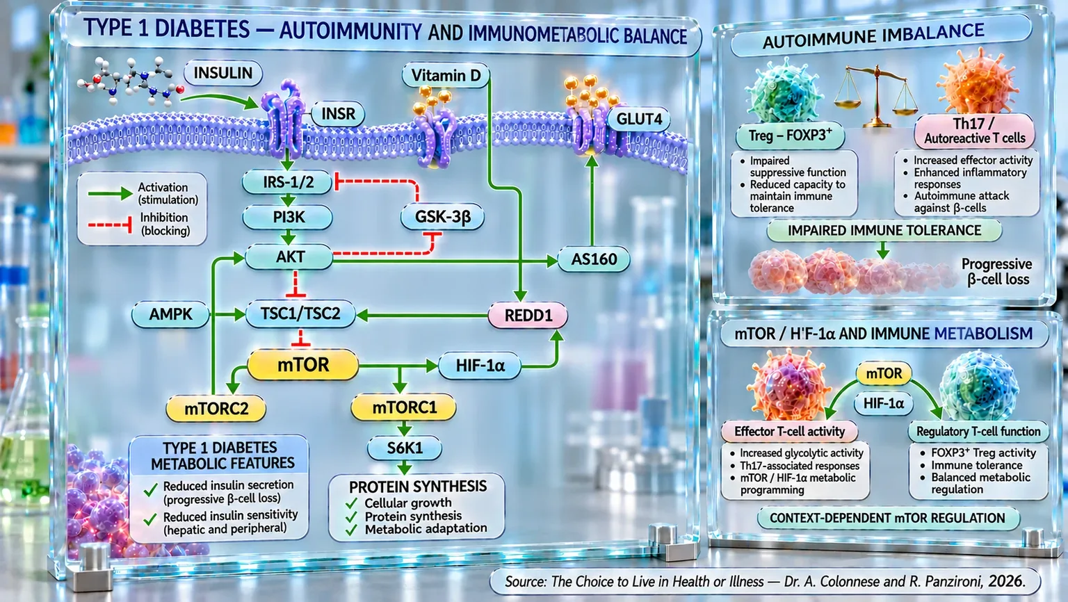
Infographic showing INSR–PI3K–AKT–mTOR–HIF-1α pathways and the balance between regulatory T cells and Th17 cells, associated with the progressive loss of β-cells.
Diagram of immunometabolic balance in type 1 diabetes: it links insulin signaling INSR–IRS-1/2–PI3K–AKT, AMPK, mTORC1/2, HIF-1α, REDD1, vitamin D, and GLUT4, while the comparison between Treg–FOXP3+ and Th17/autoreactive cells illustrates the loss of immune tolerance, inflammation, and the progressive loss of pancreatic β-cells.

ABSTRACT

Type 1 diabetes is an autoimmune disease characterized by the progressive destruction of pancreatic β-cells and the resulting insulin deficiency. In the book "The Choice to Live in Health or Disease", Dr. A. Colonnese and R. Panzironi explore the interactions between immunological tolerance, insulin sensitivity, and cell signaling, highlighting their relevance in the progression of the disease.

The autoimmune response involves autoreactive T lymphocytes and Th17 cells, while Tregs may exhibit impaired suppressive capacity even without a reduction in number. Experimental studies on the transfer and expansion of Tregs, along with early clinical experiences with autologous cells, indicate the potential interest in restoring immune regulation. In parallel, insulin resistance and elevated HOMA-IR values have also been observed in autoantibody-positive subjects, associating with clinical progression. Clamp measurements also document reduced hepatic and peripheral insulin sensitivity.

The PI3K/AKT/mTOR pathway links metabolic signals and immune differentiation: its activation supports Th1 and Th17 effector programs, while controlled modulation can favor FOXP3⁺ Tregs. Through HIF-1α, mTOR promotes glycolysis and may contribute to the persistence of autoreactive responses. HIF-1α is also activated in human pancreatic islets exposed to inflammatory stress. The regulation of AMPK/mTOR also affects autophagy and β-cell resilience, making selective modulation necessary rather than indiscriminate suppression.

In NOD models, preventive rapamycin has reduced the incidence of diabetes; combined strategies and nanoparticles containing rapamycin are under investigation to promote antigen-specific tolerance. An adequate vitamin D status in early life has been associated with lower risk, while experimental and clinical studies suggest possible immune and metabolic effects of supplementation, especially in deficient subjects. Overall, these findings outline a multifactorial interaction between autoimmunity and metabolism, indicating research targets without yet demonstrating generalizable therapeutic benefits. The distinction between preclinical evidence and clinical results remains essential.

Epidemiology of type 1 diabetes

Type 1 diabetes is an autoimmune disease characterized by the destruction of pancreatic β-cells and the resulting progressive insulin deficiency. From an epidemiological perspective, it is important to highlight some fundamental aspects regarding age, sex, and prevalence, which are useful for providing the reader with a clear picture of its significance.

First of all, globally, it is estimated that approximately 9.5 million people live with type 1 diabetes across all age groups; of these, about 1 million are children in the 0-14 age range and about 0.8 million are adolescents between 15 and 19 years old. In terms of age of onset and prevalence by age group, data show that: in the youth population (for example, in young people and adolescents), the reported prevalence in the United States is approximately 3.5 per 1,000 in the population under 20 years of age, with higher values (approximately 5 per 1,000) in the 10-17 age group.

A global analysis of the 15-39 age group estimated approximately 7.34 million cases in 2021; regarding the difference between sexes, the data do not indicate a very marked imbalance: globally, the male percentage appears slightly higher than the female percentage, but the differences are modest.

Finally, in terms of the proportion relative to all diabetes cases, although there are variations between countries, it is often noted that type 1 represents a relatively small share of total diabetes cases (often cited as between 5% and 10% globally).

T-cell imbalance

Type 1 diabetes represents one of the most emblematic manifestations of the failure of immune tolerance. Under physiological conditions, regulatory T cells (Tregs), identified by the CD4+ and CD25+ phenotype, maintain a delicate balance with effector T cells, preventing the body from reacting against its own tissues. When this balance is disrupted, as occurs in type 1 diabetes, the immune system loses the ability to distinguish between "self" and "non-self," selectively attacking the insulin-producing pancreatic β-cells. It is as if a police force, normally tasked with maintaining order, began to strike the citizens it is supposed to protect, destabilizing the entire biological society.

Experimental evidence confirms that in subjects affected by type 1 diabetes, there is not necessarily a reduction in the absolute number of Tregs, but rather a functional impairment of their suppressive capacity. One study showed that although highly activated CD4+ and CD45 Treg cells are increased in patients with type 1A diabetes, they are paradoxically incapable of exercising their regulatory function. This dysfunction is associated with a reduced capacity for residual insulin secretion, suggesting that it is not only the quantity, but above all the quality of the Tregs that determines the progression of the disease (Haseda et al., 2013) [1].

In parallel, other researchers have identified a profoundly altered immune environment in the pancreatic lymph nodes (immune structures near the pancreas that participate in the control of immune responses) of diabetic patients, dominated by an expansion of Th17 cells, known for their inflammatory activity, and by functionally defective Tregs. In this critical location, where the immune system comes into contact with pancreatic antigens, the balance between control and aggression breaks down in favor of inflammation, facilitating the destruction of pancreatic β-cells (Ferraro et al., 2011) [2].

The concept of the Th17/Treg imbalance has also been confirmed by studies on animal models, such as the one conducted on BioBreeding rats, which are predisposed to diabetes. In these animals, the excessive expansion of Th17 cells and the alteration of the peripheral T cell repertoire precede the clinical development of diabetes. Restoring immune balance through the transfer of regulatory T cells or their expansion in vivo significantly reduces the incidence of the disease, confirming that the loss of tolerance is a primary cause and not a simple consequence of the autoimmune process (Van den Brandt et al., 2010) [3].

In humans, the understanding of these mechanisms has led to therapeutic attempts aimed at restoring Treg function. Rapamycin, an immunosuppressant traditionally used in transplants, has demonstrated the ability to selectively expand CD4+CD25+ cells in vitro, while maintaining their suppressive function intact. This discovery paved the way for ex vivo cell therapy strategies based on the infusion of autologous Tregs (regulatory immune cells from the same patient) to restore immune tolerance (Battaglia et al., 2005) [4].

Subsequent clinical trials have demonstrated the feasibility of this approach: between 2012 and 2014, children with recent-onset type 1 diabetes were treated with infusions of ex vivo expanded autologous Tregs, observing not only an increase in circulating Tregs but also a significant preservation of β-cell function and a reduction in the need for exogenous insulin. In some cases, patients maintained satisfactory glycemic control without resorting to insulin for several months, an extraordinary result for a traditionally irreversible pathology (Marek-Trzonkowska et al., 2012) [5]; (Marek-Trzonkowska et al., 2013) [6]; (Marek-Trzonkowska et al., 2014) [7].

These experimental and clinical observations are confirmed in a broader context: regulatory T cells constitute the main defense mechanism against autoimmunity in general. In 2010, a researcher pointed out that quantitative or qualitative defects in these cells are common to many autoimmune diseases, including multiple sclerosis, lupus, and rheumatoid arthritis. In type 1 diabetes, their inability to suppress the response against β-cell antigens represents the pathogenetic key that links genetics, environment, and immune dysfunction (Buckner, 2010) [8].

In summary, type 1 diabetes can be considered the final result of a broken immunological balance: on one hand, a hyperactive inflammatory response driven by Th17 and effector T cells; on the other, a regulatory system incapable of containing the attack. Restoring harmony between these two poles, like a conductor bringing order to a chaotic ensemble, appears today to be one of the most promising strategies for preserving pancreatic function and changing the clinical destiny of this complex autoimmune disease.

Metabolic alterations: insulin pathway and insulin resistance

In type 1 diabetes, insulin resistance—traditionally considered a phenomenon typical of type 2 diabetes—has emerged in recent years as a key element in the pathogenesis and progression of this autoimmune form as well. It is as if, in addition to the immune system's attack on β-cells, the body must face an additional challenge: responding adequately to residual or exogenous insulin (insulin administered from the outside), thereby worsening the metabolic instability typical of the disease.

The first evidence in this regard comes from prospective studies on first-degree relatives of patients with type 1 diabetes who were already positive for autoantibodies against pancreatic islets. Some researchers have demonstrated that insulin resistance, measured via the HOMA-IR index, represents an independent risk factor for progression toward overt disease. In the same study, the ratio between the first-phase insulin response and HOMA-IR proved to be an even more powerful indicator for predicting the onset of diabetes, highlighting how an altered balance between insulin secretion and action can anticipate the diagnosis (Xu et al., 2007) [9].

Similar results had already been observed by other researchers in 2004, who highlighted that autoantibody-positive relatives who progressed toward the disease showed reduced insulin sensitivity from the preclinical stages, disproportionate to their secretory capacity. The HOMA-R/FPIR ratio (resistance index relative to secretion) proved to be the strongest predictor of progression, suggesting that the loss of glycemic control begins well before complete β-cell destruction (Fourlanos et al., 2004) [10].

These observations have been confirmed and expanded more recently by the study of another researcher who analyzed over 6,000 autoantibody-positive relatives. The HOMA-IR index and the Matsuda index, which are indicators of insulin resistance and sensitivity respectively, were strongly associated with progression from early stages (silent autoimmunity) to clinical diabetes. In particular, high HOMA-IR values and low Matsuda values increased the risk of transitioning from stage 1 to 3, even after adjustment for insulin secretion markers. This demonstrates that insulin resistance is not a simple side effect, but a factor that accelerates the transition toward manifest disease (Petrelli et al., 2024) [11].

On a pathophysiological level, insulin resistance in subjects with type 1 diabetes can be compared to a "metabolic background noise" that amplifies the effects of autoimmunity. In 2015, through a systematic review and meta-analysis of euglycemic clamp studies (a test that evaluates how effectively tissues respond to insulin), it was confirmed that in patients with type 1 diabetes, there is reduced insulin sensitivity at both the hepatic and peripheral levels, with a lower capacity to suppress endogenous glucose production and reduced glucose uptake by muscles. Lipolysis is also less inhibited, contributing to the tendency toward hyperglycemia and ketosis (Donga et al., 2015) [12].

Beyond metabolic mechanisms, other researchers have highlighted a more direct immuno-metabolic relationship: in adult subjects with recently diagnosed type 1 diabetes, anti-insulin antibody titers are inversely correlated with insulin sensitivity. In other words, the greater the autoimmune response against insulin, the lower the ability of tissues to respond to it. This link underscores how immunity and metabolism are not separate compartments, but intertwined systems that influence each other (Burkart et al., 2018) [13].

Overall, the data suggest that insulin resistance not only coexists with type 1 diabetes but actively influences its evolution. In a sense, the disease can be seen as the result of the encounter between two weaknesses: an immune system that destroys β-cells and an organism that responds less and less effectively to the available insulin. Intervening on this latter aspect, by improving insulin sensitivity through nutrition, physical activity, or insulin-sensitizing drugs, could represent an important step in the prevention and management of the early stages of the autoimmune disease.

mTOR as a central node

In type 1 diabetes, the mTOR signaling pathway acts as a metabolic “switch” that decides whether T cells will take the path of attack or the path of control: when mTOR is active, it integrates environmental signals and pushes T cells toward effector profiles (Th1, Th2, Th17); when it is attenuated, it instead favors the development of FoxP3+ regulatory T cells, the guards that restore immune order. In mouse models lacking mTOR in T cells, effector differentiation is blocked while the generation of Tregs increases, reminding us that “how much fuel” and “which signals” a cell perceives are at least as decisive as the antigen it recognizes (Delgoffe et al., 2009) [14].

This metabolic-immunological perspective also opens up therapeutic possibilities: in NOD models (Non-Obese Diabetic, a fundamental animal model for studying type 1 diabetes), metformin does not act only as a glucose-lowering agent, but remodels the immune system by suppressing mTOR in T cells and mTOR/STAT3 in dendritic cells (cells that recognize potential threats), reducing pro-inflammatory cytokines and increasing Tregs; the practical result is a delay or lower incidence of diabetes onset, as if the “volume” of inflammation were being turned down right in the lymph nodes that drain the pancreas (Suzuki et al., 2025) [15].

In parallel, intervening on mTOR can directly protect the “target organ”: in experimental models, a traditional herbal formulation (Cai tea) activates AMPK, inhibits mTOR, and enhances mitochondrial autophagy in β-cells, resulting in more intact pancreatic islets, better insulin secretion, and reduced inflammatory markers; it is like helping the cell perform “workshop cleaning,” removing damaged mitochondria and recovering energy efficiency in a hostile environment (Li et al., 2024) [16].

Taken together, these findings show that mTOR is a crossroads where immunology and metabolism meet: by modulating its signal, one can both tip the scales from effector T cells to regulatory ones and make β-cells more resilient to stress; in conclusion, this means supplementing insulin replacement with a strategy that reduces the autoimmune “impact” and strengthens the pancreas's intrinsic ability to survive the assault.

mTOR inhibitors

In type 1 diabetes, the progressive destruction of pancreatic β-cells is driven by a loss of immunological tolerance to islet antigens and the activation of autoreactive T lymphocytes. In this scenario, mTOR represents one of the main metabolic regulators of the T-cell response, as it integrates energy availability, growth signals, and stimuli from the antigen receptor, helping to determine the proliferation and differentiation of immune cells. Pharmacological inhibition of mTOR using rapamycin, or sirolimus (an immunosuppressive drug that primarily inhibits the mTORC1 complex), can therefore modify the autoimmune response by acting primarily on the balance between effector T cells and regulatory T cells. Rapamycin mainly inhibits mTORC1 and limits the proliferation of effector populations, including those associated with Th1 and Th17 phenotypes, while FOXP3+ regulatory T cells are relatively more resistant to this inhibition. This differing metabolic dependence offers a possible explanation for the immunomodulatory effect observed experimentally: reducing the mTOR signal can decrease the pressure of autoreactive cells on the pancreatic islets while simultaneously increasing the ability of Tregs to control the autoimmune response.

One of the first experimental demonstrations of the importance of mTOR in autoimmune diabetes comes from the NOD mouse model. Preventive administration of rapamycin drastically reduced the incidence of diabetes and, at the higher doses used, prevented the development of hyperglycemia during the treatment period. The effect was particularly significant when rapamycin was administered before the onset of the disease, whereas it was ineffective in reverting established diabetes. These results indicated early on that mTOR inhibition can hinder the expansion and activation phase of the autoimmune response that precedes the irreversible destruction of β-cells (Baeder et al., 1992) [17].

Subsequently, important confirmation came from a new-onset type 1 diabetes model: researchers used a combination therapy including rapamycin, an IL-2-related agonist, and a mutated IL-15 antagonist. The treatment halted the autoimmune destruction of β-cells and restored normoglycemia and immune tolerance. Particularly interesting was the reduction in the expression of cytotoxic and pro-inflammatory mediators, including IFN-γ, IL-1β, and TNF-α. The metabolic recovery was also associated with a reduction in systemic inflammation and an improvement in the insulin response. In this model, therefore, mTOR inhibition constituted a component of a strategy capable of acting simultaneously on autoimmunity and metabolic inflammation (Koulmanda et al., 2007) [18].

In type 1 diabetes, the progressive loss of pancreatic β-cells results from the interaction between β-cell vulnerability and the autoimmune response, in which autoreactive T lymphocytes acquire a highly glycolytic metabolism necessary for their activation and proliferation. mTORC1 represents one of the main regulators of this metabolic reprogramming: when the TCR signal and growth factors activate PI3K/AKT/mTOR, protein synthesis, glycolytic metabolism, and the proliferative capacity of effector lymphocytes increase. Controlled inhibition of mTORC1 can therefore reduce the expansion of effector populations and modify the ratio between autoreactive T cells and Tregs, helping to restore tolerance to β-cell antigens. The results obtained are particularly interesting: in NOD mice, this leads to less immune infiltration of the pancreatic islets, better preservation of insulin secretion, and a delay in the onset of diabetes. In parallel, FOXP3 is modified, favoring a less diabetogenic immune profile (Kong et al., 2021) [19].

A particularly significant evolution is represented by the possibility of delivering rapamycin in nanoparticles, limiting its systemic exposure and directing its effect more toward immunological tolerance. ImmTOR nanoparticles containing rapamycin have been associated with an engineered variant of IL-2 selective for Tregs. The combination increased the expansion and persistence of regulatory T cells and, in the presence of the target antigen, strongly favored the emergence of antigen-specific Tregs. In autoimmune models including type 1 diabetes, this strategy provided protection from the disease while limiting the unwanted expansion of effector immune cells that can accompany IL-2 treatment. In this case, therefore, mTOR is not simply "turned off," but used as a metabolic node to selectively orient the immune response toward tolerance (Kishimoto et al., 2023) [20].

These studies therefore suggest a biological sequence particularly useful for the chapter: PI3K/AKT → mTORC1 → glycolytic metabolism and effector lymphocyte proliferation → autoreactive response against the β-cell, while selective modulation can favor: ↓ mTORC1 in effector lymphocytes → ↓ autoreactive expansion → ↑ Treg predominance/function → greater preservation of β-cells.

The central point, however, is that mTOR should not simply be inhibited throughout the entire organism. In type 1 diabetes, the need emerges to reduce mTORC1 hyperactivity in pathogenic immune components while preserving mTORC1/mTORC2 in β-cells, where the pathway remains necessary for growth, insulin secretion, and survival. This distinction is essential because it avoids presenting mTOR inhibition as universally beneficial.

mTOR, HIF-1α and metabolic reprogramming of immune system cells

In type 1 diabetes, mTOR activation can contribute to the progression of autoimmunity by profoundly altering T-cell metabolism. Following antigen activation, mTOR promotes the expression of HIF-1α, which increases glycolysis utilization and provides effector cells with the energy and biosynthetic intermediates necessary for proliferation and the production of pro-inflammatory cytokines. This metabolic program particularly favors Th17 cells while simultaneously making the regulatory program associated with FOXP3 Treg cells less favorable. In type 1 diabetes, where autoreactive lymphocytes progressively attack pancreatic β-cells, the mTOR–HIF-1α–glycolysis axis can therefore contribute to shifting the immune balance toward a persistent pro-inflammatory response.

One of the most direct demonstrations in type 1 diabetes was provided in a 2019 study: during Th17 differentiation, treatment with metformin increased AMPK activation while simultaneously reducing mTOR phosphorylation (activation) and HIF-1α expression. In parallel, Th1 and Th17 cells decreased, while FOXP3+ Treg cells increased. In NOD mice, a spontaneous model of type 1 diabetes, metformin reduced CD4+ cells producing IFN-γ and IL-17 (immune system molecules that help activate and coordinate inflammation), while increasing regulatory populations. The study thus directly links the AMPK → mTOR → HIF-1α modulation to the reduction of pancreatic autoimmunity (Duan et al., 2019) [21].

The significance of the HIF-1α component was documented directly in the human pancreas by other researchers in 2020. By analyzing pancreatic islets from subjects with type 1 diabetes, the study authors detected significant activation of HIF-1α signaling and a roughly five-fold increase in PFKFB3, an important regulator of glycolysis. The same program was induced in human islets exposed to cytokines characteristic of the diabetic microenvironment, with an increase in lactate production. HIF-1α initially also exerted a survival effect, but at the cost of early impairment of glucose-stimulated insulin secretion (Nomoto et al., 2020) [22].

Overall, this evidence outlines a model in which mTORC1 activation represents one of the main metabolic signals capable of supporting HIF-1α in T cells. HIF-1α then enhances glycolysis and favors effector programs, especially Th17, while relatively counteracting the maintenance of FOXP3+ Treg cells. Furthermore, in the pancreatic microenvironment, HIF-1α is also activated in β-cells subjected to inflammatory stress, altering their metabolism and function. In type 1 diabetes, one can therefore outline the sequence: PI3K/AKT/mTOR activation → increase in HIF-1α → increase in glycolysis → expansion and maintenance of effector T cells → Th17/Treg imbalance → increase in autoimmune response → progressive destruction of pancreatic β-cells.

Vitamin D as an immuno-metabolic modulator

In type 1 diabetes, vitamin D acts as a “contextual” regulator: it does not extinguish the autoimmune fire on its own, but it lowers the wind that fuels it, improving the immuno-metabolic environment in which the disease originates and progresses. Prospective data suggest that early exposure makes a difference: in a Finnish birth cohort, regular supplementation in the first year of life was associated with a significantly lower risk of developing diabetes later on, with an apparent dose-response gradient; conversely, suspected rickets tripled the risk (Hyppönen et al., 2001) [23].

Regarding circulating levels, children, adolescents, and adults with type 1 diabetes show lower average concentrations of 25(OH)D compared to controls, reinforcing the idea that “vitamin D status” is part of the risk profile, even without proving causality on its own (Feng et al., 2015) [24]. A review focused on the pediatric age group confirms that, by age 15, deficiencies up to 50% lower than those of healthy peers are frequent and associated with a higher probability of disease, proposing supplementation as a potentially relevant preventive measure (Daskalopoulou et al., 2022) [25].

The logic is consistent: as described in the chapter regarding the immune system, through the modulation of the mTOR complex, calcitriol modulates adaptive immunity by reducing pro-inflammatory Th1/Th17 profiles, promotes regulatory T cells (Tregs), and “softens” dendritic cells, with possible direct effects of β-cell protection from inflammatory stress (Rak and Bronkowska, 2018) [26].

In NOD models, high but safe and prolonged dietary exposure to cholecalciferol reduces the incidence of diabetes and preserves pancreatic insulin content, accompanied by more Tregs in the pancreatic lymph nodes: it is like lowering the threshold of the immune fuse before it reaches the islets (Takiishi et al., 2014) [27].

In the pediatric age group, three months of high-unit cholecalciferol reduced HbA1c (an indicator of average glycemia over time) without altering calcium metabolism, suggesting an additive metabolic benefit when there is a baseline deficiency (Ordooei et al., 2017) [28].

In a randomized, double-blind, placebo-controlled, crossover study, 39 patients with type 1 diabetes received 4000 IU/day of vitamin D for three months. Supplementation significantly increased 25(OH)D levels and, in men, led to a greater increase in regulatory T cells (Tregs). The treatment also reduced insulin requirements and improved HbA1c (an indicator of average glycemia over time), without showing toxicity. The Treg response also varied in relation to specific vitamin D receptor genotypes (Bogdanou et al., 2017) [29].

In another retrospective study of 271 children and adolescents with type 1 diabetes, 14.8% had a deficiency and 31% had vitamin D insufficiency. In the 73 patients evaluated after three months of supplementation with cholecalciferol, 25(OH)D levels increased from 12.9 to 26.1 ng/mL, while HbA1c decreased significantly. The improvement was greater in subjects with higher initial HbA1c and lower vitamin D levels, suggesting a possible benefit on glycemic control (Giri et al., 2017) [30].

Overall, the evidence suggests that vitamin D may contribute to the modulation of type 1 diabetes by acting on both the immune and metabolic fronts. An adequate vitamin status, especially in the early stages of life, has been associated with a lower risk of disease, while deficiency is frequent in patients. Through the modulation of mTOR, vitamin D can promote Tregs and limit pro-inflammatory Th1/Th17 responses, contributing to β-cell protection. Experimental and clinical studies also indicate that supplementation, especially in deficient subjects, can be associated with an improvement in HbA1c and a reduction in insulin requirements.

Conclusions

Taken together, the analyzed scientific evidence outlines type 2 diabetes as a condition in which autoimmunity, metabolism, and cell signaling are deeply interconnected. The disease, characterized by the progressive autoimmune destruction of pancreatic β-cells and the resulting insulin deficiency, affects approximately 9.5 million people worldwide and has a significant incidence starting in childhood and adolescence.

The first step involves the loss of immunological tolerance. Evidence shows that in type 1 diabetes, the issue with Tregs is not necessarily their numerical reduction, but rather the impairment of their suppressive capacity. In parallel, the expansion of Th17 cells and autoreactive T-cell populations promotes the attack against β-cells. Experimental studies on the transfer or expansion of Tregs and early clinical experiences with autologous Tregs confirm the importance of restoring this balance to preserve β-cell function.

Associated with this immune alteration is insulin resistance, which can appear as early as the preclinical stages. In autoantibody-positive subjects, elevated HOMA-IR and reduced insulin sensitivity are associated with progression to overt disease, regardless of residual secretory capacity. Clamp studies further confirm reduced hepatic and peripheral insulin sensitivity in type 1 diabetes. The disease therefore emerges from the interaction between the progressive loss of insulin secretion and the reduced efficacy of available insulin.

In this scenario, mTOR represents a convergence point between metabolism and immunity. Its activation supports the metabolic programs of effector T cells, including Th1 and Th17, while a controlled reduction of the signal favors FOXP3+ Tregs. Modulation of AMPK/mTOR can also support autophagy and β-cell resilience.

Studies with rapamycin/sirolimus reinforce this interpretation. In NOD models, preventive inhibition of mTOR reduced the incidence of diabetes; combined strategies attenuated inflammatory mediators and favored the recovery of tolerance, while more recent approaches with rapamycin-containing nanoparticles aim to selectively expand antigen-specific Tregs. However, a fundamental principle emerges: it is not desirable to shut down mTOR indiscriminately, but rather to reduce its hyperactivity in pathogenic immune components while preserving its functions in β-cells.

The next step involves HIF-1α. mTOR can support its expression, promoting glycolysis and effector programs. The sequence reconstructed from the evidence is therefore PI3K/AKT/mTOR → HIF-1α → increased glycolysis → expansion of Th17 and relative reduction of Tregs → increased autoimmune response → progressive β-cell destruction. HIF-1α is also activated in human pancreatic islets subjected to inflammatory stress, showing how metabolic reprogramming also directly involves the β-cell.

Finally, vitamin D completes this immunometabolic picture. An adequate vitamin status in the early stages of life has been associated with a lower risk of disease, while deficiency is frequent in patients. Experimental and clinical studies also indicate possible effects on Tregs, Th1/Th17 responses, HbA1c, and insulin requirements, especially in deficient subjects. Overall, a common thread emerges: loss of tolerance → alteration of insulin sensitivity → dysregulation of mTOR/HIF-1α and cellular metabolism → predominance of effector responses → progressive β-cell damage, identifying immunometabolic balance as a fundamental key to understanding the evolution of type 1 diabetes.

References

[1] CD4+ CD45RA+ FoxP3high activated regulatory T cells are functionally impaired and related to residual insulin-secreting capacity in patients with type 1 diabetes

Haseda et al., 2013

Clin Exp Immunol. 2013 Aug;173(2):207-16.

[2] Expansion of Th17 cells and functional defects in T regulatory cells are key features of the pancreatic lymph nodes in patients with type 1 diabetes

Ferraro et al., 2011

Diabetes. 2011 Nov;60(11):2903-13.

[3] Type 1 diabetes in BioBreeding rats is critically linked to an imbalance between Th17 and regulatory T cells and an altered TCR repertoire

Van den Brandt et al., 2010

J Immunol. 2010 Aug 15;185(4):2285-94.

[4] Rapamycin selectively expands CD4+CD25+FoxP3+ regulatory T cells

Battaglia et al., 2005

Blood. 2005 Jun 15;105(12):4743-8.

[5] Administration of CD4+CD25highCD127- regulatory T cells preserves β-cell function in type 1 diabetes in children

Marek-Trzonkowska et al., 2012

Clinical Trial Diabetes Care. 2012 Sep;35(9):1817-20.

[6] Clinical application of regulatory T cells in type 1 diabetes

Marek-Trzonkowska et al., 2013

Pediatr Diabetes. 2013 Aug;14(5):322-32.

[7] Therapy of type 1 diabetes with CD4(+)CD25(high) CD127-regulatory T cells prolongs survival of pancreatic islets - results of one year follow-up

Marek-Trzonkowska et al., 2014

Clin Immunol. 2014 Jul;153(1):23-30.

[8] Mechanisms of impaired regulation by CD4+CD25+FOXP3+ regulatory T cells in human autoimmune diseases

Buckner, 2010

Nat Rev Immunol. 2010 Dec;10(12):849–859.

[9] Role of insulin resistance in predicting progression to type 1 diabetes

Xu et al., 2007

Diabetes Care. 2007 Sep;30(9):2314-20.

[10] Insulin resistance is a risk factor for progression to type 1 diabetes

Fourlanos et al., 2004

Diabetologia. 2004 Oct;47(10):1661-7.

[11] HOMA-IR and the Matsuda Index as predictors of progression to type 1 diabetes in autoantibody-positive relatives

Petrelli et al., 2024

Diabetologia. 2024 Feb;67(2):290-300.

[12] Insulin resistance in patients with type 1 diabetes assessed by glucose clamp studies: systematic review and meta-analysis

Donga et al., 2015

Eur J Endocrinol. 2015 Jul;173(1):101-9.

[13] Inverse association of insulin antibody levels with insulin sensitivity in adults with Type 1 diabetes

Burkart et al., 2018

Diabet Med. 2018 May;35(5):595-601.

[14] The mTOR kinase differentially regulates effector and regulatory T cell lineage commitment

Delgoffe et al., 2009

Immunity. 2009 Jun 19;30(6):832-44.

[15] Metformin prevents diabetes development in type 1 diabetes models via suppression of mTOR and STAT3 signaling in immune cells

Suzuki et al., 2025

Scientific Reports volume 15, Article number: 10641 (2025)

[16] Cai's herbal tea enhances mitochondrial autophagy of type 1 diabetic mellitus β cells through the AMPK/mTOR pathway and alleviates inflammatory response

Li et al., 2024

Acta Diabetol. 2024 Dec;61(12):1553-1567.

[17] Rapamycin Prevents the Onset of Insulin-Dependent Diabetes Mellitus (IDDM) in NOD Mice.

Baeder et al., 1992

Clinical and Experimental Immunology, vol. 89, no. 2, 1992, pp. 174–178.

[18] Modification of Adverse Inflammation Is Required to Cure New-Onset Type 1 Diabetic Hosts.

Koulmanda et al., 2007

Proc. Natl. Acad. Sci. U.S.A., vol. 104, no. 32, 2007, pp. 13074–13079.

[19] Mitochondrial-Encoded MOTS-c Prevents Pancreatic Islet Destruction in Autoimmune Diabetes.

Kong et al., 2021

Cell Reports, vol. 36, no. 4, 2021, article 109447.

[20] Rapamycin Nanoparticles Increase the Therapeutic Window of Engineered Interleukin-2 and Drive Expansion of Antigen-Specific Regulatory T Cells for Protection against Autoimmune Disease.

Kishimoto et al., 2023

Journal of Autoimmunity, vol. 140, 2023, article 103125.

[21] Metformin Mitigates Autoimmune Insulitis by Inhibiting Th1 and Th17 Responses while Promoting Treg Production.

Duan et al., 2019

American Journal of Translational Research, vol. 11, no. 4, 2019, pp. 2393–2402. PMID: 31105845; PMCID: PMC6511786.

[22] Activation of the HIF1α/PFKFB3 Stress Response Pathway in Beta Cells in Type 1 Diabetes.

Nomoto et al., 2020

Diabetologia, vol. 63, no. 1, 2020, pp. 149–161.

[23] Intake of vitamin D and risk of type 1 diabetes: a birth-cohort study

Hyppönen et al., 2001

Lancet. 2001 Nov 3;358(9292):1500-3.

[24] Lower serum 25 (OH) D concentrations in type 1 diabetes: A meta-analysis

Feng et al., 2015

Diabetes Res Clin Pract. 2015 Jun;108(3):e71-5.

[25] Vitamin D Deficiency as a Possible Cause of Type 1 Diabetes in Children and Adolescents up to 15 Years Old: A Systematic Review

Daskalopoulou et al., 2022

Rev Diabet Stud. 2022 Jun 30;18(2):58-67.

[26] Immunomodulatory Effect of Vitamin D and Its Potential Role in the Prevention and Treatment of Type 1 Diabetes Mellitus-A Narrative Review

Rak e Bronkowska, 2018

Molecules. 2018 Dec 24;24(1):53.

[27] Dietary supplementation with high doses of regular vitamin D3 safely reduces diabetes incidence in NOD mice when given early and long term

Takiishi et al., 2014

Diabetes. 2014 Jun;63(6):2026-36.

[28] Effect of vitamin D on HbA1c levels of children and adolescents with diabetes mellitus type 1

Ordooei et al., 2017

Minerva Pediatr. 2017 Oct;69(5):391-395.

[29] T-lymphocyte and glycemic status after vitamin D treatment in type 1 diabetes: A randomized controlled trial with sequential crossover

Bogdanou et al., 2017

Diabetes Metab Res Rev. 2017 Mar;33(3)

[30] Treating vitamin D deficiency in children with type I diabetes could improve their glycaemic control

Giri et al., 2017

BMC Res Notes. 2017 Sep 7;10:465.