
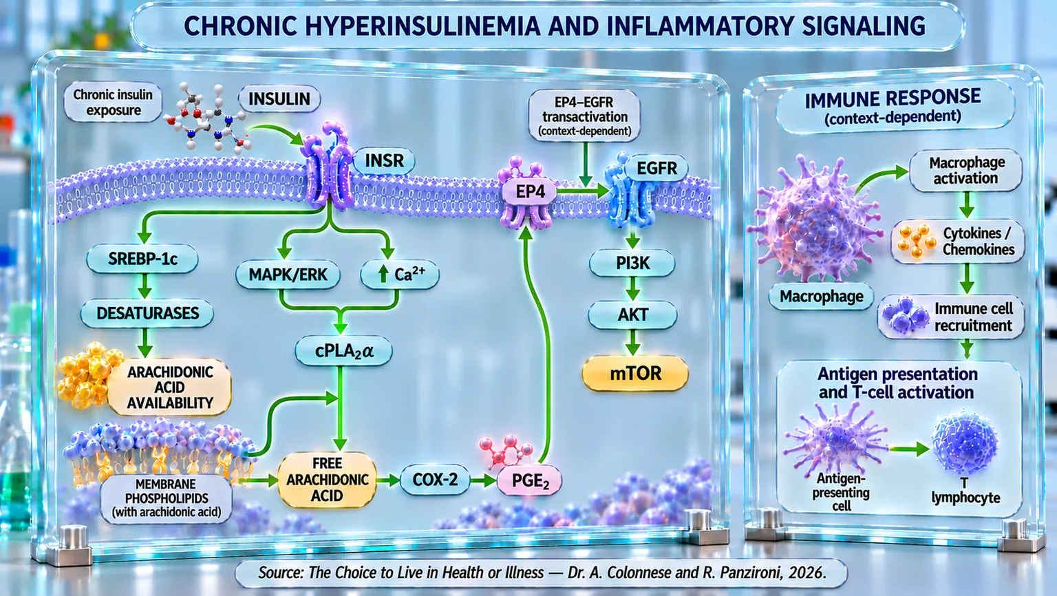
As we highlighted in the previous chapters, a growing body of scientific evidence has progressively challenged the paradigm that hyperinsulinemia is exclusively a compensatory response to insulin resistance; likewise, as we will see with the following scientific evidence, hyperinsulinemia can also become the driver for the activation and maintenance of chronic inflammation.
In fact, a broad and systematic review published in 2021 contributed decisively to this shift in perspective. By analyzing experimental and clinical data, it emerges that, in the early stages of obesity and type 2 diabetes, hyperinsulinemia often precedes both hyperglycemia and overt insulin resistance. In this context, excess insulin does not act as a neutral signal, but as a powerful modulator of cellular signaling, capable of promoting chronic inflammation, metabolic dysfunction, acceleration of aging processes, and an increased oncological risk (Zhang et al., 2021) [1].
Supporting this view, experimental studies that isolate the effect of insulin from caloric excess provide particularly robust evidence. In a 2018 study, chronic hyperinsulinemia was induced in murine models (laboratory animals, mainly mice, used in scientific studies) through continuous insulin infusion, in the absence of an increase in energy intake. Under these conditions, hyperinsulinemia proved sufficient to induce adipose tissue inflammation, characterized by an increase in pro-inflammatory immune populations, an imbalance between M1 and M2 macrophages (immune system cells), and marked extracellular matrix deposition. Conversely, partial reduction of circulating insulin, even in the presence of obesity, attenuated inflammation and improved insulin sensitivity, identifying hyperinsulinemia as a direct causal factor of metabolic inflammation (Kumar et al., 2018) [2].
The pathogenetic role of hyperinsulinemia also clearly emerges outside of classically metabolic tissues. In 2022, it was demonstrated that hyperinsulinemia associated with insulin resistance promotes the pro-inflammatory activation of microglia, the main immune population of the central nervous system. In animal and cellular models, chronic exposure to insulin induces alterations in microglial energy metabolism, mitochondrial dysfunction (cellular structures that produce the energy necessary for the cell), and polarization toward a pro-inflammatory phenotype. These changes are closely associated with the development of neuroinflammation and cognitive deficits, suggesting a direct pathogenetic link between type 2 diabetes and neurodegenerative diseases (Yang et al., 2022) [3].
Further confirmation of this model emerges from a review published in 2022, which critically analyzes the interactions between hyperinsulinemia, inflammation, and metabolic dysfunction. In this context, macrophages are identified as a central node for integrating metabolic and immune signals. Macrophages express insulin receptors and respond directly to its signaling; however, chronic exposure to high concentrations of insulin compromises physiological regulatory function, favoring the loss of anti-inflammatory signals and the enhancement of pro-inflammatory responses. This imbalance contributes to the formation of immuno-metabolic vicious cycles that sustain and amplify the progression of metabolic diseases (Püschel et al., 2022) [4].
Taken together, these studies converge on a fundamental point: chronic hyperinsulinemia emerges as an active pathogenetic determinant, capable of triggering and sustaining systemic and tissue inflammation. Inflammation, therefore, does not appear as a secondary or subsequent event, but as a consistent consequence of aberrant and non-physiological insulin signaling. This conceptual framework constitutes the necessary prerequisite for analyzing, in the following sections, the 3 phases through which excess insulin is translated into inflammatory activation.
The first event is silent but fundamental: insulin stimulates the cell to increase the production of arachidonic acid, an omega-6 polyunsaturated fatty acid present in cell membranes. This occurs through the activation of SREBP-1c (a protein that activates genes involved in the production of fats by the cell), which enhances the enzymes necessary to build lipid substrates such as triglycerides and cholesterol (Matsuzaka et al., 2002) [5]. In other words, hyperinsulinemia does not yet generate inflammation, but it creates the conditions for it to become possible.
Once accumulated in cell membranes, arachidonic acid must be released to become biologically active. Here, a second decisive step comes into play: insulin activates the MAPK cascade (a chain of signals that transmits information within the cell), increases intracellular calcium, and activates cPLA2α, the enzyme responsible for the release of free arachidonic acid (Gijón et al., 2000) [6].
Finally, the released arachidonic acid is converted into prostaglandin E2, one of the central mediators of chronic inflammation, by the action of COX2 (an enzyme that contributes to the production of substances involved in inflammation) stimulated by excess insulin through the activation of the MAPK cascade. PGE2 does not remain a simple end product: it becomes an active signal that turns on the EP4 receptor (a cellular receptor that receives the signal of prostaglandin E2 (PGE2)), reactivates EGFR (a cellular receptor that, when activated, stimulates cell growth and survival signals), and converges again on the PI3K–AKT–mTOR axis, closing a deep amplification circuit between inflammation and cell growth (Oshima et al., 2011) [7].
In the following paragraphs, we will go into more detail on each of the 3 phases described above to better explain all the biologically connected steps.