
Scientific Book

Immune defenses and their metabolic balance

The immune system protects the body through a complex network of cells, molecules, and signals that coordinate defense and immunological tolerance. T and B lymphocytes, macrophages, dendritic cells, and Th17 and Treg populations also depend on specific metabolic programs regulated by mTOR, AMPK, and other cellular pathways. In the book "The Choice to Live in Health or Disease," Dr. A. Colonnese and R. Panzironi explore the relationship between immunity, metabolism, and the development of autoimmune diseases.

Cite this article
Colonnese, A. & Panzironi, R. (2026). Immune defenses and their metabolic balance. The Choice to Live in Health or in Disease. https://lifesciencehub.info/en/la-scelta-di-vivere-in-salute-o-in-malattia/immune-defenses-and-their-metabolic-balance
The Choice to Live in Health or in Disease · ISBN 979-12-82692-03-8

DOI lsh.en.libro.6

  • Published 05/11/2026
  • Updated 05/11/2026
  • IMMUNOMETABOLISM
  • MTORC1
  • AMPK
  • HIF-1Α
  • TH17/TREG LYMPHOCYTES
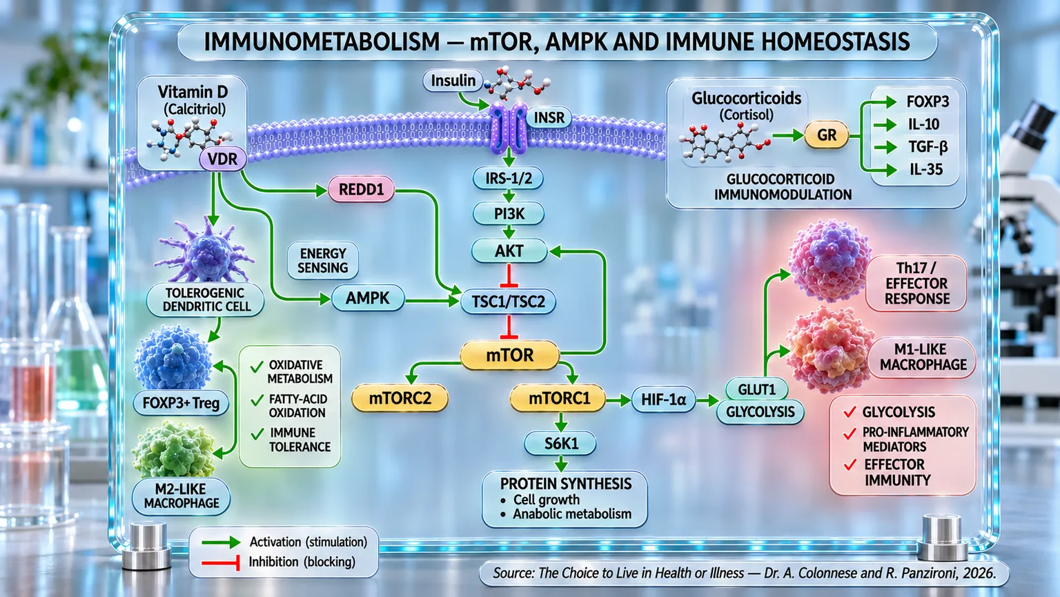
Infographic showing a cell membrane and activation or inhibition arrows between receptors, AMPK, mTOR, HIF-1α, and immune phenotypes.
Immunometabolism diagram: vitamin D and insulin modulate the AMPK–TSC1/TSC2–mTOR and mTORC1–HIF-1α circuits, while glucocorticoids act via GR. These signals orient oxidative metabolism and tolerance in dendritic cells, Tregs, and M2 macrophages, or glycolysis and effector Th17 and M1 macrophage responses.

ABSTRACT

The immune system and energy metabolism constitute functionally interconnected networks, in which nutrient availability influences cellular activation, differentiation, and regulation. In the book "La scelta di Vivere in Salute o in Malattia" (The Choice to Live in Health or Disease), Dr. A. Colonnese and R. Panzironi delve into the mechanisms of immunometabolism, highlighting the role of molecular pathways in coordinating innate and adaptive immune responses.

Pro-inflammatory M1 macrophages and effector T lymphocytes primarily utilize glycolysis, while reparative and regulatory phenotypes rely more heavily on oxidative metabolism. In this balance, mTORC1 integrates nutritional signals and growth factors, supporting the proliferation and differentiation of effector cells. Through HIF-1α, it also promotes the expression of GLUT1 and glycolytic enzymes, contributing to Th17 differentiation and the modulation of the regulatory program associated with FOXP3.

AMPK exerts complementary energy control, favoring metabolic conditions compatible with regulatory functions. The phosphorylation of TSC2 contributes to the inhibition of mTORC1, highlighting how immune balance depends on the coordinated modulation of opposing metabolic signals. Experimental studies on the inhibition of HIF-1α describe reductions in glycolysis and inflammation, associated with changes in the Th17/Treg balance and macrophage polarization.

These mechanisms support interest in immunomodulatory interventions. Rapamycin can promote FOXP3+ Treg populations and limit effector responses, while glucocorticoids influence FOXP3 and regulatory mediators such as IL-10, TGF-β, and IL-35. Calcitriol, through VDR, can also support regulatory differentiation and modify dendritic cell activity, involving REDD1 and TSC1/TSC2 in the modulation of mTOR.

Overall, a dynamic relationship emerges between cellular metabolism, immune differentiation, and tolerance. Understanding these interactions provides an interpretive basis for further exploring inflammatory and autoimmune diseases, distinguishing experimental evidence from clinical applications that are yet to be defined.

What is the immune system

In Chapter 4, we followed the insulin pathway step by step: a crucial hormonal signal that guides glucose uptake, regulates cell growth, and coordinates a complex network of intracellular activations. This pathway, seemingly confined to metabolism, is actually deeply intertwined with many other bodily functions. Among these, one of the most surprising and strategic is the immune function. In recent years, scientific research has highlighted an increasingly close link between metabolism and the immune system. Alterations in insulin signaling, such as those observed in conditions of insulin resistance, do not only affect glycemic homeostasis, but can modulate the activity of immune cells, fuel a state of chronic inflammation, and contribute to the development of metabolic and autoimmune diseases. In this chapter, we will delve into this very point: how the dysregulation of the insulin pathway impacts the immune system, paving the way for new interpretations of inflammation and many modern chronic diseases. Because today more than ever, metabolism and immunity speak the same language.

The immune system represents a complex network of cells and molecules dedicated to defending the body against pathogens such as bacteria, viruses, or transformed cells. It is an extraordinarily organized mechanism that integrates two main components: innate immunity and adaptive immunity. Among the protagonists of the first line of defense are macrophages, cells capable of recognizing and engulfing pathogenic microorganisms, destroying them through phagocytosis processes. These cells do not only perform an elimination function, but also release chemical signals, such as cytokines, capable of recruiting other immune cells and modulating the intensity of the response (Gordon and Martinez, 2010) [1]. Macrophages also show remarkable functional plasticity: based on the signals received from the environment, they can assume a pro-inflammatory (M1) or anti-inflammatory and reparative (M2) profile, a characteristic that proves crucial in healing processes and in the regulation of the inflammatory state.

Alongside these immune system cells, we also find lymphocytes, which represent the key element of adaptive immunity. T lymphocytes have the ability to recognize infected or tumor cells and eliminate them, with specialized subpopulations such as helper T lymphocytes, which coordinate the response, and cytotoxic T cells, which directly destroy target cells. B lymphocytes, on the other hand, produce antibodies, small proteins capable of binding in a highly specific manner to viruses or bacteria, neutralizing them and signaling them to other cells of the immune system (Murphy and Weaver, 2016) [2]. The collaboration between innate and adaptive immunity is not clear-cut but dynamic: macrophages, through antigen presentation, act as a bridge to T lymphocytes, favoring the development of a precise and lasting response.

In recent decades, the idea that metabolism plays a decisive role in modulating immune functions has emerged ever more clearly, so much so that it has given rise to a new field of research known as immunometabolism. Immune cells, in fact, do not use metabolic pathways only to obtain energy, but also to regulate their own activation and function. Macrophages, for example, adopt a prevalence of glycolysis in the M1 phenotype, while they use oxidative phosphorylation (the process by which mitochondria produce energy (ATP) using oxygen) and fatty acid oxidation in the M2 phenotype (O’Neill et al., 2016) [3]. This metabolic plasticity is not a simple biochemical detail, but a mechanism that determines whether the immune response will be oriented towards inflammation or towards the resolution and repair of tissues.

This close connection between immunity and metabolism has significant clinical implications. It is now well documented that in conditions of obesity and type 2 diabetes, macrophages in adipose tissues predominantly assume a pro-inflammatory profile, contributing to a state of low-grade chronic inflammation that further fuels metabolic dysfunction (Hotamisligil, 2017) [4]. This concept, seemingly abstract, actually has a concrete impact on daily health: it means that the mechanisms that regulate glycemia and insulin action are not independent of the immune system, but on the contrary, they are intertwined, as we will see shortly, in a deep and constant manner.

mTOR, HIF-1α and the immune system

When a T cell encounters its antigen (an element that the immune system can recognize as a target), it is as if it receives an alarm call: something in the body requires immediate attention. But to respond with strength and effectiveness, it needs fuel and precise instructions: this is where mTOR comes into play, the molecular complex seen previously in the insulin pathway chapter; therefore, in addition to acting as an operational hub that integrates signals from the environment, such as nutrients and growth factors, it also interacts with immunological stimuli. Among its two main complexes, mTORC1 acts as an accelerator that, once pressed, initiates the growth and transformation of naïve T cells (T lymphocytes, i.e., immune system cells that have not yet encountered the specific antigen for which they were programmed) into true soldiers – effector cells.

One of the most surprising roles of mTORC1 is to activate, even in the absence of hypoxia, the key factor HIF1α (Hypoxia-Inducible Factor 1 alpha), which was also seen previously. Normally, HIF1α is rapidly degraded when there is enough oxygen. But if mTORC1 is active, this factor is produced and stabilized anyway, as if the hypoxic environment were being “simulated.” This activation is not random: HIF1α is essential for driving a profound change in the metabolism of T cells, pushing them toward glycolysis, which is a type of cellular respiration that is very rapid but less efficient. It is like switching from a hybrid car to a turbo engine that consumes more fuel but accelerates immediately: perfect in the event of an immune emergency.

HIF1α acts on multiple fronts. On one hand, it promotes the synthesis of transporters such as GLUT1 and the activation of glycolytic enzymes, ensuring an immediate influx of energy necessary to support the proliferation and function of effector cells (Shi et al., 2011) [5]. On the other hand, it modulates their identity. T cells do not all become the same: some assume a regulatory role, maintaining balance (Tregs), while others transform into active fighters, such as Th17 cells or cytotoxic CD8+ lymphocytes. HIF1α strongly favors this second option. In fact, it promotes the production of a specific factor, fundamental for directing cells toward Th17 specialization, and simultaneously degrades FOXP3 (a protein fundamental for the functioning of regulatory T lymphocytes), the “trademark” of Tregs (Guan et al., 2017) [6].

This dual action has also been observed in experimental models of autoimmune disease: the lack of HIF1α in T cells reduced inflammation and increased the presence of Tregs, suggesting that blocking this factor could have important therapeutic implications (Dang et al., 2011) [7].

The link between mTOR and HIF1α, therefore, is not only biochemical but strategic. mTORC1 activates HIF1α to ensure that T cells not only receive energy rapidly but also transform into effectors capable of dealing with infections or inflammatory situations decisively. It is a closed but dynamic circuit: mTORC1 fuels HIF1α, which in turn switches on glycolysis and the transcription of factors that change the fate of the T cell, promoting aggressive and, in certain cases, even excessive immunity (Wang et al., 2024) [8].

In simple terms, we could say that mTOR is the switch, and HIF-1α is the transformer that turns on the high-performance engine of T cells, directing them to become tireless fighters rather than peacemakers. And like any powerful system, if left unchecked, it can contribute to chronic inflammation and autoimmunity. Precisely for this reason, today, mTOR and HIF-1α are also under special observation in the therapeutic field.

Drugs that inhibit HIF-1α

We have seen how, within the complex network that regulates our body's inflammatory responses, HIF-1α acts as a silent yet highly effective director. When activated, this transcription factor directs immune cells, particularly macrophages and T cells, toward a glycolytic metabolism, providing them with the energy needed to face emergency situations. However, when this switch remains on for too long, inflammation can become chronic, contributing to the development of autoimmune and inflammatory diseases. This is why, in recent years, numerous studies have focused on molecules capable of selectively deactivating HIF-1α, offering new therapeutic hope.

A notable example is berberine, a plant-derived compound known for centuries in traditional medicine. Recent studies have shown that berberine acts by regulating macrophage metabolism: it reduces glycolysis and promotes the shift from the M1 inflammatory phenotype to the M2 anti-inflammatory phenotype. This occurs through the activation of AMPK (the cell's energy sensor that activates when energy is scarce) and the direct inhibition of HIF-1α. In other words, berberine acts as a metabolic switch that turns off the energy supply to inflammatory cells, without eliminating them, but rather "re-educating" them to behave in a more peaceful manner (Yu et al., 2020) [9].

Costunolide, extracted from medicinal plants, has also shown similar effects in animal models of colitis. This compound promotes the degradation of HIF-1α. The result is a drastic reduction in glycolysis in Th17 cells, known for their pro-inflammatory role, and a restoration of the immune balance between Th17 and Treg cells (Lv et al., 2021) [10].

In the field of autoimmunity, lupus represents one of the most complex and difficult pathologies to treat: in an interesting study on MRL/lpr mice, well-known models for systemic lupus, the silencing of HIF-1α using interfering RNA (small RNA used by the cell to "turn off" or reduce the activity of a gene) led to a clear clinical improvement. The treated mice showed a significant reduction in Th17 cells, in the production of IL-17 (an immune system molecule that promotes and sustains inflammation), and in associated kidney damage, confirming the key role of HIF-1α in disease progression (Zhao et al., 2018) [11].

Some lesser-known compounds, such as cassiaside C, are also attracting the attention of researchers. Derived from the seeds of Cassia (a plant belonging to the legume family), this molecule has been shown to inhibit glycolysis and M1 macrophage polarization, suppressing the activation of the PI3K/AKT/mTOR pathway and the expression of HIF-1α. It is as if cassiaside C interrupts the "electric current" that fuels inflammatory activity, returning immune cells to a state of balance (Kim et al., 2022) [12].

Finally, a drug already known in anesthesiology, dexmedetomidine, has proven surprisingly useful as an inflammation modulator: in macrophages exposed to LPS (a lipopolysaccharide component), this molecule drastically reduced the production of pro-inflammatory cytokines, glucose consumption, and lactate production. These effects were attributed to the suppression of HIF-1α-mediated glycolysis, suggesting that even drugs already available on the market could find new applications in controlling immunometabolism (Meng et al., 2020) [13].

This metabolic reprogramming, however, does not occur in isolation. HIF-1α represents one of the effectors through which the cell translates specific metabolic signals into a more glycolytic and pro-inflammatory behavior, but its activity can be supported and modulated by regulatory systems located further upstream. Among these, a central role is played by mTOR, already encountered in previous chapters as one of the main sensors of nutrient availability and cellular energy.

When mTOR remains persistently activated, it can promote the expression and activity of HIF-1α, thereby helping to maintain the metabolic reprogramming that supports the activation of immune cells and the production of pro-inflammatory mediators. In this perspective, HIF-1α therefore represents not only a final target, but also the point of expression of a broader metabolic balance, in which mTOR occupies a position of particular importance.

To fully understand how an immune cell can orient itself toward a more active or, conversely, more regulatory state, it is therefore necessary to shift the focus to this higher level of control. In the following paragraphs, we will therefore analyze the relationship between mTOR and AMPK, two largely opposing metabolic systems capable of determining whether the immune response should be sustained, contained, or progressively brought back toward a condition of balance.

Metabolic Regulation of mTOR and AMPK

In the immune system, the balance between attack and tolerance is regulated by a refined system of “metabolic switches” that determine whether a cell should respond forcefully or remain silent. Two of these molecular switches are mTOR, which we discussed in depth in Chapter 4 regarding the insulin pathway, and AMPK; these are two kinases that function somewhat like the accelerator and brake of the immune system. mTOR, for example, can be imagined as a control unit that assesses whether there are enough nutrients and resources to activate the immune response. When the “traffic light” is green—that is, when glucose, amino acids, and growth factors are available—mTOR activates and promotes the growth, proliferation, and specialization of T cells into pro-inflammatory subtypes such as Th1, Th2, and Th17 (Powell and Delgoffe, 2010) [14]. This process is crucial for fighting infections or reacting to an external threat, but if it remains switched on for too long, it can fuel a chronic inflammatory response.

The activation of mTORC1 pushes T cells to switch from oxidative metabolism to a glycolytic one, similar to the “turbo engine” used by a race car: it consumes a lot, but accelerates rapidly. This strategy is ideal for cells that need to proliferate quickly, such as effector cells. However, to maintain immune homeostasis and turn off inflammation, another type of energy and another type of guidance are needed: this is where AMPK comes into play. As the other major regulator, it activates when resources are scarce, for example during fasting or metabolic stress, and promotes a more efficient metabolism based on fatty acid oxidation, which is slower but sustainable in the long term (Sun et al., 2017) [15].

In this scenario, regulatory T cells (Treg), which are responsible for “cooling down” immune reactions and maintaining tolerance, rely on more sober metabolic pathways, such as lipid oxidation, supported by AMPK. Conversely, effector cells like Th17, which are specialized in sustaining inflammation, depend on glycolysis and therefore on mTOR. This link between metabolism and immune function has also been observed in murine models (mice or rats used as models in scientific studies), where AMPK stimulation favored the generation of Treg, while mTOR activation promoted the expansion of Teff (Michalek et al., 2011) [16].

But these two regulators do not work in isolation: they interact and influence each other. AMPK can inhibit mTORC1 through the phosphorylation of TSC2, acting as an emergency brake capable of interrupting the pro-inflammatory cascade and favoring the transition toward immune tolerance (Chi, 2012) [17]. This antagonism has significant clinical implications: for example, mTOR hyperactivity has been associated with autoimmune and chronic inflammatory diseases, where the immune response becomes excessive and uncontrolled. This is why drugs like rapamycin, an mTOR inhibitor, are used as immunosuppressants (Waickman and Powell, 2012) [18].

However, one must also consider the other side of the coin. While AMPK helps contain inflammation, excessive activation of this system could compromise the immune response necessary to fight infections or tumors. Like an overly cautious commander, AMPK could prevent immune troops from intervening with force when it is truly needed (Wang et al., 2019) [19].

In oncology, for example, it has been observed that mTOR plays a crucial role in enhancing the response of T lymphocytes against tumor cells. Inhibiting mTOR indiscriminately could therefore hinder the effectiveness of immunotherapy. Consequently, the clinical challenge is to find the right balance: modulating mTOR and AMPK in a personalized way, as if adjusting the volume between two speakers, to obtain an immune response that is strong but controlled, effective but safe (Gerriets and Rathmell, 2012) [20].

In summary, mTOR and AMPK represent the two sides of the same immune coin. The former pushes the system to react, the latter invites it to calm down. And as in any good control system, health depends on the ability to switch from one state to the other with the right timing and according to the body's actual needs.

AMPK inhibits mTOR

Numerous studies have demonstrated with molecular precision how AMPK is able to inhibit mTOR activity through various mechanisms: one of the most studied is the involvement of TSC2, a protein that acts as a true biochemical brake on mTOR and which we had already "encountered" in the chapter on the insulin pathway. When AMPK is activated, for example in response to a drop in ATP levels (the cell's main source of immediately usable energy), it phosphorylates TSC2, deactivating mTORC1 and thus blocking cell growth signals. This was observed in studies on tumor cells treated with cryptotanshinone, a compound derived from Sage: AMPK activation leads to the phosphorylation of TSC2 and the inhibition of the mTOR complex, reducing the proliferation of malignant cells (Chen et al., 2017) [21].

Adiponectin, a hormone produced by adipose tissue with protective effects on the cardiovascular system, also acts through the activation of AMPK. In a model of vascular calcification, treatment with adiponectin led to the activation of AMPK, the phosphorylation of TSC2, and, consequently, the inhibition of mTORC1, reducing the differentiation of vascular smooth muscle cells. This suggests that the AMPK–TSC2–mTORC1 circuit is not only relevant in the context of tumors, but also in vascular diseases such as atherosclerosis (Zhan et al., 2014) [22].

Further confirmation comes from review studies analyzing the therapeutic potential of AMPK in cancer treatment. In particular, AMPK acts as a pleiotropic regulator (it exerts multiple effects simultaneously on different biological processes or on multiple tissues/organisms): it not only inhibits mTOR through TSC2, but also intervenes in other critical nodes of tumor growth. The result is a cell that slows its proliferation and activates controlled survival mechanisms, especially under conditions of energy deficiency (Rehman et al., 2014) [23].

In summary, AMPK acts as a conductor of cellular metabolism, capable of sensing energy levels and responding in a timely and multi-level manner to prevent excessive activation of mTOR. Through the phosphorylation of TSC2, AMPK ensures that cells do not grow uncontrollably, especially when environmental conditions do not permit it. This balance is fundamental not only for normal metabolism, but also for preventing and controlling pathological conditions such as cancer, atherosclerosis, and degenerative diseases.

Rapamycin and other mTOR inhibitors

When we talk about new therapeutic strategies to address inflammatory and autoimmune diseases, the biochemical dialogue between mTOR and AMPK emerges as a crucial point. We have seen how mTOR acts as an accelerator for the immune system, while AMPK acts as a brake: the former pushes the inflammatory response forward, while the latter balances it and, when necessary, slows it down. This finely orchestrated dynamic represents a valuable opportunity to intervene in the immune system in a targeted manner.

An emblematic example is the use of rapamycin, a specific mTOR inhibitor, which has shown a direct and selective effect on regulatory T cells (Tregs). In a pioneering study, it was demonstrated that rapamycin is capable of expanding CD4+ CD25+ FoxP3+ T cells in vitro without compromising their suppressive function. This result opened the door to the possibility of using the drug in ex vivo cell therapies (with reinfusion into the patient) to enhance immune tolerance in patients suffering from autoimmune diseases or in transplant settings (Battaglia et al., 2005) [24].

Confirming this, another study observed how rapamycin improves the clinical course of experimental autoimmune encephalomyelitis, an animal model of multiple sclerosis. This improvement was achieved through dual modulation: on one hand, a reduction in pro-inflammatory effector T cells, and on the other, an expansion of Tregs, with a favorable balance in the ratio between these two cell populations (Esposito et al., 2010) [25].

However, the strategy is not limited to mTOR inhibition. One can also act on the opposite side by boosting AMPK, which represents a sort of "anti-inflammatory switch." Natural substances and synthetic compounds are emerging as AMPK stimulators, capable of steering the immune system toward a more regulated and less aggressive response.

For example, arctigenin, a natural compound derived from the seeds of Arctium lappa, has demonstrated effective anti-inflammatory action in macrophages: it not only reduces inflammatory cytokines such as IL-1β, IL-6, and TNF-α, but also promotes polarization toward an anti-inflammatory M2 phenotype through the inhibition of the PI3K/AKT pathway (Hyam et al., 2013) [26].

In parallel, LZ205, a newly synthesized flavonoid (a natural plant-derived substance belonging to the polyphenols), has been shown to block the polarization of M1 macrophages—the pro-inflammatory ones—again through the suppression of the PI3K/AKT/mTOR cascade, resulting in a reduction of inflammation in both colitis and peritonitis models (Bai et al., 2018) [27].

Taken together, these results tell us that it is possible to intervene in the immune system either by pressing the brake (activating AMPK) or by taking the foot off the accelerator (inhibiting mTOR). From a therapeutic perspective, the combination or targeted selection of these strategies could represent a new frontier for the treatment of immune-mediated diseases, offering personalized approaches that take into account the patient's metabolic and immunological status.

Foxp3 transcription factors

When discussing immune tolerance and the regulation of the inflammatory response, the transcription factor Foxp3 represents a sort of “molecular signature” of regulatory T cells, or Tregs. Much like a conductor leading an entire symphony, Foxp3 regulates the genes responsible for the suppressive function of these cells, maintaining the balance of the immune system. Its expression is not merely a simple marker, but an essential requirement to confer identity and functional capacity upon Tregs. When Foxp3 is absent or altered, Tregs lose their control over immune responses, and severe autoimmune diseases can develop, as demonstrated in patients affected by IPEX syndrome, which is linked to mutations in the FOXP3 gene (Bacchetta et al., 2007) [28].

CD4+ CD25+ Treg cells, produced primarily in the thymus, play a central role in preventing the immune system from attacking the body’s own tissues. Foxp3 acts actively within these cells, blocking genes that stimulate inflammation and activating those that promote tolerance. If we think of the immune system as a security system, Foxp3 is the “central regulator” that prevents false alarms: it prevents the body from reacting against its own tissues in an excessive or incorrect manner (Sakaguchi et al., 2006) [29].

The metabolic environment in which these cells operate profoundly influences the stability and effectiveness of their function. The mTOR pathway, activated by an abundance of nutrients and growth signals, tends to promote the proliferation of effector T cells and can interfere with the stability of the Treg phenotype. Conversely, the activation of AMPK, a cellular sensor activated by fasting, exercise, or energy stress, creates a metabolic context favorable to Foxp3 function, promoting a more acceptable and less inflammatory response. In simple terms, we could say that AMPK “nourishes” calm and immune balance, while mTOR can fuel the activation and aggressiveness of responses (Bacchetta et al., 2007) [28].

Diet also plays an important role: dietary regimens rich in calories and nutrients, which strongly stimulate mTOR, appear to reduce the expression and stability of Foxp3, favoring an imbalance that can facilitate the development of inflammatory diseases. Conversely, caloric restriction or nutritional approaches that stimulate AMPK can increase Foxp3 expression, thereby strengthening the ability of Tregs to maintain immune homeostasis (Sakaguchi et al., 2006) [29].

This type of approach could revolutionize the management of autoimmune pathologies, where the deficiency or dysfunction of Tregs is one of the primary causes of the disease. Enhancing the Foxp3 pathway, whether through metabolic strategies (such as diet or AMPK activation) or with immunomodulatory drugs (such as rapamycin), represents a concrete opportunity to restore immunological balance and contain the aggressiveness of autoimmune responses. In other words, supporting the action of Foxp3 in Tregs is equivalent to strengthening the “natural brake” of the immune system, helping it distinguish between what should be defended and what should be tolerated.

mTOR inactivates Foxp3

The metabolic environment in which Tregs operate—and in particular the activity of key sensors such as mTOR and AMPK—directly modulates the efficacy of Foxp3. mTOR, our cellular accelerator that drives growth and effector activity, tends to reduce Foxp3 expression, especially in the early stages of Treg differentiation. It has been shown that activation of the PI3K/AKT/mTOR axis hinders Foxp3 induction: when this pathway is overactive, CD4+ cells struggle to transform into true Tregs (Sauer et al., 2008) [30]. Similarly, constitutively active AKT, if expressed before Foxp3 induction, selectively blocks the appearance of the Treg phenotype, hindering the onset of immune tolerance. This effect, however, does not influence already mature Tregs, suggesting that the epigenetic state of the cells (changes in how a gene is turned on or off) plays a protective role once Foxp3 expression is established (Haxhinasto et al., 2008) [31].

Conversely, AMPK, our “metabolic brake” activated by energy stress conditions, promotes an environment favorable to the stability and function of Tregs. When AMPK is active, for example during caloric restriction, physical exercise, or exposure to specific nutrients, it facilitates the epigenetic stability of Foxp3 and supports the suppressive profile of regulatory cells. It is a dynamic balance, a sort of molecular tug-of-war in which mTOR pushes toward an inflammatory response and AMPK attempts to restore immunological calm.

This balance is so crucial that it now represents a true therapeutic target: it has been shown that the use of rapamycin, a well-known mTOR inhibitor, allows for the selective expansion of Foxp3(+) Treg cells ex vivo without losing their identity. In just two weeks, the number of these cells increases more than tenfold, maintaining high expression of FOXP3 and CD25 and retaining the ability to suppress the proliferation of effector T cells by more than 70% (Battaglia et al., 2005) [24].

In summary, the relationship between metabolism and the immune system is directly reflected in Foxp3 expression and Treg function. mTOR and AMPK are not just two biochemical pathways: they represent two opposing strategies that our body can activate depending on environmental, dietary, and therapeutic conditions to maintain or restore immune homeostasis. The ability to modulate this balance, both through drugs like rapamycin and through lifestyle interventions, represents one of the most promising frontiers today in the prevention and treatment of autoimmune diseases.

The action of glucocorticoids on Foxp3

A further piece of the puzzle comes from glucocorticoids, drugs widely used in allergic and autoimmune contexts. Beyond their well-known anti-inflammatory effect, it has emerged that glucocorticoids directly increase the expression of FOXP3 and the production of regulatory cytokines such as IL-10, promoting the generation of Treg cells.

Particularly significant experimental confirmation came from a 2018 study conducted on murine models. Treatment with glucocorticoids led to an increase in Foxp3 expression in regulatory T cells, associated with greater production of immunosuppressive cytokines such as IL-10, TGF-β, and IL-35. Glucocorticoids therefore appear to be able to intervene not only in the survival of regulatory cells but also in their transcriptional identity and their ability to produce signals capable of containing inflammation (Ugor et al., 2018) [32].

A further advancement was achieved in 2020, when it was demonstrated that FOXP3+ Treg cells constitute an indispensable target for the anti-inflammatory action of glucocorticoids. In murine models of autoimmune and allergic inflammation, dexamethasone lost much of its therapeutic capacity when Tregs were eliminated or when the glucocorticoid receptor was selectively removed from these cells. This result significantly expands the biological significance of FOXP3: FOXP3+ Tregs do not simply represent cells influenced by glucocorticoids, but one of the main tools through which these drugs exert their anti-inflammatory action (Kim et al., 2020) [33].

During the same period, a comparative study conducted on human T cells showed that prednisolone possesses a particular ability to promote the generation of regulatory T cells. Among the various immunomodulatory agents analyzed, prednisolone and rapamycin were found to be the most capable of increasing the CD4+CD25^highFOXP3^high population and the expression of FOXP3. The cells obtained also maintained good phenotypic stability and real suppressive activity in functional tests. These results indicate that, at least under specific experimental conditions, glucocorticoids can promote not only the expression of FOXP3 but also the formation of functionally competent regulatory cells (Janyst et al., 2020) [34].

The role of FOXP3+ Tregs in the response to glucocorticoids was further confirmed in 2021 in a model of steroid-resistant allergic airway inflammation. In this study, the efficacy of dexamethasone depended directly on the ability of FOXP3+ cells to respond to the glucocorticoid signal. The modulation of the different isoforms of the glucocorticoid receptor in Tregs was, in fact, sufficient to modify sensitivity or resistance to steroid therapy. FOXP3+ regulatory T cells therefore emerge as an indispensable functional component through which glucocorticoids can limit the immune response, while their altered sensitivity may contribute to the drug resistance observed in some inflammatory conditions (Nguyen et al., 2021) [35].

The importance of Vitamin D

Vitamin D, in its active form called calcitriol (1,25(OH)2D3), acts almost like a conductor for the immune system. T lymphocytes, including regulatory T cells (Tregs), possess a specific receptor for this vitamin, known as the VDR (Vitamin D Receptor). When calcitriol binds to this receptor, it triggers a signaling cascade that leads to the modulation of the expression of hundreds of genes, many of which are directly involved in the balance between tolerance and inflammation. It is as if vitamin D can, in part, decide whether the immune orchestra should play a calm and harmonious symphony or unleash an inflammatory assault.

One of the most significant effects of calcitriol is its ability to direct naïve T cells toward a regulatory fate, transforming them into Tregs rather than pro-inflammatory effector cells such as Th1 or Th17. This orientation occurs mainly through two mechanisms: the first is the increased expression of the transcription factor FOXP3, the true hallmark of Tregs. Clinical studies have shown that vitamin D administration directly promotes the production of FOXP3, strengthening the suppressive capabilities of Tregs and the maintenance of immune homeostasis (Terrier et al., 2012) [36].

The second mechanism concerns dendritic cells, which act as a sort of "teacher" for immature T cells. In the presence of vitamin D, these cells adopt a more orderly behavior, meaning they are less prone to stimulating inflammation, thereby facilitating the transformation of T cells into regulatory cells rather than inflammatory effectors (Hafkamp et al., 2022) [37].

This dual effect produces a clear shift in the immune balance: from a pro-inflammatory state dominated by Th1 and Th17 cells, which are responsible for autoimmune diseases such as systemic lupus erythematosus, multiple sclerosis, and rheumatoid arthritis, to a state of immunological tolerance. In patients with lupus, for example, vitamin D supplementation has led to a clear reduction in Th1 and Th17 cells, accompanied by an increase in Tregs and a drop in anti-DNA autoantibodies, which are typical markers of the disease (Terrier et al., 2012) [36].

The same trend has also been observed in preterm infants, a population notoriously vulnerable to immune imbalances. In a randomized controlled trial, daily supplementation of 800 IU of vitamin D3 resulted in an increase of up to 73% in Tregs, compared to a modest 1.8% in the group receiving 400 IU (Aly et al., 2019) [38]. These data suggest not only the efficacy of vitamin D in promoting more stable immune regulation but also a clear dose-dependent effect.

In summary, vitamin D can be considered a true therapeutic lever for reorienting the immune system. Through the induction of FOXP3 and the promotion of more tolerant dendritic cells, it favors the expansion and function of Tregs, shifting the immune balance toward calm and tolerance, rather than inflammatory aggression. A simple intervention like supplementation can therefore have profound effects, both in the prevention and management of autoimmune and inflammatory diseases.

Vitamin D inhibits mTOR and activates REDD1

Vitamin D, often associated with bone health, actually has a much broader influence on our body, acting as a true conductor of the immune system and metabolism. Its active form, called calcitriol, functions like a key that opens genetic doors: once bound to the nuclear VDR (Vitamin D receptor), it enters cells and modulates the expression of numerous genes involved in immune regulation, cell growth, and the stress response.

Among the main actors that respond to this command is REDD1, a protein that, as we have already seen in the insulin pathway, enters the scene when the cell is under pressure, for example in conditions of hypoxia, exposure to free radicals (highly reactive substances that, if excessive, can cause cellular damage), or nutrient deficiency. REDD1 has a key role: it is responsible for putting the brakes on the mTORC1 complex, our cellular accelerator that promotes growth and protein synthesis. The mechanism, which was also explored previously in chapter 4, is efficient: REDD1 stimulates the TSC1/TSC2 complex, which, once activated, inhibits mTOR; the cell, therefore, stops activating and returns to a more cautious and less inflammatory state.

Vitamin D contributes directly to this process. Recent studies have shown that calcitriol can increase the expression of REDD1 in various cell types, including muscle and immune cells, precisely through the activation of the VDR receptor, which binds to specific regions of REDD1 (Lisse and Hewison, 2011) [39].

This link between vitamin D and REDD1 represents an important bridge between the environment, metabolism, and the immune response: when vitamin D is present in adequate amounts, the cell shifts toward more conservative behavior, activating protective mechanisms such as autophagy and reducing signals that would lead to uncontrolled growth or chronic inflammation.

In the field of oncology, meanwhile, it has been observed that treatment with active vitamin D leads to an increase in REDD1 and a consequent inhibition of the mTOR complex, with a reduction in cell proliferation in solid tumors. This effect is also accompanied by control of the inflammatory response, suggesting a therapeutic potential for vitamin D in autoimmune, infectious, and neoplastic diseases (Vishlaghi and Lisse, 2020) [40].

In summary, vitamin D is not just a vitamin: it is a fine regulator of cellular decisions. Through the stimulation of REDD1 and the inhibition of mTOR, it acts as a wise voice that invites cells not to burn their energy too quickly, to avoid exaggerated inflammatory responses, and to maintain the balance between growth and survival. A vitamin D deficiency, conversely, can compromise this delicate balance, favoring unfavorable metabolic conditions, inflammation, and autoimmune diseases.

Immune system disorders: the starting point

Throughout the previous chapters, a pathophysiological reality as simple as it is disruptive has strongly emerged: glycemic variability is the terrain upon which every systemic balance either flourishes or breaks down. The silent and constant director is not just the absolute value of glycemia, but precisely its variability—that is, how much blood glucose levels fluctuate throughout meals, the day, and the years. Long overlooked, postprandial glycemic variability (glycemia within 1/2/3 hours after a meal) is now emerging as the primary sign of dysfunction, long before fasting glycemia or HbA1c levels appear altered.

Downstream of these fluctuations, a chronic and insidious condition inevitably develops: compensatory hyperinsulinemia. This is not yet full-blown insulin resistance or diabetes, but a constant insulin hyper-stimulus, necessary to contain the glycemic spikes generated by an inadequate and unproductive lifestyle. This chronic excess of insulin represents the central link in metabolic maladaptation, activating deeply conserved intracellular pathways (a set of mechanisms and biochemical pathways that have remained almost unchanged throughout evolution) that are sensitive to energy availability.

Among these, as we have already seen in previous chapters, the mTOR (mechanistic Target of Rapamycin) complex plays a crucial role: it acts as a sensor and regulator of cell growth, responding to the presence of nutrients, insulin, and growth factors. Under physiological conditions, mTOR orchestrates proliferation, repair, and anabolism. But in the presence of chronic hyperinsulinemia and altered metabolic signals, mTOR can become excessively activated, contributing to inflammatory states, immune dysregulation, and autoimmunity.

Why is all this important in the context of the immune system?

To understand autoimmune or chronic inflammatory diseases, such as rheumatoid arthritis, psoriasis, inflammatory bowel diseases, etc., it is essential to start from what happens within the insulin pathway, whose main actor is mTOR itself. We are no longer dealing with an immune system that has "gone crazy" at random, but with a context in which disturbed energy signaling alters immune homeostasis, modulating: M1 and M2 (the two main functional states of macrophages), the polarization of T eff and T reg lymphocytes (the two subgroups of T lymphocytes), the chronic activation of innate immune cells (the body's first line of defense, active from birth), the persistent production of pro-inflammatory cytokines that activate the immune response, and, finally, immunological exhaustion and the loss of tolerance.

In the next chapter, we will address some of the main pathologies of the immune system, conditions that represent not only a defense dysfunction but the result of a complex interplay between metabolism and daily habits. The immune system, in fact, does not live in isolation: it constantly communicates with hormones, nutrients, and signals coming from our lifestyle. Every dietary choice, every glycemic fluctuation, and every environmental factor contributes to modulating its strength or compromising its balance.

Every pathology we will address in the following paragraphs will have its own specificity, genetics, and organ target, but all of them, without exception, will be observed with a new perspective: one that starts from the new frontier defined as "immunometabolism." This will allow us not only to explain the origin of symptoms and organ damage more coherently but also to identify concrete intervention strategies based on the modulation of insulin, mTOR, and the nutritional stimuli that influence them.

Conclusions

The scientific evidence analyzed in this chapter shows how the immune system and metabolism are deeply interconnected. The journey began by distinguishing innate immunity, in which macrophages can assume pro-inflammatory M1 or anti-inflammatory and reparative M2 phenotypes, from adaptive immunity, which is primarily entrusted to T and B lymphocytes. This organization does not depend solely on antigen recognition: immune cells actually modify their metabolism in relation to the function they must perform. M1 macrophages and effector T cells rely more heavily on glycolysis, while regulatory and reparative phenotypes rely more on oxidative metabolism and fatty acid oxidation. The concept of immunometabolism arises from this relationship, through which energy availability becomes an integral part of immune regulation.

In this system, mTOR represents one of the main integration points between nutrients, growth factors, and immunological activation: indeed, the activation of mTORC1 supports the growth and differentiation of effector T cells and promotes a glycolytic metabolism. A fundamental step occurs through HIF-1α, which mTORC1 can support even independently of hypoxia. HIF-1α increases GLUT1 and glycolytic enzymes, providing energy to effector cells, but it also intervenes in their differentiation, favoring the Th17 phenotype and counteracting the regulatory one associated with FOXP3. Consistently, the inhibition of HIF-1α has been experimentally associated with a reduction in glycolysis and inflammation, a rebalancing of Th17/Treg, and a modification of macrophage polarization.

The control exerted by mTOR is countered by that of AMPK, a sensor of cellular energy status. While mTOR promotes growth, glycolysis, and effector response, AMPK supports a metabolism more oriented toward energy efficiency and can favor conditions compatible with regulatory function. A documented mechanism involves the phosphorylation of TSC2, through which AMPK contributes to the inhibition of mTORC1. The evidence analyzed therefore shows that immune balance does not simply require the activation or inhibition of a single pathway, but a correct modulation between signals capable of supporting the immune response and systems dedicated to containing it.

This relationship explains the therapeutic interest in mTOR. Rapamycin, for example, has shown by inhibiting it that it can promote the expansion of FOXP3+ Tregs and reduce pro-inflammatory effector populations. Other compounds capable of interfering with PI3K/AKT/mTOR or promoting anti-inflammatory pathways have also shown effects on macrophage polarization, highlighting the possibility of intervening in immunity through its metabolic regulators.

A further step therefore leads to FOXP3, an essential factor for the identity and suppressive function of Tregs. The activation of the PI3K/AKT/mTOR axis can hinder the induction of FOXP3 during differentiation phases, while metabolic conditions favorable to regulation contribute to the maintenance of the Treg phenotype. Glucocorticoids also intervene in this balance: the studies reported show an increase in FOXP3 and regulatory mediators such as IL-10, TGF-β, and IL-35, and indicate FOXP3+ Tregs as important mediators of their anti-inflammatory activity.

Finally, vitamin D adds a further level of regulation: through the VDR, calcitriol can promote FOXP3, Tregs, and a more tolerogenic phenotype of dendritic cells, while simultaneously reducing the orientation toward Th1 and Th17 responses. The reported evidence also links vitamin D to the increase of REDD1, which through TSC1/TSC2 can contribute to the inhibition of mTOR.

A single common thread thus emerges: nutrients and metabolic signals → mTOR/AMPK → HIF-1α and cellular metabolism → immune cell differentiation → balance between effector cells and Tregs → FOXP3 and immune tolerance. The immune system therefore emerges not as an isolated structure, but as a network continuously influenced by the metabolic state of the organism. It is precisely this integration between metabolism and immunity that provides the conceptual basis for understanding, in the following chapters, how the loss of such balance can fit into the pathophysiology of inflammatory and autoimmune diseases.

References

[1] Alternative activation of macrophages: mechanism and functions

Gordon e Martinez, 2010

Immunity. 2010 May 28;32(5):593-604.

[2] Janeway’s Immunobiology

Murphy e Weaver, 2016

Garland Science. (2016)

[3] A guide to immunometabolism for immunologists

O'Neill et al., 2016

Nat Rev Immunol. 2016 Sep;16(9):553-65.

[4] Inflammation, metaflammation and immunometabolic disorders

Hotamisligil, 2017

Nature. 2017 Feb 8;542(7640):177-185.

[5] HIF-1αlpha-dependent glycolytic pathway orchestrates a metabolic checkpoint for the differentiation of TH17 and Treg cells

Shi et al., 2011

J Exp Med. 2011 Jul 4;208(7):1367-76.

[6] Hypoxia-inducible factor-1α: a promising therapeutic target for autoimmune diseases

Guan et al., 2017

Expert Opin Ther Targets. 2017 Jul;21(7):715-723.

[7] Control of T(H)17/T(reg) balance by hypoxia-inducible factor 1

Dang et al., 2011

Cell. 2011 Sep 2;146(5):772-84.

[8] mTOR promotes an inflammatory response through the HIF1 signaling pathway in ulcerative colitis

Wang et al., 2024

Int Immunopharmacol. 2024 Jun 15:134:112217.

[9] Anti-arthritis effect of berberine associated with regulating energy metabolism of macrophages through AMPK/ HIF-1α pathway

Yu et al., 2020

Int Immunopharmacol. 2020 Oct:87:106830.

[10] Costunolide ameliorates colitis via specific inhibition of HIF1α/glycolysis-mediated Th17 differentiation

Lv et al., 2021

Int Immunopharmacol. 2021 Aug:97:107688.

[11] RNAi Silencing of HIF-1α Ameliorates Lupus Development in MRL/lpr Mice

Zhao et al., 2018

Inflammation. 2018 Oct;41(5):1717-1730.

[12] Cassiaside C Inhibits M1 Polarization of Macrophages by Downregulating Glycolysis

Kim et al., 2022

Int J Mol Sci. 2022 Feb 1;23(3):1696.

[13] Dexmedetomidine inhibits LPS-induced proinflammatory responses via suppressing HIF1α-dependent glycolysis in macrophages

Meng et al., 2020

Aging (Albany NY). 2020 May 20;12(10):9534-9548.

[14] The mammalian target of rapamycin: linking T cell differentiation, function, and metabolism

Powell e Delgoffe, 2010

Immunity. 2010 Sep 24;33(3):301-11.

[15] Metabolism Controls the Balance of Th17/T-Regulatory Cells

Sun et al., 2017

Front Immunol. 2017 Nov 27:8:1632.

[16] Cutting edge: distinct glycolytic and lipid oxidative metabolic programs are essential for effector and regulatory CD4+ T cell subsets

Michalek et al., 2011

J Immunol. 2011 Mar 15;186(6):3299-303.

[17] Regulation and function of mTOR signalling in T cell fate decisions

Chi, 2012

Nat Rev Immunol. 2012 Apr 20;12(5):325-38.

[18] mTOR, metabolism, and the regulation of T-cell differentiation and function

Waickman e Powell, 2012

Immunol Rev. 2012 Sep;249(1):43-58.

[19] Hyperglycemia exacerbates acetaminophen-induced acute liver injury by promoting liver-resident macrophage proinflammatory response via AMPK/PI3K/AKT-mediated oxidative stress

Wang et al., 2019

Cell Death Discov. 2019 Jul 19:5:119.

[20] Metabolic pathways in T cell fate and function Gerriets e Rathmell, Trends Immunol.

2012

2012 Apr;33(4):168-73.

[21] Cryptotanshinone activates AMPK-TSC2 axis leading to inhibition of mTORC1 signaling in cancer cells

Chen et al., 2017

BMC Cancer. 2017 Jan 7;17(1):34.

[22] Adiponectin attenuates the osteoblastic differentiation of vascular smooth muscle cells through the AMPK/mTOR pathway

Zhan et al., 2014

Exp Cell Res. 2014 May 1;323(2):352-8.

[23] Role of AMP-activated protein kinase in cancer therapy

Rehman et al., 2014

Arch Pharm (Weinheim). 2014 Jul;347(7):457-68.

[24] Rapamycin selectively expands CD4+CD25+FoxP3+ regulatory T cells

Battaglia et al., 2005

Blood. 2005 Jun 15;105(12):4743-8.

[25] Rapamycin inhibits relapsing experimental autoimmune encephalomyelitis by both effector and regulatory T cells modulation

Esposito et al., 2010

J Neuroimmunol. 2010 Mar 30;220(1-2):52-63.

[26] Arctigenin ameliorates inflammation in vitro and in vivo by inhibiting the PI3K/AKT pathway and polarizing M1 macrophages to M2-like macrophages

Hyam et al., 2013

Eur J Pharmacol. 2013 May 15;708(1-3):21-9.

[27] LZ205, a newly synthesized flavonoid compound, exerts anti-inflammatory effect by inhibiting M1 macrophage polarization through regulating PI3K/AKT/mTOR signaling pathway

Bai et al., 2018

Exp Cell Res. 2018 Mar 1;364(1):84-94.

[28] Role of regulatory T cells and FOXP3 in human diseases

Bacchetta et al., 2007

J Allergy Clin Immunol. 2007 Aug;120(2):227-35; quiz 236-7.

[29] Foxp3+ CD25+ CD4+ natural regulatory T cells in dominant self-tolerance and autoimmune disease

Sakaguchi et al., 2006

Immunol Rev. 2006 Aug:212:8-27.

[30] T cell receptor signaling controls Foxp3 expression via PI3K, Akt, and mTOR

Sauer et al., 2008

Proc Natl Acad Sci U S A. 2008 Jun 3;105(22):7797-802.

[31] The AKT-mTOR axis regulates de novo differentiation of CD4+Foxp3+ cells

Haxhinasto et al., 2008

J Exp Med. 2008 Mar 17;205(3):565-74.

[32] Glucocorticoid Hormone Treatment Enhances the Cytokine Production of Regulatory T Cells by Upregulation of Foxp3 Expression

Ugor et al. Immunobiology, 2018

vol. 223, nos. 4–5, 2018, pp. 422–431.

[33] Anti-inflammatory Roles of Glucocorticoids Are Mediated by Foxp3+ Regulatory T Cells via a miR-342-Dependent Mechanism

Kim et al., 2020

Immunity, vol. 53, no. 3, 2020, pp. 581– 596.e5.

[34] Comparative Study of Immunomodulatory Agents to Induce Human T Regulatory (Treg) Cells: Preferential Treg-Stimulatory Effect of Prednisolone and Rapamycin

Janyst et al., 2020

Archivum Immunologiae et Therapiae Experimentalis, vol. 68, 2020, article 20.

[35] Cutting Edge: Steroid Responsiveness in Foxp3+ Regulatory T Cells Determines Steroid Sensitivity during Allergic Airway Inflammation in Mice

Nguyen et al., 2021

The Journal of Immunology, vol. 207, no. 3, 2021, pp. 765–770.

[36] Restoration of regulatory and effector T cell balance and B cell homeostasis in systemic lupus erythematosus patients through vitamin D supplementation

Terrier et al., 2012

Arthritis Res Ther. 2012 Oct 17;14(5):R221.

[37] Vitamin D3 Priming of Dendritic Cells Shifts Human Neutrophil-Dependent Th17 Cell Development to Regulatory T Cells

Hafkamp et al., 2022

Front Immunol. 2022 Jul 7:13:872665.

[38] Vitamin D Supplementation and T Cell Regulation in Preterm Infants: A Randomized Controlled Trial

Aly et al., 2019

J Pediatr Gastroenterol Nutr. 2019 Nov;69(5):607-610.

[39] Vitamin D: a new player in the world of mTOR signaling

Lisse e Hewison, 2011

Cell Cycle. 2011 Jun 15;10(12):1888-9.

[40] Exploring vitamin D signalling within skin cancer

Vishlaghi e Lisse, 2020

Clin Endocrinol (Oxf). 2020 Apr;92(4):273-281.